Embed Size (px)
Citation preview
RESEARCH ARTICLE Open Access
C. albicans growth, transition, biofilm formation,and gene expression modulation by antimicrobialdecapeptide KSL-WSimon Theberge1, Abdelhabib Semlali1,2, Abdullah Alamri1, Kai P Leung3 and Mahmoud Rouabhia1*
Abstract
Background: Antimicrobial peptides have been the focus of much research over the last decade because of theireffectiveness and broad-spectrum activity against microbial pathogens. These peptides also participate in inflammationand the innate host defense system by modulating the immune function that promotes immune cell adhesion andmigration as well as the respiratory burst, which makes them even more attractive as therapeutic agents. This has ledto the synthesis of various antimicrobial peptides, including KSL-W (KKVVFWVKFK-NH2), for potential clinical use. Becausethis peptide displays antimicrobial activity against bacteria, we sought to determine its antifungal effect on C. albicans.Growth, hyphal form, biofilm formation, and degradation were thus examined along with EFG1, NRG1, EAP1, HWP1,and SAP 2-4-5-6 gene expression by quantitative RT-PCR.
Results: This study demonstrates that KSL-W markedly reduced C. albicans growth at both early and late incubationtimes. The significant effect of KSL-W on C. albicans growth was observed beginning at 10 μg/ml after 5 h of contact byreducing C. albicans transition and at 25 μg/ml by completely inhibiting C. albicans transition. Cultured C. albicans underbiofilm-inducing conditions revealed that both KSL-W and amphotericin B significantly decreased biofilm formation at 2,4, and 6 days of culture. KSL-W also disrupted mature C. albicans biofilms. The effect of KSL-W on C. albicans growth,transition, and biofilm formation/disruption may thus occur through gene modulation, as the expression of variousgenes involved in C. albicans growth, transition and biofilm formation were all downregulated when C. albicans wastreated with KSL-W. The effect was greater when C. albicans was cultured under hyphae-inducing conditions.
Conclusions: These data provide new insight into the efficacy of KSL-W against C. albicans and its potential use as anantifungal therapy.
Keywords: Antimicrobial peptide, KSL-W, C. albicans, Growth, Hyphae, Gene, EFG1, NRG1, HWP1, SAPs
BackgroundThe innate defense system plays a key role in protectingthe host against microorganism-fueled infections such ascandidiasis caused by Candida albicans. C. albicans col-onizes several body sites, including the oral cavity; how-ever, as a commensal organism, it causes no apparentdamage or inflammation in the surrounding tissue [1,2].C. albicans is a polymorphic organism that adheres todifferent surfaces in the body and can grow as yeast,pseudohyphae, and hyphae [3], usually in the form ofbiofilm. C. albicans transition, biofilm formation, and
pathogenesis are under the control of various genes. TheHWP1 gene encodes the hyphal cell wall protein, whichis a hyphal-specific adhesin that is essential to biofilmformation [4]. The involvement of HWP1 in C. albicansadhesion is supported by the EAP1 gene which encodesa glucan-crosslinked cell wall protein (adhesin Eap1p).Together, these components mediate C. albicans adhe-sion to various surfaces, such as epithelial cells and poly-styrene [5]. Like many other genes, HWP1 and EAP1 aredownstream effectors of EFG1 and NRG1 as transcrip-tion factors [6,7]. EFG1 mutant strain has been shown toexhibit defects in growth, biofilm formation, and viru-lence [8], while NRG1 represses filamentous growth [3].This occurs through the DNA binding protein Nrg1p inconjunction with the global transcriptional repressor
* Correspondence: [email protected] Ecology Research Group, Faculty of Dentistry, Laval University, 2420,rue de la Terrasse, Quebec G1V 0A6, QC, CanadaFull list of author information is available at the end of the article
© 2013 Theberge et al.; licensee BioMed Central Ltd. This is an open access article distributed under the terms of the CreativeCommons Attribution License (http://creativecommons.org/licenses/by/2.0), which permits unrestricted use, distribution, andreproduction in any medium, provided the original work is properly cited.
Theberge et al. BMC Microbiology 2013, 13:246http://www.biomedcentral.com/1471-2180/13/246
Report Documentation Page Form ApprovedOMB No. 0704-0188
Public reporting burden for the collection of information is estimated to average 1 hour per response, including the time for reviewing instructions, searching existing data sources, gathering andmaintaining the data needed, and completing and reviewing the collection of information. Send comments regarding this burden estimate or any other aspect of this collection of information,including suggestions for reducing this burden, to Washington Headquarters Services, Directorate for Information Operations and Reports, 1215 Jefferson Davis Highway, Suite 1204, ArlingtonVA 22202-4302. Respondents should be aware that notwithstanding any other provision of law, no person shall be subject to a penalty for failing to comply with a collection of information if itdoes not display a currently valid OMB control number.
1. REPORT DATE 01 NOV 2013
2. REPORT TYPE N/A
3. DATES COVERED -
4. TITLE AND SUBTITLE C. albicans growth, transition, biofilm formation, and gene expressionmodulation by antimicrobial decapeptide KSL-W.
5a. CONTRACT NUMBER
5b. GRANT NUMBER
5c. PROGRAM ELEMENT NUMBER
6. AUTHOR(S) Theberge S., Semlali A., Alamri A., Leung K. P., Rouabhia M.,
5d. PROJECT NUMBER
5e. TASK NUMBER
5f. WORK UNIT NUMBER
7. PERFORMING ORGANIZATION NAME(S) AND ADDRESS(ES) United States Army Institute of Surgical Research, JBSA Fort SamHouston, TX
8. PERFORMING ORGANIZATIONREPORT NUMBER
9. SPONSORING/MONITORING AGENCY NAME(S) AND ADDRESS(ES) 10. SPONSOR/MONITOR’S ACRONYM(S)
11. SPONSOR/MONITOR’S REPORT NUMBER(S)
12. DISTRIBUTION/AVAILABILITY STATEMENT Approved for public release, distribution unlimited
13. SUPPLEMENTARY NOTES
14. ABSTRACT
15. SUBJECT TERMS
16. SECURITY CLASSIFICATION OF: 17. LIMITATION OF ABSTRACT
UU
18. NUMBEROF PAGES
14
19a. NAME OFRESPONSIBLE PERSON
a. REPORT unclassified
b. ABSTRACT unclassified
c. THIS PAGE unclassified
Standard Form 298 (Rev. 8-98) Prescribed by ANSI Std Z39-18
Tup1p to suppress hyphal formation. Elevated NRG1expression represses the expression of a number ofhypha-specific genes, although NRG1 downregulation isassociated with C. albicans filaments [3].C. albicans virulence is also mediated by proteolytic
enzymes, including secreted aspartyl proteinases (SAPs)[9,10]. The contribution of SAPs in C. albicans adher-ence, tissue damage, and evasion of host immune re-sponses has been reported [9]. SAP2 is crucial to C.albicans growth in protein-containing media [11]. SAP1and SAP3 are expressed during phenotypic switching[12,13], while SAP4, SAP5, and SAP6 are expressedupon hyphal formation [14], and SAPs 1-6 and 9-10 areinvolved in the adhesion mechanism to host cells [15].To control C. albicans pathogenesis, the host innate
immunity uses small molecules such as proteins andpeptides that display a broad antimicrobial spectrum.The number of identified potentially antimicrobial pep-tides is significant and continues to increase [16]. Anti-microbial peptides often possess common attributes,such as small size, an overall positive charge, and amphi-pathicity [17,18]; however, they also fall into a numberof distinctively diverse groups, including α-helical pep-tides, β-sheet peptides, peptides with mixed α-helicaland β-sheet structures, extended peptides, and peptidesenriched in specific amino acids [16].In humans, epithelial cells and neutrophils are the most
important cells producing antimicrobial peptides [19,20].These peptides are most often antibacterial, although anti-fungal activity has also been reported [16,21]. The majorpeptide groups known to date are the histatins, cathelici-dins, defensins, and lactoferricins [22]. The antimicrobialactivity of these peptides has been reported by differentin vitro and in vivo studies [19,20,22]. Their complex roleas well as their contribution to host defenses may be re-lated to the functional interrelationship between innateand adaptive immunity [23,24].The interest in antimicrobial peptides lies in the pos-
sible resistance of microorganisms to conventional anti-microbial strategies used against microbial pathogens inboth agriculture and medicine [25,26]. Natural anti-microbial peptides are necessary in the control of micro-bial infections. For example, the use of AMPs providedprotection against such microbial pathogens as fungalpathogens, with no reported effect on the host [27,28].Based on these promising data, a number of syntheticAMPs have been designed to overcome microbial infec-tions [29]. In the pursuit of a novel alternative antifungaltreatment, we developed a synthetic α-helical antimicro-bial decapeptide, KSL (KKVVFKVKFK), and its analogueKSL-W (KKVVFWVKFK) [30].The efficacy of KSL on a wide range of microorgan-
isms has been established [31-33], as well as its ability todisrupt oral biofilm growth [34]. KSL-W, a recently
synthesized KSL analogue, was shown to display im-proved stability in simulated oral and gastric conditionswith in vitro preserved antimicrobial activity [30]. Fur-thermore, combined with sub-inhibitory concentrationsof benzalkonium chloride, a known cationic surface-active agent [35], KSL was shown to significantly pro-mote bacterial biofilm susceptibility. We also recentlydemonstrated that KSL-W had a selective effect on C.albicans growth, while exhibiting no toxic effect on epi-thelial cells [36].As this KSL-W analogue displays a wide range of mi-
crobicidal activities, effectively kills bacteria, controlsbiofilm formation, and destroys intact biofilms, we hy-pothesized that KSL-W may also possess antifungal po-tential. Our goal was thus to investigate the ability ofKSL-W to inhibit C. albicans growth and transition fromblastospore to hyphal form. The action of KSL-W onbiofilm formation/disruption was also assessed. Finally,we examined the effect of KSL-W on various C. albicansgenes involved in its growth, transition, and virulence.
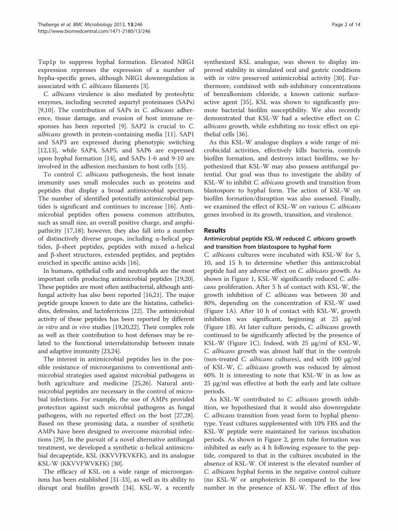
ResultsAntimicrobial peptide KSL-W reduced C. albicans growthand transition from blastospore to hyphal formC. albicans cultures were incubated with KSL-W for 5,10, and 15 h to determine whether this antimicrobialpeptide had any adverse effect on C. albicans growth. Asshown in Figure 1, KSL-W significantly reduced C. albi-cans proliferation. After 5 h of contact with KSL-W, thegrowth inhibition of C. albicans was between 30 and80%, depending on the concentration of KSL-W used(Figure 1A). After 10 h of contact with KSL-W, growthinhibition was significant, beginning at 25 μg/ml(Figure 1B). At later culture periods, C. albicans growthcontinued to be significantly affected by the presence ofKSL-W (Figure 1C). Indeed, with 25 μg/ml of KSL-W,C. albicans growth was almost half that in the controls(non-treated C. albicans cultures), and with 100 μg/mlof KSL-W, C. albicans growth was reduced by almost60%. It is interesting to note that KSL-W in as low as25 μg/ml was effective at both the early and late cultureperiods.As KSL-W contributed to C. albicans growth inhib-
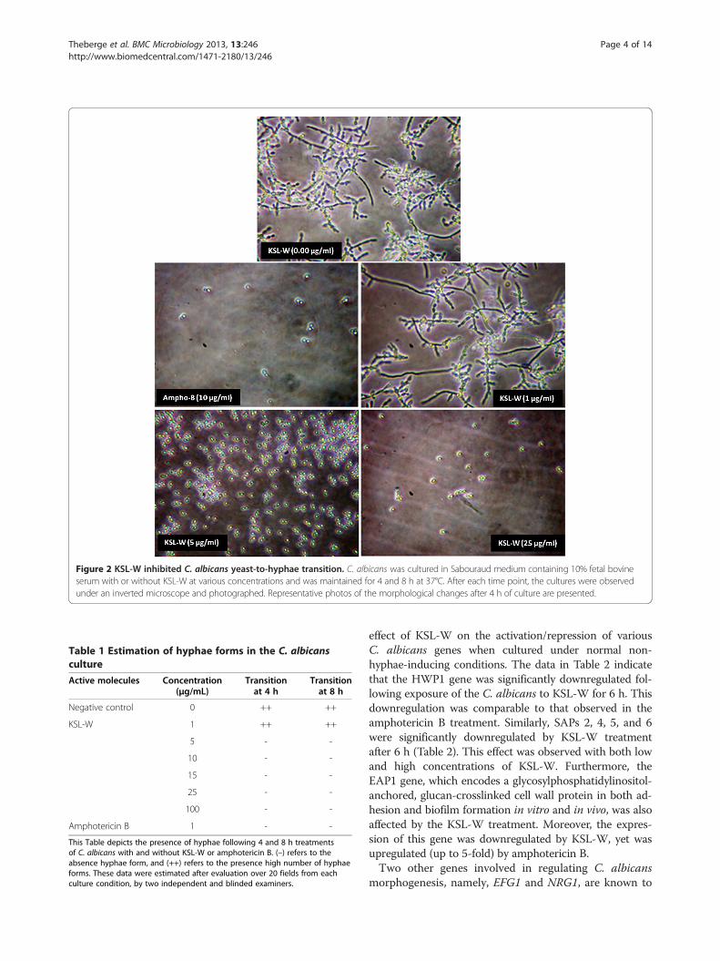
ition, we hypothesized that it would also downregulateC. albicans transition from yeast form to hyphal pheno-type. Yeast cultures supplemented with 10% FBS and theKSL-W peptide were maintained for various incubationperiods. As shown in Figure 2, germ tube formation wasinhibited as early as 4 h following exposure to the pep-tide, compared to that in the cultures incubated in theabsence of KSL-W. Of interest is the elevated number ofC. albicans hyphal forms in the negative control culture(no KSL-W or amphotericin B) compared to the lownumber in the presence of KSL-W. The effect of this
Theberge et al. BMC Microbiology 2013, 13:246 Page 2 of 14http://www.biomedcentral.com/1471-2180/13/246
antimicrobial peptide on C. albicans transition was alsodose-dependent: at 1 μg/ml, a significant number of hy-phal forms remained, and at only 5 μg/ml of KSL-W, C.albicans transition was completely inhibited (Figure 2).Semi-quantitative analyses using inverted microscope ob-servations to estimate the hyphal forms confirmed theinhibited C. albicans transition when treated with KSL-W(Table 1). The density of the hyphae was reduced as earlyas 4 h of contact with 5 μg/ml of KSL-W. This effect wasfurther supported when C. albicans was placed in contactwith KSL-W for 8 h (Table 1), thus confirming that KSL-Wdownregulated C. albicans growth and transition.
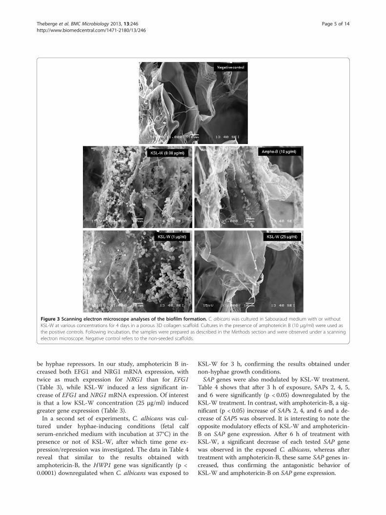
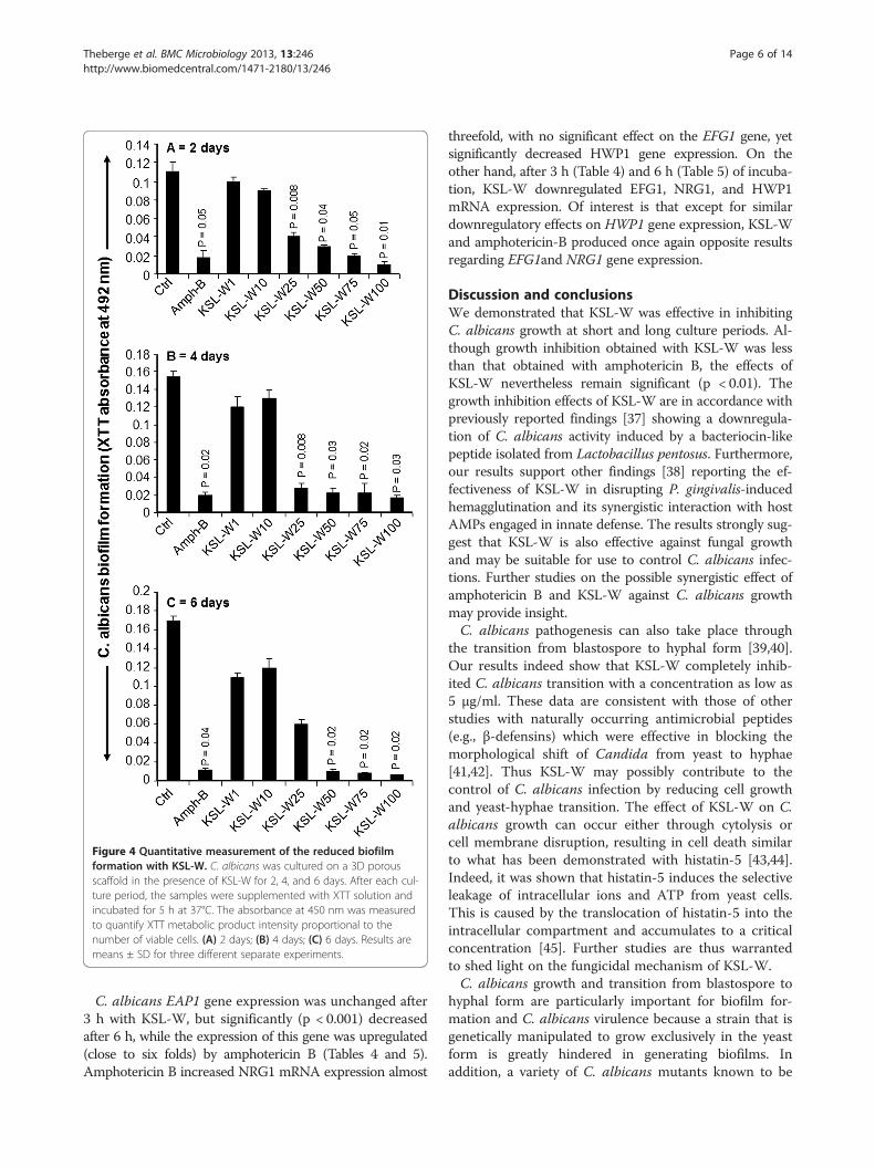
KSL-W reduced C. albicans biofilm formationAs KSL-W contributed to reducing C. albicans growthand transition, we sought to determine whether it alsodisplayed inhibitory activity against C. albicans biofilmformation. Using a biofilm-promoting scaffold, SEManalyses, and an XTT assay, we were able to demon-strate the inhibitory effect of KSL-W on biofilm forma-tion (Figure 3). SEM analyses revealed a significantdensity of C. albicans in the untreated culture, comparedto a lower density in the scaffold in the presence of KSL-W(1 and 25 μg/ml) after 4 days of culture. The de-creases obtained with the KSL-W, particularly at 25 μg/ml(Figure 3), were comparable to that obtained withamphotericin B at 10 μg/ml. To confirm these observa-tions, we performed quantitative analyses using the XTTassay. Figure 4A shows that after 2 days of culture, KSL-Wwas able to inhibit biofilm formation. This inhibitoryeffect was observed beginning at 25 μg/ml of KSL-W. Atconcentrations of 50, 75, and 100 μg/ml of KSL-W, theinhibition of C. albicans biofilm formation was compar-able to that caused by amphotericin B at 10 μg/ml. Similarresults were obtained after 4 days (Figure 4B) and 6 days(Figure 4C) of culture for biofilm formation with a persist-ent inhibitory effect of KSL-W on C. albicans biofilmformation.
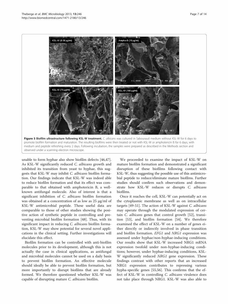
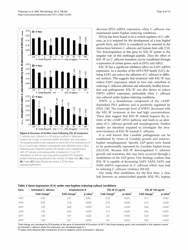
KSL-W disrupted mature C. albicans biofilmsAfter 6 days of incubation in glucose-rich Sabouraudmedium, scaffolds seeded with C. albicans strain SC5314produced mature biofilms displaying highly dense popu-lations of Candida cells (Figure 5). Significant reductionsand disruptions of the pre-formed Candida biofilmswere observed when the reference antifungal agent(amphotericin B, 10 μg/ml) was added to the maturebiofilms upon further incubation up to 6 days. Similarly,antimicrobial peptide KSL-W at 75 and 100 μg/ml alsoreduced C. albicans density in the biofilms. The ob-served reduction was noticed with KSL-W concentra-tions ranging from 25 to 100 μg/ml. Indeed, whenquantitatively investigated by XTT reduction assay, theKSL-W-treated biofilms rendered a significantly lowernumber of cells, as reflected by the lower absorbancereadings, than did the untreated control. This effect wasobserved after 2, 4, and 6 days of treatment with ampho-tericin B. Furthermore, the effect of KSL-W on the ma-ture C. albicans biofilm was comparable to that obtainedwith amphotericin B (Figure 6).
KSL-W modulated the expression of various C. albicansgenesBased on the data showing that KSL-W reducedC. albicans proliferation, transition, and biofilm forma-tion, we sought to determine the involvement, if any, ofgene regulation. For this purpose, we first investigated the
Figure 1 KSL-W inhibited C. albicans growth. The yeast wascultured in Sabouraud supplemented medium with or without KSL-W at various concentrations. The cultures were maintained for 5, 10,and 15 h at 37°C, after which time an MTT assay was performed foreach culture condition. The growth was plotted as means ± SD ofthe absorbance at 550 nm. (A) C. albicans growth with KSL-W for5 h; (B) C. albicans growth with KSL-W for 10 h; and (C) C. albicansgrowth with KSL-W for 15 h. The levels of significance for C. albicansgrowth in the presence or not of KSL-W or amphotericin B (10 μg/ml) were considered significant at P < 0 · 05.
Theberge et al. BMC Microbiology 2013, 13:246 Page 3 of 14http://www.biomedcentral.com/1471-2180/13/246
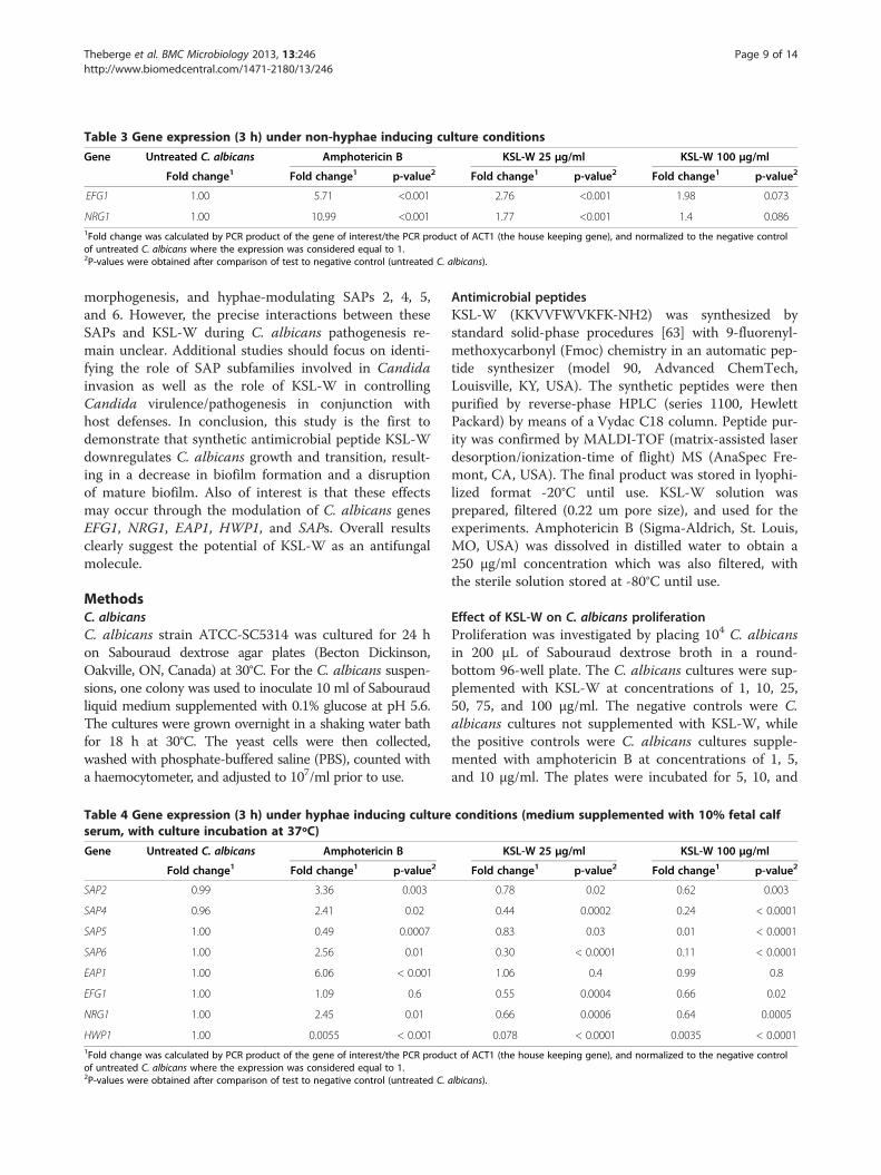
effect of KSL-W on the activation/repression of variousC. albicans genes when cultured under normal non-hyphae-inducing conditions. The data in Table 2 indicatethat the HWP1 gene was significantly downregulated fol-lowing exposure of the C. albicans to KSL-W for 6 h. Thisdownregulation was comparable to that observed in theamphotericin B treatment. Similarly, SAPs 2, 4, 5, and 6were significantly downregulated by KSL-W treatmentafter 6 h (Table 2). This effect was observed with both lowand high concentrations of KSL-W. Furthermore, theEAP1 gene, which encodes a glycosylphosphatidylinositol-anchored, glucan-crosslinked cell wall protein in both ad-hesion and biofilm formation in vitro and in vivo, was alsoaffected by the KSL-W treatment. Moreover, the expres-sion of this gene was downregulated by KSL-W, yet wasupregulated (up to 5-fold) by amphotericin B.Two other genes involved in regulating C. albicans
morphogenesis, namely, EFG1 and NRG1, are known to
Figure 2 KSL-W inhibited C. albicans yeast-to-hyphae transition. C. albicans was cultured in Sabouraud medium containing 10% fetal bovineserum with or without KSL-W at various concentrations and was maintained for 4 and 8 h at 37°C. After each time point, the cultures were observedunder an inverted microscope and photographed. Representative photos of the morphological changes after 4 h of culture are presented.
Table 1 Estimation of hyphae forms in the C. albicansculture
Active molecules Concentration(μg/mL)
Transitionat 4 h
Transitionat 8 h
Negative control 0 ++ ++
KSL-W 1 ++ ++
5 - -
10 - -
15 - -
25 - -
100 - -
Amphotericin B 1 - -
This Table depicts the presence of hyphae following 4 and 8 h treatmentsof C. albicans with and without KSL-W or amphotericin B. (–) refers to theabsence hyphae form, and (++) refers to the presence high number of hyphaeforms. These data were estimated after evaluation over 20 fields from eachculture condition, by two independent and blinded examiners.
Theberge et al. BMC Microbiology 2013, 13:246 Page 4 of 14http://www.biomedcentral.com/1471-2180/13/246
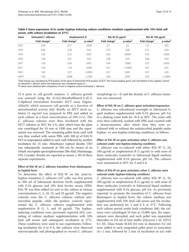
be hyphae repressors. In our study, amphotericin B in-creased both EFG1 and NRG1 mRNA expression, withtwice as much expression for NRG1 than for EFG1(Table 3), while KSL-W induced a less significant in-crease of EFG1 and NRG1 mRNA expression. Of interestis that a low KSL-W concentration (25 μg/ml) inducedgreater gene expression (Table 3).In a second set of experiments, C. albicans was cul-
tured under hyphae-inducing conditions (fetal calfserum-enriched medium with incubation at 37°C) in thepresence or not of KSL-W, after which time gene ex-pression/repression was investigated. The data in Table 4reveal that similar to the results obtained withamphotericin-B, the HWP1 gene was significantly (p <0.0001) downregulated when C. albicans was exposed to
KSL-W for 3 h, confirming the results obtained undernon-hyphae growth conditions.SAP genes were also modulated by KSL-W treatment.
Table 4 shows that after 3 h of exposure, SAPs 2, 4, 5,and 6 were significantly (p < 0.05) downregulated by theKSL-W treatment. In contrast, with amphotericin-B, a sig-nificant (p < 0.05) increase of SAPs 2, 4, and 6 and a de-crease of SAP5 was observed. It is interesting to note theopposite modulatory effects of KSL-W and amphotericin-B on SAP gene expression. After 6 h of treatment withKSL-W, a significant decrease of each tested SAP genewas observed in the exposed C. albicans, whereas aftertreatment with amphotericin-B, these same SAP genes in-creased, thus confirming the antagonistic behavior ofKSL-W and amphotericin-B on SAP gene expression.
Figure 3 Scanning electron microscope analyses of the biofilm formation. C. albicans was cultured in Sabouraud medium with or withoutKSL-W at various concentrations for 4 days in a porous 3D collagen scaffold. Cultures in the presence of amphotericin B (10 μg/ml) were used asthe positive controls. Following incubation, the samples were prepared as described in the Methods section and were observed under a scanningelectron microscope. Negative control refers to the non-seeded scaffolds.
Theberge et al. BMC Microbiology 2013, 13:246 Page 5 of 14http://www.biomedcentral.com/1471-2180/13/246
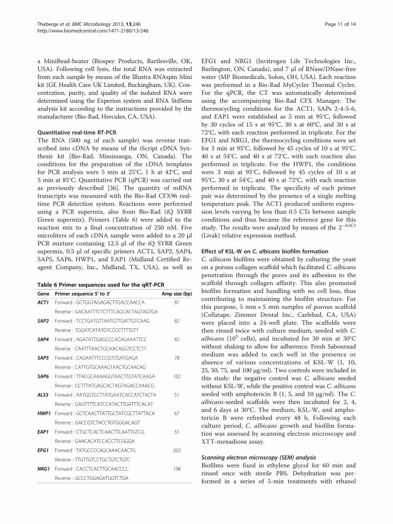
C. albicans EAP1 gene expression was unchanged after3 h with KSL-W, but significantly (p < 0.001) decreasedafter 6 h, while the expression of this gene was upregulated(close to six folds) by amphotericin B (Tables 4 and 5).Amphotericin B increased NRG1 mRNA expression almost
threefold, with no significant effect on the EFG1 gene, yetsignificantly decreased HWP1 gene expression. On theother hand, after 3 h (Table 4) and 6 h (Table 5) of incuba-tion, KSL-W downregulated EFG1, NRG1, and HWP1mRNA expression. Of interest is that except for similardownregulatory effects on HWP1 gene expression, KSL-Wand amphotericin-B produced once again opposite resultsregarding EFG1and NRG1 gene expression.
Discussion and conclusionsWe demonstrated that KSL-W was effective in inhibitingC. albicans growth at short and long culture periods. Al-though growth inhibition obtained with KSL-W was lessthan that obtained with amphotericin B, the effects ofKSL-W nevertheless remain significant (p < 0.01). Thegrowth inhibition effects of KSL-W are in accordance withpreviously reported findings [37] showing a downregula-tion of C. albicans activity induced by a bacteriocin-likepeptide isolated from Lactobacillus pentosus. Furthermore,our results support other findings [38] reporting the ef-fectiveness of KSL-W in disrupting P. gingivalis-inducedhemagglutination and its synergistic interaction with hostAMPs engaged in innate defense. The results strongly sug-gest that KSL-W is also effective against fungal growthand may be suitable for use to control C. albicans infec-tions. Further studies on the possible synergistic effect ofamphotericin B and KSL-W against C. albicans growthmay provide insight.C. albicans pathogenesis can also take place through
the transition from blastospore to hyphal form [39,40].Our results indeed show that KSL-W completely inhib-ited C. albicans transition with a concentration as low as5 μg/ml. These data are consistent with those of otherstudies with naturally occurring antimicrobial peptides(e.g., β-defensins) which were effective in blocking themorphological shift of Candida from yeast to hyphae[41,42]. Thus KSL-W may possibly contribute to thecontrol of C. albicans infection by reducing cell growthand yeast-hyphae transition. The effect of KSL-W on C.albicans growth can occur either through cytolysis orcell membrane disruption, resulting in cell death similarto what has been demonstrated with histatin-5 [43,44].Indeed, it was shown that histatin-5 induces the selectiveleakage of intracellular ions and ATP from yeast cells.This is caused by the translocation of histatin-5 into theintracellular compartment and accumulates to a criticalconcentration [45]. Further studies are thus warrantedto shed light on the fungicidal mechanism of KSL-W.C. albicans growth and transition from blastospore to
hyphal form are particularly important for biofilm for-mation and C. albicans virulence because a strain that isgenetically manipulated to grow exclusively in the yeastform is greatly hindered in generating biofilms. Inaddition, a variety of C. albicans mutants known to be
Figure 4 Quantitative measurement of the reduced biofilmformation with KSL-W. C. albicans was cultured on a 3D porousscaffold in the presence of KSL-W for 2, 4, and 6 days. After each cul-ture period, the samples were supplemented with XTT solution andincubated for 5 h at 37°C. The absorbance at 450 nm was measuredto quantify XTT metabolic product intensity proportional to thenumber of viable cells. (A) 2 days; (B) 4 days; (C) 6 days. Results aremeans ± SD for three different separate experiments.
Theberge et al. BMC Microbiology 2013, 13:246 Page 6 of 14http://www.biomedcentral.com/1471-2180/13/246
unable to form hyphae also show biofilm defects [46,47].As KSL-W significantly reduced C. albicans growth andinhibited its transition from yeast to hyphae, this sug-gests that KSL-W may inhibit C. albicans biofilm forma-tion. Our findings indicate that KSL-W was indeed ableto reduce biofilm formation and that its effect was com-parable to that obtained with amphotericin B, a well-known antifungal molecule. Also of interest is that asignificant inhibition of C. albicans biofilm formationwas obtained at a concentration of as low as 25 μg/ml ofKSL-W antimicrobial peptide. These useful data arecomparable to those of other studies showing the posi-tive action of synthetic peptide in controlling and pre-venting microbial biofilm formation [48]. Thus, with itssignificant impact in reducing C. albicans biofilm forma-tion, KSL-W may show potential for several novel appli-cations in the clinical setting. Further investigations willelucidate this effect.Biofilm formation can be controlled with anti-biofilm
molecules prior to its development, although this is notactually the case in clinical applications, as antifungaland microbial molecules cannot be used on a daily basisto prevent biofilm formation. An effective moleculeshould ideally be able to prevent biofilm formation, butmore importantly to disrupt biofilms that are alreadyformed. We therefore questioned whether KSL-W wascapable of disrupting mature C. albicans biofilm.
We proceeded to examine the impact of KSL-W onmature biofilm formation and demonstrated a significantdisruption of these biofilms following contact withKSL-W, thus suggesting the possible use of this antimicro-bial peptide to reduce/eliminate mature biofilms. Furtherstudies should confirm such observations and demon-strate how KSL-W reduces or disrupts C. albicansbiofilms.Once it reaches the cell, KSL-W can potentially act on
the cytoplasmic membrane as well as on intracellulartargets [49-51]. The action of KSL-W against C. albicansmay operate through the modulated expression of cer-tain C. albicans genes that control growth [52], transi-tion [53], and biofilm formation [54]. We thereforeexamined the effect of KSL-W on a number of genes ei-ther directly or indirectly involved in phase transitionand biofilm formation. EFG1 and NRG1 expression wasassessed under hyphae/non-hyphae-inducing conditions.Our results show that KSL-W increased NRG1 mRNAexpression twofold under non-hyphae-inducing condi-tions; however, under hyphae-inducing conditions, KSL-W significantly reduced NRG1 gene expression. Thesefindings contrast with other reports that an increasedNRG1 expression contributes to repressing varioushypha-specific genes [55,56]. This confirms that the ef-fect of KSL-W in controlling C. albicans virulence doesnot take place through NRG1. KSL-W was also able to
Figure 5 Biofilm ultrastructure following KSL-W treatment. C. albicans was cultured in Sabouraud medium without KSL-W for 6 days topromote biofilm formation and maturation. The resulting biofilms were then treated or not with KSL-W or amphotericin B for 6 days, withmedium and peptide refreshing every 2 days. Following incubation, the samples were prepared as described in the Methods section andobserved under a scanning electron microscope.
Theberge et al. BMC Microbiology 2013, 13:246 Page 7 of 14http://www.biomedcentral.com/1471-2180/13/246
decrease EFG1 mRNA expression, when C. albicans wasmaintained under hyphae-inducing conditions.EFG1p has been found to be a central regulator of C. albi-
cans, as it is required for the development of a true hyphalgrowth form, and EFG1 is considered to be essential in theinteractions between C. albicans and human host cells [7,8].The downregulation of this gene by KSL-W points to thesingular role of this antifungal peptide. Thus the effect ofKSL-W on C. albicans transition can be manifested througha repression of certain genes, such as EFG1 and NRG1.KSL-W has a significant inhibitory effect on EAP1 mRNA
expression. As a member of the GPI-CWP family [5,57], de-leting EAP1 can reduce the adhesion of C. albicans to differ-ent surfaces. This suggests that treatment with KSL-W mayreduce EAP1 expression, which in turn may contribute toreducing C. albicans adhesion and ultimately, biofilm forma-tion and pathogenesis. KSL-W was also shown to reduceHWP1 mRNA expression, particularly when C. albicanswas cultured under hyphae-inducing conditions.HWP1 is a downstream component of the cAMP-
dependent PKA pathway and is positively regulated byEFG1 [58]. The transcript level of HWP1 decreased withthe KSL-W treatment at low and high concentrations.These data suggest that KSL-W indeed impacts the ac-tivity of the cAMP–EFG1 pathway and leads to an alter-ation of C. albicans growth and morphogenesis. Furtherstudies are therefore required to investigate the inva-sion/virulence of KSL-W-treated C. albicans.It is well known that Candida pathogenesis can be
established by virtue of Candida growth and yeast-to-hyphae morphogenesis. Specific SAP genes were foundto be preferentially expressed by Candida hyphal forms[10,15,59]. Because KSL-W downregulated C. albicansgrowth and transition, this may have occurred through amodulation of the SAP genes. Our findings confirm thatKSL-W is capable of decreasing SAP2, SAP4, SAP5, andSAP6 mRNA expression in C. albicans which may leadto reducing C. albicans virulence [60-62].Our study thus establishes, for the first time, a clear
link between an antimicrobial peptide (KSL-W), hyphae
Figure 6 Decrease of biofilm mass following KSL-W treatment.C. albicans was cultured in a 3D porous scaffold in Sabouraudmedium for 6 days to promote biofilm formation and maturation.The resulting biofilms were exposed or not to KSL-W or amphotericin Bfor 2, 4, and 6 days. Medium and peptide were refreshed every 2 days.Following each treatment period, the samples were supplementedwith XTT solution and subsequently incubated for 5 h at 37°C.Absorbance at 450 nm was measured to quantify XTT metabolicproduct intensity proportional to the number of viable cells. (A) 2 days;(B) 4 days; (C) 6 days. Results are means ± SD for threeseparate experiments.
Table 2 Gene expression (6 h) under non-hyphae inducing culture conditions
Gene Untreated C. albicans Amphotericin B KSL-W 25 μg/ml KSL-W 100 μg/ml
Fold change1 Fold change1 p-value2 Fold change1 p-value2 Fold change1 p-value2
SAP2 0.99 0.57 0.001 0.24 <0.001 0.11 <0.001
SAP4 0.96 0.19 <0.001 0.29 <0.001 0.14 <0.001
SAP5 1.00 0.08 <0.001 0.16 <0.001 0.06 <0.001
SAP6 1.00 0.05 <0.001 0.14 <0.001 0.04 <0.001
EAP1 1.00 4.91 0.028 0.4 <0.001 0.29 <0.001
HWP1 1.00 0.01 <0.001 0.6 0.032 0.02 <0.0011Fold change was calculated by PCR product of the gene of interest/the PCR product of ACT1 (the house keeping gene), and normalized to the negative controlof untreated C. albicans where the expression was considered equal to 1.2P-values were obtained after comparison of test to negative control (untreated C. albicans).
Theberge et al. BMC Microbiology 2013, 13:246 Page 8 of 14http://www.biomedcentral.com/1471-2180/13/246
morphogenesis, and hyphae-modulating SAPs 2, 4, 5,and 6. However, the precise interactions between theseSAPs and KSL-W during C. albicans pathogenesis re-main unclear. Additional studies should focus on identi-fying the role of SAP subfamilies involved in Candidainvasion as well as the role of KSL-W in controllingCandida virulence/pathogenesis in conjunction withhost defenses. In conclusion, this study is the first todemonstrate that synthetic antimicrobial peptide KSL-Wdownregulates C. albicans growth and transition, result-ing in a decrease in biofilm formation and a disruptionof mature biofilm. Also of interest is that these effectsmay occur through the modulation of C. albicans genesEFG1, NRG1, EAP1, HWP1, and SAPs. Overall resultsclearly suggest the potential of KSL-W as an antifungalmolecule.
MethodsC. albicansC. albicans strain ATCC-SC5314 was cultured for 24 hon Sabouraud dextrose agar plates (Becton Dickinson,Oakville, ON, Canada) at 30°C. For the C. albicans suspen-sions, one colony was used to inoculate 10 ml of Sabouraudliquid medium supplemented with 0.1% glucose at pH 5.6.The cultures were grown overnight in a shaking water bathfor 18 h at 30°C. The yeast cells were then collected,washed with phosphate-buffered saline (PBS), counted witha haemocytometer, and adjusted to 107/ml prior to use.
Antimicrobial peptidesKSL-W (KKVVFWVKFK-NH2) was synthesized bystandard solid-phase procedures [63] with 9-fluorenyl-methoxycarbonyl (Fmoc) chemistry in an automatic pep-tide synthesizer (model 90, Advanced ChemTech,Louisville, KY, USA). The synthetic peptides were thenpurified by reverse-phase HPLC (series 1100, HewlettPackard) by means of a Vydac C18 column. Peptide pur-ity was confirmed by MALDI-TOF (matrix-assisted laserdesorption/ionization-time of flight) MS (AnaSpec Fre-mont, CA, USA). The final product was stored in lyophi-lized format -20°C until use. KSL-W solution wasprepared, filtered (0.22 um pore size), and used for theexperiments. Amphotericin B (Sigma-Aldrich, St. Louis,MO, USA) was dissolved in distilled water to obtain a250 μg/ml concentration which was also filtered, withthe sterile solution stored at -80°C until use.
Effect of KSL-W on C. albicans proliferationProliferation was investigated by placing 104 C. albicansin 200 μL of Sabouraud dextrose broth in a round-bottom 96-well plate. The C. albicans cultures were sup-plemented with KSL-W at concentrations of 1, 10, 25,50, 75, and 100 μg/ml. The negative controls were C.albicans cultures not supplemented with KSL-W, whilethe positive controls were C. albicans cultures supple-mented with amphotericin B at concentrations of 1, 5,and 10 μg/ml. The plates were incubated for 5, 10, and
Table 3 Gene expression (3 h) under non-hyphae inducing culture conditions
Gene Untreated C. albicans Amphotericin B KSL-W 25 μg/ml KSL-W 100 μg/ml
Fold change1 Fold change1 p-value2 Fold change1 p-value2 Fold change1 p-value2
EFG1 1.00 5.71 <0.001 2.76 <0.001 1.98 0.073
NRG1 1.00 10.99 <0.001 1.77 <0.001 1.4 0.0861Fold change was calculated by PCR product of the gene of interest/the PCR product of ACT1 (the house keeping gene), and normalized to the negative controlof untreated C. albicans where the expression was considered equal to 1.2P-values were obtained after comparison of test to negative control (untreated C. albicans).
Table 4 Gene expression (3 h) under hyphae inducing culture conditions (medium supplemented with 10% fetal calfserum, with culture incubation at 37ºC)
Gene Untreated C. albicans Amphotericin B KSL-W 25 μg/ml KSL-W 100 μg/ml
Fold change1 Fold change1 p-value2 Fold change1 p-value2 Fold change1 p-value2
SAP2 0.99 3.36 0.003 0.78 0.02 0.62 0.003
SAP4 0.96 2.41 0.02 0.44 0.0002 0.24 < 0.0001
SAP5 1.00 0.49 0.0007 0.83 0.03 0.01 < 0.0001
SAP6 1.00 2.56 0.01 0.30 < 0.0001 0.11 < 0.0001
EAP1 1.00 6.06 < 0.001 1.06 0.4 0.99 0.8
EFG1 1.00 1.09 0.6 0.55 0.0004 0.66 0.02
NRG1 1.00 2.45 0.01 0.66 0.0006 0.64 0.0005
HWP1 1.00 0.0055 < 0.001 0.078 < 0.0001 0.0035 < 0.00011Fold change was calculated by PCR product of the gene of interest/the PCR product of ACT1 (the house keeping gene), and normalized to the negative controlof untreated C. albicans where the expression was considered equal to 1.2P-values were obtained after comparison of test to negative control (untreated C. albicans).
Theberge et al. BMC Microbiology 2013, 13:246 Page 9 of 14http://www.biomedcentral.com/1471-2180/13/246
15 h prior to cell growth analyses. C. albicans growthwas assessed using the (3-(4,5-dimethylthiazol-2-yl)-2,5-diphenyl tetrazolium bromide) MTT assay (Sigma-Aldrich) which measures cell growth as a function ofmitochondrial activity [64]. Briefly, an MTT stock so-lution (5 mg/ml) was prepared in PBS and added toeach culture at a final concentration of 10% (v/v). TheC. albicans cultures were then incubated with theMTT solution at 30°C for 4 h, after which time the platewas centrifuged for 10 min at 1200 rpm and the super-natant was removed. The remaining pellet from each wellwas then washed with warm PBS, with 200 μl of 0.04 NHCl in isopropanol added to each well, followed by anotherincubation for 15 min. Absorbance (optical density, OD)was subsequently measured at 550 nm by means of anxMark microplate spectrophotometer (Bio-Rad, Mississauga,ON, Canada). Results are reported as means ± SD of threeseparate experiments.
Effect of KSL-W on C. albicans transition from blastosporeto hyphal formTo determine the effect of KSL-W on the yeast-to-hyphae transition, C. albicans (105 cells) was first grownin 500 ml of Sabouraud dextrose broth supplementedwith 0.1% glucose and 10% fetal bovine serum (FBS).KSL-W was then added (or not) to the culture at variousconcentrations (1, 5, 10, 15, and 25 μg/ml). The negativecontrols were the C. albicans cultures without anti-microbial peptide, while the positive controls repre-sented the C. albicans cultures supplemented withamphotericin B (1, 5, and 10 μg/ml). The hyphae-inducing conditions were previously reported [65], con-sisting of culture medium supplementation with 10%fetal calf serum and subsequent incubation at 37°C.These conditions were used in our experiments. Follow-ing incubation for 4 or 8 h, the cultures were observedmicroscopically and photographed to record C. albicans
morphology (n = 5) and the density of C. albicans transi-tion was measured.
Effect of KSL-W on C. albicans gene activation/repressionC. albicans was subcultured overnight in Sabouraud li-quid medium supplemented with 0.1% glucose, pH 5.6,in a shaking water bath for 18 h at 30°C. The yeast cellswere then collected, washed with PBS, and counted witha hemocytometer, after which time they were co-cultured with or without the antimicrobial peptide underhyphae- or non-hyphae-inducing conditions, as follows.
Effect of KSL-W on gene activation when C. albicans wascultured under non-hyphae-inducing conditionsC. albicans was co-cultured with either KSL-W (1, 25,100 μg/ml) or amphotericin B (1 μg/ml) or with none ofthese molecules (controls) in Sabouraud liquid mediumsupplemented with 0.1% glucose, pH 5.6. The cultureswere maintained at 30°C for 3 and 6 h.
Effect of KSL-W on gene activation when C. albicans werecultured under hyphae-inducing conditionsC. albicans was co-cultured with either KSL-W (1, 25,100 μg/ml) or amphotericin B (1 μg/ml) or with none ofthese molecules (controls) in Sabouraud liquid mediumsupplemented with 0.1% glucose, pH 5.6. As previouslyreported, to promote the transition of C. albicans fromblastospore to hyphal form, the culture medium wassupplemented with 10% fetal calf serum and the incuba-tion was performed for 3 and 6 h at 37°C. Followingeach culture period under both conditions [68], the cul-tures were centrifuged 10 min at 13,000 rpm, the super-natants were discarded, and each pellet was suspendedthereafter in 0.6 ml of lysis buffer (Glycerol 1 M, EDTA0.1 M). Glass beads (0.425-0.6 mm in diameter; 0.2 ml)were added to each suspended pellet prior to sonication(4 × 1 min, followed by 2 min of incubation in ice) with
Table 5 Gene expression (6 h) under hyphae inducing culture conditions (medium supplemented with 10% fetal calfserum, with culture incubation at 37ºC)
Gene Untreated C. albicans Amphotericin B KSL-W 25 μg/ml KSL-W 100 μg/ml
Fold change1 Fold change1 p-value2 Fold change1 p-value2 Fold change1 p-value2
SAP2 0.99 8.17 0.009 0.7 0.2 1.31 0.02
SAP4 0.96 2.58 0.03 0.73 0.04 0.72 0.04
SAP5 1.00 0.72 0.007 0.83 0.0004 0.56 0.006
SAP6 1.00 4.01 0.02 0.58 0.01 0.68 0.04
EAP1 1.00 6.36 0.001 0.44 0.008 0.73 0.003
EFG1 1.00 1.78 0.048 0.31 < 0.0001 0.47 0.01
NRG1 1.00 3.97 0.0005 0.37 0.001 0.37 0.05
HWP1 1.00 0.008 < 0.001 0.09 0.001 0.03 < 0.00011Fold change was calculated by PCR product of the gene of interest/the PCR product of ACT1 (the house keeping gene), and normalized to the negative controlof untreated C. albicans where the expression was considered equal to 1.2P-values were obtained after comparison of test to negative control (untreated C. albicans).
Theberge et al. BMC Microbiology 2013, 13:246 Page 10 of 14http://www.biomedcentral.com/1471-2180/13/246
a MiniBead-beater (Biospec Products, Bartlesville, OK,USA). Following cell lysis, the total RNA was extractedfrom each sample by means of the Illustra RNAspin Minikit (GE Health Care UK Limited, Buckingham, UK). Con-centration, purity, and quality of the isolated RNA weredetermined using the Experion system and RNA StdSensanalysis kit according to the instructions provided by themanufacturer (Bio-Rad, Hercules, CA, USA).
Quantitative real-time RT-PCRThe RNA (500 ng of each sample) was reverse tran-scribed into cDNA by means of the iScript cDNA Syn-thesis kit (Bio-Rad, Mississauga, ON, Canada). Theconditions for the preparation of the cDNA templatesfor PCR analysis were 5 min at 25°C, 1 h at 42°C, and5 min at 85°C. Quantitative PCR (qPCR) was carried outas previously described [36]. The quantity of mRNAtranscripts was measured with the Bio-Rad CFX96 real-time PCR detection system. Reactions were performedusing a PCR supermix, also from Bio-Rad (iQ SYBRGreen supermix). Primers (Table 6) were added to thereaction mix to a final concentration of 250 nM. Fivemicroliters of each cDNA sample were added to a 20 μlPCR mixture containing 12.5 μl of the iQ SYBR Greensupermix, 0.5 μl of specific primers ACT1, SAP2, SAP4,SAP5, SAP6, HWP1, and EAP1 (Midland Certified Re-agent Company, Inc., Midland, TX, USA), as well as
EFG1 and NRG1 (Invitrogen Life Technologies Inc.,Burlington, ON, Canada), and 7 μl of RNase/DNase-freewater (MP Biomedicals, Solon, OH, USA). Each reactionwas performed in a Bio-Rad MyCycler Thermal Cycler.For the qPCR, the CT was automatically determinedusing the accompanying Bio-Rad CFX Manager. Thethermocycling conditions for the ACT1, SAPs 2-4-5-6,and EAP1 were established as 5 min at 95°C, followedby 30 cycles of 15 s at 95°C, 30 s at 60°C, and 30 s at72°C, with each reaction performed in triplicate. For theEFG1 and NRG1, the thermocycling conditions were setfor 3 min at 95°C, followed by 45 cycles of 10 s at 95°C,40 s at 54°C, and 40 s at 72°C, with each reaction alsoperformed in triplicate. For the HWP1, the conditionswere 3 min at 95°C, followed by 45 cycles of 10 s at95°C, 30 s at 54°C, and 40 s at 72°C, with each reactionperformed in triplicate. The specificity of each primerpair was determined by the presence of a single meltingtemperature peak. The ACT1 produced uniform expres-sion levels varying by less than 0.5 CTs between sampleconditions and thus became the reference gene for thisstudy. The results were analyzed by means of the 2−ΔΔCt
(Livak) relative expression method.
Effect of KSL-W on C. albicans biofilm formationC. albicans biofilms were obtained by culturing the yeaston a porous collagen scaffold which facilitated C. albicanspenetration through the pores and its adhesion to thescaffold through collagen affinity. This also promotedbiofilm formation and handling with no cell loss, thuscontributing to maintaining the biofilm structure. Forthis purpose, 5 mm × 5 mm samples of porous scaffold(Collatape, Zimmer Dental Inc., Carlsbad, CA, USA)were placed into a 24-well plate. The scaffolds werethen rinsed twice with culture medium, seeded with C.albicans (105 cells), and incubated for 30 min at 30°Cwithout shaking to allow for adherence. Fresh Sabouraudmedium was added to each well in the presence orabsence of various concentrations of KSL-W (1, 10,25, 50, 75, and 100 μg/ml). Two controls were included inthis study: the negative control was C. albicans seededwithout KSL-W, while the positive control was C. albicansseeded with amphotericin B (1, 5, and 10 μg/ml). The C.albicans-seeded scaffolds were then incubated for 2, 4,and 6 days at 30°C. The medium, KSL-W, and ampho-tericin B were refreshed every 48 h. Following eachculture period, C. albicans growth and biofilm forma-tion was assessed by scanning electron microscopy andXTT-menadione assay.
Scanning electron microscopy (SEM) analysisBiofilms were fixed in ethylene glycol for 60 min andrinsed once with sterile PBS. Dehydration was per-formed in a series of 5-min treatments with ethanol
Table 6 Primer sequences used for the qRT-PCR
Gene Primer sequence 5′ to 3′ Amp size (bp)
ACT1 Forward : GCTGGTAGAGACTTGACCAACCA 87
Reverse : GACAATTTCTCTTTCAGCACTAGTAGTGA
SAP2 Forward : TCCTGATGTTAATGTTGATTGTCAAG 82
Reverse : TGGATCATATGTCCCCTTTTGTT
SAP4 Forward : AGATATTGAGCCCACAGAAATTCC 82
Reverse : CAATTTAACTGCAACAGGTCCTCTT
SAP5 Forward : CAGAATTTCCCGTCGATGAGA 78
Reverse : CATTGTGCAAAGTAACTGCAACAG
SAP6 Forward : TTACGCAAAAGGTAACTTGTATCAAGA 102
Reverse : CCTTTATGAGCACTAGTAGACCAAACG
ALS3 Forward : AATGGTCCTTATGAATCACCATCTACTA 51
Reverse : GAGTTTTCATCCATACTTGATTTCACAT
HWP1 Forward : GCTCAACTTATTGCTATCGCTTATTACA 67
Reverse : GACCGTCTACCTGTGGGACAGT
EAP1 Forward : CTGCTCACTCAACTTCAATTGTCG 51
Reverse : GAACACATCCACCTTCGGGA
EFG1 Forward : TATGCCCCAGCAAACAACTG 202
Reverse : TTGTTGTCCTGCTGTCTGTC
NRG1 Forward : CACCTCACTTGCAACCCC 198
Reverse : GCCCTGGAGATGGTCTGA
Theberge et al. BMC Microbiology 2013, 13:246 Page 11 of 14http://www.biomedcentral.com/1471-2180/13/246
solutions of increasing concentration (50, 70, 90, andtwice at 100%). The dehydrated biofilms were kept over-night in a vacuum oven at 25°C, after which time theywere sputter-coated with gold, examined, and imaged(n = 4) under a JEOL 6360 LV SEM (Soquelec, Montréal,QC, Canada) operating at a 30 kV accelerating voltage.
XTT reduction assayTo support the hypothesis that KSL-W quantitatively af-fects C. albicans biofilms, an XTT reduction assay wasperformed on the KSL-W-treated and control biofilmsat defined time points. XTT assay is one of the mostuseful and accurate methods to investigate microbialbiofilm formation. The metabolic activity of the biofilmcells was measured as a reflection of viable cell count.To do so, C. albicans biofilms formed in the porousscaffold with or without KSL-W treatments for 2, 4, and6 days were subjected to an XTT assay. Fifty microlitersof XTT salt solution (1 mg/ml in PBS; Sigma-Aldrich)and 4 μl of menadione solution (1 mM in acetone;Sigma-Aldrich) were added to wells containing 4 ml ofsterile PBS. The biofilms were then added to the mixtureand the plates were incubated at 37°C for 5 h, afterwhich time the supernatant was collected to measurethe XTT formazan at 492 nm by means of an xMark mi-croplate spectrophotometer (Bio-Rad, Mississauga, ON,Canada).
Effect of KSL-W on the disruption of mature C. albicansbiofilmsMature C. albicans biofilms were obtained by culturingC. albicans (105) on a porous 3D collagen scaffold for6 days at 30°C in Sabouraud liquid medium supple-mented with 0.1% glucose at pH 5.6. The culturemedium was refreshed every 2 days. At the end of the 6-day culture period, the biofilms were treated (or not)with KSL-W (75 and 100 μg/ml). Amphotericin B-treated biofilms (1, 5, and 10 μg/ml) were used as thepositive controls. The biofilms were continuously incu-bated (or not) with either KSL-W or amphotericin B for2, 4, and 6 days, with medium changing every day.KSL-W and amphotericin B were also refreshed at eachmedium changing. Following each incubation period,SEM and XTT analyses were performed, as describedabove.
Statistical analysisEach experiment was performed at least four times, withexperimental values expressed as means ± SD. The stat-istical significance of the differences between the control(absence of KSL-W) and test (presence of KSL-W oramphotericin B) values was determined by means of aone-way ANOVA. Posteriori comparisons were per-formed using Tukey’s method. Normality and variance
assumptions were verified using the Shapiro-Wilk testand the Brown and Forsythe test, respectively. All of theassumptions were fulfilled. P values were declared sig-nificant at ≤ 0.05. The data were analyzed using the SASversion 8.2 statistical package (SAS Institute Inc., Cary,NC, USA).
Authors’ contributionsMR, KPL, and AS designed the experiments, supervised the research andwrote the paper. ST, AA, and AS performed the experiments and dataanalyses and contributed to the writing of the paper. Each author read andapproved the final manuscript.
AcknowledgementsThis study was supported financially by the United States Army MedicalResearch and Materiel Command (Award number ERMS No. 12304006) andby a grant from the Fonds Émile-Beaulieu, a Université Laval foundation. Theauthors also thank Ms. Claire Kingston (Traduction CFK) for proofreading andediting this manuscript.
DOD DisclaimerOne of the authors (KPL) is a United States Government employee. The workpresented is part of his official duties. The opinions or assertions containedherein are the personal views of these authors and are not to be construedas official or reflecting the views of the United States Army or Department ofDefense.
Author details1Oral Ecology Research Group, Faculty of Dentistry, Laval University, 2420,rue de la Terrasse, Quebec G1V 0A6, QC, Canada. 2Department ofBiochemistry, Genome Research Chair, College of Science King SaudUniversity, Riyadh, Kingdom of Saudi Arabia. 3Dental and Trauma ResearchDetachment, US Army Institute of Surgical Research, JBSA Fort Sam Houston,San Antonio, TX, USA.
Received: 5 July 2013 Accepted: 4 November 2013Published: 7 November 2013
References1. Arendorf TM, Walker DM: The prevalence and intra-oral distribution of
Candida albicans in man. Arch Oral Biol 1980, 25:1–10.2. Cannon RD, Chaffin WL: Oral colonization by Candida albicans. Crit Rev
Oral Biol Med 1999, 10:359–383.3. Sudbery P, Gow N, Berman J: The distinct morphogenic states of Candida
albicans. Trends Microbiol 2004, 12:317–324.4. Nobile CJ, Nett JE, Andes DR, Mitchell AP: Function of Candida albicans
adhesin Hwp1 in biofilm formation. Eukaryot Cell 2006, 5:1604–1610.5. Li F, Palecek SP: EAP1, a Candida albicans gene involved in binding
human epithelial cells. Eukaryot Cell 2003, 2:1266–1273.6. Sohn K, Urban C, Brunner H, Rupp S: EFG1 is a major regulator of cell wall
dynamics in Candida albicans as revealed by DNA microarrays.Mol Microbiol 2003, 47:89–102.
7. Stoldt VR, Sonneborn A, Leuker CE, Ernst JF: Efg1p, an essential regulatorof morphogenesis of the human pathogen Candida albicans, is amember of a conserved class of bHLH proteins regulatingmorphogenetic processes in fungi. EMBO J 1997, 16:1982–1991.
8. Lo HJ, Köhler JR, DiDomenico B, Loebenberg D, Cacciapuoti A, Fink GR:Nonfilamentous C. albicans mutants are avirulent. Cell 1997, 90:939–949.
9. Schaller M, Borelli C, Korting HC, Hube B: Hydrolytic enzymes as virulencefactors of Candida albicans. Mycoses 2005, 48:365–377.
10. Décanis N, Tazi N, Correia A, Vilanova M, Rouabhia M: Farnesol, a fungalquorum-sensing molecule triggers Candida albicans morphologicalchanges by down-regulating the expression of different secretedaspartyl proteinase genes. Open Microbiol J 2011, 5:119–126.
11. Naglik JR, Challacombe SJ, Hube B: Candida albicans secreted aspartylproteinases in virulence and pathogenesis. Microbiol Mol Biol Rev 2003,67:400–428.
12. Hube B, Naglik J: Candida albicans proteinases: resolving the mystery of agene family. Microbiology 2001, 147:1997–2005.
Theberge et al. BMC Microbiology 2013, 13:246 Page 12 of 14http://www.biomedcentral.com/1471-2180/13/246
13. White TC, Miyasaki SH, Agabian N: Three distinct secreted aspartylproteinases in Candida albicans. J Bacteriol 1993, 175:6126–6133.
14. White TC, Agabian N: Candida albicans secreted aspartyl proteinases:isoenzyme pattern is determined by cell type, and levels are determinedby environmental factors. J Bacteriol 1995, 177:5215–5221.
15. Albrecht A, Felk A, Pichova I, Naglik JR, Schaller M, de Groot P, Maccallum D,Odds FC, Schäfer W, Klis F, Monod M, Hube B:Glycosylphosphatidylinositol-anchored proteases of Candida albicanstarget proteins necessary for both cellular processes and host-pathogeninteractions. J Biol Chem 2006, 281(2):688–694.
16. van der Weerden NL, Bleackley MR, Anderson MA: Properties andmechanisms of action of naturally occurring antifungal peptides. Cell MolLife Sci 2013, 70(19):3545–3570.
17. Fjell CD, Hiss JA, Hancock RE, Schneider G: Designing antimicrobialpeptides: form follows function. Nat Rev Drug Discov 2012, 11:37–51.
18. Seo MD, Won HS, Kim JH, Mishig-Ochir T, Lee BJ: Antimicrobial peptidesfor therapeutic applications: a review. Molecules 2012, 17:12276–12286.
19. Campbell EL, Serhan CN, Colgan SP: Antimicrobial aspects ofinflammatory resolution in the mucosa: a role for proresolvingmediators. J Immunol 2011, 187:3475–3481.
20. Lehrer RI, Lu W: alpha-Defensins in human innate immunity. Immunol Rev2012, 245:84–112.
21. Mehra T, Koberle M, Braunsdorf C, Mailander-Sanchez D, Borelli C, et al: Alternativeapproaches to antifungal therapies. Exp Dermatol 2012, 21:778–782.
22. Zhu S: Discovery of six families of fungal defensin-like peptides providesinsights into origin and evolution of the CSalphabeta defensins.Mol Immunol 2008, 45:828–838.
23. Batoni G, Maisetta G, Brancatisano FL, Esin S, Campa M: Use ofantimicrobial peptides against microbial biofilms: advantages and limits.Curr Med Chem 2011, 18:256–279.
24. Dziarski R, Gupta D: Review: Mammalian peptidoglycan recognitionproteins (PGRPs) in innate immunity. Innate Immun 2010, 16:168–174.
25. Taraszkiewicz A, Fila G, Grinholc M, Nakonieczna J: Innovative strategies toovercome biofilm resistance. Biomed Res Int 2013, 2013:150653.doi: 10.1155/2013/150653.
26. Cota-Arriola O, Cortez-Rocha MO, Burgos-Hernandez A, Ezquerra-Brauer JM,Plascencia-Jatomea M: Controlled release matrices and micro/nanoparticlesof chitosan with antimicrobial potential: development of new strategies formicrobial control in agriculture. J Sci Food Agric 2013, 93:1525–1536.
27. Dhople V, Krukemeyer A, Ramamoorthy A: The human beta-defensin-3, anantibacterial peptide with multiple biological functions. Biochim BiophysActa 2006, 1758:1499–1512.
28. Joly S, Maze C, McCray PB Jr, Guthmiller JM: Human beta-defensins 2 and3 demonstrate strain-selective activity against oral microorganisms. J ClinMicrobiol 2004, 42:1024–1029.
29. Mooney C, Haslam NJ, Pollastri G, Shields DC: Towards the improveddiscovery and design of functional peptides: common features ofdiverse classes permit generalized prediction of bioactivity. PLoS One2012, 7:e45012.
30. Na DH, Faraj J, Capan Y, Leung KP, DeLuca PP: Stability of antimicrobialdecapeptide (KSL) and its analogues for delivery in the oral cavity.Pharm Res 2007, 24:1544–1550.
31. Hong SY, Park TG, Lee KH: The effect of charge increase on the specificityand activity of a short antimicrobial peptide. Peptides 2001, 22:1669–1674.
32. Oh JE, Hong SY, Lee KH: Structure-activity relationship study: shortantimicrobial peptides. J Pept Res 1999, 53:41–46.
33. Concannon SP, Crowe TD, Abercrombie JJ, Molina CM, Hou P, et al:Susceptibility of oral bacteria to an antimicrobial decapeptide. J MedMicrobiol 2003, 52:1083–1093.
34. Leung KP, Crowe TD, Abercrombie JJ, Molina CM, Bradshaw CJ, et al:Control of oral biofilm formation by an antimicrobial decapeptide. J DentRes 2005, 84:1172–1177.
35. Baker PJ, Coburn RA, Genco RJ, Evans RT: The in vitro inhibition ofmicrobial growth and plaque formation by surfactant drugs. J PeriodontalRes 1978, 13:474–485.
36. Semlali A, Leung KP, Curt S, Rouabhia M: Antimicrobial decapeptide KSL-Wattenuates Candida albicans virulence by modulating its effects on Toll-like receptor, human β-defensin, and cytokine expression by engineeredhuman oral mucosa. Peptides 2011, 32(5):859–867.
37. Okkers DJ, Dicks LM, Silvester M, Joubert JJ, Odendaal HJ: Characterizationof pentocin TV35b, a bacteriocin-like peptide isolated from Lactobacillus
pentosus with a fungistatic effect on Candida albicans. J Appl Microbiol1999, 87:726–734.
38. Dixon DR, Jeffrey NR, Dubey VS, Leung KP: Antimicrobial peptideinhibition of Porphyromonas gingivalis 381-induced hemagglutination isimproved with a synthetic decapeptide. Peptides 2009, 30:2161–2167.
39. Raines SM, Rane HS, Bernardo SM, Binder JL, Lee SA, et al: Deletion ofVacuolar Proton-translocating ATPase Voa Isoforms Clarifies the Role ofVacuolar pH as a Determinant of Virulence-associated Traits in Candidaalbicans. J Biol Chem 2013, 288:6190–6201.
40. Ariyachet C, Solis NV, Liu Y, Prasadarao NV, Filler SG, et al: SR-Like RNA-BindingProtein Slr1 Affects Candida albicans Filamentation and Virulence.Infect Immun 2013, 81:1267–1276.
41. Décanis N, Savignac K, Rouabhia M: Farnesol promotes epithelial celldefense against Candida albicans through Toll-like receptor 2expression, interleukin-6 and human beta-defensin 2 production.Cytokine 2009, 45:132–140.
42. Zhang J, Silao FG, Bigol UG, Bungay AA, Nicolas MG, et al: Calcineurin isrequired for pseudohyphal growth, virulence, and drug resistance inCandida lusitaniae. PLoS One 2012, 7:e44192.
43. Koshlukova SE, Araujo MWB, Baev D, Edgerton M: Released ATP is anextracellular cytotoxic mediator in salivary histatin 5-induced killingofCandida albicans. Infect Immun 2000, 68:6848–6856.
44. Vylkova S, Jang WS, Li W, Nayyar N, Edgerton M: Histatin 5 initiates osmoticstress response in Candida albicans via activation of the Hog1 mitogen-activated protein kinase pathway. Eukaryot Cell 2007, 6:1876–1888.
45. Jang WS, Bajwa JS, Sun JN, Edgerton M: Salivary histatin 5 internalizationby translocation, but not endocytosis, is required for fungicidal activityin Candida albicans. Mol Microbiol 2010, 77:354–370.
46. Ramage G, Vandewalle K, Wickes BL, Lopez-Ribot JL: Characteristics of biofilmformation by Candida albicans. Rev Iberoam Micol 2001, 18:163–170.
47. Banerjee M, Uppuluri P, Zhao XR, Carlisle PL, Vipulanandan G, et al:Expression of UME6, a key regulator of Candida albicans hyphaldevelopment, enhances biofilm formation via Hgc1- and Sun41-dependent mechanisms. Eukaryot Cell 2013, 12:224–232.
48. da Silva BR, de Freitas VA, Carneiro VA, Arruda FV, Lorenzon EN, et al:Antimicrobial activity of the synthetic peptide Lys-a1 against oralstreptococci. Peptides 2013, 42C:78–83.
49. Beckloff N, Laube D, Castro T, Furgang D, Park S, et al: Activity of anantimicrobial peptide mimetic against planktonic and biofilm cultures oforal pathogens. Antimicrob Agents Chemother 2007, 51:4125–4132.
50. Patrzykat A, Friedrich CL, Zhang L, Mendoza V, Hancock RE: Sublethalconcentrations of pleurocidin-derived antimicrobial peptides inhibitmacromolecular synthesis in Escherichia coli. Antimicrob AgentsChemother 2002, 46:605–614.
51. Mason AJ, Chotimah IN, Bertani P, Bechinger B: A spectroscopic study ofthe membrane interaction of the antimicrobial peptide Pleurocidin.Mol Membr Biol 2006, 23:185–194.
52. Bauerova V, Pichova I, Hruskova-Heidingsfeldova O: Nitrogen source andgrowth stage of Candida albicans influence expression level of vacuolaraspartic protease Apr1p and carboxypeptidase Cpy1p. Can J Microbiol2012, 58:678–681.
53. Cleary IA, Lazzell AL, Monteagudo C, Thomas DP, Saville SP: BRG1 andNRG1 form a novel feedback circuit regulating Candida albicans hyphaformation and virulence. Mol Microbiol 2012, 85:557–573.
54. Nobile CJ, Fox EP, Nett JE, Sorrells TR, Mitrovich QM, et al: A recentlyevolved transcriptional network controls biofilm development inCandida albicans. Cell 2012, 148:126–138.
55. Murad AM, Leng P, Straffon M, Wishart J, Macaskill S, et al: NRG1 repressesyeast-hypha morphogenesis and hypha-specific gene expression inCandida albicans. EMBO J 2001, 20:4742–4752.
56. Braun BR, Kadosh D, Johnson AD: NRG1, a repressor of filamentousgrowth in C.albicans, is down-regulated during filament induction.EMBO J 2001, 20:4753–4761.
57. Li F, Svarovsky MJ, Karlsson AJ, Wagner JP, Marchillo K, et al: Eap1p, anadhesin that mediates Candida albicans biofilm formation in vitro andin vivo. Eukaryot Cell 2007, 6:931–939.
58. Sharkey LL, McNemar MD, Saporito-Irwin SM, Sypherd PS, Fonzi WA: HWP1functions in the morphological development of Candida albicans down-stream of EFG1, TUP1, and RBF1. J Bacteriol 1999, 181:5273–5279.
59. Staniszewska M, Bondaryk M, Siennicka K, Kurek A, Orlowski J, et al: In vitrostudy of secreted aspartyl proteinases Sap1 to Sap3 and Sap4 to Sap6
Theberge et al. BMC Microbiology 2013, 13:246 Page 13 of 14http://www.biomedcentral.com/1471-2180/13/246
expression in Candida albicans pleomorphic forms. Pol J Microbiol 2012,61:247–256.
60. Lian CH, Liu WD: Differential expression of Candida albicans secretedaspartyl proteinase in human vulvovaginal candidiasis. Mycoses 2007,50:383–390.
61. Hube B, Monod M, Schofield DA, Brown AJ, Gow NA: Expression of sevenmembers of the gene family encoding secretory aspartyl proteinases inCandida albicans. Mol Microbiol 1994, 14:87–99.
62. Puri S, Kumar R, Chadha S, Tati S, Conti HR, et al: Secreted asparticprotease cleavage of Candida albicans Msb2 activates Cek1 MAPKsignaling affecting biofilm formation and oropharyngeal candidiasis.PLoS One 2012, 7:e46020.
63. Hong SY, Oh JE, Kwon M, Choi MJ, Lee JH, et al: Identification andcharacterization of novel antimicrobial decapeptides generated bycombinatorial chemistry. Antimicrob Agents Chemother 1998, 42:2534–2541.
64. Denizot F, Lang R: Rapid colorimetric assay for cell growth and survival.Modifications to the tetrazolium dye procedure giving improvedsensitivity and reliability. J Immunol Methods 1986, 89:271–277.
65. Li L, Zhang C, Konopka JB: A Candida albicans temperature-sensitive cdc12-6mutant identifies roles for septins in selection of sites of germ tubeformation and hyphal morphogenesis. Eukaryot Cell 2012, 11:1210–1218.
doi:10.1186/1471-2180-13-246Cite this article as: Theberge et al.: C. albicans growth, transition, biofilmformation, and gene expression modulation by antimicrobial decapeptideKSL-W. BMC Microbiology 2013 13:246.
Submit your next manuscript to BioMed Centraland take full advantage of:
• Convenient online submission
• Thorough peer review
• No space constraints or color figure charges
• Immediate publication on acceptance
• Inclusion in PubMed, CAS, Scopus and Google Scholar
• Research which is freely available for redistribution
Submit your manuscript at www.biomedcentral.com/submit
Theberge et al. BMC Microbiology 2013, 13:246 Page 14 of 14http://www.biomedcentral.com/1471-2180/13/246