Embed Size (px)
Citation preview
International Scholarly Research NetworkISRN MicrobiologyVolume 2012, Article ID 749694, 5 pagesdoi:10.5402/2012/749694
Research Article
Bacterial Growth on Chitosan-Coated Polypropylene Textile
D. Erben,1 V. Hola,2 J. Jaros,3, 4 and J. Rahel1, 5
1 Department of Physical Electronics, Faculty of Science, Masaryk University, 611 37 Brno, Czech Republic2 Institute for Microbiology, Faculty of Medicine, Masaryk University and St. Anne’s Faculty Hospital in Brno,656 91 Brno, Czech Republic
3 Department of Biology, Faculty of Medicine, Masaryk University, 625 00 Brno, Czech Republic4 Centre of Cellular Therapy and Tissue Replacements, 2nd Faculty of Medicine, Charles University, 150 06 Prague, Czech Republic5 Department of Experimental Physics, Comenius University, 842 48 Bratislava, Slovakia
Correspondence should be addressed to V. Hola, [email protected]
Received 19 September 2011; Accepted 10 November 2011
Academic Editors: P. Di Martino, G. Mauriello, and J. D. Stopforth
Copyright © 2012 D. Erben et al. This is an open access article distributed under the Creative Commons Attribution License,which permits unrestricted use, distribution, and reproduction in any medium, provided the original work is properly cited.
Biofouling is a problem common in all systems where microorganisms and aqueous environment meet. Prevention of biofoulingis therefore important in many industrial processes. The aim of this study was to develop a method to evaluate the ability ofmaterial coating to inhibit biofilm formation. Chitosan-coated polypropylene nonwoven textile was prepared using dielectricbarrier discharge plasma activation. Resistance of the textile to biofouling was then tested. First, the textile was submerged into agrowth medium inoculated with green fluorescein protein labelled Pseudomonas aeruginosa. After overnight incubation at 33◦C,the textile was observed using confocal laser scanning microscopy for bacterial enumeration and biofilm structure characterisation.In the second stage, the textile was used as a filter medium for prefiltered river water, and the pressure development on the in-flowside was measured to quantify the overall level of biofouling. In both cases, nontreated textile samples were used as a control. Theresults indicate that the chitosan coating exhibits antibacterial properties. The developed method is applicable for the evaluationof the ability to inhibit biofilm formation.
1. Introduction
Biofilm formation causes serious problems in areas such asmedicine, chemistry, mariculture, and many branches ofindustry such as food, papermaking, and water treatmentindustry [1, 2]. Undesirable biofouling is found in filtrationsystems, air-conditioning systems, medical equipment, foodprocessing facilities, and so forth [3, 4]. Prevention andrepair of biofilm damage is very expensive and often lowefficient. Biofilms are also increasingly blamed for persistentinfectious diseases. Biofil-related diseases include dentalcaries, cystic fibrosis pneumonia, otitis, infectious kidneystones, and many more [5].
Biofilms also constitute a niche for microorganisms. Themajor problem is the extreme resistance of microbes to un-friendly conditions, for example, heat, extreme pH, antibi-otics, and disinfectants [5, 6]. Biofilm bacteria can often sur-vive as high as 1000 to 1500 times higher concentrationsof disinfectants and antibiotics compared to planktonic cells[7]. Therefore, it is desirable to prevent or minimise biofilm
formation at its start. Development of novel materials withfunctional coating exhibiting high resistance to biofilm for-mation is a very promising solution.
Chitosan is a natural polysaccharide processed from chit-in obtained mainly from shrimp and crab shells. Its mono-mers are 2-amino-2-deoxy-D-glucose (glucosamine) and N-acetylglucosamine, so it is a partially N-deacetylated chitin.Chitosan has a huge variety of useful physico-chemical andbiological properties such as nontoxicity for human organ-ism, chelating and film-forming ability, and antimicrobialproperties [8].
The antimicrobial activity of chitosan was observedagainst a wide variety of microorganisms [9, 10]. Inhibitoryeffects against numerous fungi and bacteria were observed[11, 12]. Chitosan was proposed as an antimicrobial agent forfood preservation [11, 13], wound dressing [14], and otherpurposes.
The mechanism of antimicrobial activity is not fully un-derstood at present time [15]. Several hypotheses have beenproposed. The leakage of intracellular components caused
2 ISRN Microbiology
by cell permeabilization due to the interaction between pos-itively charged chitosan and negatively charged cell mem-branes was observed [16]. Another possible mechanisminvolves binding of chitosan with DNA, which inhibitstranscription [17].
A major obstacle hampering the chitosan coating ontopolymer substrates is their generally poor wettability, whichadversely affects the adhesion. Conventional “wet chemistry”methods used for imparting the desired adhesive prop-erties present both health and environmental concerns andincrease production cost considerably. The application ofnonthermal plasmas is an efficient low-cost alternative solu-tion. The interaction of reactive particles and radiation ofshort wavelengths with material introduced into plasma cansignificantly change its properties. A number of differentprocesses such as activation, deposition, crosslinking, andgrafting can take place due to introduction of a variety offunctional groups and modification of the surface free energy[18]. Recently, we were able to address the problem of chi-tosan coating adhesion by our genuine atmospheric pressureplasma source—DCSBD (diffuse coplanar surface barrierdischarge) [19]. This method was used to coat a thin film ofchitosan on the surface of a polypropylene nonwoven textile.
The aim of this study was to develop a method to evaluatethe ability of chitosan coating to inhibit biofilm formation. Itis based on the actual evaluation of biofouling level by mea-suring the pressure increase during filtering of contaminatedwater with the tested textile sample. The method has shownto be simple to carry out, giving some reasonably repro-ducible quantitative results of biofilm formation resistance.
Confocal laser scanning microscopy (CLSM) has beenused for further evaluation of the antimicrobial properties ofthe textile. This allows the observation of the biofilm withoutsignificantly damaging it, giving information about structureand cell distribution.
2. Materials and Methods
2.1. Chitosan Coating Procedure. Using an implementationof the DCSBD plasma system into a continuous textile treat-er, we are able to quickly prepare large amounts of chitosan-coated PP fabric. The DCSBD used is a modification ofthe device described elsewhere [20], allowing a treatmentof thicker textile if needed. The electrode system consistsof 11 pairs of 200 mm long and 2 mm wide silver stripelectrodes with mutual distance of 1.5 mm, which aredeposited on a panel of 96% Al2O3 ceramics. The electricalinsulation of the electrodes from the inner side is mediatedby transformer oil, which is also used for cooling. The elec-trodes are energized by 14 kHz sinusoidal voltage suppliedby a high-voltage generator. The DCSBD electrode systemis mounted in a prototype of continuous feed roller. In thiscase, the plasma generator was operated at 2 W/cm2, and thesamples were passed through plasma for 5 s.
A nonwoven spun-bonded PP fabric of 50 g/m2 was used(PEGAS NONWOVENS s.r.o.). Immediately after the plasmatreatment, the textile was immersed into a chitosan solutionand stirred for 1 hour at 60◦C. The solution was prepared by
f
P
S
Figure 1: A schematic diagram of the arrangement of the biofilmtesting device showing a cross-section of one chamber. (S) pressuretransducer, (P) centrifugal pump, (f) filter samples. Double arrowsshow the direction of the flow.
dissolving 30 g of chitosan powder (ALDRICH Inc., mediummolecular weight, 75–85% deacetylated) into 1500 mL of0.6% (v/v) acetic acid solution. Afterwards, the textile wasleft to dry at ambient temperature and then rinsed vigorouslyin 3 L of demineralized water twice to remove free chitosan,then again dried at ambient temperature. Finally, we haveverified the efficiency of the coating procedure, which can befound elsewhere [19].
The biofouling testing device consisted of polyethylene(PE) water tank with two centrifugal pumps drawing waterthrough PE tubing simultaneously into two cylindricalfiltration chambers with samples, from where the waterreturned through PE tubing to the tank. The chamberswere made of two parts machined from acetal copolymerERTACETAL C (Quadrant AG). By screwing them together,the circular filter samples could be fixed in tightly (Figure 1).The chambers were fixed to a chemical support stand byclamps in an invariable height. Pressure transducers TSA(Gefran S.p.A, 0–25 kPa) were used to measure a relativepressure of water on the inflow side of both chambers.Centrifugal pumps Micra (SICCE S.p.A., up to 400 L/h, 0.6 mH2O pressure head) were used.
The connection between the two parts of the chamberswas sealed with a silicone tension ring, all other connectionswere sealed with PTFE sealing tape. The connections ofseparate parts were of brass-or nickel-coated steel, and thepressure sensors were of stainless steel. The signals from thepressure transducers (converting pressure to electric current)were digitalized in a 8 bit A–D converter, transferring the datato a PC. A computer programme was written for logging ofthe values in required time intervals.
The water tank was filled with about 1.75 L of natural sur-face water (without any sterilisation and/or filtration). Watercontaining wide range of microbial species was desirable.Therefore it was taken from river Svratka several hundredmeters below Brno dam. The water was prefiltered through
ISRN Microbiology 3
1
1.2
1.4
1.6
1.8
2
2.2
2.4
2.6
2.8
3
3.2
3.4
3.6
0 20 40 60 80 100 120
p 1(k
Pa)
t (h)
(a)
1
1.2
1.4
1.6
1.8
2
2.2
2.4
2.6
2.8
3
3.2
3.4
3.6
0 20 40 60 80 100 120
p 2(k
Pa)
t (h)
(b)
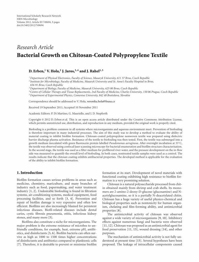
Figure 2: Pressure development in experiment 3. (a) reference, (b) chitosan-coated sample. The discrete character of measured values iscaused by digitalization in the 8 bit A–D converter (256 possible values). The fitted lines show adjacent averaging fit as described in the text.
a piece of untreated PP using sintered glass frit to removelarger suspended solids, thus minimizing nonmicrobial filterfouling. A 1,5 g addition of D-glucose (Natura a.s.) wasdissolved to increase the exopolysaccharide production. Fivecircular pieces of textile of 55 mm diameter were fixedinto each chamber. One chamber was used with referencesamples (no treatment), the second with chitosan-coatedsamples. The chambers were connected to the water tank,fixed to the support stand, the pumps were started, andthe pressure values were logged in 10-minute intervals. Toensure similar conditions for all experiments, pH valueswere measured at the beginning of each experiment, andtemperature was occasionally measured too. The starting pHvalues were between 7.45 and 8.03, and the temperaturestabilized in about 2 hours between 27.7◦C and 29.7◦C.The experiments ran from 72 to 117 hours. To verify thatthe pressure development was not caused by nonbiologicalfouling or other phenomena, one run with water disinfectedby addition of methylene blue was conducted. The pressuresduring this run remained constant for four days.
Because of high oscillation of the pressures, the measuredfunctions were fitted with adjacent averaging smoothingcalculated from series of 20 adjacent points. The minimalvalue was calculated as an average of 9 measured valuesaround the minimum. To obtain pressure difference, wesubtracted this minimal value from the final value, whichwas calculated as an average of 9 final values. The finalvalue was chosen because in experiment 3 the pressuresreached their maxima in approximately 2 days and thensignificantly dropped. Since we are interested in long-termstable biofouling, the final value is more important than thetemporary maximum.
2.2. Confocal Laser Scanning Microscopy. For the preparationof samples for microscopy, chitosan-coated and reference
Table 1: Pressure differences for three experiments. Δp1 is for refer-ence sample, Δp2 for chitosan-coated sample. Standard deviationsare displayed.
Exp. # Time/h Start pH Δp1/Pa Δp2/Pa
1 72 8.03 1547± 78 1050± 55
2 96 7.92 702± 67 554± 53
3 117 8.01 1109± 70 664± 66
samples of about 2 cm2 were cut and put together into100 mL of sterile TSB medium (15 g medium per L litre ofdemineralized water; HiMedia). Since chitosan coating couldbe thermally damaged during sterilisation in an autoclave,the samples were sterilised by 70% (v/v) ethanol solution for5 minutes [21]. The medium was inoculated with 1 mL ofGFP labeled Pseudomonas aeruginosa (generously providedby W. Kim, USA) solution with optical density 0.5 ofMcFarland standard and incubated at 33◦C for 22 hours ataerobic conditions with occasional shaking. After that, thesamples were shortly rinsed (shaking) in 100 mL of tap water,which was repeated thrice to remove unattached bacteria.Immediately after that, they were observed via Olympus IX81microscope in CLSM mode, using a 405 nm excitation laser.
3. Results
3.1. Biofouling Device. Three runs on the biofilm testing de-vice were run, and the results are summarized in Table 1.
For experiment 3, graphs of the pressure developmentsare shown (Figure 2), since it was the longest run and thusshows the largest part of the biofouling process including thefinal decline of pressure that we assume is corresponding tothe death phase and partial release of biofilm, as was shownin our previous studies [22].
4 ISRN Microbiology
(a)
10 µm
(b)
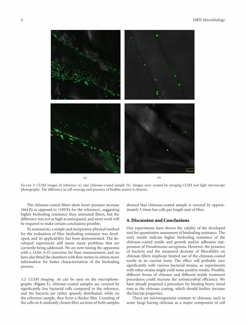
Figure 3: CLSM images of reference (a) and chitosan-coated sample (b). Images were created by merging CLSM and light microscopyphotographs. The difference in cell coverage and presence of biofilm matrix is obvious.
The chitosan-coated filters show lower pressure increase(664 Pa as opposed to 1109 Pa for the reference), suggestinghigher biofouling resistance than untreated filters, but thedifference was not as high as anticipated, and more work willbe required to make certain conclusions possible.
To summarize, a simple and inexpensive physical methodfor the evaluation of filter biofouling resistance was devel-oped, and its applicability has been demonstrated. The de-veloped experiment still meets many problems that arecurrently being addressed. We are now testing the apparatuswith a 14 bit A–D converter for finer measurement, and wehave also fitted the chambers with flow meters to obtain moreinformation for better characterization of the biofoulingprocess.
3.2. CLSM Imaging. As can be seen on the microphoto-graphs (Figure 3), chitosan-coated samples are covered bysignificantly less bacterial cells compared to the reference,and the bacteria are rather sparsely distributed, while onthe reference sample, they form a thicker film. Counting ofthe cells on 8 randomly chosen fibre sections of both samples
showed that chitosan-coated sample is covered by approx-imately 5 times less cells per length unit of fibre.
4. Discussion and Conclusions
Our experiments have shown the validity of the developedtool for quantitative assessment of biofouling resistance. Theearly results indicate higher biofouling resistance of thechitosan-coated textile and growth and/or adhesion sup-pression of Pseudomonas aeruginosa. However, the presenceof bacteria and the measured decrease of filterability onchitosan filters implicate limited use of the chitosan-coatedtextile in its current form. The effect will probably varysignificantly with various bacterial strains, so experimentswith other strains might yield some positive results. Possibly,different forms of chitosan and different textile treatmentprocedures could increase the antimicrobial efficiency. Wehave already prepared a procedure for binding heavy metalions to the chitosan coating, which should further increasethe biocide properties.
There are microorganisms resistant to chitosan, such assome fungi having chitosan as a major component of cell

ISRN Microbiology 5
walls [23]. Also, it is known that some bacteria are efficientproducers of chitosanases—enzymes that attack chitosan byendohydrolysis of beta-1, 4-linkages between D-glucosamineresidues in a partly acetylated chitosan [24]. According tothe study, the production of chitosanases can be significantlyinactivated by the presence of Cu2+ ions. The copper ionsalso have a biocide effect. This might speak for use ofchitosan-coated filters with adsorbed Cu as a material havinghigher antimicrobial effect. We are proceeding to test thishypothesis.
To summarize, these early results support the hypothesisof bacterial growth/attachment suppression and furtherresearch into the application of a thin-film chitosan coatingas an antibiofouling agent, which we are currently conduct-ing.
Abbreviations
A–D: Analog-digitalCLSM: Confocal laser scanning microscopyDCSBD: Diffuse coplanar surface barrier dischargeGFP: Green fluorescent proteinPE: PolyethylenePP: PolypropylenePTFE: PolytetrafluoroethyleneTSB: Tryptone soya broth.
Acknowledgments
This work was supported in part by Project no.KAN101630651 of the Czech Academy of Sciences, by theProject no. 2A-3TP1/126 of the Ministry of Industry andTrade, CZ, by Project no. 9678 of the Ministry of Health, CZ,and by Project 1M0528 (activity 88VM01) of the Ministry ofEducation, CZ.
References
[1] C. G. Kumar and S. K. Anand, “Significance of microbialbiofilms in food industry: a review,” International Journal ofFood Microbiology, vol. 42, no. 1-2, pp. 9–27, 1998.
[2] M. Ludensky, “Control and monitoring of biofilms in indus-trial applications,” International Biodeterioration and Biodeg-radation, vol. 51, no. 4, pp. 255–263, 2003.
[3] R. B. Simmons, L. J. Rose, S. A. Crow, and D. G. Ahearn, “Theoccurrence and persistence of mixed biofilms in automobileair conditioning systems,” Current Microbiology, vol. 39, no. 3,pp. 141–145, 1999.
[4] J. S. Baker and L. Y. Dudley, “Biofouling in membrane sys-tems: a review,” Desalination, vol. 118, no. 1–3, pp. 81–89,Conference on Membranes in Drinking and Industrial WaterProduction, 1998.
[5] C. A. Fux, J. W. Costerton, P. S. Stewart, and P. Stoodley, “Sur-vival strategies of infectious biofilms,” Trends in Microbiology,vol. 13, no. 1, pp. 34–40, 2005.
[6] K. Lewis, “Riddle of biofilm resistance,” Antimicrobial Agentsand Chemotherapy, vol. 45, no. 4, pp. 999–1007, 2001.
[7] J. W. Costerton, “Introduction to biofilm,” International Jour-nal of Antimicrobial Agents, vol. 11, no. 3-4, pp. 217–221, 1999.
[8] M. Rinaudo, “Main properties and current applications ofsome polysaccharides as biomaterials,” Polymer International,vol. 57, no. 3, pp. 397–430, 2008.
[9] M. Kong, X. G. Chen, K. Xing, and H. J. Park, “Antimicrobialproperties of chitosan and mode of action: a state of the artreview,” International Journal of Food Microbiology, vol. 144,no. 1, pp. 51–63, 2010.
[10] D. Raafat and H. G. Sahl, “Chitosan and its antimicrobialpotential—a critical literature survey,” Microbial Biotechnol-ogy, vol. 2, no. 2, pp. 186–201, 2009.
[11] J. Rhoades and S. Roller, “Antimicrobial actions of degradedand native chitosan against spoilage organisms in laboratorymedia and foods,” Applied and Environmental Microbiology,vol. 66, no. 1, pp. 80–86, 2000.
[12] S. Roller and N. Covill, “The antifungal properties of chitosanin laboratory media and apple juice,” International Journal ofFood Microbiology, vol. 47, no. 1-2, pp. 67–77, 1999.
[13] S. Roller and N. Covill, “The antimicrobial properties of chi-tosan in mayonnaise and mayonnaise-based shrimp salads,”Journal of Food Protection, vol. 63, no. 2, pp. 202–209, 2000.
[14] F. L. Mi, Y. B. Wu, S. S. Shyu, A. C. Chao, J. Y. Lai, and C. C.Su, “Asymmetric chitosan membranes prepared by dry/wetphase separation: a new type of wound dressing for controlledantibacterial release,” Journal of Membrane Science, vol. 212,no. 1-2, pp. 237–254, 2003.
[15] E. I. Rabea, M. E. T. Badawy, C. V. Stevens, G. Smagghe, andW. Steurbaut, “Chitosan as antimicrobial agent: applicationsand mode of action,” Biomacromolecules, vol. 4, no. 6, pp.1457–1465, 2003.
[16] N. R. Sudarshan, D. G. Hoover, and D. Knorr, “Antibacterialaction of chitosan,” Food Biotechnology, vol. 6, no. 3, pp.257–272, 1992.
[17] Y. C. Chung and C. Y. Chen, “Antibacterial characteristics andactivity of acid-soluble chitosan,” Bioresource Technology, vol.99, no. 8, pp. 2806–2814, 2008.
[18] S. Kaplan, “Plasma processes for wide fabric, film and non-wovens,” Surface and Coatings Technology, vol. 186, no. 1-2,pp. 214–217, 2004.
[19] J. Rahel, V. Prochazka, M. Zahoran, and D. Erben, “Removalof copper metal ions from aqueous solutions by plasmamade chitosan filter,” Chemicke Listy, vol. 102, no. 16, pp.1432–1435, 2008.
[20] M. Simor, J. Rahel’, P. Vojtek, M. Cernak, and A. Brablec,“Atmospheric-pressure diffuse coplanar surface discharge forsurface treatments,” Applied Physics Letters, vol. 81, no. 15, pp.2716–2718, 2002.
[21] W. A. Rutala, D. J. Weber, and Healthcare Infection ControlPractices Advisory Committee (HICPAC), “Guideline for dis-infection and sterilization in healthcare facilities,” 2008, http://www.cdc.gov/hicpac/Disinfection Sterilization/toc.html.
[22] V. Hola, F. Ruzicka, and M. Votava, “The dynamics of Staph-ylococcus epidermis biofilm formation in relation to nutrition,temperature, and time,” Scripta Medica Facultatis MedicaeUniversitatis Brunensis Masarykianae, vol. 79, no. 3, pp.169–174, 2006.
[23] C. R. Allan and L. A. Hadwiger, “The fungicidal effect ofchitosan on fungi of varying cell wall composition,” Exper-imental Mycology, vol. 3, no. 3, pp. 285–287, 1979.
[24] X. A. Gao, W. T. Ju, W. J. Jung, and R. D. Park, “Purificationand characterization of chitosanase from Bacillus cereus D-11,” Carbohydrate Polymers, vol. 72, no. 3, pp. 513–520, 2008.