Embed Size (px)
Citation preview
Vol. 40: 259-265. 1987 MARINE ECOLOGY - PROGRESS SERIES Mar. Ecol. Prog. Ser.
Published October 28
Lipid and exopolysaccharide production during hydrocarbon growth of a marine bacterium from
the sea surface
Madeleine Goutx, Stephanie Mutaftshiev, Jean-Claude Bertrand
Centre d'oceanologie de Marseille, URA 41, Facult6 des Sciences de Luminy, F-13288 Marseille Cedex 9, France Laboratoire de Chimie et de Biochimie Moleculaire, Centre National de la Recherche Scientifique, Chernin Joseph Aiguiers.
13009 Marseille. France
ABSTRACT: The marine bacterium Alcaligenes sp. PHY 9 L.86 was isolated from hydrocarbon-polluted sea-surface waters and grown on 0.1 % tetradecan in batch cultures. Lipid composition of cell pellets and supernatants were examined throughout growth, using thin layer chromatography coupled with flame ionization detection Cellular and extracellular carbohydrate and protein contents were esti- mated. Bacterial growth on hydrocarbon induced the production of extracellular emulsifying agents (biosurfactants). Formation of foams was observed in the culture medium at early stationary phase; it was related to high emulsifying activity and maximum extracellular lipid production (in particular, free fatty acids and triglycerides). Specific staining for acid polysaccharides revealed the formation of exopolysaccharid fibers associated with vesicles, the size of which depended on the growth phase. Surface-active agents produced by Strain PHY 9 L.86 explain the foam formation. Our results stress the role of biosurfactants in the biodegradation of hydrocarbon in the marine environment.
INTRODUCTION carbon substrate; it was therefore selected for detailed investigation of the production of en~ulsifying agents.
In coastal areas of the Gulf of Marseille (France), In this study, a qualitative and quantitative analysis which is chronically polluted with hydrocarbons, foams of compounds released into the culture medium by can frequently be observed at the sea surface. The Strain PHY 9 L.86 was performed at various phases of ability of these foams to emulsify crude oil was noted the bacterial culture grown on hydrocarbon substrate. by Rambeloarisoa et al. (1984). Emulsifying agents are responsible for foam formation. They can be either synthetic emulsifiers (detergents) or tensioactive MATERIAL AND METHODS molecules of biological origin (biosurfactants). The fact that detergents have not been detected suggests a Organisms and growth conditions. The bacterial biological origin of the foams. Strain PHY 9 L.86 was identified as Alcaligenes sp.
Studies by Rambeloarisoa et al. (1984) showed that according to the classification system proposed by hydrocarbons and hydrocarbonoclastic bacteria Buchanan & Gibbons (1974). accumulated extensively in these foams (lo7 to 108 Bacteria were grown a t 30 "C in 2 1 Fernback flasks bacteria ml-l). The ability of bacteria growing on hy- containing 300 m1 of mineral salt medium with the drocarbon substrates to produce emulsifying agents is following composition (in g 1-' distilled water): Tris well known (Rosenberg 1986). Thus, the production of (hydroxymethyl amino methane) (6.05), NaC1 (23), KC1 surface-active molecules by bacteria could be an im- (0.?5), CaClz (1.47), NH,C1 (3.?4), MgS04 .7H20 (12.3), portant factor involved in foam formation. A mixed NaHPO, (0.4 mM). The pH was adjusted to 7.8 with bacterial community (EM4) composed of 8 bacterial Titrosol (Merck) (4 N). strains which degraded crude oil very effectively was Tetradecane (0.1 % , v/v) was provided as sole carbon isolated from foams (Rambeloarisoa e t al. 1984). and energy source. Aeration was provided by agitation Among this community, the marine Strain PHY 9 L.86 on a reciprocal shaker (96 rpm). Growth was monitored exhibited a maximum efficiency for degrading hydro- by following absorbance at 450 nm with a Shimadzu
8 Inter-Research/Printed in F. R. Germany
260 Mar Ecol. Prog. Ser
UV-visible spectrophotometer, by viable count estimates on agar medium plates (f~ltered seawater enriched with 15 g I-' Bjo-Trypcase [Merieux], 5 g 1-' Phytone [Merieux], 15 g 1-' Agar [Difco]) or by estimat- ing the protein content using the method of Lowry et al. (1951).
Whole contents of the flasks were harvested at vari- ous phases of growth, either for electron microscopical studies or chemical analyses.
Emulsifying activity of the 6 0 0 0 ~ g supernatant was measured as a function of the degree of stability of an emulsion obtained after 5 rnin mechanical agitation of a reaction system or after exposure to sonic oscillation (Zosim et al. 1982) with 2 impulses for 1 rnin at 9 kHz each. The reaction system contained a determined quantity of supernatant corresponding to an amount of 0.09 m g total sugars (estimated according to the method of Dubois et al. 1956) (the quantity of liquid used could vary between culture media, but the sugar concentration remained constant), and 0.1 m1 of tetra- decane and mineral salt medium without NH4C1 to a final volume of 10 ml. The stability of the emulsion over time was measured at 610 nm (Roy et al. 1979) at 20 "C. A similar reaction system prepared from synthetic sea- water (i.e. without biosurfactants) served as control.
Electron microscopy. Ahquots of cultures were sam- pled under the tetradecane upper layer and prepared for electron microscopy analysis. The techniques used were adapted to allow for the high fragility of the surface materials (exopolysaccharides, vesicules, etc). Since the classical methods of preparation for electron microscopic observation of thin sections turned out to be inappropriate, we used either negative staining with 1 ?'o potassium phosphotungstate (PTA) and 1 O/O uranyl acetate (UA), with or without previous stabilization of the bactenal surface structures with ruthenium red/ glutaraldehyde (G/RR/UA) which specifically reveals acidic polysaccharides (Mutaftshiev et al. 1982). A car- bon-coated Formvar grid was deposited on top of a drop of a bactenal cell suspension culture, and left (2 min) for adsorption of the bacteria and their exostruc- tures on the carbon surface. Free-standing material was removed by blotting with filter paper. Grids were treated with a mixture (v/v) of 1 % glutaraldehyde and 1 % ruthenium red, and then contrasted with 0.1 O/O uranyl acetate. Grids were stained for 5 rnin and examined with a Hitachi 600 electron microscope at 75 kV.
Analytical methods. Centrifugation ( 6 0 0 0 ~ g for 15 min, twice, at 5 "C) of cultures ylelded 2 fractions desi.g- nated cell pellets and supernatant. The 2 freeze-dried fractions were analysed for protein, carbohydrate and lipid content. Protein was estimated according to the method of Lowry'et al. (1951) with bovine serum albu- min as standard. Carbohydrate was estimated using
the phenol-sulfuric acid method of Dubois et al. (1956). h p i d was extracted according to the method of Bligh &
Dyer (1959). Aliquots of the lyophilized fractions were suspended in 0.8 volumes of distilled water, 1 volume of chloroform and 2 volumes of methanol. The mixture was stirred overnight under nitrogen. After addition of 1 volume chloroform and 1 volume distilled water and stirring for 10 min, the mixture was filtered through GF/C glass fiber filters. Filtrates were allowed to sepa- rate into 2 phases. The bottom phase was collected as the Lipid extract. The filter was again extracted using the same procedure and the extracts combined.
Lipids were separated by thn-layer chromatography coupled with flame ionization detection (TLC/FID) using a Iatroscan apparatus TH 10 (Iatron Laboratories, Tokyo). The Iatroscan was operated wlth a hydrogen flow of 75 kg cm-2, a n air flow of 2000 m1 min-' and a scanning speed of 0.32 cm S-' (30 tooth gear). The Iatroscan was connected to a Shimadzu-chromatopac CRlB Integrator. Lipid classes were separated on chromarods S I1 using several developments and par- tial scans, as proposed by Pamsh & Ackman (1983). In the first development (40 rnin hexane-diethyl ether- formic acid 99: 1 : 0.1), the less polar lipids (hy- drocarbons [HC], wax esters [WE] and free fatty acids [FFA]) moved away from the point of application. There were scanned and the scan was stopped manu- ally. The remaining neutral Lipids (triacyglycerides [TG]; fatty alcohols [Alc]; diacylglycerides [DG]; sterols [ST]) were separated in the second development (40 rnin in hexane-diethyl ether-formic acid 80 : 20 : 0.1). Again, a partial scan was performed. This was followed by a short development in acetone (3 cm above the origin). This development separates monoglycerides, glycolipids and pigments from total phospholipids which do not move in acetone. In this set of samples, the pigments were visibly separated from monogly- cerides and glycolipides. Lipids were identified on the basis of their ability to CO-chromatograph with authen- tic standards purchased from Sigma LTP Corp. Lipids were quantified with reference to calibration curves performed for each rod and each class of compound. Each rod was considered as an isolated analytical unit as recommended by Delmas et al. (1984). Calibrations were performed at 5 levels with loads in the range 0.5 to 20 klg.
RESULTS
Bacterial growth and emulsifying activity
No growth of Strain PHY 9 L.86 was obtained at sodium chloride concentrations between 0 and 100 mM. Maximum growth was achieved at a sodium
Goutx et al.: Lipid and exopolysaccharide production in a marine bacterium 261
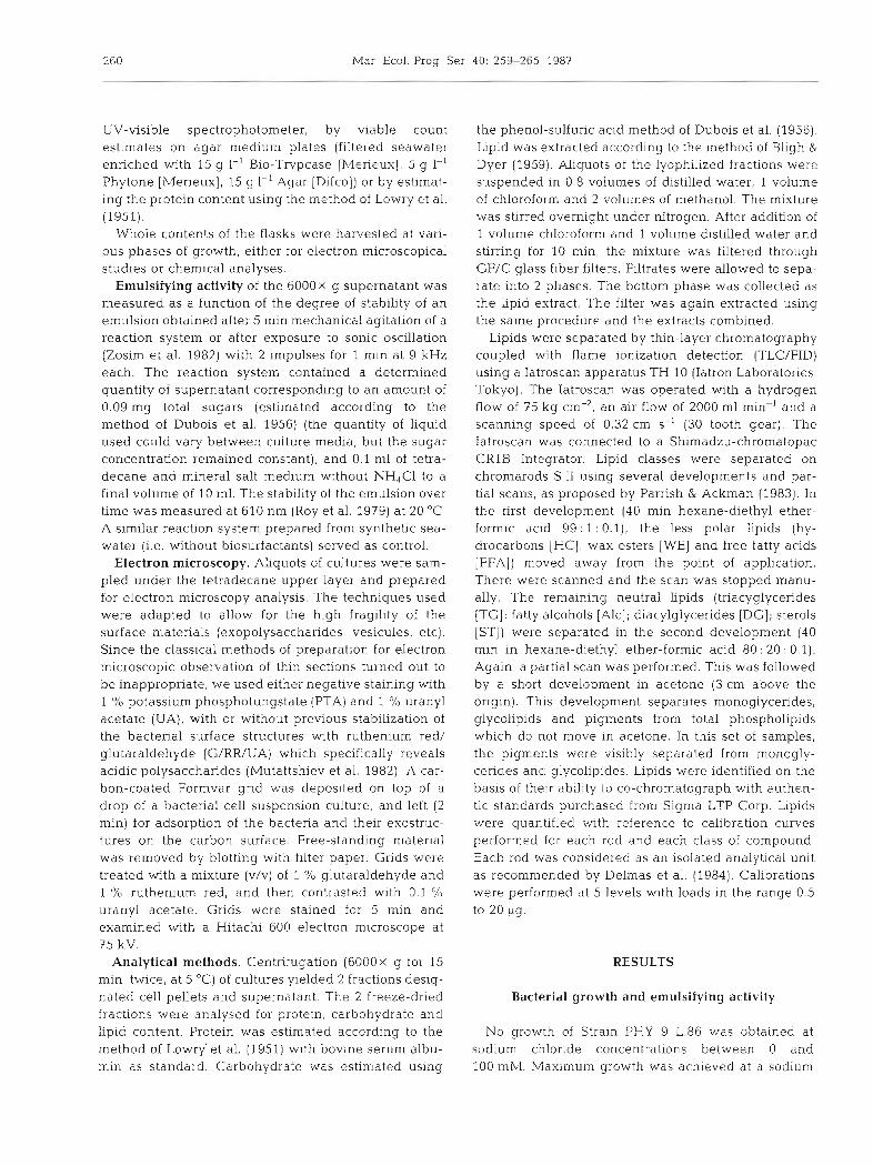
chloride concentration of 400 mM; Alcaligenessp. PHY strate, the optimum rate of cell yield was achieved 9 is thus a marine bacterium according to the definition between T3 and T,. Foaming appeared at Tg and of Larsen (1986). Typical growth curves are given in remained stable in cultures until the end of the statio- Fig. 1 Cultures were harvested at log phases (T1, T2), nary phase.
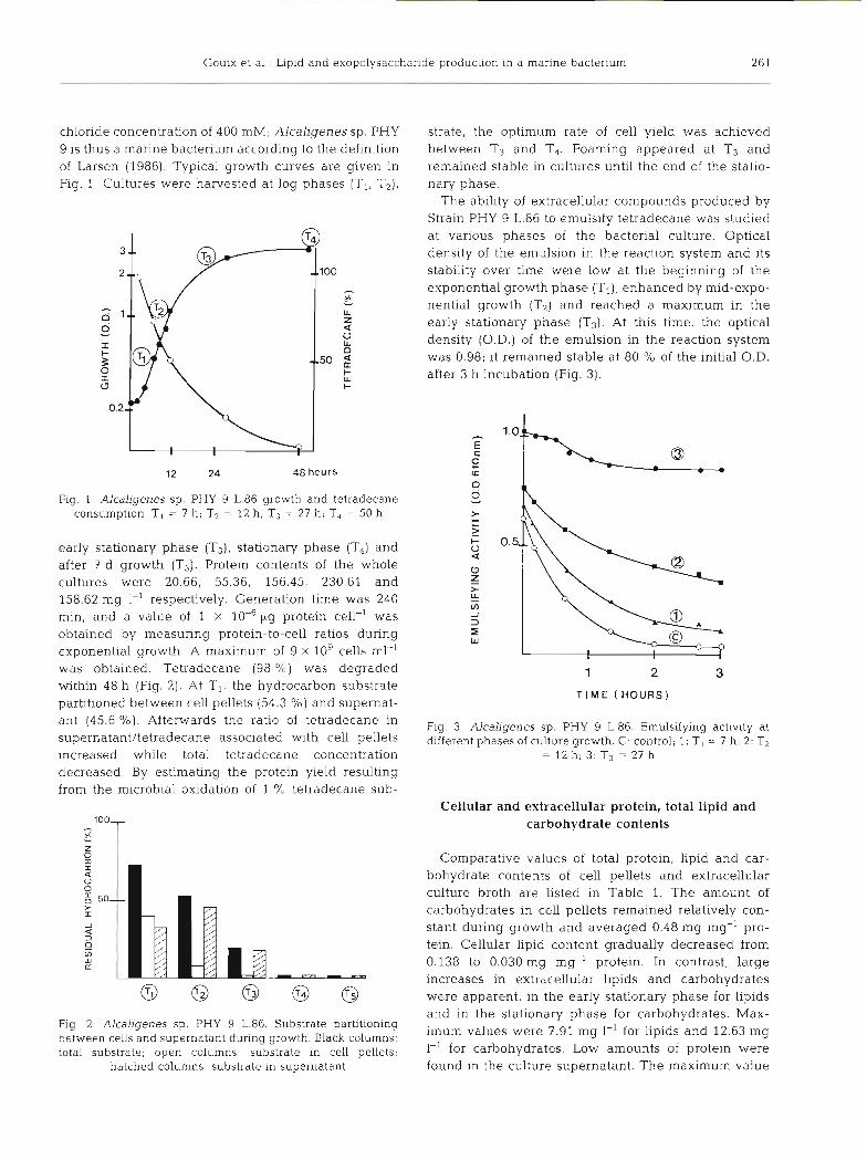
The ability of extracellular compounds produced by Strain PHY 9 L.86 to emulsify tetradecane was studied at various phases of the bacterial culture. Optical density of the emulsion in the reaction system and its stability over time were low at the beginning of the exponential growth phase (T,), enhanced by mid-expo- nential growth (T2) and reached a maximum in the early stationary phase (T3). At this time, the optical density (O.D.) of the emulsion in the reaction system was 0.98; it remained stable at 80 O/O of the initial O.D. after 3 h incubation (Fig. 3).
12 24 48 hours
Fig. 1 Alcaligenes sp. PHY 9 L.86 growth and tetradecane consumption T1 = 7 h; T2 = 12 h ; T j = 27 h ; T4 = 50 h
early stationary phase (T3), stationary phase (T4) and after 7 d growth (T5). Protein contents of the whole cultures were 20.66, 55.36, 156.45, 230.61 and 158.62 mg 1-' respectively. Generation time was 240 min, and a value of 1 X lo4 pg protein cell-' was 3 1
Z obtained by measuring protein-to-cell ratios during W
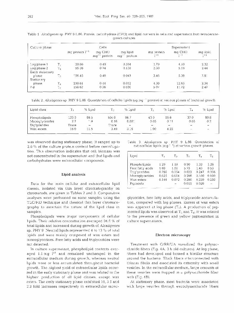
exponential growth. A maximum of 9 X 10' cells ml-' was obtained. Tetradecane (98 %) was degraded 1 2 3 within 48 h (Fig. 2). At T1, the hydrocarbon substrate
T I M E ( H O U R S ) partitioned between cell pellets (54.3 %) and supernat- ant (45'6 %)' Afterwards the ratio of tetradecane in Pig, 3, Alc-ljgenes sp. PHY CJ L.86. Emulsifying activity at supernatant/tetradecane associated with cell pellets different phases of culture growth. C: control; 1: T, = 7 h ; 2: T2 increased while total tetradecane concentration = 12 h; 3: T3 = 27 h
decreased. By estimating the protein yield resulting from the microbial oxidation of 1 % tetradecane sub-
Cellular and extracellular protein, total lipid and carbohydrate contents
Comparative values of total protein, lipid and car- bohydrate contents of cell pellets and extracellular culture broth are listed in Table 1. The amount of carbohydrates in cell pellets remained relatively con- stant during growth and averaged 0.48 mg mg-' pro- tein. Cellular lipid content gradually decreased from 0.138 to 0.030 mg mg-' protein. In contrast, large increases in extracellular lipids and carbohydrates were apparent, in the early stationary phase for lipids and in the stationary phase for carbohydrates. Max-
Fig 2. Alcaligenes sp. PHY 9 L.86. Substrate partitioning between cells and supernatant during growth. Black columns: iillum values were 7.91 mg 1-I for lipids and 12.63 mg
total substrate; open columns: substrate in cell pellets; 1-I for protein were hatched columns: substrate in supernatant found in the culture supernatant. The maximum value
262 Mar. Ecol. Prog. Ser. 40: 259-265, 1987
Table 1 AlcaLigenes sp. PHY 9 L.86. Protein, carbohydrate (CHO) and lipid contents in cells and supernatant from tctrddecane- grown cultures
Culture phase Cells Supernatant rng proteln 1-' mg CH0 rng lipid rng protein mg CH0 mg llpid
mg-' protein mg-' protein I - ' 1-' I- '
Log phase 1 T 1 20.66 0.49 0.104 1.79 4.10 2 32 Logphase 2 T2 55.36 0.74 0.138 2.30 5.18 3.44 Early stationary
phase T3 156.45 0.40 0.043 3.46 5.36 7.91 Stationary
phase T4 230.61 0.44 0.032 4.30 12.63 3.34 7 d T5 158.62 0.36 0.030 5.07 11.72 2.47
Table 2. AlcaLigenes sp. PHY 9 L.86. Quantitation of cellular lipids (vg mg-' protein) at various phases of bacterial growth
Lipid class T 1 O/O lipid T2 % lipid T3 % lipid T4 % lipid
Phospholipids 120.0 86.5 104.0 96.7 43.0 95.6 32 0 99.8 Monoglycerides 2.7 1.9 0.16 0.001 0.05 0.11 0.05 0.2 Diglycerides Trace - Trace - - - - Wax esters 16.0 11.5 3.40 3.16 1.90 4.22 - -
was observed during stationary phase; it ranged up to Table 3. Alcaligenes sp. PHY 9 L.86. Quantitation of 2.6 % of the culture protein content before centrifuga- extracellular lipids (rng l-') at various growth phases
tion. This observation indicates that cell biomass was not concentrated in the supernatant and that lipids and carbohydrates were extracellular compounds.
Lipid analysis
Data for the main cellular and extracellular lipid classes, isolated via thin layer chromatography on chromarods, are given in Tables 2 and 3. Comparative analyses were performed on some samples using the TLC/FID technique and classical thin layer chromato- graphy to ascertain the nature of the lipid class in samples.
Phospholipids were major components of cellular lipids. Their relative concentration averaged 94.6 "/o of total Lipids and increased during growth of Alcaligenes sp. PHY 9. Neutral lipids represented 4 to 13 O/b of total lipids and were mainly composed of wax esters and monoglycerides. Free fatty acids and tnglycendes were not detected.
In culture supernatant, phospholipid contents aver- aged 1.1 mg 1-' and remained unchanged in the extracellular medium during growth, whereas neutral lipids more or less accumulated throughout bacterial growth. The highest yleld of extracellular lipids occur- red in the early stationary phase and was related to the highest production of all lipid classes, except wax esters. The early stationary phase exhibited 10, 3.5 and 2.3 fold increases respectively in extracellular mono-
Lipid Ti T2 T3 T4 Ts
Phospholipids 1.29 1.19 0.90 1.33 1.26 Free fatty acids 1.69 1.50 5.75 1.40 0.53 Triglycerides 0.290 0.254 0 633 0.247 0.338 Monoglycerides 0.025 0.004 0.298 0.100 0.100 Wax esters 0.146 0.872 0.280 0.239 0.250 Pigments - 0.011 0.026 -
glycerides, free fatty acids, and triglyceride accumula- tion, compared with log phases. Excess of wax esters was apparent at log phase (T1). A production of pig- mented lipids was observed at Tg and T,; it was related to the presence of green and yellow pigmentation in culture supernatants.
Electron microscopy
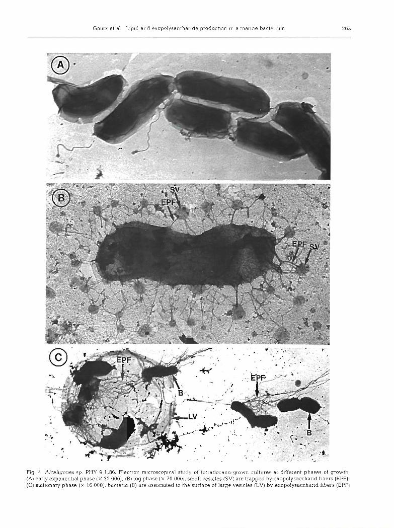
Treatment with G/RR/UA visualized the polysac- charide fibers (Fig. 4A, 3 h old cultures). At log phase, fibers h.ad developed and formed a fibrillar structure around the bacteria. Thick fibers interconnected with thinner fibnls and associated its extremity with small vesicles. In the extracellular medium, large amounts of these vesicles were trapped in a polysaccharide fiber web (Fig. 4B).
At stationa.ry phase, most bacteria were associated with large vesicles through exopolysaccharide fibers
7,:
'
'b
. ?., .
1 ;; , :- :;A , .,:j2:,;
1'
' .
$=
P
1:
264 Mar Ecol. Prog Ser.
(Fig. 4C). Sheets, that collapsed as the preparation was dried, made up the envelope of these large vesicles. The spherical conformation of these large vesicles was confirmed by a freeze-etching study (Mutaftshiev pers. comm.). We plan to investigate these structures further.
Relations between extracellular compounds and morphological structures during cell growth
Comparative studies of extracellular compounds and morphological structures, employing electron micro- scopy, suggest that several types of extracellular fac- tors are involved in tetradecane assimilation during growth of Alcaligenes sp. PHY 9 L.84. (1) At log phase the presence of small vesicles linked to the cells through short exopolysacharid fibers (Fig. 4B) is related to the highest amount of lipids and tetradecane associ- ated with cell pellets (Tables 1 & 2). These vesicles therefore could be microemulsions resulting from emulsification process of the substrate by the lipid compunds. (2) Enhancement of 'emulsified' tetra- decane (Fig. 2 ) , as well of extracellular lipids and car- bohydrates (Table l) , can be related to the dispersion of microemulsions into exopolysaccharid fibrillar matenal in the supernatant. At stationary phase, the presence of large vesicular structures are correlated with the max- imum concentrations of extracellular carbohydrates and pigments (Fig. 4C).
DISCUSSION
The marine bacterium Alcaligenes sp. PHY 9 L.86, isolated from surface foams, appears to be well adapted to tetradecane substrate. The strain released organic compounds into the medium which provided the culture broth with emulsifying activities and foaming proper- ties. These were due to lipids and carbohydrates. Pro- duction of such compounds has been reported for many microorganisms growing on hydrocarbon substrates (reviewed by Rosenberg 1986). Numerous authors con- sidered the emulsification of hydrocarbons by surface- active agents to be an essential step in hydrocarbon biodegradation, particularly in the marine environment (Re~sfeld et al. 1972, Gutnick & Rosenberg 1977, Flood- gate 1984, Bartha 1986, Mattei et al. 1986); however few data exist on detailed characteristics of extracellu- lar lipid classes released by manne bactena.
Using TLC/FID, w e have emphasized the dynamics of lipid production in relation to the rate of hydrocarbon consumption. This analytical technique, whi.ch pro- vides synoptic Lipid class data from small samples, enables an easy and rapid sample work up procedure that we found particularly useful for studying the
repartition and dynamics of lipid production through- out bacterial growth. There exists a good relationship between lipid production and emulsifying activity. Increasing Iipid production is related to increasing emulsifying activity. Very low activity was estimated in the early growth phase when most tetradecane was associated with cells. Thus, direct cell contact with the substrate can be an initiating step at the beginning of growth. The emulsifying activity was enhanced during the bacterial growth and was maximal in the early stationary phase. Extracellular lipid content including fatty acids - already described as good emulsifiers (Rosenberg 1986) - attained maximum values at this stage of growth.
Using electron microscopy with specific staining for acid polysaccharides, we have revealed the precise structural organisation of carbohydrates into large exopolysaccharid fibers. The particular methodology for sample treatment used by us, involving neither centrifugation nor filtration, prevented disruption of fragile structures. Thus, we obtained more details on the structural relations between exopolysaccharids and lipids during the bacterial growth. Exopolysaccharid fibers could act as a n emulsion stabilizer. Maximum exopolysaccharid production occurred in the stationary phase with fibers adhering to large vesicles of mem- brane-type organisation and was obviously related to foam formation. The production of an oil emulsifying agent, polysaccharide in nature, was described by Floodgate (1978) for a manne bacterium which degraded oil rapidly, and by Reisfeld et al. (1972) and Zuckerberg et al. (1979) who characterized the well known 'emulsan' as a n anionic heteropolysaccharide containing fatty acid side chains, synthesized by Acinetobacter calcoaceticus RAG-1 growing in sea- water.
During the growth of Alcaligenes sp. PHY 9 L.86 on tetradecane, we observed that the bacteria pass through various phases in which the physlcal organisa- tion of extracellular factors tends to optimize substrate assimilation. An maximum biodegradation rate was achieved when the microbial population had provided an amount of molecules sufficient to emulsify the sub- strate and to stabilize the emulsion. Very high concen- tration of hydrocarbons (100 to 180 mg I-') were found in the area from which A1~alige~e.s sp. PHY 9 L.86 was isolated. Hence, the ability for this strain to release surface-active compounds during hydrocarbon growth can be considered an important factor in foam forma- tion.
In open seawater, where concentrations of cells are low, an effective emulsification process is unlikely to prevail (Rosenberg 1986). In contrast, In the surface microlayer petroleum hydrocarbons accumulate to a greater extent than in the underlllng water column
Goutx et al.: Lipid and exopolysaccharide production in a marine bacterium 265
(Marty & Saliot 1976, Gear ing & G e a r i n g 1982). An Miles. J. A. R. (ed.) Microbial ecology. Springer-Verlag,
a n n u a l i n p u t of 5 X 10³ t c r u d e o i l i n t h e form of slicks
h a s recently b e e n es t imated by Burns & Saliot (1986) for t h e Medi te r ranean Sea . Numerous hydrocarbon
utilizers h a v e b e e n isolated (Crow e t al. 1976), a n d
recen t s tud ies a p p e a r to demons t ra te tha t this biomass
is highly act ive (Carlucci e t al. 1985, William e t al .
1986). Therefore , i n this par t of the mar ine environ-
m e n t , the emulsification process that w e observed in vitro c a n b e effective a n d m a y represent a n advan tage
for microorganisms growing on insoluble substra tes .
Moreover , the inpu t of surface-active compounds in this a rea could modify the physical characteristics of
the ocean surface. Fur ther investigations a r e n e e d e d to
quantify this particular aspect of microbial activity i n
the sea surface microlayer.
Acknowledgements We gratefully acknowledge the assist- ance of Dr Richard (Institut Pasteur, Paris) in the identification of Alcaligenes sp. This work was supported by a grant from Elf Aquitaine Society.
LITERATURE CITED
Bartha, R. (1986). Biotechnology of petroleum pollutant biodegradation. Microb. Ecol. 12: 155-172
Bligh, E. G., Dyer, W J. (1959). A rapid method of total lipid extraction and purification. Can. J. Biochem. Physiol. 37: 911-917
Buchanan, R. E., Gibbons, N. E. (ed.) (1974). Bergeys's manual of determinative bacteriology, 8th edn. Williams and Wil- kins, Baltimore
Burns, K. A., Saliot. A. (1986). Petroleum hydrocarbons in the Mediterranean Sea: a mass balance. Mar Chem. 20. 141-157
Carlucci, A F., Craven, D. B., Henrich, S. M. (1985). Surface fllm mlcroheterotrophs: amino acid metabolism and solar radlatlon effects on their activities. Mar. Biol. 85: 13-22
Crow, S. A , Cook, W. L., Ahearn, D. G., Bourquin, A M' (1976). llicrobial population in coastal surface slicks. In: Sharplcy J. M , Kaplan, A . M. (ed.) Proceedings of the 3rd International Biodegradation Symposium. Applied Science Publishers, London, p. 93-98
Delmas, R. P., Parrish, C. C . , Ackman, R. G. (1984). Determi- nation of lipid class concentrations in sea water by thin- layer chromatography with flame ionization detection. Analyt. Chem. 56: 1272-1277
Dubois, M., Gilles, K. A., Hamilton, J. K., Rebers, P. A., Smith, F. (1956). Colorimetric method for determination of sugars and related substances. Analyt. Chem. 28: 350-356
Floodgate, G. D. (1978). The formation of oil emulsifying agents in hydrocarbonclastic bacteria. In: Loutit, M. W.,
Berlin. p. 82-85 Floodgate. G. D. (1984). The fate of petroleum in marine
ecosystems. In: Atlas, R. M. (ed.) Petroleum microbiology. MacMillan, New York. p. 355-397
Gearing, P. J., Gearing, J. N. (1982). Transport of no. 2 fuel oil between water column, surface microlayer and atmos- phere in controlled ecosystems. Mar environ. Res. 6 : 133-143
Gutnik, D., Rosenberg, E. (1977). Oil tankers and pollution: a microbiological approach. A. Rev. Microbiol. 31: 379-396
Larsen, H. (1986). Halophilic and halotolerant microorgan- Isms An overvlew and historical perspect.ive. FEMS (Fed- eration of European microbiological Societies) Microb. Rev. 39- 3-7
Lowry, 0. H., Rosebrough, N. J., Farr, A. L. , Randall, R. J . (1951). Proteln measurement with the Folin phenol rea- gent. J. biol Chem. 95: 2102-2107
Marty, J . C., Saliot, A. (1976). Hydrocarbons (normal alkanes) in the surface microlayer of sea water. Deep Sea Res. 23: 863-873
Mattei, G., Rambeloarisoa, E., Giusti, G., Rontani, J. F., Bertrand, J . C. (1986). Fermentation procedure of a crude oil in continuous culture on sea water. Appl. Microbiol. Biotechnol. 23: 302-304
Mutaftshiev. S., Vasse, J., Truchet. G. (1982). Exostructures of Rhizobiurn meliloti. FEMS (Federation of European Mi- crobiological Societies) Microb. Letters 13: 171-175
Parrish, C. C., Ackman, R. G. (1983). Chromarod separations for the analysis of marine lipid classes by Iatroscan thin- layer chromatography flame ionization detection. J. Chromat. 262: 103-1 12
Rambeloarisoa, E., Rontani, J. F., Ciusti, G., Duvjnak, Z.. Bertrand, J. C. (1984). Degradation of crude oil by a mixed population of bacteria isolated from sea surface foams. Mar Biol. 83: 69-81
Relsfeld, A.. Rosenberg, E., Gutnick, D. (1972). Microbial degradation of crude oil: factors affecting the dispersion in sea water by mixed and pure cultures. Appl. Microbiol. 24: 363-368
Rosenberg, E . (1986). Microbial surfactants. CRC Crit. Rev Biotechnol 3: 109-132
Roy, P. K., Singh, H. D., Bhagat, S. D., Baruah, J . N. (1979). Characterization of hydrocarbon emulsification and sol- ubilization occurring during the growth of Endomycops~s Iipolytlca on hydrocarbon. Biotechnol. Bioeng. 21: 955-974
William, P. M., Carlucci. A. F., Henrich, S. M,, van Veet, E. S. , Horrigan, F. G. O., Red. F. M. H., Robertson, K. J. (1986). Chemical and microbiological studies of sea surface film in the southern Gulf of California and of the West Coast of Baja California. Mar. Chem. 19: 17-98
Zosim, Z., Gutnick, D., Rosenberg, E. (1982). Properties of hydrocarbon in water emulsions stab~lized by Acinetobac- ter RAG-l emulsan. Biotechnol. Bioeng. 24: 281-292
Zuckerberg, A.. Diver, A., Peen, Z., Gutnick, D. L., Rosenberg, E. (1979). Emulsifier of Artl~robacter RAG-l. chemical and physical properties. Appl. environ. Microbiol. 37: 414420
This article was presented by Dr A. Bianchi; it was accepted for printing on August 5, 1987