Embed Size (px)
Citation preview
1
Republic of the Philippines Department of Health
OFFICE OF THE SECRETARY
Interim Guidelines No. 22
Clinical Management of Suspected and Confirmed Human Pandemic (H1N1) 2009 Infection
This document is intended for use by health care practitioners who manage suspected
or confirmed cases of pandemic (H1N1) 2009 infection. It contains clinical management
guidelines for the following:
1. Outpatient treatment 2. Management of hospitalized cases
It highlights areas of care critical in the management of pandemic (H1N1) 2009 infection
and is not intended to replace routine care. Appropriate infection control measures
should be adhered to at all times.
Section Page
Management in the Outpatient Setting
I. Case Definitions 3
II. When to Perform Swabbing 3
III. Clinical Management in the Outpatient Setting 4
IV. Criteria for Admission 5
Management of Hospitalized Patients
I. When to Suspect Pandemic (H1N1) 2009 Infection among Hospitalized Patients 6
II. Diagnostic Work-up 6
III. General Treatment Considerations 7
IV. Antiviral Therapy 8
V. Criteria for Discharge 10
VI. Infection Control Measures 11
VII. Post-mortem Care 12
VIII. List of Annexes 13
_________________________________________________________________________________________________________
Bldg. 1, San Lazaro Compound, Rizal Avenue, Sta. Cruz, 1003 Manila · Trunk Line 743-8301 Direct Line: 711-9501 Telefax: 743-1829; 743-1786 · URL: http:/www.doh.gov.ph: email: [email protected]
2
Annexes
Annex 1. Guidelines on Collection and Handling of Specimens 14
Annex 2. Guidelines for Home Care 17
Annex 3. Guidelines on the Management of Community-Acquired Pneumonia in Adults (CAP)
18
Annex 4. Guidelines on the Management of Children Hospitalized for Pediatric Community-Acquired Pneumonia (PCAP)
25
Annex 5. Guidelines on the Management of Sepsis in Adults and Children 29
Annex 6. Guidelines on the Management of Pregnant Women with Confirmed or Suspected Pandemic (H1N1) Infection
33
Annex 7. Algorithm for the Clinical Management of Hospitalized Patients Suspected and Confirmed Cases of Influenza A (H1N1) 2009 Infection
38
Annex 8. Patient Care Checklist for Pandemic (H1N1) 39
Annex 9. Post-mortem care 40
Annex 10. Task Force on the Clinical Management of Pandemic (H1N1) 2009 Infection 42
Annex 11. References 44
Tables and Figures
Table 1: Management of Suspected or Confirmed Cases in the Outpatient Setting 4
Table 2A: Dosing Recommendations for Oseltamivir in the Treatment or Chemoprophylaxis of Pandemic (H1N1) 2009 Infection
9
Table 2B: Dosing Schedule of Oseltamivir in Renal Insufficiency 9
Figure 1: Collection of nasopharyngeal swab specimen 14
Figure 2: Collection of oropharyngeal swab specimen 15
Table 3: Classification of Community-Acquired Pneumonia by Risk Stratification 18
Figure 3. Algorithm for the Management-Oriented Risk Stratification of CAP in Immunocompetent Adults
19
Table 4: Risk Stratification and Empiric Antibiotic Therapy for CAP 21
Table 5: Usual Recommended Dosages of Antibiotics in 50-60 KBW Adults with Normal Liver and Renal Functions
22
Table 6: Suggested Initial Antibiotic Regimens for Pneumonia in Children 27
Table 7: Dosage of Antibiotics for Pneumonia in Children 27
Table 8: Systemic Inflammatory Response Syndrome (SIRS) Criteria 29
3
MANAGEMENT IN THE OUTPATIENT SETTING
Most cases have had uncomplicated illness of limited duration. Hospitalization is therefore not required for a great majority of patients who fulfil the case definition below.
I. CASE DEFINITIONS
Case Under Observation (CUO) or suspected case of pandemic (H1N1) 2009:
A person presenting with fever (temp 37.80C or higher) AND typical acute respiratory influenza-like illness (e.g., cough, sore throat, rhinorrhea; others - body aches, headache, fatigue, vomiting and diarrhea) in the absence of a KNOWN cause other than influenza
Confirmed pandemic (H1N1) 2009 case:
A symptomatic patient whose respiratory specimen was reported as positive for pandemic (H1N1) 2009 virus by the Research Institute for Tropical Medicine (RITM) or other accredited government or private laboratories
Close contact:
Defined as having cared for or lived with a suspected or confirmed case, or having been in a setting where there is high likelihood of exposure to respiratory droplets from infected persons within a distance of 3 to 6 feet (1 to 2 meters).
II. WHEN TO PERFORM SWABBING
In general, the swabbing of outpatient cases is not recommended.
As part of DOH surveillance, a nasopharyngeal or oropharyngeal swab may be obtained for the following (based on DOH Interim Guidelines No. 16: “Major Policy Changes from Containment to Mitigation Response to the Influenza A (H1N1) Virus Threat”):
� CUOs identified at various ports of entry in the country � Investigation of first suspected cases of influenza-like illness (ILI) in a specific
community or institution experiencing an initial cluster of ILI cases:
• A purposive sampling for nasopharyngeal or oropharyngeal swabbing to determine whether that cluster is infected with pandemic (H1N1) 2009 can be conducted among:
1. The first 5 to 10 cases that were reported during an outbreak among Philippine Integrated Disease Surveillance and Response (PIDSR) surveillance sites and sentinel sites doing lab-based surveillance (refer to DOH Interim Guidelines No. 21: “On the Shift of Reporting from Influenza A (H1N1) Enhanced Surveillance to ILI Surveillance”); or, 2. The first 5 to 10 cases that consulted for ILI at a health facility
4
� Random sampling of persons in clusters (at least 2 cases) of ILI with unusual symptoms or severity (i.e. severe acute respiratory infection or SARI)
� Investigation of ILI in persons at high risk of developing complications because of
other medical conditions or problems Refer to Annex 1. Guidelines on Collection and Handling of Specimens
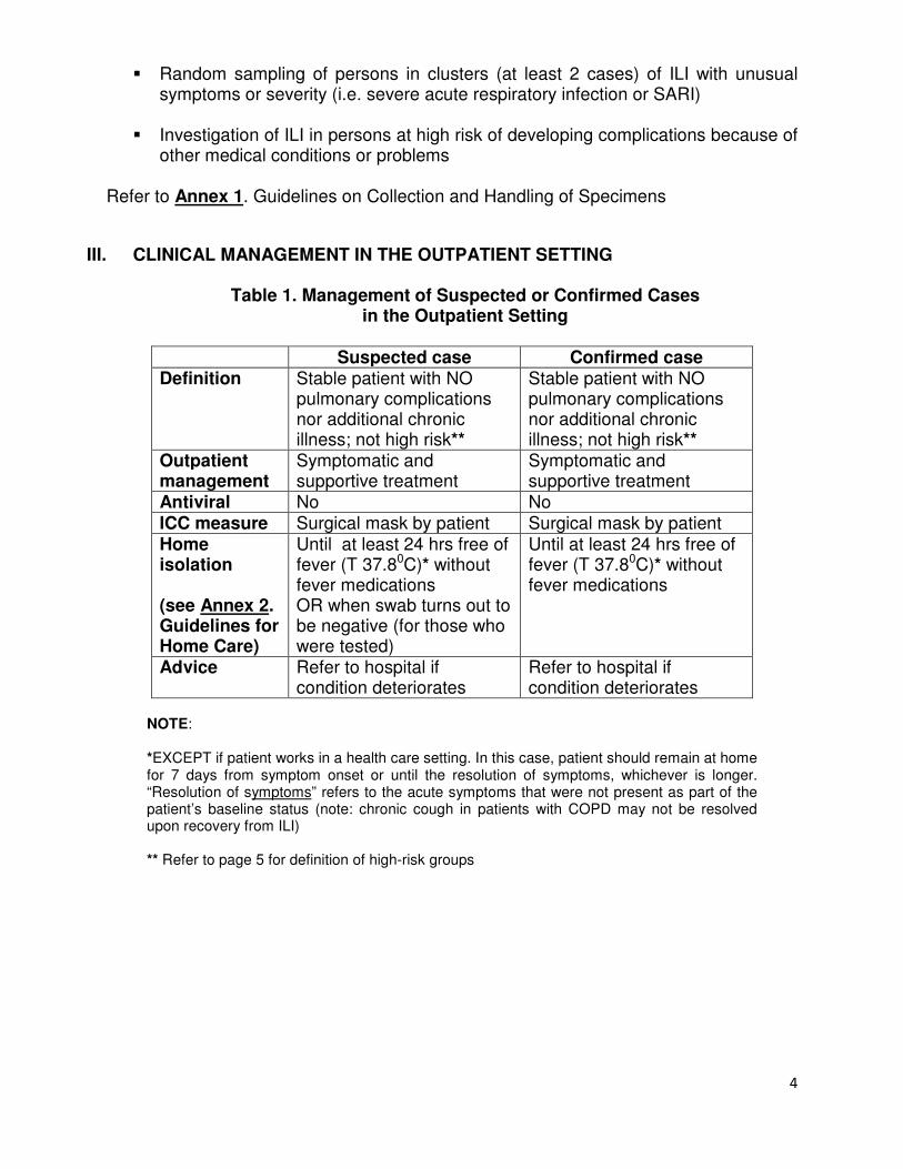
III. CLINICAL MANAGEMENT IN THE OUTPATIENT SETTING
Table 1. Management of Suspected or Confirmed Cases
in the Outpatient Setting
Suspected case Confirmed case Definition Stable patient with NO
pulmonary complications nor additional chronic illness; not high risk**
Stable patient with NO pulmonary complications nor additional chronic illness; not high risk**
Outpatient management
Symptomatic and supportive treatment
Symptomatic and supportive treatment
Antiviral No No ICC measure Surgical mask by patient Surgical mask by patient Home isolation (see Annex 2. Guidelines for Home Care)
Until at least 24 hrs free of fever (T 37.80C)* without fever medications OR when swab turns out to be negative (for those who were tested)
Until at least 24 hrs free of fever (T 37.80C)* without fever medications
Advice Refer to hospital if condition deteriorates
Refer to hospital if condition deteriorates
NOTE: *EXCEPT if patient works in a health care setting. In this case, patient should remain at home for 7 days from symptom onset or until the resolution of symptoms, whichever is longer. “Resolution of symptoms” refers to the acute symptoms that were not present as part of the patient’s baseline status (note: chronic cough in patients with COPD may not be resolved upon recovery from ILI) ** Refer to page 5 for definition of high-risk groups
5
IV. CRITERIA FOR ADMISSION
The following are indications for hospitalization among patients presenting with ILI:
1. Unstable condition – dyspnea, hypoxemia, hemodynamic instability
(e.g.,hypotension, tachycardia), altered level of consciousness and confusion,
syncope, severe dizziness, etc
2. Signs of sepsis and/or pneumonia (see Annex 3. Management of Community-
Acquired Pneumonia or CAP, Annex 4. Management of Pediatric CAP and
Annex 5. Management of Sepsis in Adults and Children)
3. Inability to eat and/or take oral fluids
4. Urgent need for work-up for alternative/additional diagnoses
5. Presence of unstable/uncontrolled co-morbidities which increase the risk of
severe illness - chronic pulmonary (including asthma), cardiovascular (except
hypertension), renal, hepatic, hematologic, neurologic or metabolic disorders
(including diabetes mellitus), immunosuppression, cancer, malnutrition
The following high-risk groups need not be admitted but will require close
monitoring for possible complications. They may be given oseltamivir based on
clinician’s evaluation:
1. Patients age < 5 yrs and > 60 yrs, without co-morbidities
2. Pregnant women (see Annex 6. Management of Pregnant Women with
Confirmed or Suspected Pandemic (H1N1) 2009 Infection)
3. Those with stable co-morbidities
4. Obese patients – defined as body mass index [BMI] more than 30
6
MANAGEMENT OF HOSPITALIZED PATIENTS
Refer to Annex 7. Algorithm for Management of Hospitalized Patients
I. WHEN TO SUSPECT PANDEMIC (H1N1) 2009 INFECTION AMONG HOSPITALIZED PATIENTS
Consider pandemic (H1N1) 2009 infection in hospitalized patients admitted for other
reasons who:
� have history of acute influenza-like illness [manifesting as fever (temp 37.80C or
higher) plus cough, sore throat, stuffy or runny nose, muscle aches, GI
symptoms] OR
� have moderate/severe or non-resolving or unusual manifestations* of pneumonia
(see Annex 3 and Annex 4) or sepsis (see Annex 5 ) OR
� have contact or exposure in the past 10 days to a confirmed or suspect case of
pandemic (H1N1) 2009 OR
� history of travel abroad or to an affected area in the Philippines
*unusual manifestations = negative radiologic evidence of pneumonia but with
clinical signs such as inspiratory rales, rhonchi, and wheezes; interstitial pattern of
infiltrates; non-resolving or progressing pneumonia even after 72 hours of CAP
management; pneumonia with extrapulmonary manifestations; ILI or pneumonia with
unexplained seizures or mental status changes (especially in children)
II. DIAGNOSTIC WORK-UP
1. Nasopharyngeal and oropharyngeal swabs for H1N1 Real Time - Polymerase Chain Reaction (RT-PCR) (Annex 1. Guidelines on Collection and Handling of Specimens)
2. For intubated patients, at least 1-2 ml of endotracheal aspirate (ETA) for H1N1 RT-PCR should be obtained in addition to a nasopharyngeal swab and placed in the same virus transport medium (VTM) as the nasopharyngeal swab
3. Chest x-ray
4. Pulse oximetry and/or ABGs
5. Complete blood count with platelet count
6. Other recommended laboratory tests/procedures, as indicated, include:
a. Blood culture/sensitivity b. Gram stain and culture/sensitivity of sputum (for older children and adults)
7
7. Additional tests may be done, taking into account underlying co-morbidities and alternative diagnoses (e.g. creatinine and electrolytes for those with diarrhea; and BUN and creatinine for those with renal problems)
III. GENERAL TREATMENT CONSIDERATIONS (Refer to Annex 8 Patient Care
Checklist)
1. Supportive care in the form of antipyretics (paracetamol for fever or pain) and
fluids for rehydration should be provided.
2. Salicylates should not be given.
3. Signs of possible clinical deterioration such as difficulty in breathing, chest pain,
altered level of consciousness and confusion should be watched for.
4. Oxygen therapy should be given to correct hypoxemia.
5. If pneumonia is considered, antimicrobial therapy and other measures should be
started in accordance with guidelines for empiric treatment of community-
acquired pneumonia (Refer to Annex 3 and Annex 4).
6. Seasonal influenza and past pandemics have been associated with an increased
risk of staphylococcal pneumonia, so this association should be considered in the
choice of antimicrobials (Refer to Annex 3 and Annex 4). Wherever possible,
results of microbiologic studies should be used to guide continued therapy for
suspected bacterial co-infection.
7. If signs of sepsis are noted, the guidelines for management of sepsis should be
followed (Annex 5).
8. Pregnant women constitute a high risk group requiring special care (Refer to
Annex 6).
9. Co-morbid and other underlying conditions should be managed individually.
8
IV. ANTIVIRAL THERAPY
1. Treatment
Persons with suspected or confirmed pandemic (H1N1) 2009 infection who
present with uncomplicated febrile illness do not need antiviral treatment with
oseltamivir (Tamiflu).
Antiviral treatment is recommended for the following:
a. All hospitalized patients suspected/confirmed to have pandemic (H1N1) 2009
infection. This will include patients without history of ILI but have pneumonia
which is moderate/ severe, non-resolving or with unusual manifestations.
b. All hospitalized patients with ILI and clinical and/or radiologic signs of
pneumonia (i.e. adults with Low, Moderate, and High Risk CAP, see Annex 3. CAP; or, for children with severe pneumonia, see Annex 4. PCAP).
c. All hospitalized patients with ILI and are at high risk for complications of
influenza (i.e., < 5 and > 60 yr of age, pregnant, with co-morbidities or obese)
even if illness is mild
Oseltamivir may be discontinued if NP/throat swab test result is negative for both
pandemic (H1N1) 2009 virus and seasonal influenza A.
2. Chemoprophylaxis
Post-exposure prophylaxis may be considered for:
a. Close contacts (i.e. 3-6 feet or 1-2 meters), who are at high risk for
complications (pregnant, co-morbidities, or <5 or >60 yrs), of confirmed cases
b. HCWs with unprotected (no PPE) close contact exposure (within 3 to 6 feet)
to a confirmed case, within 10 days after the last known unprotected
exposure.
Pre-exposure prophylaxis is NOT recommended.

9
Table 2A. Dosing Recommendations for Oseltamivir in the Treatment or Chemoprophylaxis of Pandemic (H1N1) 2009 Infection
AGE GROUP TREATMENT* CHEMOPROPHYLAXIS
ADULTS 75 mg capsule BID x 5
days
75 mg capsule OD x 5-7 days
after last known exposure
Children 12 months and older
< 15 kg 30 mg BID x 5 days 30 mg OD x 5-7 days after last
known exposure
15-23 kg 45 mg BID x 5 days 45 mg OD x 5-7 days after last
known exposure
24-40 kg 60 mg BID x 5 days 60 mg OD x 5-7 days after last
known exposure
> 40 kg 75 mg BID x 5 days 75 mg OD x 5-7 days after last
known exposure
6-11 months 25 mg BID x 5 days 25 mg OD x 5-7 days after last
known exposure
3-5 months 20 mg BID x 5 days 20 mg OD x 5-7 days after last
known exposure
< 3 months 12 mg BID x 5 days NOT recommended
*For patients with severe or progressive illness, consideration may be given to the use of higher doses of
oseltamivir up to 150 mg bid, and longer duration of treatment depending on clinical response.
Refer to the following table for Renal Dosing:
Table 2B: Dosing Schedule of Oseltamivir in Renal Insufficiency
Creatinine clearance (ml/min)
Treatment Prevention
90 – 60 75 mg twice a day 75 mg once a day
60 -30 75 mg twice a day 75 mg once a day
30 -15 75 mg once a day 75 mg every other day
< 15 and dialysis Not defined Not defined
Note: For hemodialysis, 30 mg on non-dialysis days; for CAPD, 30 mg 1-2x/wk
10
V. CRITERIA FOR DISCHARGE
The patient may be discharged:
� Once afebrile and stable for at least 24 hours
� Chronic or underlying conditions are controlled
THERE IS NO NEED TO REPEAT NASOPHARYNGEAL/THROAT SWAB COLLECTION.
Refer to Annexes 3, 4 and 5 for other criteria for discharge.
Discharge Instructions
� Home isolation should continue up to 24 hrs after resolution of fever (T 37.80C)
without fever medications. For workers in a health care setting, home isolation should be maintained 7 days from onset of symptoms or until resolution of the acute symptoms (i.e. in patients with chronic pulmonary
diseases, such as uncontrolled asthma, COPD, etc, the resolution of cough is not
a criterion for discontinuation of home isolation), whichever is longer.
� Infection control measures (cough etiquette, hand hygiene, use of mask when in
the company of others, social distancing) should continue until all symptoms are
resolved.
� Emphasize the importance of hand hygiene as a routine practice even when well.
� Advise consultation if any household member develops similar symptoms,
especially if he/she belongs to a high risk group.
11
VI. INFECTION CONTROL MEASURES
Based on DOH Interim Guidelines No. 2: “Infection Control and use of Personal
Protective Equipment in Influenza A (H1N1) Virus Infection”, “it is critical that health
care workers use appropriate infection control precautions when caring for patients
with influenza like symptoms, particularly in areas affected by outbreaks of pandemic
(H1N1) 2009 virus infection, in order to minimize the possibility of transmission
among themselves, to other health care workers, patients and visitors”:
1. The Use of Masks: This enables an individual with influenza-like symptoms to cover their mouth and nose to help contain respiratory droplets. For specific work activities that involve contact with people who have ILI, such as escorting a person with ILI, interviewing a person with ILI, providing assistance to an individual with ILI, the following are recommended:
• Health workers should try to maintain a distance of 2 meters or 6 feet or more from the person with ILI;
• Health workers should keep their interactions with ill persons as brief as possible;
• The ill person should be asked to follow good cough etiquette and hand hygiene and to wear a facemask, if able, and if one is available;
• Health workers at increased risk of severe illness from influenza infection (i.e. those > 60 yrs without co-morbidities; pregnant women; those with stable co-morbidities and the obese) should avoid people with ILI (possibly by temporary reassignment); and,
• Where health workers cannot avoid close contact with persons with ILI, some may choose to wear a facemask or N95 respirator on a voluntary basis.
2. Personal Hygiene: Health workers should practice frequent and proper hand
washing with soap and water for at least 15-20 seconds and may use alcohol-
based sanitizers. In addition, proper cough etiquette should be observed by
covering the mouth when sneezing or coughing with a handkerchief, tissue paper
or sleeves. Used tissue papers should be disposed of properly.
3. Considerations in the Health Care Setting:
• Isolate the patient in a single room. If a single room is not available, cohort patients separately in designated multi-bedrooms or wards; beds should be placed 1-2 meters apart and preferably separated by a physical barrier (e.g. curtain, partition).
• Reinforce standard precautions with droplet and contact precautions.
• Use appropriate Personal Protective Equipment (PPE) for all those entering patients’ rooms.
• Limit the number of Health Care Workers (HCW) who have direct contact with the patient(s); HCWs with direct contact with H1N1
12
patients should not look after other patients. The number of other hospital employees (e.g. cleaners, laboratory personnel) with access to the patient’s room should also be limited. Designated HCWs should all be properly trained in infection control precautions.
• Restrict the number of visitors and provide them with appropriate PPE and instruct them on its use.
• Ask HCWs with direct patient contact to monitor their own temperature twice daily and report any febrile event to hospital authorities. Any HCW who develops a fever (>37.8oC) and, upon investigation, has had direct patient contact should undergo naso-/oropharyngeal swab for pandemic Influenza A (H1N1) 2009 and should not report back to work while ill. If confirmed positive, isolation at home is maintained until 7 days from onset of symptoms or until acute symptoms are resolved. The affected HCW should be treated with oseltamivir if he/she has significant risk factors for the development of complications.
• Provide post-exposure prophylaxis (i.e. oseltamivir 75 mg daily orally for 5-7 days from last known exposure) to any HCW who has had potential patient contact within the last 7 days with droplets from a patient without having had adequate PPE. HCWs who are unwell should not be involved in direct patient care since they are more vulnerable and may be more likely to develop severe illness.
• Dispose waste properly by placing it in sealed, impermeable bags which should be clearly labelled “Biohazard” and disposed of according to existing laws. Linen and reusable materials that have been in contact with patients should be handled separately and disinfected.
For more information on Infection Control, refer to DOH Interim Guidelines No. 2:
Infection Control and Use of Personal Protective Equipment and also to Annex 8.
Patient Care Checklist.
VII. POST MORTEM CARE
Refer to Annex 9. Post-mortem Care
13
VIII. LIST OF ANNEXES
Annex 1. Guidelines on Collection and Handling of Specimens
Annex 2. Guidelines for Home Care
Annex 3. Guidelines on the Management of Community Acquired Pneumonia in
Adults (CAP)
Annex 4. Guidelines on the Management of Children Hospitalized for Pediatric
Community Acquired Pneumonia (PCAP)
Annex 5. Guidelines on the Management of Sepsis in Adults and Children
Annex 6. Guidelines on the Management of Pregnant Women with Confirmed or
Suspected Pandemic (H1N1) 2009 Infection
Annex 7. Algorithm for the Clinical Management of Hospitalized Patients Suspected
and Confirmed Cases of Influenza A (H1N1) 2009 Infection
Annex 8. Patient Care Checklist for Pandemic (H1N1)
Annex 9. Post-mortem care
Annex 10. Task Force on the Clinical Management of Pandemic (H1N1) 2009
Infection
Annex 11. References
14
ANNEX 1. GUIDELINES ON COLLECTION AND HANDLING OF SPECIMENS GENERAL GUIDELINES
1. Only personnel who have been properly trained on specimen collection should be allowed to obtain specimens.
2. Prior to any collection, appropriate personnel protective equipment must be worn (gloves, N95 respirator, gown, goggles).
3. Fill out the NEC Case Report Form (CRF) and the Molecular Biology Lab (MBL) request form completely and legibly.
4. Coordinate with the DOH - Centers for Health Development and/or the National Epidemiology Center (7438301 local 1903).
COLLECTION OF SPECIMENS All specimen collecting materials (VTM, Dacron Swabs) are available upon request from the Virology Department of RITM, San Lazaro Hospital and Lung Center of the Philippines.
1. Allow VTM to thaw at room temperature before specimen collection is performed. Prepare one (1) VTM per patient.
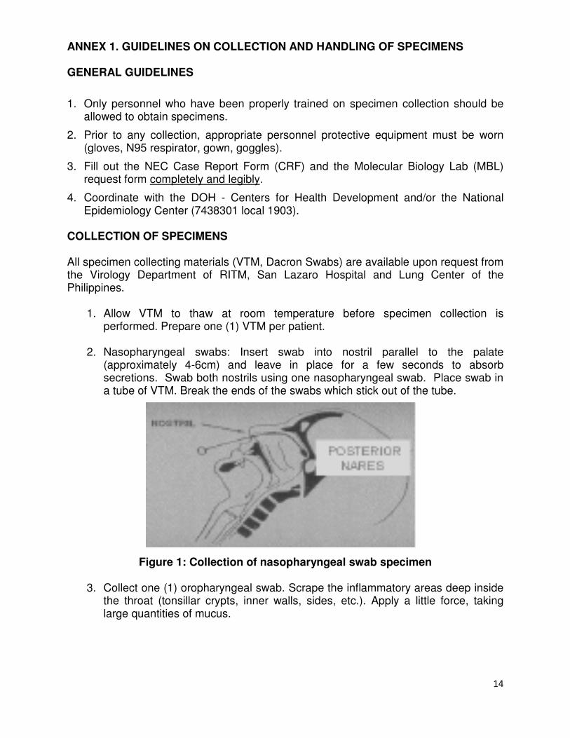
2. Nasopharyngeal swabs: Insert swab into nostril parallel to the palate
(approximately 4-6cm) and leave in place for a few seconds to absorb secretions. Swab both nostrils using one nasopharyngeal swab. Place swab in a tube of VTM. Break the ends of the swabs which stick out of the tube.
Figure 1: Collection of nasopharyngeal swab specimen
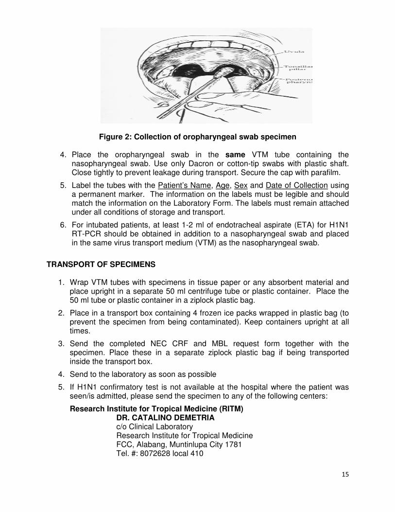
3. Collect one (1) oropharyngeal swab. Scrape the inflammatory areas deep inside the throat (tonsillar crypts, inner walls, sides, etc.). Apply a little force, taking large quantities of mucus.
15
Figure 2: Collection of oropharyngeal swab specimen
4. Place the oropharyngeal swab in the same VTM tube containing the nasopharyngeal swab. Use only Dacron or cotton-tip swabs with plastic shaft. Close tightly to prevent leakage during transport. Secure the cap with parafilm.
5. Label the tubes with the Patient’s Name, Age, Sex and Date of Collection using a permanent marker. The information on the labels must be legible and should match the information on the Laboratory Form. The labels must remain attached under all conditions of storage and transport.
6. For intubated patients, at least 1-2 ml of endotracheal aspirate (ETA) for H1N1 RT-PCR should be obtained in addition to a nasopharyngeal swab and placed in the same virus transport medium (VTM) as the nasopharyngeal swab.
TRANSPORT OF SPECIMENS
1. Wrap VTM tubes with specimens in tissue paper or any absorbent material and place upright in a separate 50 ml centrifuge tube or plastic container. Place the 50 ml tube or plastic container in a ziplock plastic bag.
2. Place in a transport box containing 4 frozen ice packs wrapped in plastic bag (to prevent the specimen from being contaminated). Keep containers upright at all times.
3. Send the completed NEC CRF and MBL request form together with the specimen. Place these in a separate ziplock plastic bag if being transported inside the transport box.
4. Send to the laboratory as soon as possible
5. If H1N1 confirmatory test is not available at the hospital where the patient was seen/is admitted, please send the specimen to any of the following centers:
Research Institute for Tropical Medicine (RITM) DR. CATALINO DEMETRIA
c/o Clinical Laboratory Research Institute for Tropical Medicine FCC, Alabang, Muntinlupa City 1781 Tel. #: 8072628 local 410
16
Lung Center of the Philippines (LCP) MS. SUSAN CASTRO MS. SIMONETTE LAXINA MS. MONIQUE BARILE Lung Center of the Philippines Tel. No.: 9246101 local 286 or 386
San Lazaro Hospital (SLH)
MS. SUSAN LEAÑO STD AIDS Central Collaborating Laboratory (SACCL)
San Lazaro Hospital, Manila Tel. No.: 7114117
The Medical City (TMC)
DR. RAUL DESTURA Section of Molecular Diagnostics and Clinical Laboratories The Medical City Tel No: 6356789 local 6415
Vicente Sotto Memorial Medical Center (VSMMC)
DR. MARILYN ZARRAGA Head Laboratory Department Tel. No.: 032-2539891 (trunkline)
6. Specimens must be transported to accredited testing facilities under cold transport (Ice box). If unable to submit samples on the day of collection, samples may be stored in the freezer and must be removed only on the day of transport under cold environment. Avoid repeated freeze-thawing. These samples are ONLY applicable for PCR testing and not for viral culture.
17
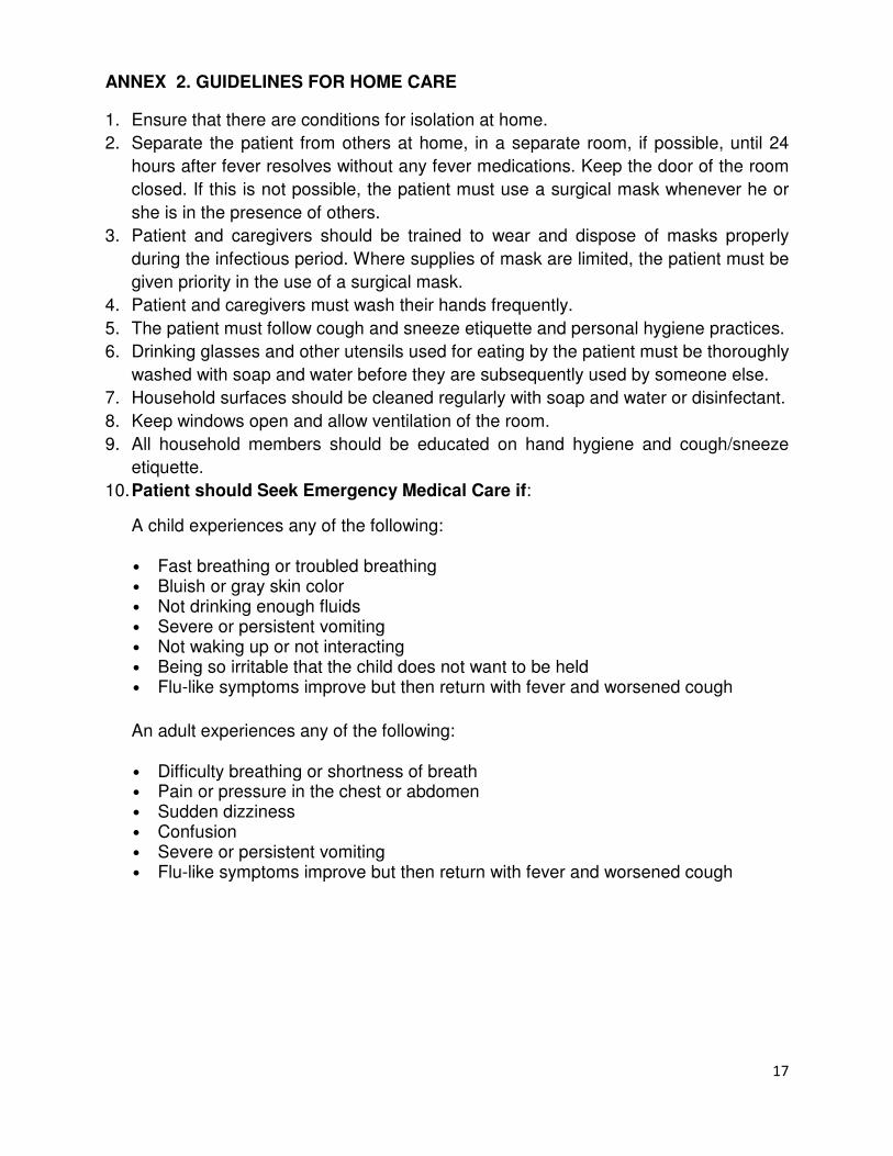
ANNEX 2. GUIDELINES FOR HOME CARE
1. Ensure that there are conditions for isolation at home.
2. Separate the patient from others at home, in a separate room, if possible, until 24
hours after fever resolves without any fever medications. Keep the door of the room
closed. If this is not possible, the patient must use a surgical mask whenever he or
she is in the presence of others.
3. Patient and caregivers should be trained to wear and dispose of masks properly
during the infectious period. Where supplies of mask are limited, the patient must be
given priority in the use of a surgical mask.
4. Patient and caregivers must wash their hands frequently.
5. The patient must follow cough and sneeze etiquette and personal hygiene practices.
6. Drinking glasses and other utensils used for eating by the patient must be thoroughly
washed with soap and water before they are subsequently used by someone else.
7. Household surfaces should be cleaned regularly with soap and water or disinfectant.
8. Keep windows open and allow ventilation of the room.
9. All household members should be educated on hand hygiene and cough/sneeze
etiquette.
10. Patient should Seek Emergency Medical Care if:
A child experiences any of the following:
• Fast breathing or troubled breathing • Bluish or gray skin color • Not drinking enough fluids • Severe or persistent vomiting • Not waking up or not interacting • Being so irritable that the child does not want to be held • Flu-like symptoms improve but then return with fever and worsened cough
An adult experiences any of the following:
• Difficulty breathing or shortness of breath • Pain or pressure in the chest or abdomen • Sudden dizziness • Confusion • Severe or persistent vomiting • Flu-like symptoms improve but then return with fever and worsened cough
18
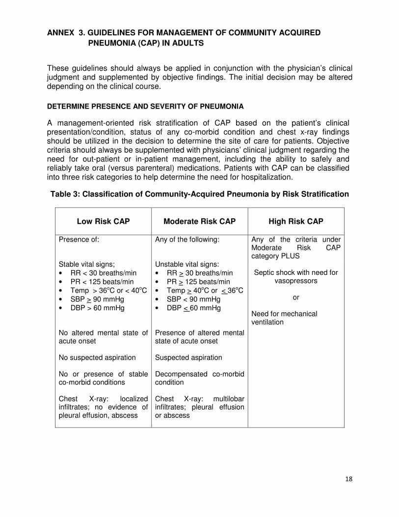
ANNEX 3. GUIDELINES FOR MANAGEMENT OF COMMUNITY ACQUIRED PNEUMONIA (CAP) IN ADULTS
These guidelines should always be applied in conjunction with the physician’s clinical judgment and supplemented by objective findings. The initial decision may be altered depending on the clinical course.
DETERMINE PRESENCE AND SEVERITY OF PNEUMONIA
A management-oriented risk stratification of CAP based on the patient’s clinical presentation/condition, status of any co-morbid condition and chest x-ray findings should be utilized in the decision to determine the site of care for patients. Objective criteria should always be supplemented with physicians’ clinical judgment regarding the need for out-patient or in-patient management, including the ability to safely and reliably take oral (versus parenteral) medications. Patients with CAP can be classified into three risk categories to help determine the need for hospitalization.
Table 3: Classification of Community-Acquired Pneumonia by Risk Stratification
Low Risk CAP
Moderate Risk CAP
High Risk CAP
Presence of: Any of the following: Any of the criteria under Moderate Risk CAP category PLUS
Stable vital signs;
• RR < 30 breaths/min
• PR < 125 beats/min
• Temp > 36oC or < 40oC
• SBP > 90 mmHg
• DBP > 60 mmHg No altered mental state of acute onset No suspected aspiration No or presence of stable co-morbid conditions Chest X-ray: localized infiltrates; no evidence of pleural effusion, abscess
Unstable vital signs:
• RR > 30 breaths/min
• PR > 125 beats/min
• Temp > 40oC or < 36oC
• SBP < 90 mmHg
• DBP < 60 mmHg Presence of altered mental state of acute onset Suspected aspiration Decompensated co-morbid condition Chest X-ray: multilobar infiltrates; pleural effusion or abscess
Septic shock with need for
vasopressors
or
Need for mechanical ventilation
19
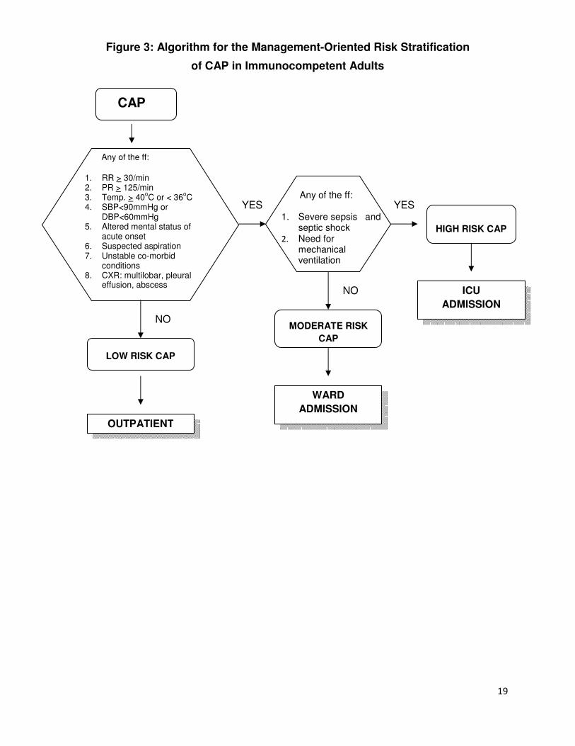
Figure 3: Algorithm for the Management-Oriented Risk Stratification
of CAP in Immunocompetent Adults
Any of the ff:
1. RR > 30/min 2. PR > 125/min 3. Temp. > 40
oC or < 36
oC
4. SBP<90mmHg or DBP<60mmHg
5. Altered mental status of acute onset
6. Suspected aspiration 7. Unstable co-morbid
conditions 8. CXR: multilobar, pleural
effusion, abscess
LOW RISK CAP
Any of the ff:
1. Severe sepsis and septic shock
2. Need for mechanical ventilation
HIGH RISK CAP
CAP
YES
NO
OUTPATIENT
MODERATE RISK CAP
NO
WARD ADMISSION
YES
ICU ADMISSION
20
DIAGNOSTICS
1. For Low Risk CAP, microbiologic studies (example: sputum GS/CS) are
OPTIONAL.
2. In moderate and high risk CAP, blood culture and gram stain/culture of
respiratory specimens should be done.
3. When possible, tests to document the presence of Legionella sp. are
recommended in hospitalized patients.
4. Invasive procedures to obtain specimens for gram stain and culture such as transtracheal, transthoracic biopsy, bronchoalveolar lavage and protected brush specimens are options for non-resolving pneumonia, immunocompromised patients and when no adequate respiratory specimen can be sent despite sputum induction and routine diagnostic test.
5. Emerging etiologies: the existence of new and emerging causes of pneumonia (e.g. pandemic (H1N1) 2009, SARS) should be sought during outbreaks or when there are epidemiologic clues that point to their existence. Rapid influenza diagnostic tests (RIDTs) in respiratory clinical specimens have low over-all sensitivity to detect pandemic (H1N1) 2009 (40-69%). If the presence of pandemic (H1N1) 2009 is suspected in patients with high risk pneumonia, a definitive determination with RT-PCR should be conducted.
MANAGEMENT
I. START ANTIBIOTICS
For patients requiring hospitalization, empiric therapy should be initiated as soon as possible after a diagnosis of CAP is made. Antimicrobial management of a patient with CAP is based on: 1) an assessment of pneumonia severity; 2) the ability of the patient to comply with oral therapy; and, 3) the social circumstances and available care for an individual.
21
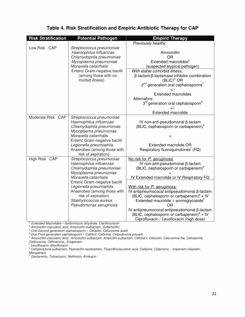
Table 4. Risk Stratification and Empiric Antibiotic Therapy for CAP
Risk Stratification Potential Pathogen Empiric Therapy Low Risk CAP
Streptococcus pneumoniae Haemophilus influenzae Chlamydophila pneumoniae Mycoplasma pneumoniae Moraxella catarrhalis Enteric Gram-negative bacilli
(among those with co-morbid illness)
Previously healthy:
Amoxicillin OR
Extended macrolidesa
(suspected atypical pathogen) With stable comorbid illness: β-lactam/β-lactamase inhibitor combination
(BLIC)b
OR 2
nd generation oral cephalosporins
c
+/- Extended macrolides
Alternative: 3
rd generation oral cephalosporin
d
+/- Extended macrolide
Moderate Risk CAP Streptococcus pneumoniae Haemophilus influenzae Chlamydophila pneumoniae Mycoplasma pneumoniae Moraxella catarrhalis Enteric Gram-negative bacilli Legionella pneumophila Anaerobes (among those with
risk of aspiration)
IV non-anti-pseudomonal β-lactam
(BLIC, cephalosporin or carbapenem)e
+
Extended macrolide OR Respiratory fluoroquinolones
f (FQ)
High Risk CAP Streptococcus pneumoniae
Haemophilus influenzae Chlamydophila pneumoniae Mycoplasma pneumoniae Moraxella catarrhalis Enteric Gram-negative bacilli Legionella pneumophila Anaerobes (among those with
risk of aspiration) Staphylococcus aureus Pseudomonas aeruginosa
No risk for P. aeruginosa: IV non-anti-pseudomonal β-lactam
(BLIC, cephalosporin or carbapenem)e
+ IV Extended macrolide or IV Respiratory FQ
With risk for P. aeruginosa: IV antipneumococal antipseudomonal β-lactam
(BLIC, cephalosporin or carbapenem)g + IV
Extended macrolide + aminoglycosideh
OR IV antipneumococal antipseudomonal β-lactam
(BLIC, cephalosporin or carbapenem)g + IV
Ciprofloxacin / Levofloxacin (high dose) a
Extended Macrolides – Azithromycin dihydrate, Clarithromycin b Amoxicillin-clavulanic acid, Amoxicillin-sulbactam, Sultamicillin
c Oral Second generation cephalosporin – Cefaclor, Cefuroxime axetil
d Oral Third generation cephalosporin – Cefdinir, Cefixime, Cefpodoxime proxetil
e Amoxicillin-clavulanic acid, Amoxicillin-sulbactam, Ampicillin-sulbactam, Cefotiam, Cefoxitin, Cefuroxime Na, Cefotaxime,
Ceftizoxime, Ceftriaxone,, Ertapenem f Levofloxacin, Moxifloxacin
g Cefoperazone-sulbactam, Piperacillin-tazobactam, Ticarcillinclavulanic acid, Cefipime, Cefpirome, , Imipenem-cilastatin,
Meropenem h Gentamicin, Tobramycin, Netilmicin, Amikacin
22
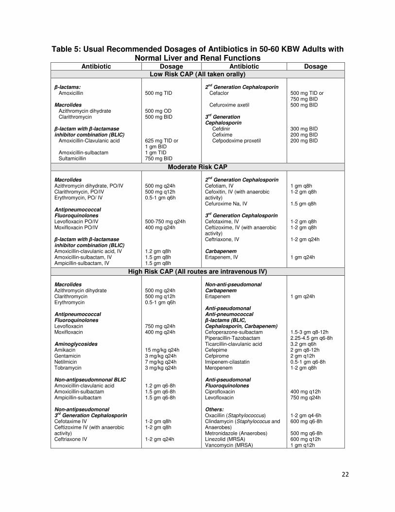
Table 5: Usual Recommended Dosages of Antibiotics in 50-60 KBW Adults with Normal Liver and Renal Functions
Antibiotic Dosage Antibiotic Dosage Low Risk CAP (All taken orally)
β-lactams: Amoxicillin Macrolides Azithromycin dihydrate Clarithromycin β-lactam with β-lactamase inhibitor combination (BLIC) Amoxicillin-Clavulanic acid Amoxicillin-sulbactam Sultamicillin
500 mg TID 500 mg OD 500 mg BID 625 mg TID or 1 gm BID 1 gm TID 750 mg BID
2
nd Generation Cephalosporin
Cefaclor Cefuroxime axetil 3
rd Generation
Cephalosporin Cefdinir Cefixime Cefpodoxime proxetil
500 mg TID or 750 mg BID 500 mg BID 300 mg BID 200 mg BID 200 mg BID
Moderate Risk CAP Macrolides Azithromycin dihydrate, PO/IV Clarithromycin, PO/IV Erythromycin, PO/ IV Antipneumococcal Fluoroquinolones Levofloxacin PO/IV Moxifloxacin PO/IV β-lactam with β-lactamase inhibitor combination (BLIC) Amoxicillin-clavulanic acid, IV Amoxicillin-sulbactam, IV Ampicillin-sulbactam, IV
500 mg q24h 500 mg q12h 0.5-1 gm q6h 500-750 mg q24h 400 mg q24h 1.2 gm q8h 1.5 gm q8h 1.5 gm q8h
2
nd Generation Cephalosporin
Cefotiam, IV Cefoxitin, IV (with anaerobic activity) Cefuroxime Na, IV 3
rd Generation Cephalosporin
Cefotaxime, IV Ceftizoxime, IV (with anaerobic activity) Ceftriaxone, IV Carbapenem Ertapenem, IV
1 gm q8h 1-2 gm q8h 1.5 gm q8h 1-2 gm q8h 1-2 gm q8h 1-2 gm q24h 1 gm q24h
High Risk CAP (All routes are intravenous IV) Macrolides Azithromycin dihydrate Clarithromycin Erythromycin Antipneumococcal Fluoroquinolones Levofloxacin Moxifloxacin Aminoglycosides Amikacin Gentamicin Netilmicin Tobramycin Non-antipseudomnonal BLIC Amoxicillin-clavulanic acid Amoxicillin-sulbactam Ampicillin-sulbactam Non-antipseudomonal 3
rd Generation Cephalosporin
Cefotaxime IV Ceftizoxime IV (with anaerobic activity) Ceftriaxone IV
500 mg q24h 500 mg q12h 0.5-1 gm q6h 750 mg q24h 400 mg q24h 15 mg/kg q24h 3 mg/kg q24h 7 mg/kg q24h 3 mg/kg q24h 1.2 gm q6-8h 1.5 gm q6-8h 1.5 gm q6-8h 1-2 gm q8h 1-2 gm q8h 1-2 gm q24h
Non-anti-pseudomonal Carbapenem Ertapenem Anti-pseudomonal Anti-pneumococcal β-lactams (BLIC, Cephalosporin, Carbapenem) Cefoperazone-sulbactam Piperacillin-Tazobactam Ticarcillin-clavulanic acid Cefepime Cefpirome Imipenem-cilastatin Meropenem Anti-pseudomonal Fluoroquinolones Ciprofloxacin Levofloxacin Others: Oxacillin (Staphylococcus) Clindamycin (Staphylococus and Anaerobes) Metronidazole (Anaerobes) Linezolid (MRSA) Vancomycin (MRSA)
1 gm q24h 1.5-3 gm q8-12h 2.25-4.5 gm q6-8h 3.2 gm q6h 2 gm q8-12h 2 gm q12h 0.5-1 gm q6-8h 1-2 gm q8h 400 mg q12h 750 mg q24h 1-2 gm q4-6h 600 mg q6-8h 500 mg q6-8h 600 mg q12h 1 gm q12h
23
II. PROVIDE SUPPORTIVE CARE
1. Oxygen
2. Hydration
3. Bronchodilator/nebulization, if wheezing is present
III. MONITORING RESPONSE TO ANTIBIOTIC THERAPY
Most patients with uncomplicated bacterial pneumonia will respond to treatment within 24-72 hours; re-evaluation of patients, therefore, should be done after 72 hours of initiating therapy. A patient is considered to have responded to treatment if fever decreases within 72 hours, temperature normalizes within 5 days and respiratory signs, particularly tachypnea, return to normal. In patients with low risk CAP who are clinically improving, a follow-up chest x-ray is not necessary. Follow-up cultures of blood and sputum are not also indicated for patients who respond to therapy. In hospitalized patients, de-escalating initial empiric broad spectrum or combination IV therapy to a single narrow spectrum IV or oral agent based on available laboratory data is recommended as early as 72 hours following initiation of empirical treatment. Switch therapy to an oral agent will allow early discharge from the hospital and will lead to cost-savings.
Indications for de-escalation of antibiotic therapy
1. Resolution of fever for > 24 hours (temperature <37.8°C) 2. Resolution of respiratory distress (normalization of RR < 24 breaths/min) 3. Resolution of hypotension (systolic BP >90 mmHg) and tachycardia 4. Absence of hypoxemia 5. Etiologic agent is not a high risk (virulent/resistant) pathogen (e.g. Legionella,
Staphylococcus aureus or gram negative enteric bacilli) 6. No unstable co-morbid condition or life-threatening complication such as MI,
CHF, complete heart block, new atrial fibrillation, supraventricular tachycardia, etc
7. No signs of organ dysfunction 8. Improving white cell count 9. No bacteremia 10. Patient is clinically hydrated, taking oral fluids and able to take oral medications 11. Normal mental status
24
IV. CRITERIA FOR DISCHARGE
During the 24 hours before discharge, the patient should be clinically stable with the
following characteristics (unless this represents the baseline status):
1. Resolution of fever (temperature of <37.8 O
C) without fever medications 2. Heart rate < 100 beats/min 3. Resolution of hypotension 4. Resolution of tachypnea 5. Oxygen saturation >90 6. Clinically hydrated and taking oral fluids 7. With a functioning gastrointestinal tract
25
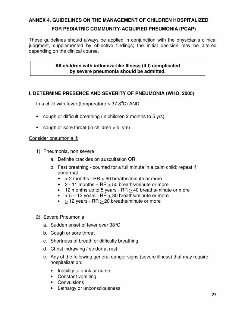
ANNEX 4. GUIDELINES ON THE MANAGEMENT OF CHILDREN HOSPITALIZED
FOR PEDIATRIC COMMUNITY-ACQUIRED PNEUMONIA (PCAP) These guidelines should always be applied in conjunction with the physician’s clinical judgment, supplemented by objective findings; the initial decision may be altered depending on the clinical course.
I. DETERMINE PRESENCE AND SEVERITY OF PNEUMONIA (WHO, 2005)
In a child with fever (temperature > 37.80C) AND
• cough or difficult breathing (in children 2 months to 5 yrs)
• cough or sore throat (in children > 5 yrs)
Consider pneumonia if:
1) Pneumonia, non severe
a. Definite crackles on auscultation OR
b. Fast breathing - counted for a full minute in a calm child; repeat if abnormal
• < 2 months - RR > 60 breaths/minute or more
• 2 - 11 months – RR > 50 breaths/minute or more • 12 months up to 5 years - RR > 40 breaths/minute or more
• > 5 – 12 years - RR > 30 breaths/minute or more
• > 12 years - RR > 20 breaths/minute or more
2) Severe Pneumonia
a. Sudden onset of fever over 38°C
b. Cough or sore throat
c. Shortness of breath or difficulty breathing
d. Chest indrawing / stridor at rest
e. Any of the following general danger signs (severe illness) that may require hospitalization:
• Inability to drink or nurse
• Constant vomiting
• Convulsions
• Lethargy or unconsciousness
All children with influenza-like Illness (ILI) complicated by severe pneumonia should be admitted.
26
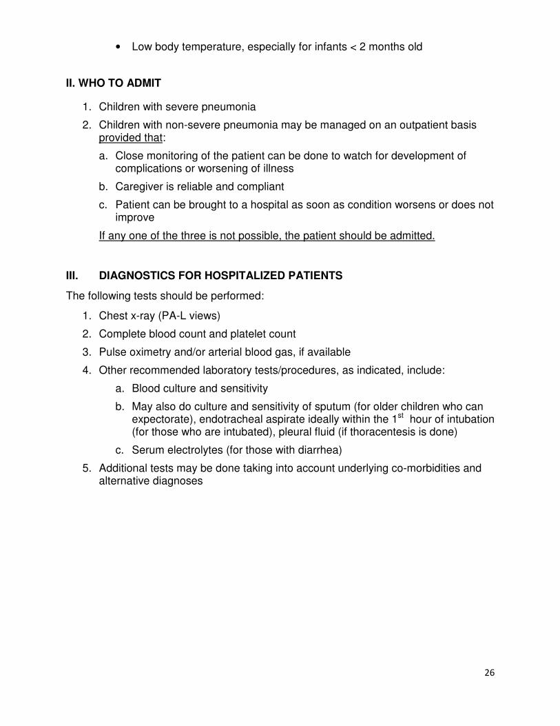
• Low body temperature, especially for infants < 2 months old
II. WHO TO ADMIT
1. Children with severe pneumonia
2. Children with non-severe pneumonia may be managed on an outpatient basis provided that:
a. Close monitoring of the patient can be done to watch for development of complications or worsening of illness
b. Caregiver is reliable and compliant
c. Patient can be brought to a hospital as soon as condition worsens or does not improve
If any one of the three is not possible, the patient should be admitted.
III. DIAGNOSTICS FOR HOSPITALIZED PATIENTS
The following tests should be performed:
1. Chest x-ray (PA-L views)
2. Complete blood count and platelet count
3. Pulse oximetry and/or arterial blood gas, if available
4. Other recommended laboratory tests/procedures, as indicated, include:
a. Blood culture and sensitivity
b. May also do culture and sensitivity of sputum (for older children who can expectorate), endotracheal aspirate ideally within the 1st hour of intubation (for those who are intubated), pleural fluid (if thoracentesis is done)
c. Serum electrolytes (for those with diarrhea)
5. Additional tests may be done taking into account underlying co-morbidities and alternative diagnoses
27
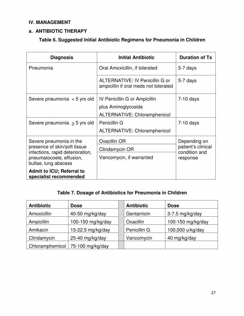
IV. MANAGEMENT
a. ANTIBIOTIC THERAPY
Table 6. Suggested Initial Antibiotic Regimens for Pneumonia in Children
Diagnosis Initial Antibiotic Duration of Tx
Pneumonia
Oral Amoxicillin, if tolerated 5-7 days
ALTERNATIVE: IV Penicillin G or ampicillin if oral meds not tolerated
5-7 days
Severe pneumonia < 5 yrs old IV Penicillin G or Ampicillin
plus Aminoglycoside
ALTERNATIVE: Chloramphenicol
7-10 days
Severe pneumonia > 5 yrs old Penicillin G
ALTERNATIVE: Chloramphenicol
7-10 days
Severe pneumonia in the presence of skin/soft tissue infections, rapid deterioration, pneumatocoele, effusion, bullae, lung abscess
Admit to ICU; Referral to specialist recommended
Oxacillin OR Depending on patient’s clinical condition and response
Clindamycin OR
Vancomycin, if warranted
Table 7. Dosage of Antibiotics for Pneumonia in Children
Antibiotic Dose Antibiotic Dose
Amoxicillin 40-50 mg/kg/day Gentamicin 3-7.5 mg/kg/day
Ampicillin 100-150 mg/kg/day Oxacillin 100-150 mg/kg/day
Amikacin 15-22.5 mg/kg/day Penicillin G 100,000 u/kg/day
Clindamycin 25-40 mg/kg/day Vancomycin 40 mg/kg/day
Chloramphemicol 75-100 mg/kg/day
28
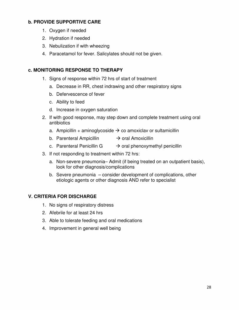
b. PROVIDE SUPPORTIVE CARE
1. Oxygen if needed
2. Hydration if needed
3. Nebulization if with wheezing
4. Paracetamol for fever. Salicylates should not be given.
c. MONITORING RESPONSE TO THERAPY
1. Signs of response within 72 hrs of start of treatment
a. Decrease in RR, chest indrawing and other respiratory signs
b. Defervescence of fever
c. Ability to feed
d. Increase in oxygen saturation
2. If with good response, may step down and complete treatment using oral antibiotics
a. Ampicillin + aminoglycoside � co amoxiclav or sultamicillin
b. Parenteral Ampicillin � oral Amoxicillin
c. Parenteral Penicillin G � oral phenoxymethyl penicillin
3. If not responding to treatment within 72 hrs:
a. Non-severe pneumonia– Admit (if being treated on an outpatient basis), look for other diagnosis/complications
b. Severe pneumonia – consider development of complications, other etiologic agents or other diagnosis AND refer to specialist
V. CRITERIA FOR DISCHARGE
1. No signs of respiratory distress
2. Afebrile for at least 24 hrs
3. Able to tolerate feeding and oral medications
4. Improvement in general well being
29
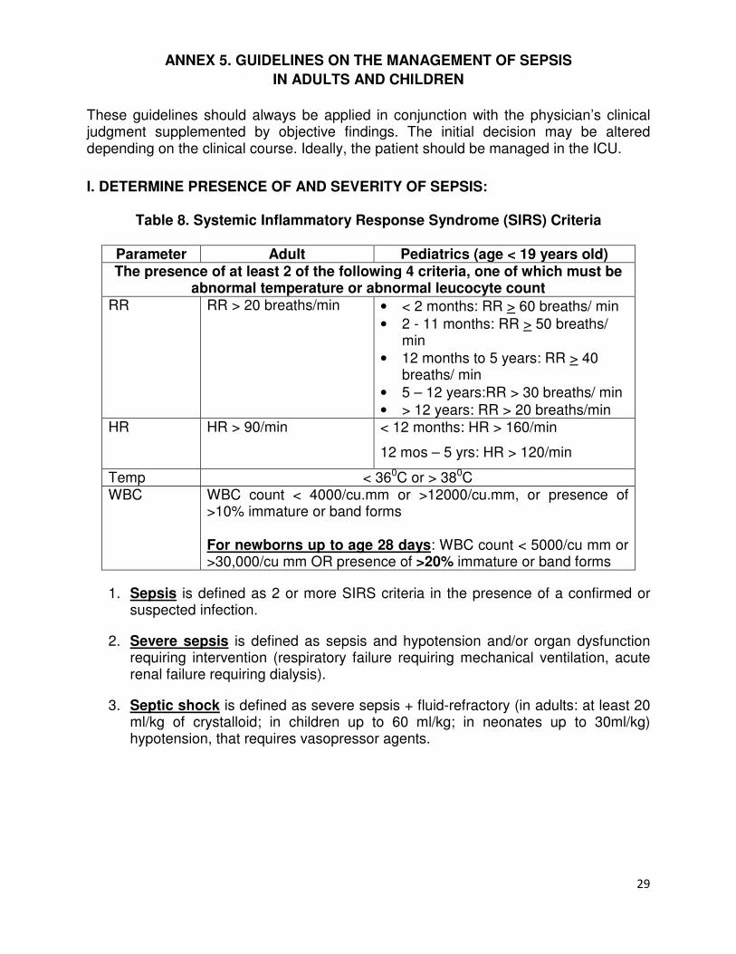
ANNEX 5. GUIDELINES ON THE MANAGEMENT OF SEPSIS IN ADULTS AND CHILDREN
These guidelines should always be applied in conjunction with the physician’s clinical judgment supplemented by objective findings. The initial decision may be altered depending on the clinical course. Ideally, the patient should be managed in the ICU.
I. DETERMINE PRESENCE OF AND SEVERITY OF SEPSIS:
Table 8. Systemic Inflammatory Response Syndrome (SIRS) Criteria
Parameter Adult Pediatrics (age < 19 years old) The presence of at least 2 of the following 4 criteria, one of which must be
abnormal temperature or abnormal leucocyte count RR RR > 20 breaths/min • < 2 months: RR > 60 breaths/ min
• 2 - 11 months: RR > 50 breaths/ min
• 12 months to 5 years: RR > 40 breaths/ min
• 5 – 12 years:RR > 30 breaths/ min
• > 12 years: RR > 20 breaths/min HR HR > 90/min < 12 months: HR > 160/min
12 mos – 5 yrs: HR > 120/min
Temp < 360C or > 380C WBC WBC count < 4000/cu.mm or >12000/cu.mm, or presence of
>10% immature or band forms
For newborns up to age 28 days: WBC count < 5000/cu mm or >30,000/cu mm OR presence of >20% immature or band forms
1. Sepsis is defined as 2 or more SIRS criteria in the presence of a confirmed or suspected infection.
2. Severe sepsis is defined as sepsis and hypotension and/or organ dysfunction requiring intervention (respiratory failure requiring mechanical ventilation, acute renal failure requiring dialysis).
3. Septic shock is defined as severe sepsis + fluid-refractory (in adults: at least 20 ml/kg of crystalloid; in children up to 60 ml/kg; in neonates up to 30ml/kg) hypotension, that requires vasopressor agents.
30
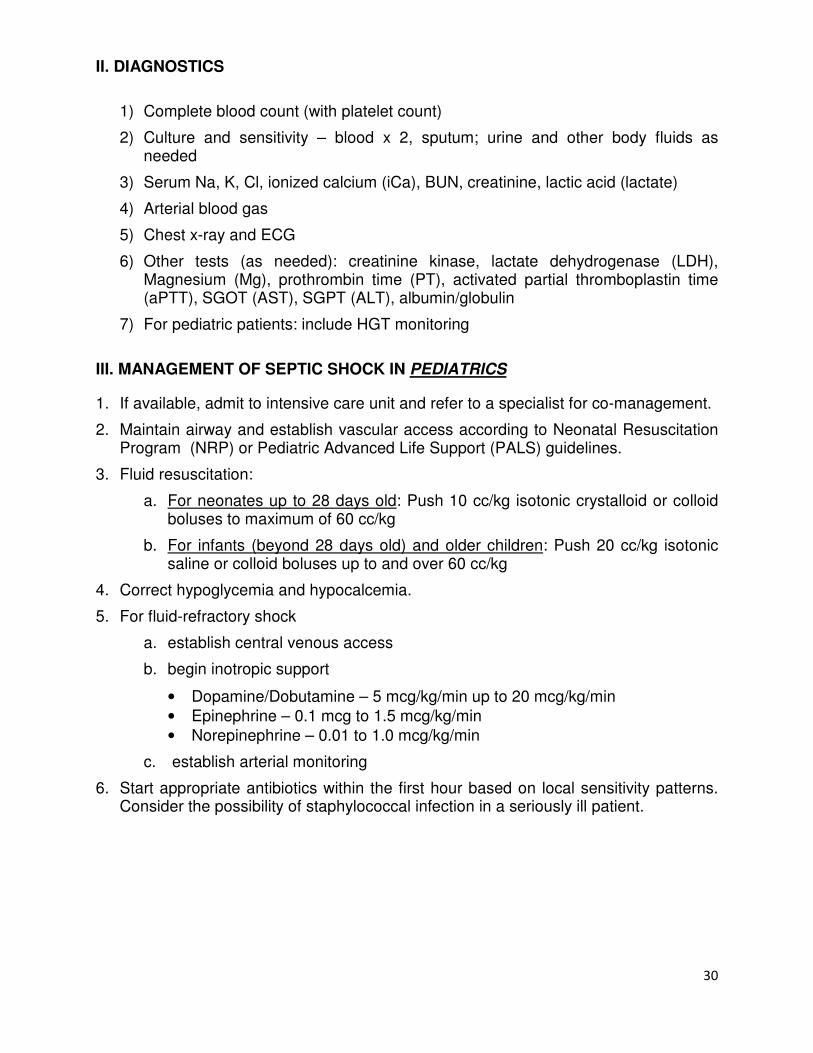
II. DIAGNOSTICS
1) Complete blood count (with platelet count)
2) Culture and sensitivity – blood x 2, sputum; urine and other body fluids as needed
3) Serum Na, K, Cl, ionized calcium (iCa), BUN, creatinine, lactic acid (lactate)
4) Arterial blood gas
5) Chest x-ray and ECG
6) Other tests (as needed): creatinine kinase, lactate dehydrogenase (LDH), Magnesium (Mg), prothrombin time (PT), activated partial thromboplastin time (aPTT), SGOT (AST), SGPT (ALT), albumin/globulin
7) For pediatric patients: include HGT monitoring
III. MANAGEMENT OF SEPTIC SHOCK IN PEDIATRICS
1. If available, admit to intensive care unit and refer to a specialist for co-management.
2. Maintain airway and establish vascular access according to Neonatal Resuscitation Program (NRP) or Pediatric Advanced Life Support (PALS) guidelines.
3. Fluid resuscitation:
a. For neonates up to 28 days old: Push 10 cc/kg isotonic crystalloid or colloid boluses to maximum of 60 cc/kg
b. For infants (beyond 28 days old) and older children: Push 20 cc/kg isotonic saline or colloid boluses up to and over 60 cc/kg
4. Correct hypoglycemia and hypocalcemia.
5. For fluid-refractory shock
a. establish central venous access
b. begin inotropic support
• Dopamine/Dobutamine – 5 mcg/kg/min up to 20 mcg/kg/min
• Epinephrine – 0.1 mcg to 1.5 mcg/kg/min
• Norepinephrine – 0.01 to 1.0 mcg/kg/min
c. establish arterial monitoring
6. Start appropriate antibiotics within the first hour based on local sensitivity patterns. Consider the possibility of staphylococcal infection in a seriously ill patient.
31
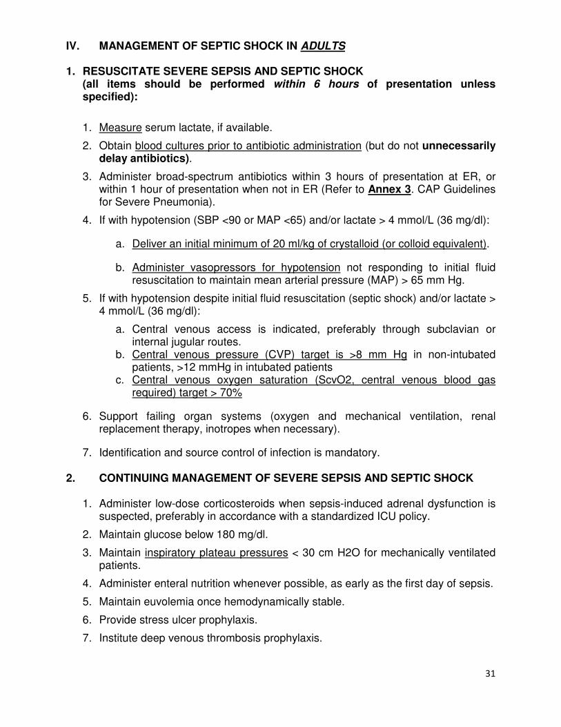
IV. MANAGEMENT OF SEPTIC SHOCK IN ADULTS 1. RESUSCITATE SEVERE SEPSIS AND SEPTIC SHOCK
(all items should be performed within 6 hours of presentation unless specified):
1. Measure serum lactate, if available.
2. Obtain blood cultures prior to antibiotic administration (but do not unnecessarily delay antibiotics).
3. Administer broad-spectrum antibiotics within 3 hours of presentation at ER, or within 1 hour of presentation when not in ER (Refer to Annex 3. CAP Guidelines for Severe Pneumonia).
4. If with hypotension (SBP <90 or MAP <65) and/or lactate > 4 mmol/L (36 mg/dl):
a. Deliver an initial minimum of 20 ml/kg of crystalloid (or colloid equivalent).
b. Administer vasopressors for hypotension not responding to initial fluid resuscitation to maintain mean arterial pressure (MAP) > 65 mm Hg.
5. If with hypotension despite initial fluid resuscitation (septic shock) and/or lactate > 4 mmol/L (36 mg/dl):
a. Central venous access is indicated, preferably through subclavian or internal jugular routes.
b. Central venous pressure (CVP) target is >8 mm Hg in non-intubated patients, >12 mmHg in intubated patients
c. Central venous oxygen saturation (ScvO2, central venous blood gas required) target > 70%
6. Support failing organ systems (oxygen and mechanical ventilation, renal replacement therapy, inotropes when necessary).
7. Identification and source control of infection is mandatory.
2. CONTINUING MANAGEMENT OF SEVERE SEPSIS AND SEPTIC SHOCK
1. Administer low-dose corticosteroids when sepsis-induced adrenal dysfunction is suspected, preferably in accordance with a standardized ICU policy.
2. Maintain glucose below 180 mg/dl.
3. Maintain inspiratory plateau pressures < 30 cm H2O for mechanically ventilated patients.
4. Administer enteral nutrition whenever possible, as early as the first day of sepsis.
5. Maintain euvolemia once hemodynamically stable.
6. Provide stress ulcer prophylaxis.
7. Institute deep venous thrombosis prophylaxis.
32
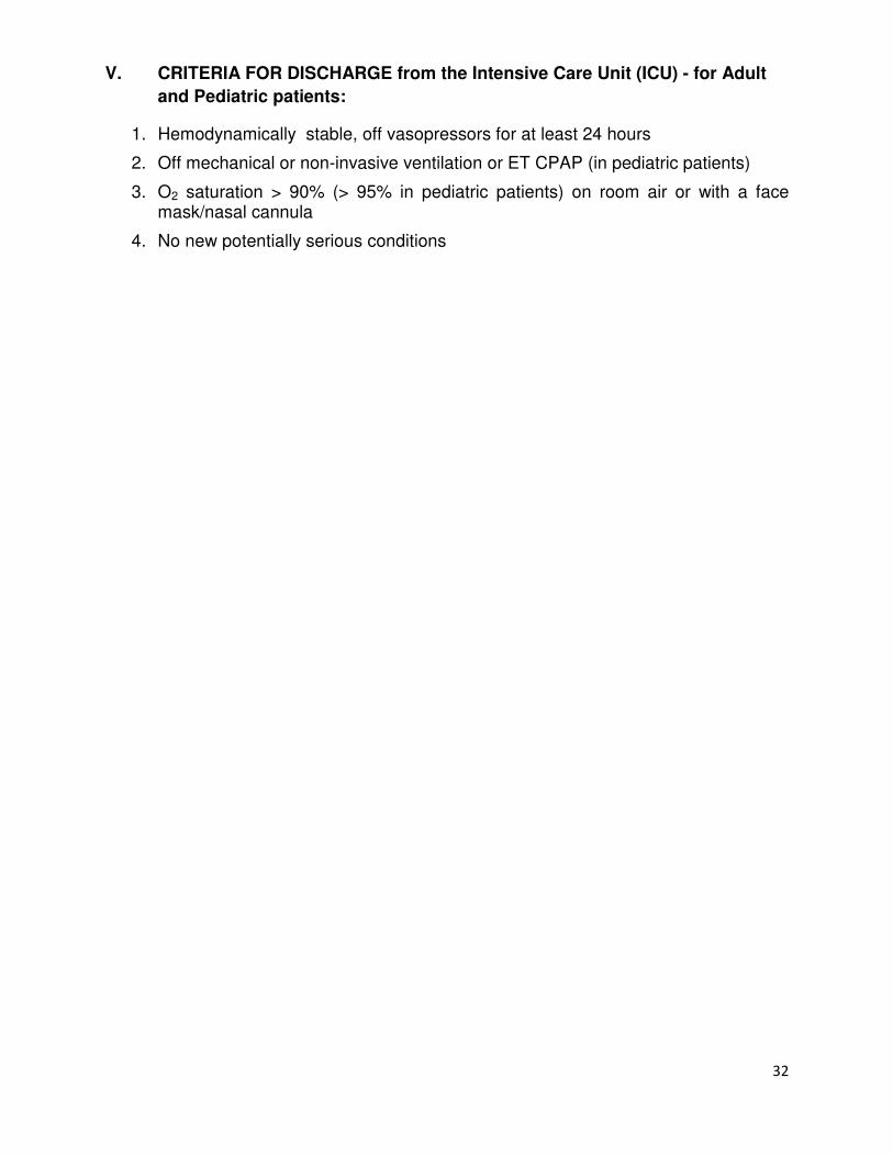
V. CRITERIA FOR DISCHARGE from the Intensive Care Unit (ICU) - for Adult and Pediatric patients:
1. Hemodynamically stable, off vasopressors for at least 24 hours
2. Off mechanical or non-invasive ventilation or ET CPAP (in pediatric patients)
3. O2 saturation > 90% (> 95% in pediatric patients) on room air or with a face mask/nasal cannula
4. No new potentially serious conditions
33
ANNEX 6. GUIDELINES ON THE MANAGEMENT OF PREGNANT WOMEN WITH CONFIRMED OR SUSPECTED PANDEMIC (H1N1) 2009 INFECTION
These guidelines should always be applied in conjunction with the physician’s clinical judgment, supplemented by objective findings; the initial decision may be altered depending on the clinical course.
I. GENERAL CONSIDERATIONS
1. A key concept for obstetric settings is to keep healthy pregnant women, in both inpatient and outpatient settings, separated from persons who are ill or potentially ill. Facilities should have a mechanism to identify and segregate ill patients, visitors and staff.
2. Prenatal care, labor and delivery services should be provided to healthy pregnant women in settings where the risk of exposure to pandemic (H1N1) 2009 flu has been minimized.
3. Healthy pregnant women and infants who have not been in close contact with persons with suspected, probable, or confirmed pandemic (H1N1) 2009 can be managed in the usual way in compliance with established infection control guidance.
II. SUSPECT INFLUENZA IN THE PRESENCE OF THE FOLLOWING:
1. Fever (temperature of 100o F {37.8oC} or greater) and typical acute respiratory influenza-like illness (e.g., cough, sore throat, rhinorrhea) in the absence of a KNOWN cause other than influenza
2. Other symptoms - body aches, headache, fatigue, vomiting and diarrhea
BASIC PRINCIPLES
Pregnant women with influenza are at higher risk of serious illness, including death, as well as adverse pregnancy outcomes.
Newborns of women with influenza are expected to be at increased risk of severe illness.
34
III. CRITERIA FOR ADMISSION
Generally, pregnant patients with fever require close monitoring for possible
complications. They may be given oseltamivir based on clinician’s evaluation.
Pregnant women with ILI should be admitted to the hospital for management if any of the following are present:
1. Unstable condition – dyspnea, hypoxemia, hemodynamic instability (e.g.,hypotension, tachycardia), altered level of consciousness and confusion, syncope, severe dizziness, etc
2. Signs of sepsis and/or pneumonia (see Annex 3. Management of Community Acquired Pneumonia or CAP, Annex 4. Management of Pediatric CAP and Annex 5. Management of Sepsis in Adults and Children)
3. Inability to eat and/or take oral fluids 4. Urgent need for work-up for alternative/additional diagnoses or need for close
monitoring of fetal status and well-being using appropriate tests (e.g., biophysical profile, fetal heart rate, fetal movement, etc.) as well as monitoring for and control of preterm labor
5. Presence of unstable/uncontrolled co-morbidities which increase the risk of severe illness - chronic pulmonary (including asthma), cardiovascular (except hypertension), renal, hepatic, hematologic, neurologic, or metabolic disorders (including diabetes mellitus), immunosuppression, cancer, malnutrition
IV. DIAGNOSTICS
1. Nasopharyngeal and oropharyngeal swabs for RT-PCR for pandemic (H1N1) 2009 virus
2. Maternal and fetal well-being studies, as indicated: biophysical profile, pelvic ultrasound, electronic tocomonitor
3. Additional tests may be done taking into account underlying co-morbidities and alternative diagnoses
V. MANAGEMENT
A. ANTIVIRAL
1. Treatment a. Pregnancy should not be considered a contraindication to oseltamivir use. b. Early treatment with influenza antiviral medications (preferably within 48
hours of onset) is recommended for pregnant women with suspected pandemic (H1N1) 2009 virus infection. However, antiviral medications are recommended for high risk persons, including pregnant women, presenting for care more than 48 hours after illness onset, particularly for those who require hospitalization.
c. Clinicians should not wait for test results to initiate treatment since these medications work best if started as early as possible after illness onset.
35
d. Oseltamivir is the drug of choice for treatment of pregnant women because of its systemic activity.
e. Recommended duration of treatment is 5 days.
2. Prophylaxis a. Pregnant women appear to be at higher risk for severe complications from
pandemic (H1N1) 2009 virus infection and the benefits of treatment or chemoprophylaxis with oseltamivir or zanamivir outweigh the theoretical risks of antiviral use.
b. Post exposure antiviral chemoprophylaxis can be considered for pregnant women who are close contacts of persons with laboratory confirmed pandemic (H1N1) 2009 virus infection.
c. Recommended duration of chemoprophylaxis is 5-7 days after the last known exposure to person with suspected or laboratory confirmed pandemic (H1N1) 2009
d. Close monitoring for influenza like illness in exposed pregnant women is recommended.
B. ANTIBIOTIC THERAPY
1. Antibiotics are given for patients with confirmed or clinically-suspected bacterial
complication like pneumonia or sepsis 2. In general, the following classes of antibiotics are considered safe in pregnancy:
amino-penicillins, cephalosporins, carbapenems, macrolides and extended-spectrum penicillins.
C. PROVIDE SUPPORTIVE CARE
1. Oxygen if needed
2. Hydration if needed
3. Nebulization if with wheezing
4. Fever in pregnant women should be treated because of the risk that hyperthermia appears to pose to the fetus. Paracetamol appears to be the best option for treatment of fever during pregnancy.
5. Bed rest on left lateral decubitus position
D. MONITORING RESPONSE TO THERAPY
The following are considered signs of good response:
1. Lysis of fever 2. Resolution of cough 3. Improvement in the sense of well-being
36
VI. CRITERIA FOR DISCHARGE 1. Afebrile for at least 24 hours
2. Good fetal status
3. No preterm labor
VII. CARE OF NEWBORNS OF INFECTED MOTHERS
1. The newborn should be considered to be potentially infected if delivery occurs during the 2 days before through 7 days after illness onset in the mother
2. The newborn should be closely monitored for signs and symptoms of influenza. 3. If signs or symptoms develop, testing should be performed, infection control
measures should be continued, and treatment with anti-influenza medications should be considered.
VIII. BREASTFEEDING 1. Women who are not ill with influenza should be encouraged to initiate
breastfeeding early and feed frequently. Ideally, babies should receive most of their nutrition from breast milk. Eliminate unnecessary formula supplementation, so the infant can receive as much maternal antibodies as possible
2. If possible, only adults who are not sick should care for infants, including providing feedings.
3. Sick women who are able to express their milk for bottle feedings by a healthy family member should be encouraged to do so. Antiviral medication treatment or prophylaxis is not a contraindication for breastfeeding.
4. Careful adherence to hand hygiene and cough etiquette is critical, especially for sick women who do not have anyone to help with infant care while they are ill.
5. Women with influenza-like illness should use facemasks when providing infant care and feedings
6. Instruct parent and caretakers on how to protect their infant from the spread of microorganisms, like pandemic (H1N1) 2009 virus, that cause respiratory illnesses:
a. Practice hand hygiene and cough etiquette at all times b. Keep the infant away from persons who are ill and out of crowded areas. c. Limit sharing of toys and other items that have been in infants' mouths. Wash
thoroughly with soap and water any items that have been in infants' mouths. 7. Infants who are ill may still breastfeed providing their condition allows it.
IX. INFECTION CONTROL MEASURES
1. Isolate the ill mother from healthy pregnant women. 2. Place a surgical mask on the ill mother during labor and delivery, if tolerable, in
order to decrease exposure of the newborn, healthcare personnel, and other labor and delivery patients to potentially infectious respiratory secretions.
3. The mother who has ILI at delivery should consider avoiding close contact with her infant until the following conditions have been met: she has received antiviral
37
medications for 48 hours, her fever has fully resolved, and she can control coughs and secretions.
4. Before these conditions are met, the newborn should be cared for in a separate room by another person who is well, and the mother should be encouraged and assisted to express her milk.
5. As soon as all conditions are met, the mother should be encouraged to wear a facemask, change to a clean gown or clothing, adhere to strict hand hygiene and cough etiquette when in contact with her infant, and begin breastfeeding (or if not able to breastfeed, bottle feeding)
6. Protective measures should be continued, both in the hospital setting and at home, for at least 7 days after the onset of influenza symptoms
X. OTHER WAYS TO REDUCE RISK FOR PREGNANT WOMEN
There is no vaccine available yet to prevent pandemic (H1N1) 2009 virus infection; however, the risk might be reduced by taking steps to reduce the chance of being exposed to respiratory infections. These steps include:
1. Frequent hand washing. 2. Minimizing contact with sick individuals. 3. Having ill persons stay home (except to seek medical care). 4. Having ill persons cover coughs. 5. Avoiding, whenever possible, crowded settings in communities having outbreaks
of pandemic (H1N1) 2009 virus. 6. Using facemasks and respirators correctly if they are used
XI. PREGNANT HEALTH CARE WORKERS 1. All health care workers with direct patient care, including pregnant women,
should follow standard precautions with all patients, regardless of infection status.
2. Pregnant health workers who will likely be in direct contact with patients with confirmed, probable, or suspected pandemic (H1N1) 2009 (e.g., nurse, physician, or respiratory therapist caring for hospitalized patients), should consider reassignment to lower-risk activities.
3. If reassignment is not possible, pregnant women should avoid participating in procedures that may generate increased small-particle aerosols of respiratory secretions in patients with known or suspected influenza, including the following procedures:
a. Endotracheal intubation b. Bronchoscopy c. Open airway suctioning d. Cardiopulmonary resuscitation (CPR) e. Diagnostic sputum induction
38
High-risk groups:
- Age <5 and > 60yrs - Pregnant (see
Pregnancy guidelines) - Co-morbidities - Obese
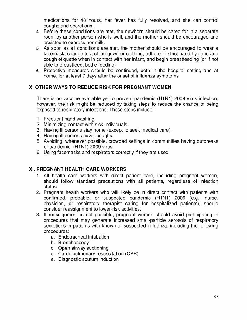
ANNEX 7. ALGORITHM FOR THE CLINICAL MANAGEMENT OF HOSPITALIZED SUSPECTED AND CONFIRMED CASES OF INFLUENZA A (H1N1) 2009 INFECTION
YES Treat as pneumonia &/or
sepsis (see CAP/PCAP and
Sepsis guidelines) and start
oseltamivir*
Start oseltamivir*
Manage symptomatically
YES
Hospitalized suspected/confirmed cases
of pandemic (H1N1) 2009 Infection
Clinical findings of
pneumonia &/or
hypoxemia &/or sepsis
Assess clinically
Collect naso- /oropharyngeal swab
CBC, chest x-ray, pulse oximetry
Follow-up daily
NO
Discharge once stable
and afebrile ≥ 24 hours
Re-evaluate and
manage accordingly**
NB: Please see guidelines for criteria for
admission of adult and pediatric patients
YES
* May discontinue oseltamivir if swab is negative; if swab is positive, complete 5 days treatment
** May repeat chest x-ray and pulse oximetry and assess for presence of pneumonia/hypoxemia and signs of sepsis and other focus of infection
Deteriorating?
NO
NO
39
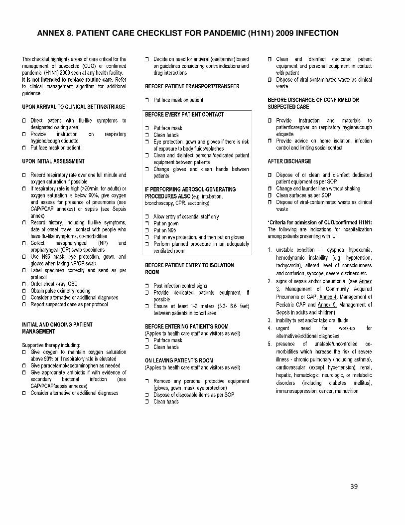
ANNEX 8. PATIENT CARE CHECKLIST FOR PANDEMIC (H1N1) 2009 INFECTION
40
ANNEX 9. POST-MORTEM CARE
Objective: Safely handle human remains during autopsy procedures to prevent
transmission of pandemic (H1N1) 2009 virus.
I. Transport of Deceased Persons
1. Transport of deceased persons does not require any additional precautions when bodies have been secured in a transport bag. Hand hygiene should be performed after completing transport.
2. Standard precautions should be practiced when handling deceased persons and preparing bodies for autopsy or transfer to mortuary services.
3. Appropriate use of PPE (e.g., gowns, gloves, masks, and/or eye protection) is recommended. Hand hygiene should be performed after PPE is removed.
II. Family Contact with the Deceased
1. Direct contact with the body is discouraged; however, necessary contact may be allowed as long as hands are cleaned immediately with soap and water or alcohol-based handrub.
2. Consider limiting contact (i.e. touching, no kissing) to close family members only.
III. Autopsy Procedures
1. In general, standard precautions and safety measures consistent with any autopsy procedure should be observed when handling human remains infected with pandemic (H1N1) 2009 virus.
2. Additional respiratory protection is needed during an autopsy procedure that generates aerosols (e.g. use of oscillating saws).
3. The number of personnel participating in post mortem examinations should be limited to the minimum required.
IV. Personal protective equipment (PPE)
1. Wear standard autopsy PPE including a scrub suit worn under an impervious gown or apron, eye protection (e.g. goggles, face shield), double surgical gloves with an interposed layer of cut-proof synthetic mesh gloves, surgical mask or respirator, and shoe covers.
2. Add respiratory protection if aerosols might be generated. This includes N-95 or N-100 disposable particulate respirators or powered air purifying respirator (PAPR).
3. Remove PPE before leaving the autopsy suite and dispose in accordance with facility policies and procedures. Observe hand hygiene after removal of PPE.
V. Engineering controls
1. Whenever possible, perform autopsies on human remains infected with pandemic (H1N1) 2009 virus in autopsy settings that have an adequate air-handling system. This includes a minimum of six (old construction) to twelve
41
(new construction) air changes per hour (ACH), negative pressure relative to adjacent areas as per recommendations for airborne infection isolation rooms (AIIRs) and direct exhaust of air to the outside or passed through a HEPA filter if air is recirculated. Exhaust systems around the autopsy table should direct air (and aerosols) away from health care workers performing the procedure (e.g., exhaust downward). For autopsies, local airflow control (e.g. laminar flow systems) can be used to direct aerosols away from personnel; however, this safety feature does not eliminate the need for appropriate PPE.
2. Use containment devices whenever possible. Use biosafety cabinets for the handling and examination of smaller specimens. When available, use vacuum shrouds for oscillating saws to contain aerosols and reduce the volume released into the ambient air environment.
3. Protective outer garments should be removed when leaving the immediate autopsy area and discarded in appropriate laundry or waste receptacles, either in an antechamber to the autopsy suite or immediately inside the entrance if an antechamber is unavailable.
4. Hand hygiene is recommended immediately after PPE removal.
VI. Prevention of percutaneous injuries
• Follow standard safety procedures for preventing percutaneous injuries during autopsy.
.
42
ANNEX 10. TASK FORCE ON THE CLINICAL MANAGEMENT OF PANDEMIC (H1N1) 2009 INFECTION
Chair: Dr. Remigio M. Olveda Vice-chair: Dr. Mediadora C. Saniel Members:
PPS/PIDSP Dr. Cynthia Aguirre Dr. Fatima Gimenez Dr. May Montellano Dr. Anna Ong-Lim Dr. Beatriz Quiambao Dr. Charissa Fay Tabora PCCP Dr. Abundio Balgos Dr. Roberto Barzaga Dr. Benilda Galvez Dr. Ma. Liza Garcia PPS/PAPP Dr. Cesar Ong PPS/PICS Dr. Benjamin Lim POGS Dr. Ricardo Manalastas PSMID Dr. Marissa Alejandria Dr. Regina Berba Dr. Jennifer Chua Dr. Manolito Chua Dr. Efren Dimaano Dr. Edna Edrada Dr. Kate Leyritana Dr. Jodor Lim Dr. Minda Manalo Dr. Myrna Mendoza Dr. Cecilia Montalban Dr. Maria Fe Tayzon PHICS Ms. Dominga Gomez Dr. Melecia Velmonte

43
DOH Dr. Yolanda Oliveros Dr. Eric Tayag Dr. Lyndon Lee Suy Dr. Vito Roque Others Dr. Jose Emmanuel Palo Dr. Aguedo Troy Gepte IV
44
ANNEX 11: REFERENCES
1. Carcillo JA, Davis AL, Zaritsky A. Role of early fluid resuscitation in pediatric septic shock. JAMA 1991;266:1242-5.
2. Ceneviva G. Paschall JA, Maffei F, et al. Hemodynamic support in fluid-refractory pediatric septic shock. Pediatrics 1998;102:e19.
3. Centers for Disease Control and Prevention (CDC). Considerations for Pregnant Women who are More Likely to be Exposed to Novel H1N1 Flu (Siwne flu) at work: Information for Women in Education, Child Care and Health Care. May 3, 2009.
4. Centers for Disease Control and Prevention (CDC). Interim Guidance for Infection Control for Care of Patients with Confirmed or Suspected Novel Influenza A (H1N1) Virus Infection in a Healthcare Setting. May 13, 2009.
5. Centers for Disease Control and Prevention (CDC). Interim Guidance for Clinicians on the Prevention and Treatment of Novel Influenza A (H1N1) Virus Infection in Infants and Children. May 13, 2009.
6. Centers for Disease Control and Prevention (CDC). Post-mortem Care and Safe Autopsy Procedures for Novel (H1N1) Influenza. May 28, 2009.
7. Centers for Disease Control and Prevention (CDC). Pregnant Women and Novel Influenza A (H1N1) Virus: Considerations for Clinicians. June 30, 2009.
8. Centers for Disease Control and Prevention (CDC). Considerations Regarding Novel H1N1 Flu Virus in the Obstetrical Setting. July 6, 2009.
9. Centers for Disease Control and Prevention (CDC). Interim Recommendations for Facemask and Respirator Use to Reduce Novel Influenza A (H1N1) Virus Transmission. August 5, 2009.
10. Centers for Disease Control and Prevention (CDC). Recommendations for the Amount of Time Persons with Influenza-like Illness should be away from others. August 5, 2009.
11. Centers for Disease Control and Prevention (CDC).. Home Care Guidance: Physician Directions to Patient/Parent. August 5, 2009.
12. Centers for Disease Control and Prevention (CDC).. Evaluation of rapid influenza diagnostic tests for detection of novel influenza A (H1N1) Virus - United States, 2009. CDC, MMWR Morb Mortal Wkly Rep. 2009 Aug 7;58(30):826-9.
13. Committee on Infectious Diseases, American Academy of Pediatrics. Red Book: 2009 Report of the Committee on Infectious Diseases, 28th ed.
14. Dellinger RP, Levy MM, Carlet JM, et al: Surviving Sepsis Campaign: International guidelines for management of severe sepsis and septic shock: 2008 [published correction appears in Crit Care Med 2008; 36:1394-1396]. Crit Care Med 2008; 36:296-327. http://www.survivingsepsis.org.
15. DOH H1N1 interim guideline number 16: “Major Policy Changes from Containment to Mitigation Response to the Influenza A (H1N1) Virus Threat”. http://www.doh.gov.ph.
16. DOH H1N1 interim guideline number 17: “On Revised Clinical Case Management of Influenza A (H1N1) Virus Infection”. http://www.doh.gov.ph.
45
17. Institute for Healthcare Improvement. Sepsis. IHI.org. http://www.ihi.org/IHI/Topics/CriticalCare/Sepsis/. Accessed July 12, 2009.
18. Goldstein B, Giroir B, Randolph A. International pediatric sepsis consensus conference: Definition of sepsis and organ dysfunction in pediatrics. Pediatr Crit Care Med 2005;6:2-8.
19. Pan American Health Organization (PAHO) Health Surveillance and Disease Management Area, Communicable Disease unit, Viral Disease Team. PAHO-CDC Generic Protocol for Influenza Surveillance. December 2006.
20. Philippine Academy of Pediatric Pulmonologists (PAPP). Pediatric Community Acquired Pneumonia guidelines, 2008.
21. Randolph AG. International sepsis forum on sepsis in infants and children. Pediatr Crit Care Med 2005;6:S1-S164
22. SCCM/ESICM/ACCP/ATS/SIS Consensus definitions of Sepsis, 1992.
23. Society for Health Care Epidemiology of America (SHEA). Interim Guidance on Infection Control Precautions for Novel Swine-Origin Influenza A H1N1 in Healthcare Facilities. June 2009.
24. Wheeler AP, Bernard GR. Treating patients with severe sepsis. N Engl J Med 1999;340:207-14.
25. World Health Organization (WHO). Pocketbook of Hospital Care for ChiIdren. Guidelines for the Management of Common Illnesses with Limited Resources, 2005.
26. World Health Organization (WHO). Interim Guidance on Infection Prevention and Control in Health Care in Providing Care for Confirmed or Suspected A (H1N1) Swine Influenza Patients. April 29, 2009.
27. World Health Organization (WHO). Clinical Management of Human Infection with New Influenza a: (H1N1) virus: initial guidance. May 21, 2009.
28. World Health Organization (WHO). Options for Use of Antivirals for Influenza A (H1N1) cases. June 12, 2009.
29. World Health Organization (WHO). Patient Care Checklist: New influenza A (H1N1). June 2009.
30. World Health Organization (WHO). Pandemic Influenza in Pregnant Women. Pandemic (H1N1) briefing Note 5. July 31, 2009.
31. World Health Organization (WHO). Guidelines for Pharmacological Management of Pandemic (H1N1) 2009 Influenza and other Influenza Viruses. August 20, 2009