Embed Size (px)
DESCRIPTION
sdfghjkl
Citation preview
REPERFUSION INJURY
Frank Nami, M.D.
Saint Barnabas Medical Center
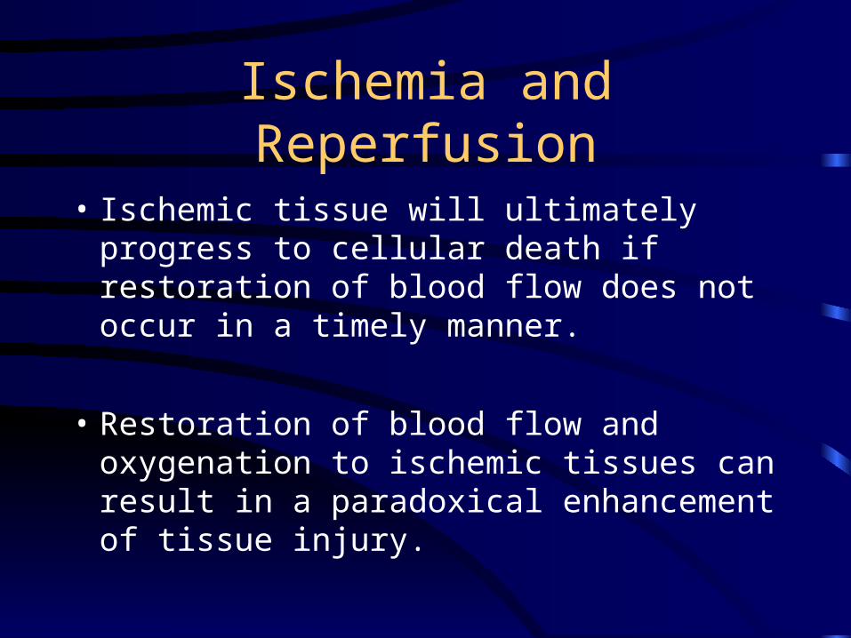
Ischemia and Reperfusion
• Ischemic tissue will ultimately progress to cellular death if restoration of blood flow does not occur in a timely manner.
• Restoration of blood flow and oxygenation to ischemic tissues can result in a paradoxical enhancement of tissue injury.
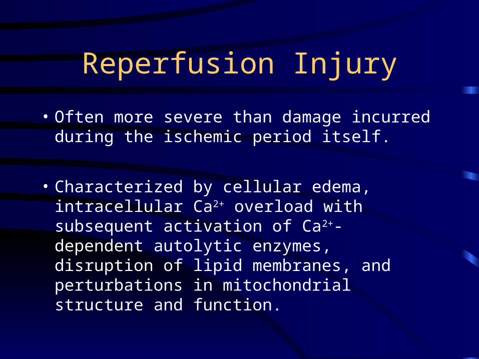
Reperfusion Injury
• Often more severe than damage incurred during the ischemic period itself.
• Characterized by cellular edema, intracellular Ca2+ overload with subsequent activation of Ca2+- dependent autolytic enzymes, disruption of lipid membranes, and perturbations in mitochondrial structure and function.
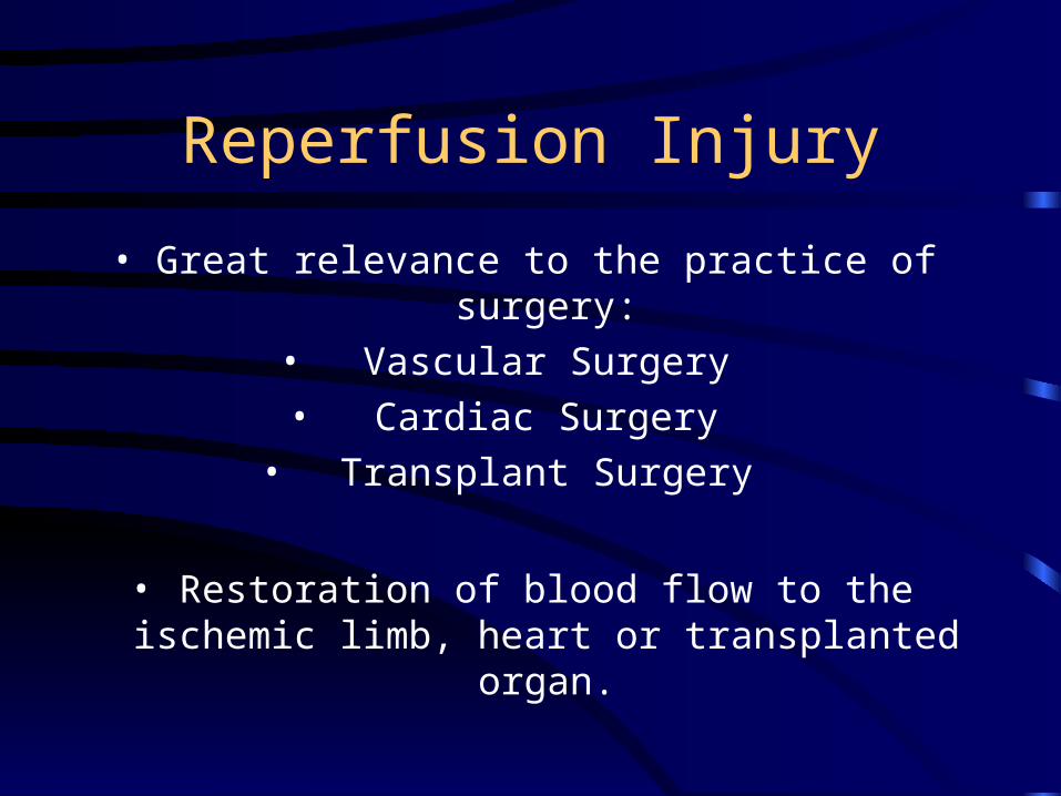
Reperfusion Injury
• Great relevance to the practice of surgery:
• Vascular Surgery
• Cardiac Surgery
• Transplant Surgery
• Restoration of blood flow to the ischemic limb, heart or transplanted organ.
Mediators of Reperfusion Injury
• Endothelial Cell
• Oxygen Free Radicals
• Polymorphonuclear Cells (PMNs)
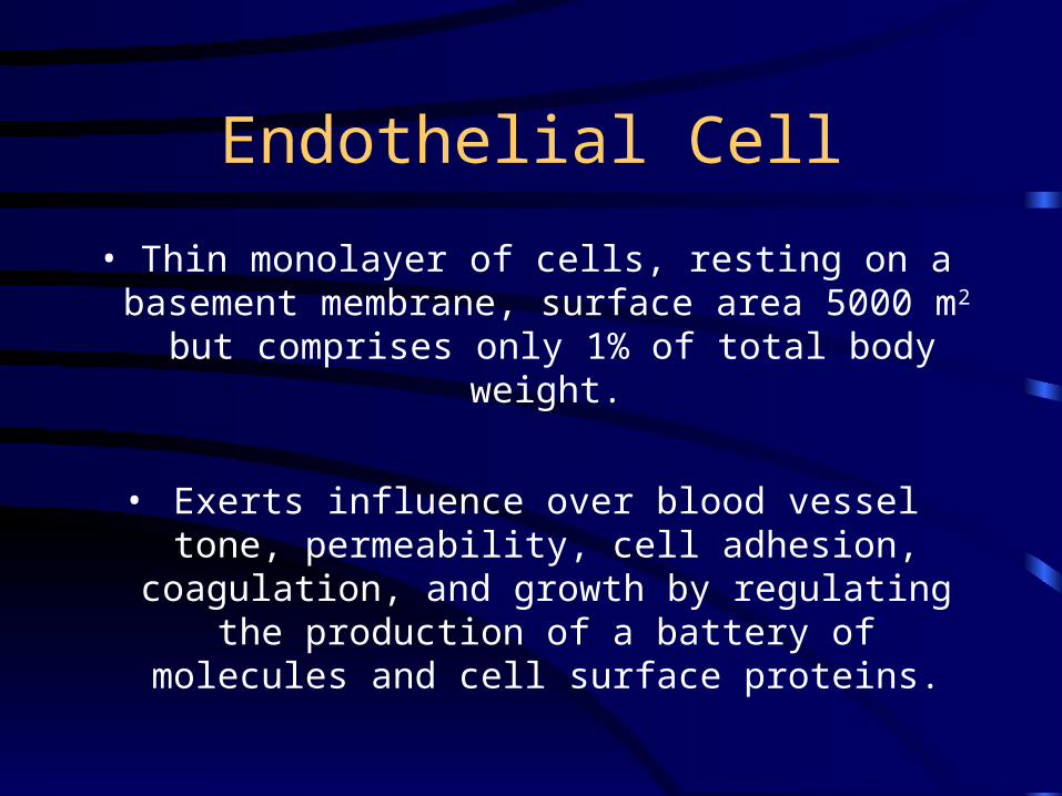
Endothelial Cell
• Thin monolayer of cells, resting on a basement membrane, surface area 5000 m2
but comprises only 1% of total body weight.
• Exerts influence over blood vessel tone, permeability, cell adhesion, coagulation, and
growth by regulating the production of a battery of molecules and cell surface proteins.
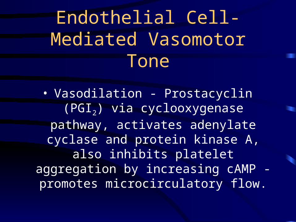
Endothelial Cell-Mediated Vasomotor Tone
• Vasodilation - Prostacyclin (PGI2) via cyclooxygenase pathway, activates
adenylate cyclase and protein kinase A, also inhibits platelet aggregation by increasing cAMP - promotes microcirculatory flow.
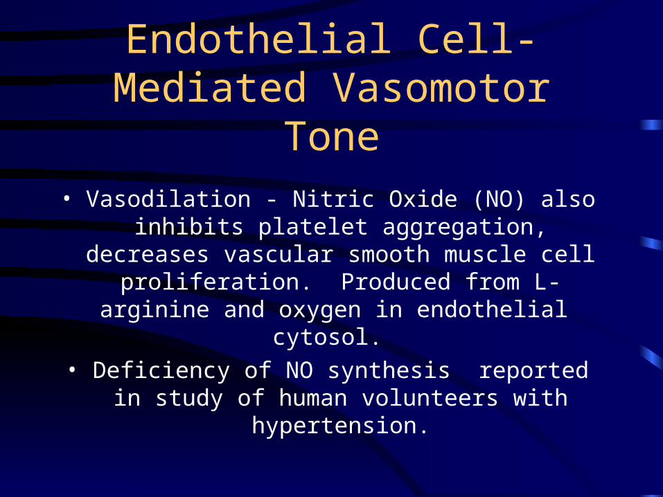
Endothelial Cell-Mediated Vasomotor Tone
• Vasodilation - Nitric Oxide (NO) also inhibits platelet aggregation, decreases vascular smooth
muscle cell proliferation. Produced from L-arginine and oxygen in endothelial cytosol.
• Deficiency of NO synthesis reported in study of human volunteers with hypertension.
Endothelial Cell-Mediated Vasomotor Tone
• Patients with diabetes, atherosclerosis, hypercholesterolemia, or cigarette smoking
also exhibit deficient NO synthesis.
• Adenosine - also a vasodilator, inhibits platelet and neutrophil aggregation.
Endothelial Cell-Mediated Vasomotor Tone
• Vasoconstriction - Thromboxane A2 (TXA2) via thromboxane synthase, opposes prostacyclin and produces platelet
adherence.
• Endothelin-1, most potent vasoconstrictor known, counteracts NO.
Endothelial Cell-Mediated Cell Adhesion
• Mediators formed during reperfusion induce endothelial cells to express intercellular
adhesion molecules (ICAM 1 and 2), endothelial leukocyte adhesion molecule
(ELAM) and selectins.• These receptors bind the CD11/CD18 complex
on activated, facilitating PMN adherence to and migration across endothelium.
Endothelial Cell
• Secretes an abundance of soluble factors which promote vasoconstriction, platelet
aggregation, PMN plugging of capillaries, and increased vascular permeability.
• Factors include: Platelet aggregating factor (PAF), LTB4, TXA2 and endothelin.
Endothelial Cell
• End result : perfusion of the microcirculation is severely compromised, which manifests as the classic “no-reflow”
phenomenon of reperfusion injury.
Oxygen Free Radicals
• Three different molecules to be aware of:
• Superoxide anion O2-
• Hydrogen peroxide H2O2
• Hydroxyl radical .OH
Oxygen Free Radicals
• Reperfusion stimulates xanthine oxidase which is activated in ischemic endothelial cells to generate superoxide radicals.
• PMNs also generate oxygen free radicals.
Oxygen Free Radicals
• These toxic moieties are rapidly generated at the onset of reperfusion and cause
widespread damage to cellular macromolecules.
• Peroxidation of lipid membranes, protein degradation, nucleic acid damage,
cytochrome inactivation and neutralization of nitric oxide.

Oxygen Free Radicals
• Most damaging effect is on lipid membranes, impairs normal fluidity and
permeability of cell membranes leading to cellular edema, massive Ca2+ and Na+
overload and cell lysis.
Oxygen Free Radicals
• Oxygen free radical scavengers and antioxidants have been shown both
experimentally and clinically to ameliorate reperfusion injury.

Oxygen Free Radicals
• Natural protective enzyme systems to reduce free radical damage include superoxide dismutase, catalase, and
glutathione peroxidase.
• Most important endogenous antioxidant is glutathione. N-acetylcysteine is an artificial
glutathione precursor.
Activated PMNs
• Inflict damage to reperfused endothelial and parenchymal cells.
• Release a host of destructive proteolytic enzymes, including elastase, collagenase,
gelatinase, lysozyme, and cathepsin G.
Activated PMNs
• Source of oxygen free radicals by virtue of a superoxide generating NAD oxidase.
• Produce hypochlorous acid by activity of myeloperoxidase.
Reduction of Reperfusion Injury
• Allopurinol - inhibitor of xanthine oxidase has been shown to have protective effects.
• Desferrioxamine - an iron chelator, removes an essential cofactor for the generation of hydroxyl radical.
Reduction of Reperfusion Injury
• Vitamin E - prevents neutrophil accumulation and attenuates tissue damage in ischemic-reperfused human skeletal muscle.
• N-acetylcysteine - pretreatment 30 minutes before infrarenal aortic clamping may help prevent reperfusion injury.
Ischemia, Reperfusion Injury and Compartment Syndrome
• Elevated pressure within a confined tissue space.
• High energy injuries.
• Pain out of proportion to injury.
• Most commonly occurs in the leg.
Ischemia, Reperfusion Injury and Compartment Syndrome
• Four compartments in leg:
• Anterior: anterior tibial artery, deep peroneal nerve, extensor muscles of toes and foot
• Lateral: superficial peroneal nerve, peroneal brevis and longis muscle

Ischemia, Reperfusion Injury and Compartment Syndrome
• Deep posterior: tibial nerve, posterior tibial artery, peroneal artery, deep toe and foot flexor muscles
• Superficial posterior: superficial foot flexor muscles
• Examining leg, document sensation at first web space (deep peroneal nerve), dorsum of foot (superficial peroneal nerve) and plantar surface of foot (tibial nerve)
Ischemia, Reperfusion Injury and Compartment Syndrome
• Pressure threshold at which fasciotomy is indicated has been debated.
• 30-40 mm Hg
• Can use arterial pressure transducer, IV tubing, 3 way stopcock, 20 ml syringe, a 16Ga needle.
• Four compartments should be measured