Embed Size (px)
Citation preview

1
Rehabilitation in Sudden Onset Disasters
RP 06
Edited by Pete Skelton and Alice Harvey

2
EditorsAlice HARVEYPeter SKELTON
ContributorsWCPTADAPTACPINBACPARInterburnsBAHT
AcknowledgmentsMary Jane Cole, RuthAnn Fanstone, Zoe Clift, Diana Hiscock, Felicity Gillham, Emma Cook, Sue Paddison, and Amelia Shaw. Thanks to content reviewers Eric Weerts, Sylvain Rouaud, Berangere Gohy, Steve Mannion, Jo Armostrong, Jo Woodrow, Fiona Stephenson, Letitia Rabey, Paula Dimarco, Amy Hughes, Anne Vicker, Pip Joubert, Anna Rose, Kathryn Sizer, Julia Earle, Joy Rendell, Laura bockholtz, Julie Dixon, Berangere Gohy and Catherine Sykes.
1st Edition, September 2015 Handicap International/ UK Emergency Medical Team
Photograph CreditsCover: © William DANIELS / Handicap InternationalAll images used within chapters are used with the permission of the chapter authors.
You are free to Share — copy and redistri-bute the material in any medium or format, under the following terms:
Attribution: You must give appropriate credit, provide a link to the license, and indicate if changes were made. You may do so in any reasonable manner, but not in any way that suggests the licensor endorses you or your use.
Non Commercial : You may not use the material for commercial purposes.
No Derivatives: If you remix, transform, or build upon the material, you may not distribute the modified material.
238
Chapter
8Acquired Brain InjuryThis module was developed by Association of Chartered Physiotherapists in Neurology (ACPIN) members with a special interest in brain injury, working in collaboration with Handicap International.
The acquired brain injury chapter aims to provide an overview of how to provide acute rehabilitation for patients with brain injury in an austere, emergency situation, when working as part of a medical team. It is based on best available evidence in the UK, with consideration for particular challenges seen in a humanitarian environment.
Introduction
The focus of the chapter will be on acquired brain injury (ABI) and therefore covers patients who have had a traumatic brain injury as well as those whose brain injury occurs from other circumstances (e.g. hypoxia). An acquired brain injury (ABI) is defined as:
“Damage to the brain, which occurs after birth and is not related to a congenital or a degenerative disease. These impairments may be temporary or permanent and cause partial or functional disability or psychosocial maladjustment.”
The term Traumatic brain injury (TBI) is used to specifically describe a brain injury that has occurred when a sudden trauma causes damage to the brain. This can be as a result
of the head suddenly and violently hitting an object, or when an object pierces the skull and enters brain tissue. (World Health Organization, 1996). As such, an acquired brain injury will include TBI, hypoxic brain injury, brain tumour, brain haemorrhage and inflammatory diseases of the brain, such as encephalitis. For the purpose of this chapter, the term ABI will be used in order to encompass all aspects of brain injury that may be found in a humanitarian setting.The term Stroke (or Cerebrovascular accident (CVA)) is often excluded from ABI in literature as it has its own specific guidelines. For the purposes of this manual, only where there is a clear distinction to be made will stroke management be discussed separately from ABI management otherwise ABI and stroke will be discussed together.
ABI in sudden onset disasters
Learning outcome
To understand the aetiology and incidence of brain injury in a humanitarian situation.
239
There is often a lack of clarity around the presentation of brain injury patients in disasters, particularly in the data and available research, often with little differentiation made between head injury (such as simple lacerations) and brain injury. As a result, there is very limited useful data on the number or severity of brain injury following disasters, and almost no data on the long term outcomes of patients with brain injuries.
Traumatic Brain injuries can result from falling debris, such as in earthquakes, or falling or flying debris in wind storms. Hypoxic injuries may result from near drownings from Tsunamis or windstorms. Mild injuries may frequently be missed due to focus on other life threatening injuries and polytrauma, while in areas where rescue is slow and pre-hospital care poor, there is a low likelihood of severe traumatic brain injury cases surviving extraction/evacuation.
A review in the Lancet by Batels et al found that brain injuries were a leading cause of death in disasters. For example, 30% of people affected by the earthquake in Taiwan in 1999 reportedly died from head injuries. In survivors, scalp lacerations account for between 43% and 65% of head injuries, while 4% are concussions. Most earthquake-induced head injuries were mild (55%) or mild-to-moderate (85%) in severity, while skull fractures were seen in between 8% and 28% of people with head injuries. Basal skull fractures have a reported frequency of 11%. Because many injuries are minor, surgical intervention is not usually required.
Bhatti et al (2008) examined cases received in a major trauma centre following the Pakistan earthquake. Of significance, they had intensive care and ventilator capability and were a key referral centre. They found that delayed evacuation contributed to high number of deaths (only 35% of head injuries were received in first 72 hours). 8.5% of all patients received had head injuries, and the majority of wounds were contaminated. The overall mortality increased from 3.3% to 7% in one year follow-up.
Where health care systems are overwhelmed, rescue is difficult or there is limited access to ventilators, the low survival rates of severely injured patients is likely to be exacerbated. In addition, a lack of ventilators in low income countries, coupled with limited access to specialists, means that the quality of acute and after care for patients with severe injuries is likely to be limited. For example, in Indonesia there are 140 neurosurgeons for 250 Million people, while in the USA there are 3,500 neurosurgeons for 299million.
Disasters also disrupt existing health care systems, limiting access to medication and normal care for non-communicable diseases such as hypertension, while at the same time increasing stressors in populations. As a result, it is very likely that there will be an increase in CVAs following disasters (e.g. Mateen et al 2010).
While the purpose of this manual is to cover sudden onset disasters, it is worthy of note that those working in conflict situations may see a significantly higher number of ABI due to penetrating trauma and blast injury.
Classification of ABI
Learning outcomes
To understand the classification of ABI
To be able to identify a deteriorating patient and know what action to take
To be able to identify some possible differential diagnoses

240
Recap of Anatomy
Acquired Brain Injury
Anatomy
There are two cerebral hemispheres (right and left) of the brain. Each cerebral hemisphere is comprised of four lobes: frontal, temporal, parietal and occipital. The two cerebral hemispheres are separated by the great longitudinal fissure which contains the corpus callosum. The corpus callosum is made up of commissural fibres that connect corresponding regions of the two hemispheres.
The brain also has the following other important structures: brain stem (comprised of the medulla oblongata, pons and midbrain); cerebellum and the ventricular system.
The ventricles are chambers within the brain that connect with the spinal cord. It is here where cerebrospinal fluid (CSF) is produced and circulates.
The brain and spinal cord are covered by a 3 layers of protective membranes, collectively called the meninges. These layers are: dura mater (the most external layer lying between the brain and skull); arachnoid mater (the middle layer), and the pia mater (the closet lining of the brain).
CerebellumBrain Stem
Occipitallobe
Parietal lobeFrontal lobe
Temporal lobe

241
Anatomy
These layers enclose capillaries and CSF and provide a cushion and protection to the delicate brain tissue.
The skull acts as a ‘closed box’ which exerts a normal pressure referred to as Intracranial Pressure (ICP). This ICP is normally finely controlled to ensure adequate brain tissue perfusion with oxygen and nutrients. Any insult to the brain tissue such as a bleed would disrupt this normal ICP, causing a rise in ICP. This rise results in the compromise of brain tissue perfusion and thus can cause further insult to brain tissue.
With a head injury of a traumatic nature you should always consider there may be an insult to the cervical spine. NICE guidelines recommend you consider the mechanism of injury and nature of neurological limb deficit or paraesthesia; and see how this may fit with a corresponding injury. For example, following the Haiti earthquake the patient
below, with a history of a closed head injury was referred to physiotherapy for massage due to a torticollis:
242
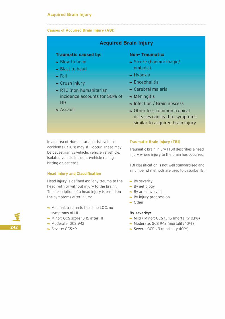
Traumatic caused by: Blow to head Blast to head Fall Crush injury RTC (non-humanitarian incidence accounts for 50% of HI)
Assault
Non- Traumatic: Stroke (haemorrhagic/embolic)
Hypoxia Encephalitis Cerebral malaria Meningitis Infection / Brain abscess Other less common tropical diseases can lead to symptoms similar to acquired brain injury
Acquired Brain Injury
Causes of Acquired Brain Injury (ABI)
In an area of Humanitarian crisis vehicle accidents (RTC’s) may still occur. These may be pedestrian vs vehicle, vehicle vs vehicle, isolated vehicle incident (vehicle rolling, hitting object etc.).
Head Injury and Classification
Head injury is defined as: “any trauma to the head, with or without injury to the brain”.The description of a head injury is based on the symptoms after injury:
Minimal: trauma to head, no LOC, no symptoms of HI
Minor: GCS score 13-15 after HI Moderate: GCS 9-12 Severe: GCS <9
Traumatic Brain Injury (TBI)
Traumatic brain injury (TBI) describes a head injury where injury to the brain has occurred.
TBI classification is not well standardised and a number of methods are used to describe TBI:
By severity By aetiology By area involved By injury progression Other
By severity: Mild / Minor: GCS 13-15 (mortality 0.1%) Moderate: GCS 9-12 (mortality 10%) Severe: GCS < 9 (mortality 40%)
Acquired Brain Injury
243
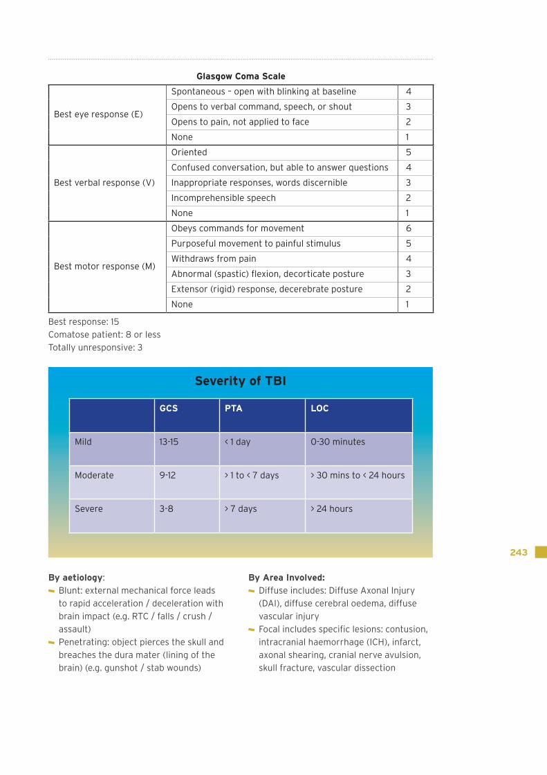
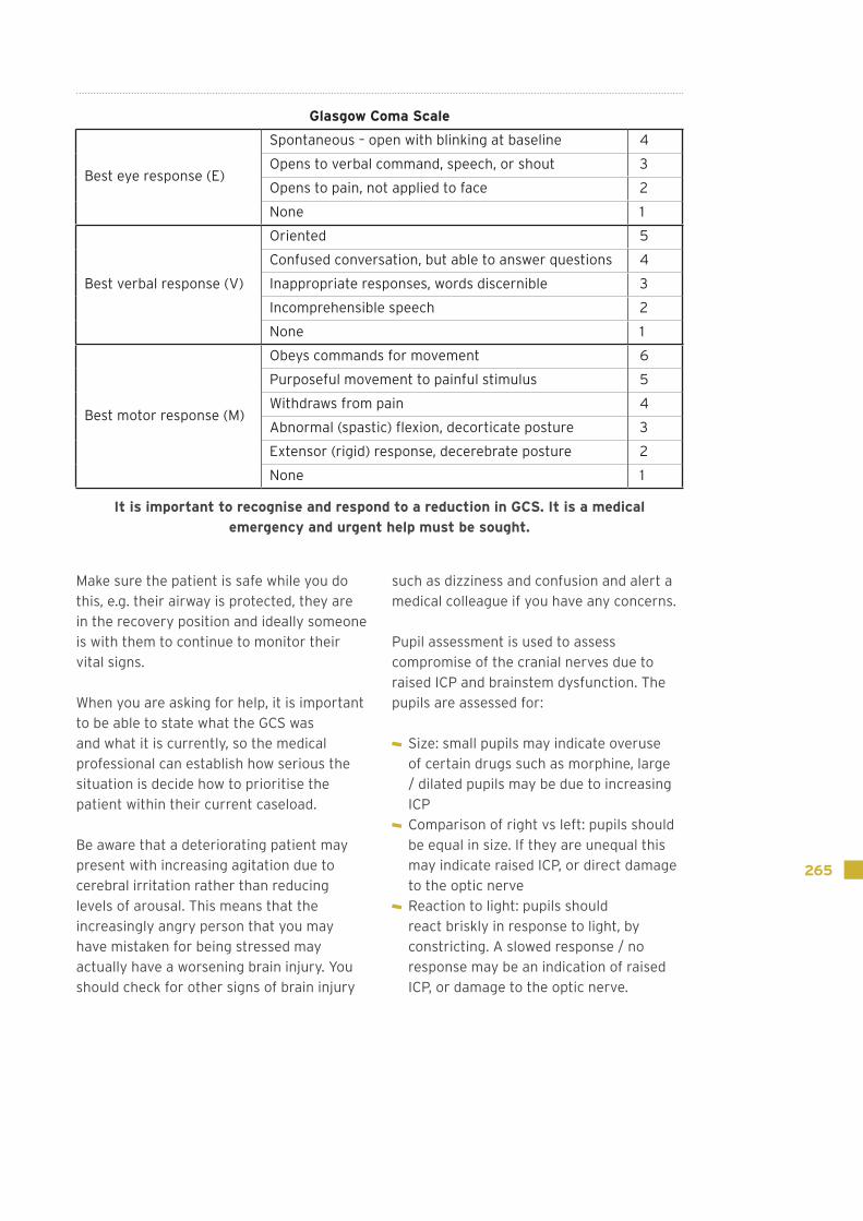
Glasgow Coma Scale
Best eye response (E)
Spontaneous – open with blinking at baseline 4
Opens to verbal command, speech, or shout 3
Opens to pain, not applied to face 2
None 1
Best verbal response (V)
Oriented 5
Confused conversation, but able to answer questions 4
Inappropriate responses, words discernible 3
Incomprehensible speech 2
None 1
Best motor response (M)
Obeys commands for movement 6
Purposeful movement to painful stimulus 5
Withdraws from pain 4
Abnormal (spastic) flexion, decorticate posture 3
Extensor (rigid) response, decerebrate posture 2
None 1
Best response: 15Comatose patient: 8 or lessTotally unresponsive: 3
Severity of TBI
GCS PTA LOC
Mild 13-15 < 1 day 0-30 minutes
Moderate 9-12 > 1 to < 7 days > 30 mins to < 24 hours
Severe 3-8 > 7 days > 24 hours
By aetiology: Blunt: external mechanical force leads to rapid acceleration / deceleration with brain impact (e.g. RTC / falls / crush / assault)
Penetrating: object pierces the skull and breaches the dura mater (lining of the brain) (e.g. gunshot / stab wounds)
By Area Involved: Diffuse includes: Diffuse Axonal Injury (DAI), diffuse cerebral oedema, diffuse vascular injury
Focal includes specific lesions: contusion, intracranial haemorrhage (ICH), infarct, axonal shearing, cranial nerve avulsion, skull fracture, vascular dissection

244
By Injury Progression Primary: due to immediate forces (occurs at time of injury, cannot be altered) e.g. skull fracture, contusion, haemorrhage, axonal shearing
Secondary: due to evolving pathophysiological processes and their consequences, the multitude of neurobiological cascades that are altered or initiated at cellular level in response to the primary injury e.g. cerebral oedema, raised ICP, haemorrhage, seizure, ischaemia, infection
Other:Open: penetration to the brain / breach of the dura mater
Closed: no breach of dura mater / skull
Skull fractures may be open or closed. They most commonly involve the temporal,
occipital or base of skull due to the initial impact and / or transmission of forces through the skull.
Generally GCS is used as a measure of severity of Brain Injury at the time of incident but also any persisting change in GCS (SIGN guidelines).
It is very unlikely that you will see patients in the severe category, as these patients will not survive. Usual levels of mortality for moderate TBI is 10%, this is likely to be much higher in humanitarian areas due to difficulty gaining access to specialist units.
Using LOC as a guide within your initial subjective assessment may be a useful indicator of potential deficits even if these are not immediately apparent.
Types of injury and relevance
TBI: Types of Injury and relevance
Skull fracture Haematomas (EDH/SDH) Haemorrhagic injury (SAH / intracerebral / contusions / IVH)
Diffuse Axonal Injury Hypoxic / Anoxic / Cerebral oedema (diffuse swelling)
Blast / penetrating
* Concussion
EDH
SAH SAH IVH Diffuse Swelling
Contusion/Hematoma DAI
Acquired Brain Injury

245
Skull fracture: A number of different types may occur: simple (small linear fracture, tend to be closed); compound (comprised of many pieces of bone, open or closed); depressed (fracture has indented so that the shape of the skull is lost).
Skull fractures may be: Open (more likely) or closed (only if dura is not breached). If open they be compromising the brain tissue underneath and causing contusion / haemorrhage and the fracture should be elevated if this is occurring.
Base of skull – may be associated with CSF leak from nose (rhinorrhoea) or ears (otorrhoea), and are of relevance due to incidence of infection that may occur sometime later, presenting either as meningitis or cerebral abscess
Skull fractures may be identified by extracranial bleeding, swelling and pain, obvious deformity, bruising (of significance for complex facial / BOS fractures are Racoon/Panda eyes or Battles sign)
Haematomas: Haematoma lying above dura (extradural) or within the dura (sub dural): Injury is caused by pressure effect on surrounding areas of an expanding haematoma, leading to ischaemia and structural deformity rather than direct damage to the brain parenchyma. Haemorrhagic injury can also lead to ischaemia / infarct.
Of note is that a patient with minor HI, particularly in the elderly, can present a few weeks later with a Chronic SDH which causes a slow onset of neurological deficit, as bleeding can continue to occur within the subdural space.
Haematoma within brain parenchyma: Haemorrhagic injury that occurs to the brain parenchyma directly by damage causing contusions (bleeding into / bruising of brain tissue), can be coup (same side of impact) or contre-coup (opposite side of impact).
Often contusions / haemorrhage are seen in frontal and temporal areas due to the brain being forced forwards against the skull vault and the bony prominences that make up the inner surface of the skull.
These result in swelling and ischaemia of the focal area of brain tissue, and as they enlarge may cause pressure effects on surrounding areas of the brain. Therefore subsequent neurological deficits may occur.
Other Haemorrhagic injury: Traumatic subarachnoid haemorrhage (SAH) is common, where there is bleeding within the sub-arachnoid space. Intraventricular haemorrhage (IVH) is bleeding into the ventricles. Both SAH and IVH can cause secondary hydrocephalus and is frequently associated with other injuries, and both can cause neurological deficit.
Diffuse axonal injury: disruption of axons (shearing of white matter) usually caused by acute deceleration injury (RTC). Commonly associated with more severe TBI, and significant neurological deficit.
Hypoxic injury / Anoxia: Hypoxia (reduced oxygen levels to the brain) caused by inadequate respiration / ventilation possibly as a result of reducing conscious level or other severe injuries to the thorax
Cerebral oedema: a secondary injury, related to hypoxia, increased CO2, and the secondary pathological processes that occur following primary brain injury
Concussion / Mild TBI
A term used interchangeably with mild TBI and minimal / minor head injury. There is no consensus regarding a definition. There appears to be general agreement that mild TBI / concussion is due to a blunt or mechanical force that results in some type of transient confusion, disorientation or loss of consciousness lasting not more than 30 minutes, and possibly associated with transient neurobehavioural deficits and a GCS no worse than 13-15.

246
It is likely that the majority of patients who suffer TBI have minor TBI / concussion but will not require admission.
Mild TBI can be described as 1 or more of the following:
any loss of consciousness up to 30 min any loss of memory for events immediately before or after the accident for as much as 24 h
any alteration of mental state at the time of the accident (eg, feeling dazed, disoriented, or confused)
OR:
focal neurologic deficits that might or might not be transient, but where the severity of the injury does not exceed loss of consciousness exceeding 30 min
posttraumatic amnesia longer than 24 h a Glasgow Coma Scale score falling below 13 after 30 min.
Even with a mild brain injury, there is still an interruption in the physiology of the brain and this can lead to a number of symptoms.
Symptoms of a mild TBI can be physical, behavioural, emotional or cognitive and may be missed in patients with other significant traumatic injuries (such as crush / limb fractures)
Persistent symptoms following mild TBI might occur in 10% to 15% of patients and can include post-traumatic headache, sleep disturbance, disorders of balance, cognitive impairments, fatigue, and mood disorders.
Although these symptoms should resolve, persistent post-concussive symptoms can
result in functional disability, stress, time away from work / school and reduced quality of life (Marshall et al, 2012). This can be exacerbated by the patient attempting to return to “normal” too quickly, which often results in the symptoms worsening.
It is important that all patients suspected of having a head injury undergo an assessment in order that these potential symptoms can be highlighted to the patient and their families and management strategies commenced as early as possible where necessary.
Signs and Symptoms of Mild TBI
Headache Sleep disturbance Disorientation Dizzy/nauseous Fatigue Irritability Altered mood Difficulty concentrating Difficulty remembering
Acquired Brain Injury

247
It is also important that the patient and their family are provided with information, advice and reassurance at the time of their brain injury, as unless they have other injuries requiring medical management they will be discharged.
As some of the symptoms of mild TBI are associated with stress, people who are caught in a humanitarian crisis could be misdiagnosed as having a stress response to the event and their mild head injury is missed. This is where your observational
skills will be necessary. Don’t dismiss someone as medically well if they are distressed but appear uninjured. Look at their face, is there any bruising? This could be a sign of an underlying head injury, particularly those with black eyes (panda eyes) who could have a simple broken nose or a more serious fractured base of skull.
Do they have any wounds to the head or, if they will allow you to feel, can you feel any lumps or bumps? Again, these could be signs of an underlying head injury.
Increased drowsiness (feeling sleepy for >1hr when normally would be awake)
GCS: Sustained drop of GCS / drop of 3 points / GCS < 13
Altered respiratory pattern / signs of aspiration
Problems with eyesight / double vision / photophobia / nystagmus
Deteriorating unremitting/ headache significantly worse in mornings
Vomiting (being sick) Seizures (also known as convulsions or fits)
CSF leak / sign of infection Double incontinence
Onset / worsening of neurological deficit:
Weakness of one or more limbs (pronator drift)
Communication problems (difficulty with speech or comprehension)
Behavioural / cognitive changes
Changes in size / reactivity of pupils , failure of upward gaze
Changes in CVS / Respiratory status: HR / BP / RR
Loss of balance / co-ordination, or problems walking
Signs and Symptoms of TBI of concern Deteriorating: Mild / Moderate / Severe
Patient information leaflets are included for those with concussion and mild to moderate ABI.

248
Deterioration following TBI
Anyone with a mild head injury is at risk of deterioration as the brain starts to swell. Signs and symptoms that indicate deterioration in their condition include:
unconsciousness, or lack of full consciousness (e.g having difficulty keeping eyes open)
drowsiness (feeling sleepy)that goes on for longer than 1 hour when they would normally be wide awake
problems understanding or speaking loss of balance or difficulty walking weakness in one or more limb problems with eyesight painful headaches that won’t go away vomiting seizures (also known as convulsions / fits) clear fluid coming out of the ear / nose bleeding from one or both ears
The onset of any of these symptoms should be treated with concern, and necessitate discussion / review by a medical member of the team.
Therefore, following a mild TBI the family need to be provided with information about signs to look out for that indicates a patient is showing early signs of deterioration of their brain injury. They also need information on their longer term strategies to overcome the effects of a mild TBI.
In the UK, all A&E departments or GP practices will hand out a head injury alert card for signs and symptoms to look out for following a mild brain injury. A leaflet based on these has been developed for the UKIETR. If any of the symptoms occur, the advice is to seek medical assistance immediately (e.g. NICE 2014).
As well as observing for signs of deterioration, the patient is also advised: not to be alone in the first few days (NICE, 2014); to make sure they have a phone / a method available to contact medical help quickly; they have plenty of rest and avoid stressful situations; not to take alcohol or social drugs; not to take any medication that has not been prescribed (especially sleeping pills / sedatives / tranquillisers).
In a humanitarian crisis where the patient may be separated from family or friends, it is important to consider where they can be looked after that doesn’t mean that they are using an acute bed that a more seriously injured patient needs, but they are still safe.
It is best practise for someone who has suffered a mild TBI to be followed up by a specialist (in the UK it is usually a specialist head injury nurse), at one week post injury. These are used to review symptoms, identify ongoing problems and initiate referrals to appropriate services (often neuropsychology). Access to such specialist services is unlikely to be available in a Humanitarian situation, and therefore the team will need to decide how to / who requires follow up.
Functional Anatomy
To assist your assessment, identification of problems and clinical reasoning it is useful to understand basic functional neuroanatomy as specific areas of the brain control and regulate specific functions (physical and cognitive).
Acquired Brain Injury
249
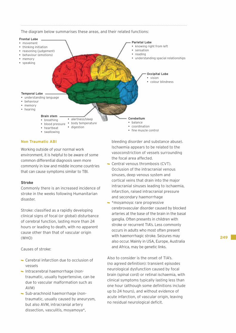
The diagram below summarises these areas, and their related functions:
Non Traumatic ABI
Working outside of your normal work environment, it is helpful to be aware of some common differential diagnosis seen more commonly in low and middle income countries that can cause symptoms similar to TBI.
StrokeCommonly there is an increased incidence of stroke in the weeks following Humanitarian disaster.
Stroke: classified as a rapidly developing clinical signs of focal (or global) disturbance of cerebral function, lasting more than 24 hours or leading to death, with no apparent cause other than that of vascular origin (WHO)
Causes of stroke:
Cerebral infarction due to occlusion of vessels
Intracerebral haemorrhage (non-traumatic, usually hypertensive, can be due to vascular malformation such as AVM)
Sub-arachnoid haemorrhage (non-traumatic, usually caused by aneurysm, but also AVM, intracranial artery dissection, vasculitis, moyamoya*,
bleeding disorder and substance abuse). Ischaemia appears to be related to the vasoconstriction of vessels surrounding the focal area affected.
Central venous thrombosis (CVT). Occlusion of the intracranial venous sinuses, deep venous system and cortical veins that drain into the major intracranial sinuses leading to ischaemia, infarction, raised intracranial pressure and secondary haemorrhage
*moyamoya: rare progressive cerebrovascular disorder caused by blocked arteries at the base of the brain in the basal ganglia. Often presents in children with stroke or recurrent TIA’s. Less commonly occurs in adults who most often present with haemorrhagic stroke. Seizures may also occur. Mainly in USA, Europe, Australia and Africa, may be genetic links.
Also to consider is the onset of TIA’s.(no agreed definition): transient episodes neurological dysfunction caused by focal brain (spinal cord) or retinal ischaemia, with clinical symptoms typically lasting less than one hour (although some definitions include up to 24 hours), and without evidence of acute infarction, of vascular origin, leaving no residual neurological deficit.
Frontal Lobe• movement• thinking initiation• reasoning (judgement)• behaviour (emotions)• memory• speaking
Temporal Lobe• understanding language• behaviour• memory• hearing
Brain stem• breathing• blood pressure• heartbeat• swallowing
Parietal Lobe• knowing right from left• sensation• reading• understanding spacial relationships
Occipital Lobe• vision• colour blindness
Cerebellum• balance• coordination• fine muscle control
• alertness/sleep• body temperature• digestion

250
Of relevance for your assessment are the following:
Time: Time since onset may be relevant in humanitarian settings as the patient may have travelled a distance before receiving medical attention. Also relevant to time and the differential diagnosis is:
the order of onset of the symptoms since the onset of symptoms have these remained constant, or have they progressed, or are they fluctuating?
Is the level of consciousness remaining the same, or does it fluctuate, or is it worsening?
Were there are any signs of TIA
previously? Was headache sudden worst headache ever (SAH often described as if hit on the back of the head)
Is headache remaining constant or increasing?
Is headache worse in the morning?
Vascular functional anatomy relevant to StrokeAs Stroke is commonly related to the vascular circulation a brief overview of the functional impact of certain vessels may be useful (especially when considering signs and symptoms when predicting outcome).
The signs and symptoms of stroke are as follows in the diagram below:
Non haemorrhagic FAST:
Facial weakness Arms Speech (Time) / time since onset
Weakness of one side of body Blurred vision / loss of sight Confusion Dizziness / unsteadiness Headache Swallowing problems LOC
HaemorrhagicAs non haemorrhagic but also:
characterised by sudden onset of severe persistent headache
vomiting LOC Stiff neck Irritability Poor memory Fluctuating neurological deficit
Signs and Symptoms of Stroke
Acquired Brain Injury
251
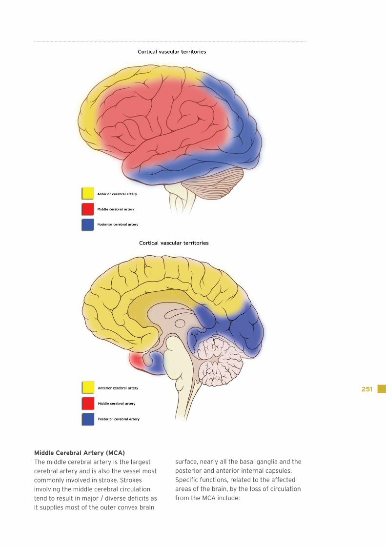
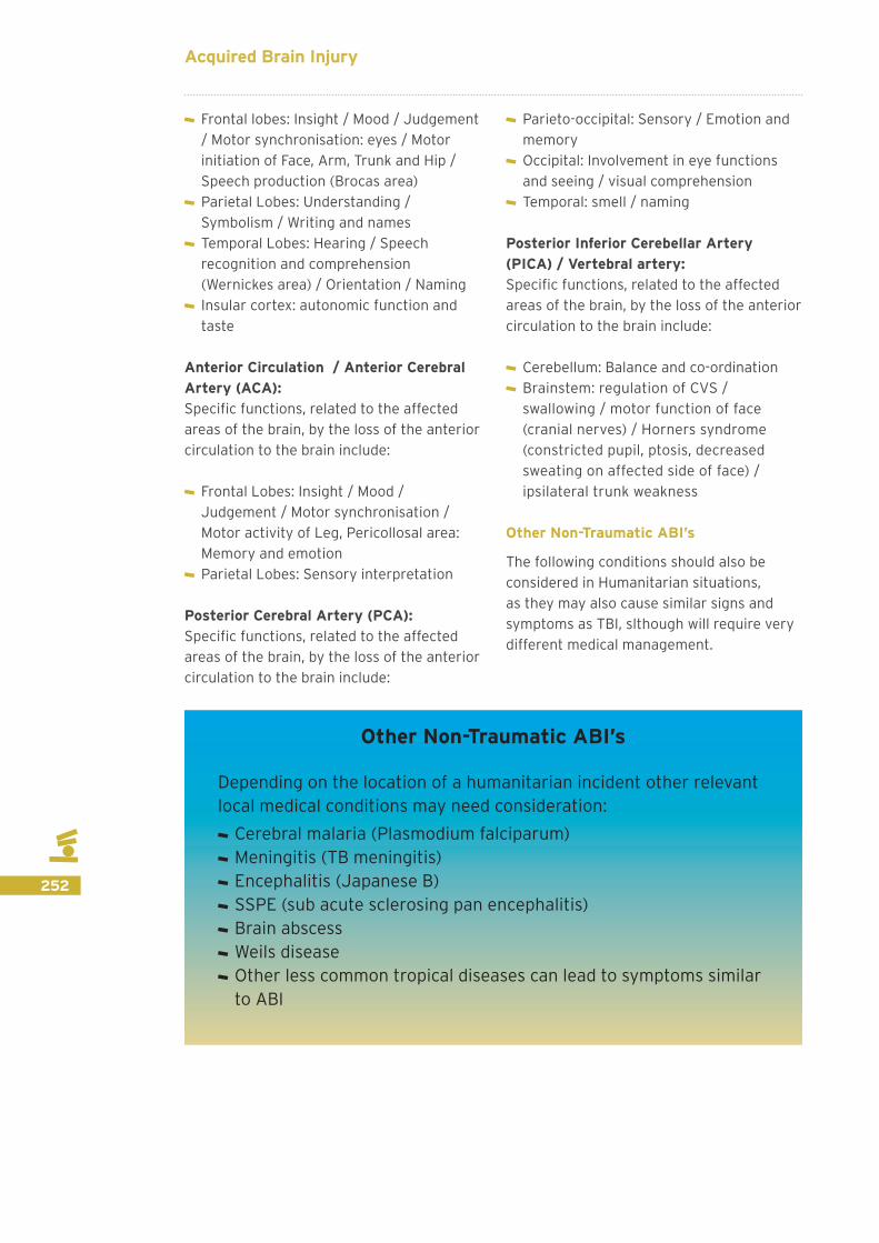
Middle Cerebral Artery (MCA)The middle cerebral artery is the largest cerebral artery and is also the vessel most commonly involved in stroke. Strokes involving the middle cerebral circulation tend to result in major / diverse deficits as it supplies most of the outer convex brain
surface, nearly all the basal ganglia and the posterior and anterior internal capsules.Specific functions, related to the affected areas of the brain, by the loss of circulation from the MCA include:
252
Frontal lobes: Insight / Mood / Judgement / Motor synchronisation: eyes / Motor initiation of Face, Arm, Trunk and Hip / Speech production (Brocas area)
Parietal Lobes: Understanding / Symbolism / Writing and names
Temporal Lobes: Hearing / Speech recognition and comprehension (Wernickes area) / Orientation / Naming
Insular cortex: autonomic function and taste
Anterior Circulation / Anterior Cerebral Artery (ACA):Specific functions, related to the affected areas of the brain, by the loss of the anterior circulation to the brain include:
Frontal Lobes: Insight / Mood / Judgement / Motor synchronisation / Motor activity of Leg, Pericollosal area: Memory and emotion
Parietal Lobes: Sensory interpretation
Posterior Cerebral Artery (PCA):Specific functions, related to the affected areas of the brain, by the loss of the anterior circulation to the brain include:
Parieto-occipital: Sensory / Emotion and memory
Occipital: Involvement in eye functions and seeing / visual comprehension
Temporal: smell / naming
Posterior Inferior Cerebellar Artery (PICA) / Vertebral artery:Specific functions, related to the affected areas of the brain, by the loss of the anterior circulation to the brain include:
Cerebellum: Balance and co-ordination Brainstem: regulation of CVS / swallowing / motor function of face (cranial nerves) / Horners syndrome (constricted pupil, ptosis, decreased sweating on affected side of face) / ipsilateral trunk weakness
Other Non-Traumatic ABI’s
The following conditions should also be considered in Humanitarian situations, as they may also cause similar signs and symptoms as TBI, slthough will require very different medical management.
Other Non-Traumatic ABI’s
Depending on the location of a humanitarian incident other relevant local medical conditions may need consideration:
Cerebral malaria (Plasmodium falciparum) Meningitis (TB meningitis) Encephalitis (Japanese B) SSPE (sub acute sclerosing pan encephalitis) Brain abscess Weils disease Other less common tropical diseases can lead to symptoms similar to ABI
Acquired Brain Injury
253
The most common are described below:
Cerebral malaria: There are 3 strains of malaria: Faciparum, Vivax and Malariae.
Cerebral malaria is a complication of the infection Plasmodium falciparum (and not the other strains). It is only found in areas where malaria is endemic and is most common in children. It is part of a multi-organ disease. Little is known about the actual pathogenesis.
It is most common in children and leads to reduced consciousness, vomiting and behaviour change along with the high fever associated with malaria. If treated with anti-malarials then the symptoms are reversible, but if the malaria is left untreated then permanent brain damage is possible.
Meningitis: bacterial (worse type) or viralThe presentation of meningitis is similar to cerebral malaria and other forms of acquired brain injury (reduced consciousness, vomiting, increased tone, seizures, paralysis, cognitive and behavioural change) but, unlike stroke or TBI, will be associated with high fever and photosensitivity.
TB meningitis should be suspected if the patient has had contact with TB and has a history of night fevers, cough and weight loss – it will also not respond as well to standard antibiotic or antiviral treatment – TB treatment is required. Again swift diagnosis and treatment will reduce the possibility of lasting brain damage
Encephalitis: Japanese encephalitis may be common in humanitarian areas, and is spread by mosquito bites.
Commonly symptoms start with flu like symptoms with high fever, headache, nausea, vomiting, and joint pain. This is
followed by the onset of some / all of the following: confusion, disorientation, seizures, LOC, photophobia, inability to speak, inability to initiate movement, stiff neck, hallucinations, loss of vision, involuntary eye movements, rash (which may be specific to the virus (herpes simplex virus causes characteristic blisters on the skin, around eyes and in the mouth)
As this is usually viral in nature, treatment is more difficult. Again if a fever is present then encephalitis should be on your differential diagnosis list.
SSPE: SSPE is caused by the measles virus, and there is commonly a long time period between the occurrence of measles and the onset of SSPE. Initially symptoms may include mild mental deterioration such as memory loss and changes in behaviour (irritability), followed by: disturbances in motor function; myoclonus (involuntary jerking movements of head; trunk of limbs); and seizures. There is a progression to the loss of ability to walk due to spasticity / spasms, blindness may occur and eventually there is a progression to coma, Persistent Vegetative State (PVS) and death (1 - 3 years prognosis).
Brain abscess: A brain abscess is a local collection of infected material from either a local (usually ear, nose, tooth – linked to poor dental hygiene), remote, or direct (secondary to infected skull fracture / following cranial surgery) source of infection
Presentation is similar to meningitis and encephalitis and often escalates over a period of 2-4 weeks, with fever, headache, vomiting and neurological symptoms. Treatment is with antibiotics (IV) if the source is bacterial. However abscesses can be caused by parasites, protoza and fungi.
254
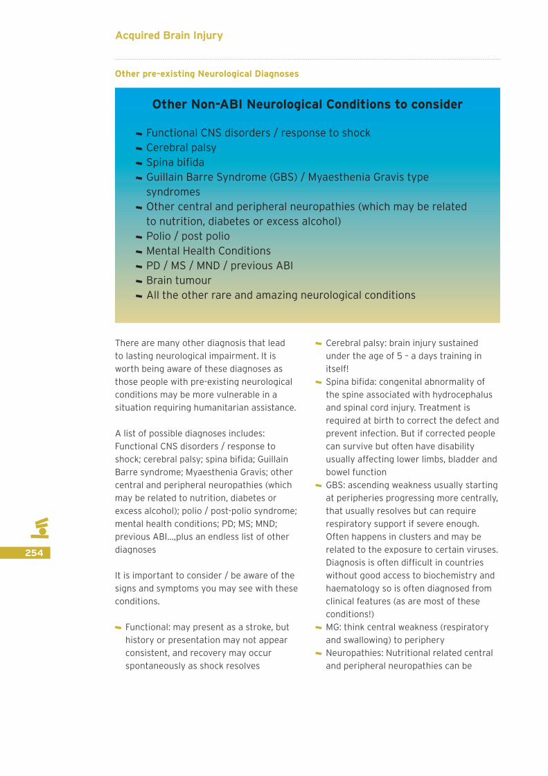
Other pre-existing Neurological Diagnoses
There are many other diagnosis that lead to lasting neurological impairment. It is worth being aware of these diagnoses as those people with pre-existing neurological conditions may be more vulnerable in a situation requiring humanitarian assistance.
A list of possible diagnoses includes: Functional CNS disorders / response to shock; cerebral palsy; spina bifida; Guillain Barre syndrome; Myaesthenia Gravis; other central and peripheral neuropathies (which may be related to nutrition, diabetes or excess alcohol); polio / post-polio syndrome; mental health conditions; PD; MS; MND; previous ABI…,plus an endless list of other diagnoses
It is important to consider / be aware of the signs and symptoms you may see with these conditions.
Functional: may present as a stroke, but history or presentation may not appear consistent, and recovery may occur spontaneously as shock resolves
Cerebral palsy: brain injury sustained under the age of 5 – a days training in itself!
Spina bifida: congenital abnormality of the spine associated with hydrocephalus and spinal cord injury. Treatment is required at birth to correct the defect and prevent infection. But if corrected people can survive but often have disability usually affecting lower limbs, bladder and bowel function
GBS: ascending weakness usually starting at peripheries progressing more centrally, that usually resolves but can require respiratory support if severe enough. Often happens in clusters and may be related to the exposure to certain viruses. Diagnosis is often difficult in countries without good access to biochemistry and haematology so is often diagnosed from clinical features (as are most of these conditions!)
MG: think central weakness (respiratory and swallowing) to periphery
Neuropathies: Nutritional related central and peripheral neuropathies can be
Other Non-ABI Neurological Conditions to consider
Functional CNS disorders / response to shock Cerebral palsy Spina bifida Guillain Barre Syndrome (GBS) / Myaesthenia Gravis type syndromes
Other central and peripheral neuropathies (which may be related to nutrition, diabetes or excess alcohol)
Polio / post polio Mental Health Conditions PD / MS / MND / previous ABI Brain tumour All the other rare and amazing neurological conditions
Acquired Brain Injury

255
related to excess alcohol but also lack of Vitamin B12 in the diet. Signs include ataxia, peripheral weakness characterised by foot drop / loss of hand dexterity and strength and sensory loss
Polio: peripheral neurological weakness following exposure to the polio virus, now rare as new onset but may find patients post-polio.
Mental Health Conditions: Following a sudden onset disaster people may present with behavioural changes, mood changes
or cognitive problems – be aware this could be a pre-existing mental health problem, a newly acquired brain injury or an acute (and common) psychological stress response to the situation they are in.
PD/MS/MND/ABI – People in a sudden onset disaster or humanitarian crisis may well be suffering with these long term progressive conditions already. Therefore evaluation of any recent alteration / recent deterioration may be required.
Medical Management of ABI
Learning Outcome
To understand potential medical and surgical management of ABI in disasters
Medical Investigations In the UK the gold standard for a patient with suspected TBI or stroke is a CT Scan of the brain, initially, within 30 mins of admission. If required admission to a neuro-surgery / hyper acute stroke unit, and initiation of interventions should occur within 4 hours of injury. In a disaster setting this is unlikely to be possible!
In the field, x-ray can help identify skull fractures and clear the Cx spine.
Close monitoring of the CVS and regular neurological observations are critical (GCS and limb function).
Blood tests where available may be useful to monitor infection markers, sodium/potassium levels, and malaria testing
Monitoring of levels of sodium and potassium following TBI is important as these commonly can be deranged due to pituitary / endocrine dysfunction. Symptoms can include Diabetes Insipidus (DI) (diabetic type S/S with high urine output (which is dilute), and thirst.
Sodium and potassium levels may also become either excessively high / low. This can cause cardiac arrhythmias, confusion,
loss of consciousness or muscle tremors / fasciculation and weakness. In severe cases which are uncorrected this can lead to death.
Other results of importance are Hb (anaemia may affect levels of fatigue), and other trace elements such as magnesium and selenium which are becoming increasingly recognised as important for neuroprotection post-injury and for normal CNS function.

256
Surgical Management
In the trauma field hospital setting, there is unlikely to be neurosurgery specialty, although Burr hole procedures may be possible in a field hospital setting with a surgeon with the right experience. Normally however, acute ABI patients should be
triaged appropriately or transferred to a local facility or to a specialist (type 3) field hospital. As rehabilitation professionals may still come across patients who have received more complex interventions, they should therefore still be familiar with the following:
Burr Holes - used for drainage of CSDH / as emergency evacuation of SDH / EDH: 2 holes drilled into the skull and haematoma is evacuated by irrigation through the holes
Craniotomy – opening made to allow for removal or evacuation of any space occupying clot/lesion. The bone flap is then replaced.
Decompressive craniectomy – section of skull is removed to reduce pressure in the skull and thus reduce ICP. This is not replaced immediately and allows an extended time period for raised ICP to resolve.
In the UK it would be replaced at later stage by a titanium plate that has been specifically manufactured. In other areas of the world
the piece of skull that has been removed maybe frozen, or can be stored into the abdomen wall (maximum of 40 days), so that it can be retrieved and used in the future to cover the defect. In many cases in humanitarian settings the skull defect will not be repaired. This puts the unprotected brain under the skin at risk of further injury. In the UK patients are provided with a protective hat / baseball cap / helmet.
There is Level 1 evidence that in adults, standard trauma craniectomy is more effective than limited craniectomy in lowering elevated ICP and leading to better outcomes at 6 months. There is Level 1 evidence that in children, decompressive craniectomy reduces elevated ICP but does not significantly improve clinical outcomes post-ABI.
Non-specialised Neurosurgical management
Burr Hole Craniotomy Decompressive craniectomy
What you are unlikely to see: ICP bolt External Ventricular Drain Shunt Ventriculostomy Clipping or coiling of aneurysm
Acquired Brain Injury
257
There is Level 3 evidence that resection of a larger bone flap results in greater decreases in ICP reduction after craniectomy, better patient outcomes and leads to fewer post-surgical complications.
You may rarely see “salvage” bifrontal craniectomies.
You are unlikely to see the following as these are specialist interventions, but it is still useful to have a basic understanding:
ICP bolt: device which monitors ICP via a burr hole into the subdural space. Normal 7-15mmHg in supine.
External Ventricular Drain (EVD): enables temporary external drainage of CSF thus can reduce ICP, divert infectious or blood stained CSF. Specific instructions will be present: for the level the EVD is positioned at, above or below, the zero point (usually the external auditory meatus)the time periods it will be draining / closed. During any respiratory treatments, position change or mobility practice you must follow specific restrictions given.
There is Level 1 evidence that cerebrospinal fluid drainage decreases intracranial pressure in the short term.
*If a patient has had an LP for CSF removal there may be a period of bed rest recommended immediately following (usually up to 4 hours). This is usually to reduce the onset of low pressure headache caused by the ICP change. However in cases of raised ICP removing CSF will reduce pressure, and headache may resolve so earlier mobilisation may be allowed.
Longer term management of hydrocephalus:
1) Shunt; long term internalised drainage method of CSF. Not often considered in acute early stages.
2) Ventriculostomy; small drainage hole placed within the third ventricle, acts as a secondary drainage or diversion for CSF around any blockage.
Clipping or coiling of aneurysm: Method to block off an aneurysm in order to reduce potential risk or further bleeding or rupture of the aneurysm
Medical Management
During the initial stages of any brain injury, the primary injury to the CNS is irreversible. However the brain remains extremely vulnerable to secondary complications, most of which will result in ischaemia, which may / may not be reversible.
TBI and stroke can result in raised Intracranial Pressure (ICP). High intracranial pressure is one of the most frequent causes of death and disability following severe head injury. It is defined as an ICP reading greater than 20 mmHg within any intracranial space (subdural, intraventricular, extradural, or intraparenchymal compartments) (Sahuqillo & Arikan, 2006).
An uncontrolled increase in intracranial pressure results in: compression / shift of brain tissue, compromise of vascular and CSF circulation and ultimately ischaemia and infarction. In the worst case this leads to “coning” where the pressure causes the brain to herniate downwards through the foramen magnum causing brainstem death.
There is a relationship between ICP and Cerebral Perfusion Pressure. Cerebral perfusion pressure is the net pressure gradient causing adequate cerebral blood flow to the brain. Normal CPP is 70-90 mmHg, and must be >70mmHg in adults and > 60mmHg in children to prevent ischaemia
In the humanitarian environment changes in ICP and CPP can be monitored through neurological observation: specifically GCS, pupil size / reaction to light, and motor deficits.
258
The medical management in the field hospitals will be supportive, and should aim to reflect / follow appropriate guidelines where they exist, for example NICE guidelines for Stroke / TBI, as far as possible within the limitations of the environment and available medical specialties.
Overall Aims of medical management:
To identify if a patient requires transfer to a specialist unit if one is available and arrange as appropriate / possible
Determine prognosis (will medical care be supportive, palliative, rehabilitative)
To maintain / stabilise neurological status Prevention of neurological deterioration / maintenance of neurological status
Prevention of neurological deterioration and maintenance of the neurological status is achieved by ensuring an environment maintaining homeostasis in the following ways:
Management of Blood Pressure: management regime will depend on diagnosis and either a set figure for MAP may be used (90mmHg), or a target “range” for systolic pressure (>180mmHg)
Fluid management: to maintain hydration and yet ensure excessive hydration is prevented (maintenance of neutral fluid balance)
Nutrition may be difficult to achieve effectively. Following ABI there is a significant increase in metabolic rate due to autonomic dysfunction, and the increased need for energy to assist the brain to recover. Metabolism may increase by over 100%. There will be difficulty optimising
Medical Management
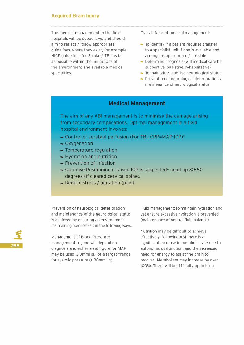
The aim of any ABI management is to minimise the damage arising from secondary complications. Optimal management in a field hospital environment involves:
Control of cerebral perfusion (For TBI: CPP=MAP-ICP)* Oxygenation Temperature regulation Hydration and nutrition Prevention of infection Optimise Positioning if raised ICP is suspected– head up 30-60 degrees (If cleared cervical spine).
Reduce stress / agitation (pain)
Acquired Brain Injury
259
nutritional intake if there are swallowing deficits. Ng tubes may be available, however specialist feeds are unlikely therefore you may need to be inventive in how foods are turned into liquids.
Positioning: 30° – 60° degrees head up (to optimise cerebral circulation and perfusion)
Maintenance of adequate oxygenation: the ideal range is Sa02> 95% (in the UK PaO2 should be > 12)
Maintenance of temperature within a normal range
Maintenance of blood glucose concentration between 4-11mmol/litre
Prevention of secondary complications: infection – swallowing, early mobilisation* post-stroke; pressure area damage – early / appropriate mobilisation
Treatment of infection: might include debridement of wounds
Use of medication: particularly for pain, seizures, temperature, blood pressure management, antibiotics, sedation in situations where a patient’s behaviour is putting them / others at risk
*Mobilisation: Some types of ABI may require a more conservative approach to mobilisation for example non-traumatic / spontaneous SAH due to: potential for re-bleeding (most likely day 0-1, following this significant risk continues, although reduces gradually with time. By 4 weeks chance of re-bleed is 3%). Vasospasm is most likely to occur between days 5-21
Therefore an initial period of bed rest may be advised for 7 – 10 (possibly up to 21) days, followed by a structured and gradual progression in mobility while monitoring neurological status.
Rehabilitation Assessment
Learning outcome
To be aware of specific rehabilitation assessment considerations for patients with ABI.
Just as in the UK, you are only expected to work to your level of expertise, knowledge and competence. The following is a guideline to remind you of some of the basic assessments and problems that you may identify.
If an assessment is needed that you are not competent to complete, ask and see if
there is anyone else available / who will be available in the future to complete it.
One of the first steps may also be to evaluate where else there may be neuro rehabilitation facilities / specialists, or to identify they don’t and may not exist for some time.

260
Assessment: Database - key factors
HPC : How long ago, mechanism / onset of symptoms Other relevant history: progression of symptoms / deterioration,
morning headaches, vomiting, photophobia or seizures, PMH :
any previous major illnesses or surgery any current illness/surgery
DH : on any CVS or diabetic medications?
SH: includes relevant cultural information Previous lifestyle: family role (where are other members of
family now), work/ education, daily routine, drug or alcohol use, level of independence, language and literacy, home environment/ location (check if still able to return)
The assessment of a patient in a humanitarian crisis where possible should still be the same as the assessment you would complete in the UK and include:
HPC – mechanism of injury can potentially help ascertain area of brain damage, for example an obvious blow to the side of the head would indicate likely tempero-parietal lobe damage; a high speed injury (eg RTC) can result in a diffuse axonal injury which leads to more non-specific impairment; a cardiac arrest or history involving someone being trapped for any length of time could lead to a hypoxic brain injury, again this has a less defined pattern to it. Knowing the nature of the injury can go some way to predicting rehab potential.
PMH – as with all major trauma, premorbid status will affect rehab outcome
DH – these can give you an indication of any PMH that hasn’t been disclosed, for example patients may fail to report high blood pressure, but are on beta blockers, or they may have lost their medication / forgotten about it. Also anticoagulants such as aspirin may be indicated if embolic stroke, but contraindicated if TBI or brain haemorrhage
Amending medication isn’t our role as AHPs, but an awareness of medication and their actions / interactions is useful and you need to be aware why someone on aspirin may be continuing to deteriorate
Acquired Brain Injury

261
SH: What is the next step will it be D/C to a temporary camp, their home or onto another hospital?
What outcome are we aiming for (function vs quality)? If we don’t achieve the functional outcome required for them to return to their normal role / lifestyle, what will the psychological effect be on them or their family? If they are the main wage earner, how will the family manage?
What cultural implications are there re:
disability - family desire or obligation to take over care / “look after” patient rather than encourage return to independence, impact on them of having someone with a disability in the family
motivation - to participate in rehabilitation
Other social factors that may impact on physical symptoms include:
Smoking / alcohol / drug use: Are they agitated, shaky and / or confused?
Do they have ABI / cerebral irritation or are they just withdrawing from nicotine and a nicotine patch but are unable to express this?
Can we support this medically e.g alcohol withdrawal protocol / basic support with multivitamins etc?
Objective Assessment and the ICF
Objective assessment
Impairment
Function
Independence
Ability to participate in activity
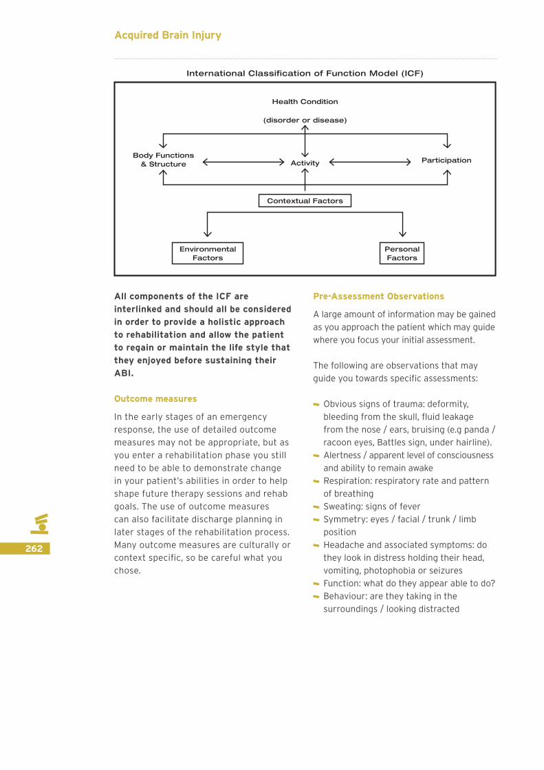
The International Classification of Functioning, Disability and Health, known more commonly as ICF, is a classification of health and health-related domains (WHO, 2001).
The ICF allows the clinician to look beyond the diagnosis and biological or medical dysfunction and assess the impact on quality of life through the ability to carry
out certain activities, such as walk, or feed oneself, and whether the patient is able to continue to participate in social activities.
The ICF also helps the clinician to help set patient-centred goals and identify barriers to participation that might be easily overcome, such as providing a grab rail so the patient is able to continue to take a shower independently.
262
Contextual Factors
Activity ParticipationBody Functions
& Structure
Health Condition
(disorder or disease)
International Classification of Function Model (ICF)
Environmental Factors
Personal Factors
All components of the ICF are interlinked and should all be considered in order to provide a holistic approach to rehabilitation and allow the patient to regain or maintain the life style that they enjoyed before sustaining their ABI.
Outcome measures
In the early stages of an emergency response, the use of detailed outcome measures may not be appropriate, but as you enter a rehabilitation phase you still need to be able to demonstrate change in your patient’s abilities in order to help shape future therapy sessions and rehab goals. The use of outcome measures can also facilitate discharge planning in later stages of the rehabilitation process. Many outcome measures are culturally or context specific, so be careful what you chose.
Pre-Assessment Observations
A large amount of information may be gained as you approach the patient which may guide where you focus your initial assessment.
The following are observations that may guide you towards specific assessments:
Obvious signs of trauma: deformity, bleeding from the skull, fluid leakage from the nose / ears, bruising (e.g panda / racoon eyes, Battles sign, under hairline).
Alertness / apparent level of consciousness and ability to remain awake
Respiration: respiratory rate and pattern of breathing
Sweating: signs of fever Symmetry: eyes / facial / trunk / limb position
Headache and associated symptoms: do they look in distress holding their head, vomiting, photophobia or seizures
Function: what do they appear able to do? Behaviour: are they taking in the surroundings / looking distracted
Acquired Brain Injury

263
Assessment
This is a brief overview of basic assessments which will help identify: the main problems quickly; developing complications and progress
Primary survey: Airway Breathing: RR / pattern / rhythm / cough /
Ausc / expansion Circulation: HR / BP / Sa02 Disability: GCS / Pupils / Neuro Ax Exposure: observation / inspection
Airway:If the patient has a low GCS, is having a seizure or is post-ictal they are at risk of obstructing their airway. A decision will have to be made re: protection of airway as intubation and ventilation is unlikely to be available. Assessment of the presence of a cough reflex is useful as an absent cough reflex may indicate severe neurological compromise. If the patient is unable to protect their airway use positioning (place on their side in recovery position) and / or if you have one, and are competent, then use a guedel airway or NPA.
Airway obstruction may also occur if someone has low GCS and has vomited / is unable to clear secretions, has a reduced cough reflex or has aspirated due to swallowing difficulty.
Therefore you will need to identify if suction is available? If so, is it appropriate, and who else is able to carry this out / who can be trained? If suction is not available, discuss with the team what other methods are there to maintain a clear airway if needed?
Breathing:Respiratory pattern gives a large amount of information, it can indicate respiratory distress, and it can also indicate compromise of the CNS. Altered respiratory patterns due to neurological compromise or associated problems include: reduced RR; increased respiratory rate; waxing / waning patterns of breathing; large sighs; altered I:E ratios that vary in cyclical patterns e.g. a large deep breath followed by apnoea followed by fast shallow breaths followed by slower ramping breaths followed by a deep breath to repeat the cycle; hiccoughs and periods of apnoea can be an indication of seizure activity or brainstem dysfunction and may be due to raised ICP.
Paradoxical patterns can be a result of reduced intercostal innervation due to central neuropathies such as MG, or in the case of severe GBS. Paradoxical patterns can also be seen with dense hemiplegia or post-SAH.
264
You may also need to identify with your assessment if the patient is in respiratory distress due to a primary respiratory problem (pneumonia) or due to respiratory muscle fatigue?
Other useful information includes: are they able to cough and clear secretions effectively? If yes, prompt them. If no, is it because they don’t have the strength? Is their cognition impaired and therefore they don’t realise that they have a problem? Or are they too drowsy?
Easy interventions:
If there is respiratory distress, you need to make an immediate intervention consider the following:
Use of side to side / semi-prone positioning if safe to do so.
With TBI avoid head down positioning, 30-60° head up is optimal.
Manual techniques such as vibrations / percussion may not be tolerated as well as shaking.
Should they be / can they be moved to a critical care unit, or a higher level field hospital?
Circulation:
Identify if the cardiovascular system appears stable. Is the systolic pressure staying consistently within set limits set by the medical team (if these exist) / within acceptable limits (usually …), or is the blood pressure variable particularly if this is not linked to other interventions. Also observe the heart rate: is this consistently within normal limits, or is there reduced or increased heart rate?
Signs of instability in the cardiovascular system may be an indication of early brainstem compression, or sepsis secondary to infection or may be a result of severe fever.
Disability: GCS / pupils / Neurological examination
The main methods to assess neurological disability include:
Assessment of GCS Assessment of pupils Neurological examination
The primary method of assessing consciousness is the Glasgow Coma Scale (GCS). This is available at http://www.glasgowcomascale.org/ It is useful to monitor trends, which can alert the clinician as to whether the patient is better, worse or the same.
Acquired Brain Injury
265
Make sure the patient is safe while you do this, e.g. their airway is protected, they are in the recovery position and ideally someone is with them to continue to monitor their vital signs.
When you are asking for help, it is important to be able to state what the GCS was and what it is currently, so the medical professional can establish how serious the situation is decide how to prioritise the patient within their current caseload.
Be aware that a deteriorating patient may present with increasing agitation due to cerebral irritation rather than reducing levels of arousal. This means that the increasingly angry person that you may have mistaken for being stressed may actually have a worsening brain injury. You should check for other signs of brain injury
such as dizziness and confusion and alert a medical colleague if you have any concerns.
Pupil assessment is used to assess compromise of the cranial nerves due to raised ICP and brainstem dysfunction. The pupils are assessed for:
Size: small pupils may indicate overuse of certain drugs such as morphine, large / dilated pupils may be due to increasing ICP
Comparison of right vs left: pupils should be equal in size. If they are unequal this may indicate raised ICP, or direct damage to the optic nerve
Reaction to light: pupils should react briskly in response to light, by constricting. A slowed response / no response may be an indication of raised ICP, or damage to the optic nerve.
Glasgow Coma Scale
Best eye response (E)
Spontaneous – open with blinking at baseline 4
Opens to verbal command, speech, or shout 3
Opens to pain, not applied to face 2
None 1
Best verbal response (V)
Oriented 5
Confused conversation, but able to answer questions 4
Inappropriate responses, words discernible 3
Incomprehensible speech 2
None 1
Best motor response (M)
Obeys commands for movement 6
Purposeful movement to painful stimulus 5
Withdraws from pain 4
Abnormal (spastic) flexion, decorticate posture 3
Extensor (rigid) response, decerebrate posture 2
None 1
It is important to recognise and respond to a reduction in GCS. It is a medical emergency and urgent help must be sought.

266
Neurological assessment:
The use of clinical reasoning following the initial subjective and observational assessments should direct you towards choosing the most appropriate assessments for the likely diagnosis / immediate needs of the patient. More specific and formalised testing may be required for certain conditions such as SCI.
Using your basic skills a neurological / neuromusculoskeletal assessment of the following should be completed:
Motor Asessment: A “pronator drift” test is a quick method to identify if there is a subtle onset of neurological weakness as a result of CNS injury.
Method – ask patient to hold both arms up in front of them, turn palms towards ceiling and then close eyes and keep arms still and
steady. If there is CNS deficit causing subtle weakness, on one side (possibly both sides) the arm will pronate and drift downwards
This can be used at every assessment to identify progression / improvement of symptoms. If there is progression of weakness then the planned mobilisation / completion of a rehabilitation session may need to be re-evaluated, and medical review may be required.
Tone: Identify if there is low / increased tone evident. Assess by feeling how a limb feels as you hold and move it (heavy and easy to move / heavy and resisting). Indicates how relevant strength testing may be.
Muscle tone can be measured with the Modified Ashworth Scale:
Neuro Assessment
Motor Function: Pronator drift test Tone Muscle strength Co-ordination Reflexes
Sensation: Sensory neglect Light touch / pin prick / temp
/ 2 point discrimination Proprioception
Acquired Brain Injury

267
Muscle strength: The Oxford MRC scale should be used to assess strength in both upper and lower limbs. Trunk strength can be assessed using a bridging exercise, or completion of a sit up type task.
Co-ordination: The following basic co-ordination test can b completed: Upper Limb – finger to nose test (past pointing); Lower Limb – heel slide along shin from ankle to knee
Sensation: Is useful to assess first for Sensory neglect.
A quick test to identify inattention and possibly global problem is as follows:
Eyes shut. Stroke a section of one arm – can they feel sensation? Stroke same section of the other arm – can they feel the same
sensation? Stroke same section of both arms together – can they identify sensation on both sides equally?
Repeat in another section then complete the test on the lower limbs.
For assessment of light touch and pin prick use dermatomes. These tests may be difficult with a language barrier. Remember to ask patient to close eyes during testing
Proprioception: There are a variety of methods used to test proprioception. The easiest are using the distal phalanx of thumb and hallux: move the distal section and ask if the patient can identify if the movement is up or down; a larger position test of joint / limb proprioception: place the limb which is affected into a position for unaffected side to copy.
Grade Description
0 No increase in muscle tone
1Slight increase in musle tone, manifested by a catch or by minimal resistance at the end of the range of motion (ROM) when the affected part(s) is moved in flexion or extension
1 + Slight increase in muscle tone, manifested by a catch, followed by minimal resistance throughout the remainder (less than half) of the ROM
2 More marked increase in muscle tone through most of the ROM, but affected part(s) easily moved
3 Considerable increase in muscle tone, passive movement difficult
4 Affected part(s) rigid in flexion or extension
9 Unable to test

268
Other Physical Elements of a Neurological Assessment
These elements are important to identify additional rehabilitation problems and to form a holistic approach to treatment.
Swallow: If you do not feel able to assess swallow identify if there is anyone in the team trained to carry out a swallow assessment. If not, evaluate if you think the patient is either exhibiting signs / likely to show the following signs of aspiration:
Facial weakness Drowsiness Are they maintaining / coping their own secretions?
Do they cough / struggle / alter respiratory rate or pattern if you observe them taking a drink
Evidence of chest infection (which may be recurrent)
If there appear to be swallowing problems you will need to discuss with the team appropriate management options. “Risk” feeding / drinking may continue with close observation if signs of aspiration / development of chest infection
Other physical elements of Neuro Ax:
Swallow – check with patient/family for difficulties Speech / comprehension Sensory elements:
Vision: visual acuity / visual field loss Hearing
Vestibular: Gaze stability / smooth pursuit Saccades Herdmanns
Speech: Simple methods to evaluate if there are speech problems include: is patient attempting to speak, or is speech affected by facial injury or reduced consciousness; is the speech slurred? (family may be helpful in identifying problems)?
You may need assistance from locals to identify if patient is talking sense / comprehending the spoken word
Expressive dysphasia: Understanding is retained and patient will follow commands, but not speak, or speak using nonsensical words/sentences. Reading and writing may also be impaired.
When testing comprehension in patients with expressive dysphasia, you should ask them to carry out non-verbal tasks, such as ‘stick your tongue out’.
Receptive dysphasia: Able to talk fluently, but out of context to the conversation, or jargon, indicating that they haven’t understood the words spoken to them. Do not obey simple commands.
Acquired Brain Injury
269
Some people may have expressive and receptive dysphasia, and this is usually associated with hemiplegia and visual field deficit.
To test for dysphasia assess:
spontaneous speech through general conversation. Use simple questions like ‘What is your name?’ and ‘Where do you live?’
naming objects by pointing to objects you want the patient to name (use items familiar to them with local help if required)
Ask them to follow a 3 stage command: “with your right hand, touch your right knee, and then your nose”
Other assessments may depend on level of literacy as these involve spelling words, reading, or writing
Vision: Does the patient have a pre-morbid visual field deficit? Do they usually wear glasses? If so, where are they? If they are lost/broken, is it possible to get a new pair? Do they have a new visual deficit? If so can they explain it e.g. blurring / double vision (diplopia)
Visual field loss: *do not confuse with visual neglect (discussed in section on cognition)
Homonymous hemianopia – physical loss of visual field to the same side, in both eyes, in the vertical plane.
Quadrantanopia – visual field loss in one quadrant, in both eyes
Other field loss / altered perception of vision
Simple testing for visual field loss: Use index finger of right and left hand. Start with them either side of the patients head, behind their visual field, then as you begin to move your fingers forward towards the front of the patient, ask the patient to tell when they can see you fingers but all the time they must look at your nose. Someone with a hemianopia will only see both fingers when they are in the same half of the visual field.
Testing for a quadrantanopia is the same, but you will have to start with your fingers higher up or lower down. Cognitively intact people can be taught compensatory strategies to overcome this.
Other field loss / altered perception of vision:
Diplopia: if they have double vision, they may benefit from an eye patch. Remember to advise the patient to swap eyes so they don’t develop dependency on one eye.
Are the eyes deviated to one side? Can the patient follow an object and bring their eyes beyond midline to the opposite side? If not, they have a visual inattention. You may be able to identify this through function e.g. ignoring items to one side, attending only to one side, bumping into things
If there is altered vision / perception compensatory strategies will need to be taught. People who aren’t able to learn compensatory strategies will be unaware of objects on their affected side. This has safety implications if the patient is able to mobilise independently. It will be important to educate caregivers about the visual field deficit and teach them strategies to help keep the patient safe.
Vestibular: It is useful to be aware that a patient with a skull fracture may have no specific limb deficit but can experience dizziness and balance problems. This is more common in temporal, occipital and base of skull fractures. It is useful to complete a vestibular screen to identify any issues. Be aware of any changes of hearing as a result of the injury and follow up to ENT specialists may be warranted.
Presentation of dizziness may be due to:
Disruption of otoconia causing BPPV Interference with the ear canal due to the site of fracture
Disputation of the neuronal networking as a result of TBI

270
Vestibular assessments include the following tests for: Gaze stability; Saccades and Herdmanns.
Gaze stability – if they focus on a point in front of them and you observe them: are their eyes still, is there any convergence or divergence of the gaze of each eye
Can they move their eyes from side to side in the horizontal plane, vertically up and down, and in an “H”: Is the movement smooth or jumping? Do they have a nystagmus (an involuntary to and fro, or in a rotary movement in any plane? May indicate a problem with the cerebellum / vestibular systems
Saccades - head stays still while they move their eye horizontally from side to side as quickly as they can for 1 minute: can they complete for 1 minute, do they feel dizzy / nauseous / vomit, does the head remain still
Herdmanns – focus on a point in front while turning head from side to side as quickly as possible for 1 minute: can they complete one minute, do they feel dizzy / nauseous / vomit, does the head remain in a horizontal plane during the movement or does it “wobble”.
Neuro Assessment: Functional
Balance: Sitting: static / dynamic Standing: static / dynamic
Moving / Mobility: Moving in bed Moving in/out of bed Sitting to standing Walking
ADL’s Eating and Drinking Washing/ Dressing/ Toileting Cooking, Childcare, Ability to complete other usual tasks
Functional Neurological Assessment
Assessments of function such as ability to balance, move and complete activities of daily living should also be undertaken.
Functional and / or specific outcome measures can be used to complete an objective assessment of functional ability.
Acquired Brain Injury

271
Non-physical Neurological Assessment
These include assessment of the higher cognitive, executive and perceptual functions on the brain. When assessing cognitive skills it is helpful to consider them as a hierarchy, alongside mood, behaviour, language and communication. For example
if someone is having great difficulties maintaining sustained attention during an assessment or rehabilitation session, and is constantly distracted / asking repeated questions, this will likely impact on the cognitive skills higher up he hierarchy.
Neuro Assessment: Non-physical
Executive Function
Memory
Praxis
Information processing
Visual/perceptual processing
Attention
Sensory Registration
Mood and Behaviour Language and Communication
Cognitive Hierarchy:
Non-physical assessments can be divided into assessments of:
Cognition Psychological Perception Behavioural
Dyspraxia / apraxia are also commonly included within these groups of tests although they are an evaluation of the quality of a movement, which is closely linked to perception of movement.

272
Cognition is an encompassing term for a variety of higher mental processes, such as thinking, imaging, speaking, acting and planning (Ward, 2010).
Attention is the process whereby certain information is selected for processing while other information is discarded. If you have a patient who has impaired attention,
they will find therapy difficult in a noisy environment because they won’t be able to filter out other stimuli and so won’t be able to give therapy their full attention – sensory overload. Even in a quiet environment, they may have trouble sustaining attention, so you need to make your treatment sessions short to maximise on learning opportunity.
Cognition Attention / concentration Speed of processing information
Memory Recall: immediate STM/
delayed LTM Anterograde/ Retrograde
Amnesia Executive function
Insight into condition/ deficits
Ability to initiate/ terminate Planning and organization Problem solving and
abstract thinking Safety awareness/ Judgement
Psychological Mood
Perceptual problems Sensory / Visual inattention:
Visuo-spatial loss / spatial awareness
Visual inattention Agnosia:
auditory / visual / body / tactile
Dyspraxia / apraxiaBehavioural problems
Agitation / Apathy / Fatigue Aggression / Lability Personality change Disinhibition
Neuro Assessment: Cognition / Perception / Behavioural/ Psychological
Acquired Brain Injury

273
At the top left is the cancellation test, where the patient has to put a line through all the lines on the piece of paper. As can be seen, none of the lines to the left have been crossed through, indicating a left-sided visual inattention.
At the bottom right are examples of copying pictures and also drawing freehand, all which show a left-sided visual inattention.
Memory:
There are many different types of memory:
Short term memory refers to information that is currently held ‘in mind’. It has limited capacity. STM is important for long-term learning. In other words, if you have an ABI patient with poor STM, their recovery is likely to be slow and their prognosis is poor as they can’t retain information and build on it between treatment sessions (i.e. no carry over).
Long term memory is information that is stored. It may not be presently accessed or consciously accessible. LTM has unlimited capacity.
Following a brain injury, patients may be unable to recall events leading up to the injury (retrograde amnesia) or have difficulty forming new memories, and therefore be unable to learn new tasks (anterograde amnesia).
ABI patients could present with post-traumatic amnesia; a period of amnesia occurring after the injury (anterograde amnesia). This can last days or weeks and the longer it lasts, the more severe the brain injury.
Perceptual problems:
Sensory / visual inattention: Can be assessed / identified during function
Hemispatial / visual inattention: An attention disorder that prevents a patient attending to stimuli on one side (usually affecting ability to attend to the left side). Can be inattentive to sensory stimulus of any kind on the affected side, for example touch, sound, recall or memory of objects / places that would normally be on one side of the body, or visual.
Assessments for inattention
CopyingModel Patient's copy
Spontaneous drawing:

274
Examples: ignoring items to one side, attending only to one side, bumping into things
Can occur with / without homonymous hemianopia
Identify the following: Are the eyes deviated to one side? Can the patient follow an object and bring their eyes beyond midline to the opposite side? If not, they have a visual inattention.
Homonymous Heminanopia (HH) – a loss of visual field to the same side in both eyes where compensation can be made for loss of vision e.g. turning head to shave both sides of face
If both HH and visual inattention occur together the prognosis is worse as
compensatory strategies are difficult to put in place.
Agnosia: loss of ability to recognise objects, people, shapes, sounds, smell even though there is no loss of the specific sense, or evidence of memory loss. For assessment use locally sourced items that the patient would normally recognise.
Dyspraxia: reduced ability to co-ordinate, perform, plan and carry out movements even when there is no movement / motor disorder. E.g. difficulty dressing in the right order
Below are some examples of cognitive and perceptual tests that can be modified and adapted depending on the setting that you are in and the patient’s circumstances.
Severity of TBI
Domain Examples
Orientation Time: Age, Day/night, Month or Season (dry/rainy), Year, LOS/days post-op Place: Name of town Person: Ability to identify accompanying NOK or familiar staff member
Attention "Recite months of years backwards" Difficulties complying with simple 1 or 2 step commands during session
Memory I am going to tell you a list of items that I usually get from the market, you must try and remember them for me. They are: cooking oil, soap, rice and eggs (replace with culturally appropriate equivalents). Now repeat this list to me three times "Ask again after 5-10 mins."
No carryover between therapy sessions e.g. use of transfer board technique
Perceptual "Show me your right foot/left hand/with your right hand touch your left shoulder"
Language "Tell me the names of as many different animals as you can in 1 minutes"
Executive function
Often hard to identify on a bedside screen but observed through function. For e.g. person appears indifferent or surprised when experiencing difficulties during fx tasks, difficultues problem solving in a novel task, family reporting personality change, apathy etc.
Refer to the ABI cheat sheet for more information on what to be aware of during your assessment
Acquired Brain Injury

275
Behaviour:
Behaviour may change as a result of the head injury, particularly if the injury is in the frontal lobes. Behavioural changes can be anything from disinhibition (e.g. taking clothes off in public), swearing when previously did not do this, and aggression, to apathy and ‘laziness’, or someone who was previously aggressive / motivated becoming childlike
It is important to establish what is normal for the patient, and remember that the family may find the patient’s behaviour distressing.
Don’t forget the individual may also have experienced significant psychological trauma! Access to psychology, neuropsychology or psychiatry is likely to be limited initially, but may become available at a later date. Educating and supporting the patient and family members is critical.
It is important that you try and educate the patient to exhibit behaviour that is socially acceptable, rather than let them do as they please just because they have had a brain injury.
Patients who are agitated often attempt to wander around and may inadvertently be a danger to themselves or others. They frequently will also be in a confused / disorientated state; have memory problems; and will not be aware they have had a TBI, they won’t remember they can’t walk and so are at risk of falls as they try to get up.
Agitation may increase when they have a basic need to do something: for example to toilet, have a drink / eat; when in pain, and they may become aggressive and hit out at other people trying to stop them from moving around.
They usually need 1:1 supervision and a quieter environment. Often it may be safest to initially nurse on the floor and to establish a regular toileting / daily regime of activities, and consistent team of staff involved in their care.
It is important to note whether the agitation is getting worse, or if the patient is getting significantly quieter/drowsy as these are signs that the patient is deteriorating as the brain swells. The first 48 - 72 hours post-ABI are most crucial for monitoring this.
Overstimulation may make them worse. Therefore limit attempts to reason, and work with / alongside them as long as it is safe to do so. In the early stages concentration will also be reduced and often the effort and physical effects of attempting to get up, will quickly lead to a return to bed.
As well as being unable to concentrate on the task or being unable to motivate themselves to complete the task, patients may also be too tired to complete the task.
Remember the brain is healing, and will be using lots of calories to do this as the metabolic rate increases significantly (the autonomic reaction is likened to that of someone with severe burns).
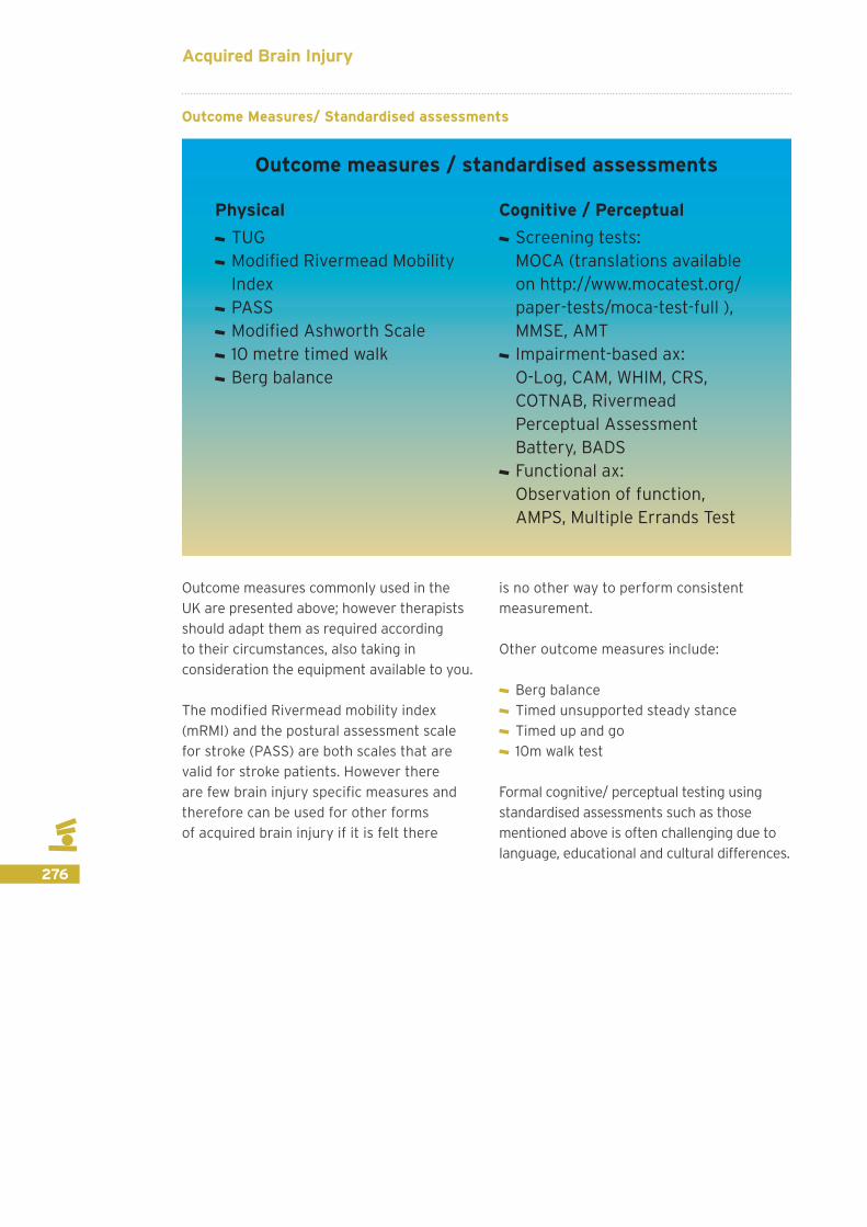
276
Outcome Measures/ Standardised assessments
Outcome measures commonly used in the UK are presented above; however therapists should adapt them as required according to their circumstances, also taking in consideration the equipment available to you.
The modified Rivermead mobility index (mRMI) and the postural assessment scale for stroke (PASS) are both scales that are valid for stroke patients. However there are few brain injury specific measures and therefore can be used for other forms of acquired brain injury if it is felt there
is no other way to perform consistent measurement.
Other outcome measures include:
Berg balance Timed unsupported steady stance Timed up and go 10m walk test
Formal cognitive/ perceptual testing using standardised assessments such as those mentioned above is often challenging due to language, educational and cultural differences.
Physical TUG Modified Rivermead Mobility Index
PASS Modified Ashworth Scale 10 metre timed walk Berg balance
Cognitive / Perceptual Screening tests:
MOCA (translations available on http://www.mocatest.org/paper-tests/moca-test-full ), MMSE, AMT
Impairment-based ax: O-Log, CAM, WHIM, CRS,
COTNAB, Rivermead Perceptual Assessment Battery, BADS
Functional ax: Observation of function,
AMPS, Multiple Errands Test
Outcome measures / standardised assessments
Acquired Brain Injury
277
Rehabilitation Treatment
As with all good clinical practice, your assessment should lead to your problem list, which in turn will shape your treatment plan and help you set short and long term goals.
These will obviously vary depending on the severity of the brain injury, social and family roles of the patient and the culture that you
Learning outcomes
To be aware of specific rehabilitation treatment considerations for patients with ABI
are in. (For example, in Gaza we encountered several SCI patients who were fully independent, but their culture meant that that, where the patient was male, everything that could be done was done for the patient and they weren’t expected or given the opportunity to be independent).
Treatment
The aim of any treatment should be to allow the patient to achieve their maximum potential
Mild TBI may only need advice, help setting expectations and relatively simple interventions
Severe TBI need specialist rehabilitation where possible
Rehabilitation is described by the World Health Organisation as a way of assisting an individual with a disability to achieve and maintain maximum physical, cognitive, social and psychological function, participation in society and quality of living. (WHO, 2011)
Where the patient has suffered from a mild TBI, the treatment may be limited to assessment, giving advice and setting expectations, and possibly organising relatively simple interventions. (BSRM core standards, 2013)
There is now a substantial body of trial-based evidence and other research to support both the effectiveness and cost-effectiveness of specialist rehabilitation. Despite their longer length of stay, the cost of providing early specialist rehabilitation for patients with complex needs is rapidly offset by longer-term savings in the cost of community care, making this a highly cost efficient intervention (eg Turner-Stokes et al 2005)
A key component of the rehabilitation should be that it is multidisciplinary approach with regular ward rounds, MDT meetings, case conferences and provision of support to family and carers (BSRM, 2013). Being multidisciplinary also means that the patient will have all aspects of their rehabilitation addressed within the same facility (Turner-stokes et al, 2005) with all partners working to the same patient centred goals.
Treatment and Rehabilitation
It is unlikely that patients will present only with an acquired brain injury following a sudden onset disaster so before commencing rehabilitation ensure that any contaminant injuries have been treated/ stabilised and consider what influence these may have on your treatment choices. Likely other injuries are fractures, amputations and especially spinal cord injury that can be easily missed.
The first choice of treatment may be positioning to prevent postural problems,
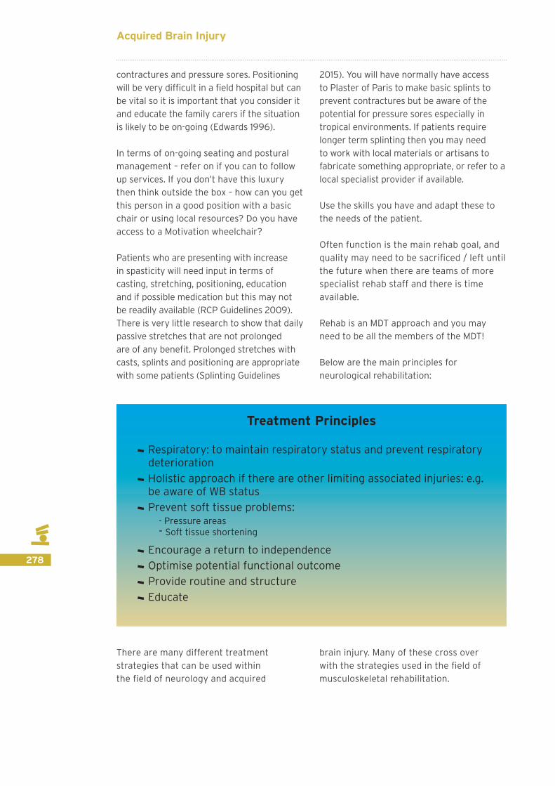
278
contractures and pressure sores. Positioning will be very difficult in a field hospital but can be vital so it is important that you consider it and educate the family carers if the situation is likely to be on-going (Edwards 1996).
In terms of on-going seating and postural management – refer on if you can to follow up services. If you don’t have this luxury then think outside the box – how can you get this person in a good position with a basic chair or using local resources? Do you have access to a Motivation wheelchair?
Patients who are presenting with increase in spasticity will need input in terms of casting, stretching, positioning, education and if possible medication but this may not be readily available (RCP Guidelines 2009). There is very little research to show that daily passive stretches that are not prolonged are of any benefit. Prolonged stretches with casts, splints and positioning are appropriate with some patients (Splinting Guidelines
2015). You will have normally have access to Plaster of Paris to make basic splints to prevent contractures but be aware of the potential for pressure sores especially in tropical environments. If patients require longer term splinting then you may need to work with local materials or artisans to fabricate something appropriate, or refer to a local specialist provider if available.
Use the skills you have and adapt these to the needs of the patient.
Often function is the main rehab goal, and quality may need to be sacrificed / left until the future when there are teams of more specialist rehab staff and there is time available.
Rehab is an MDT approach and you may need to be all the members of the MDT!
Below are the main principles for neurological rehabilitation:
Treatment Principles
Respiratory: to maintain respiratory status and prevent respiratory deterioration
Holistic approach if there are other limiting associated injuries: e.g. be aware of WB status
Prevent soft tissue problems: - Pressure areas - Soft tissue shortening
Encourage a return to independence Optimise potential functional outcome Provide routine and structure Educate
There are many different treatment strategies that can be used within the field of neurology and acquired
brain injury. Many of these cross over with the strategies used in the field of musculoskeletal rehabilitation.
Acquired Brain Injury

279
Rehabilitation should be aimed at improving postural control, muscle activity and balance (RCP 2012). This can be achieved through prescription of specific exercises to address the underlying problems but patients with brain injury respond particularly well to exercises that enable them to work against gravity (sit them up, and stand them as soon as possible, and work in these positions) and to experience movement. Specific exercises are not described in this manual, but the principles of exercise prescription should be adhered to and trying to make exercises as functional and meaningful as possible will help with recovery, motivation and compliance.
Exercises that allow repetition of specific functional tasks have good research to back them up (e.g. repeated reach to grasp in a standing position) (French et al 2007). Physical fitness training and strengthening are not contraindicated in brain injury (NICE 2013).
Repeated task practise with assistance and guidance of functional tasks is appropriate.
Use aids and equipment to enhance function where appropriate – again use local resources if possible. If you are the sole rehabilitation provider it is likely that in an austere environment your treatment will need to involve treatments combining PT and OT tasks. Upper limb treatment may be essential to improving functional ability.
There is good research to back up the use of low tech treatments to improve upper limb function including mirror box treatment and mental imagery treatment. For patients with reasonable upper limb activity modified constraint induced movement techniques are a possibility but this requires a very specific set of circumstances (Thieme et al 2012, Zimmerman 2008, Wolf et al 2006) that are unlikely to be available early after a sudden onset disaster.
Specific treatment of sensory impairment is important as this can lead to long term problems – research suggests that you should try to enhance impaired sensory mechanisms (e.g. Light touch or proprioception) rather than working on sensory mechanisms that have been totally lost (e.g. Pain) (Carey et al 2011).
Physical Treatment strategies
Physical: main principle is to regain postural control and movement
Use whichever treatment principles you are familiar and confident with: strengthening/facilitation/repeated tasks/combination of techniques/exercise therapy/stretching/splinting/aping/sensory rehab/vestibular rehab/core stability/balance/encourage movement
Positioning/postural management Seating Use practise of functional tasks such as transfers / sitting to standing/squatting
Identify equipment that is required/available/can be constructed safely For high muscle tone, MDT management including pharmaceutical management should be considered
For low muscle tone, be aware of subluxation risk of shoulder and wrist.
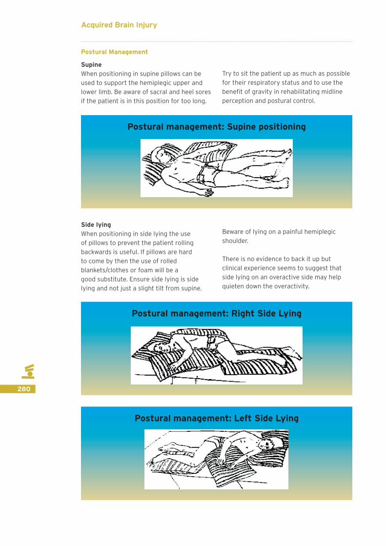
280
Postural Management
SupineWhen positioning in supine pillows can be used to support the hemiplegic upper and lower limb. Be aware of sacral and heel sores if the patient is in this position for too long.
Try to sit the patient up as much as possible for their respiratory status and to use the benefit of gravity in rehabilitating midline perception and postural control.
Side lyingWhen positioning in side lying the use of pillows to prevent the patient rolling backwards is useful. If pillows are hard to come by then the use of rolled blankets/clothes or foam will be a good substitute. Ensure side lying is side lying and not just a slight tilt from supine.
Beware of lying on a painful hemiplegic shoulder.
There is no evidence to back it up but clinical experience seems to suggest that side lying on an overactive side may help quieten down the overactivity.
Postural management: Supine positioning
Postural management: Right Side Lying
Postural management: Left Side Lying
Acquired Brain Injury

281
SittingSitting is a good position to help with reorientation of midline and balance. Care
needs to be taken to ensure the hemiplegic shoulder is well supported.
Cognitive Treatment Strategies:
Cognitive Treatment Strategies
Cognitive: main principle is to establish some normality and provide a daily structured regime
Integrate ADL’s into daily rehabilitation Practise tasks Grade / pace level of activity Teach compensatory strategies Consider impact on physical and sensory deficits e.g. ability to use mobility aids, manage UL weakness, visual loss
Consider physical and cognitive fatigue leading to performance fluctuations
Educating the patient and their family is key!!
Postural management: Sitting
The main principle of addressing cognition is ensuring the patient and family are given information on their impairments and how this may impact on their everyday routine/ activities. External compensatory strategies are key and need to be used consistently and understood
by the wider family, as not only can there sometimes be unhelpful cultural beliefs surrounding cognitive impairments in certain cultures, cognition also impacts on the ability of the patient to engage in the treatment and management of their physical impairments.

282
Other Treatment Strategies:
Swallowing and Nutrition:You may need to work alongside the medical and nursing teams as well as family to identify an appropriate treatment plan. Physical activity and rehabilitation may need to be modified if nutritional needs are not being met.
Communication:Examples of communication boards can be given to family / locals who may be
able to produce something similar that is more relevant to the patient in the context of their location and family life etc. A “This is Me” booklet could also be produced in conjunction with family members to assist the patient and the rehabilitation team.
Education is important to the family and anyone else who is available to assist with rehabilitation.
Examples of communicationaids for people with aphasia
Acquired Brain Injury

283
Emotional:Reassurance and motivation may be key rehabilitation strategies that are required for both the patient and family members.
Key to rehabilitation is communication and provision of advice for the patient and their family. Examples of the types of information leaflet and advice that may be required are below:
Education and Patient Information
Mild TBI: Concussion leaflet Advice on where to go / who to contact if a patient deteriorates HI Advice leaflet Home exercise / Structured Activity programme Information for family:
- Seizures - Secondary complications such as hydrocephalus - Spasticity management - Postural management - Pain and sensory rehabilitation - Management of the shoulder - Speech and communication - Swallow - Cognitive, emotional and behavioural problems
Contact details for future appointments
Discharge
Learning Outcomes
To be aware of deficits of brain injury that are not physical
To be aware of the challenges when discharging a patient with an ABI in a post disaster setting
To feel confident to give advice and information to patients and their families/carers on discharge from the field hospital or other care environment

284
Treatment and discharge planning
Establish likely goals to be achieved in the timeframe you are avialable
Establish the next step in the patient journey and the most likely discharge destination (long term goals)
Identify problem list Choose appropriate outcomes measures Set and implement treatment plan Arrange equipment if possible Plan for discharge
Discharge planning should be considered at first contact with the patient as this will give you time to find out what resources are available locally in terms of equipment and on-going treatment. There may also be pressure in an emergency setting to move medically stable patients on from acute facilities.
Ensuring safe follow up and ongoing rehabilitation is the gold standard. Options in the UK usually include specialised or generic inpatient rehabilitation units, community based or outpatient rehabilitation programs. However these may not exist, or the services may yet be operational. Early identification of available options is important, and will determine the length of stay of the patient in your facility.
UK best practice dictates that when leaving an inpatient facility, patients should have access to out-patient or community follow-up (Turner-Stokes et al, 2011) however in an austere emergency environment this may be challenging.
Discharge planning in an emergency setting is key to this, due to the high risk that patients will be lost to follow up. Ensuring appropriate equipment and care provisions for patients on discharge is also difficult – liaison with local disability organisations or governmental or non-governmental service providers will be key.
Where possible patients need to be put in touch with local services that can offer on-going support (if available). It is important to keep their details for future referrals should services become available in the future.
When planning discharge for patients with a brain injury there are some important factors to remember. There are many sequalae of brain injury and in the circumstances of a sudden onset disaster/humanitarian crisis some of the more subtle symptoms may go unrecognised.
Therefore the education of the patient and their carers is vital.
Acquired Brain Injury
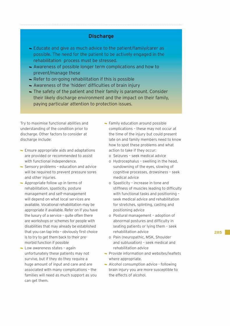
285
Discharge
Educate and give as much advice to the patient/family/carer as possible. The need for the patient to be actively engaged in the rehabilitation process must be stressed.
Awareness of possible longer term complications and how to prevent/manage these
Refer to on-going rehabilitation if this is possible Awareness of the ‘hidden’ difficulties of brain injury The safety of the patient and their family is paramount. Consider their likely discharge environment and the impact on their family, paying particular attention to protection issues.
Try to maximise functional abilities and understanding of the condition prior to discharge. Other factors to consider at discharge include:
Ensure appropriate aids and adaptations are provided or recommended to assist with functional independence.
Sensory problems – education and advice will be required to prevent pressure sores and other injuries.
Appropriate follow up in terms of rehabilitation, spasticity, posture management and self-management will depend on what local services are available. Vocational rehabilitation may be appropriate if available. Refer on if you have the luxury of a service – quite often there are workshops or schemes for people with disabilities that may already be established that you can tap into – obviously first choice is to try to get them back to their pre-morbid function if possible
Low awareness states – again unfortunately these patients may not survive, but if they do they require a huge amount of input and care and are associated with many complications – the families will need as much support as you can get them.
Family education around possible complications – these may not occur at the time of the injury but could present late on and family members need to know how to spot these problems and what action to take if they occur:
o Seizures – seek medical advice o Hydrocephalus – swelling in the head,
sundowning of the eyes, slowing of cognitive processes, drowsiness – seek medical advice
o Spasticity – increase in tone and stiffness of muscles leading to difficulty with functional tasks and positioning – seek medical advice and rehabilitation for stretches, splinting, casting and positioning advice
o Postural management – adoption of abnormal postures and difficulty in seating patients or lying them – seek rehabilitation advice
o Pain (neuropathic, MSK, Shoulder and subluxation) – seek medical and rehabilitation advice
Provide information and websites/leaflets where appropriate.
Alcohol consumption advice - following brain injury you are more susceptible to the effects of alcohol.
286
Finally there are aspects of brain injury that may fall outside of the domain of the rehabilitation professional in normal practice but that should be covered prior to discharge. These may not always present acutely and should be discussed with the patient/carer/family and advice given on how to manage these impairments or if possible who to contact for on-going care or advice:
Visual problems are common and often beyond the scope of an optician – specialist referral to orthoptics or opthalmology would be the gold standard but this is unlikely to be feasible in many situations.
Psychological support to manage changes in emotion or behaviour as well as issues with anxiety and depression would be ideal but simply acknowledging that this may occur may be as good as it gets depending on the situation.
Cognitive deficits are often missed or overlooked in the rapid acute stages of brain injury and are either too subtle to be noticed or are not assessed for. Again ideal intervention would be a combination of on-going OT and psychology input with local services. If this is not possible then education around the impairments and the use of basic strategies to overcome them (e.g. Memory strategies) is important.
Aspiration risk and malnutrition: SLTs and dieticians tend to be few and far between in less developed countries – refer on if possible, if not then masses of education around aspiration risk and modified diets may fall to you. Unfortunately in patients with severely impaired swallows in very poorly resourced environments the long term prognosis is poor as they are likely to suffer from aspiration pneumonia or malnutrition. If the patient is being fed, check the position – it is common that carers will feed the patient in supine, greatly increasing the risk of aspiration.
Speech impairments can have huge cultural implications so giving as much understanding and any strategies you have to the patient are likely to be of benefit.
Incontinence – education is the key especially in settings where equipment and adaptations to prevent or manage bowel and bladder incontinence may be difficult to access. The importance of hygiene is vital in order to prevent pressure sores developing
Fatigue – perhaps one of the least well know but most debilitating aspects of brain injury (especially mild injuries with only limited other symptoms) – this does tend to improve but can be all encompassing so should be addressed with education/advice prior to discharge
Mental capacity and vulnerability – patients who have sustained a brain injury may lack the capacity to make certain decisions (capacity is assessed on a decision by decision basis; so a patient may have the capacity to decide if they want to drink tea or coffee but not have the capacity to decide on a suitable discharge destination). Capacity is assessed by the patient’s ability to take on board information, retain that information, weigh up the consequences of a decision and to communicate a decision. If a person lacks capacity they are vulnerable and can be easily exploited so family may need to support them with certain decisions (be aware family are often the ones that exploit!). There have been situations when people with brain injury have been exploited to join extremist or military organisations.
Sexual Dysfunction – a real taboo topic and it may be that in the fleeting circumstances and culture that you are in it would be inappropriate to address this subject with certain patients. But where appropriate information and advice is important as is the reassurance that their symptoms are related to their brain injury. Specialist referral would be great but very unlikely to be widely available.
Acquired Brain Injury
287
Finally if nothing else is possible when it comes to discharging patients with brain injury at least try and reassure the patient or their family that the symptoms they are reporting are not unusual and are likely to be
as a result of their brain injury. Simply having this knowledge can be empowering. But where possible refer on to local rehabilitation services/Community based Rehabilitation teams or to DPOs for ongoing support.
TWO Key Messages from the training:
1. How to spot a deteriorating patient2. The importance of educating the patient and their family about
the long term consequences and complications of brain injury

288
Core Recommended Texts
Edwards, S (1996) Neurological Physiotherapy A problem solving Approach, London: Churchill Livingstone
Mateen, F (2010) Neurological disorders in complex humanitarian emergencies and natural disasters. ANN NEUROL 2010; 68:282–294
Stokes, M. (1998) Neurological Physiotherapy. London: Mosby
WHO (2004) Guidelines for Essential Trauma Care. http://www.who.int/violence_injury_prevention/publications/services/guidelines_traumacare/en/
References for brain injury management
British Society of Rehabilitation Medicine (2013) Specialist rehabilitation in the trauma pathway: BSRM core standards, London: Royal College of Physicians
Carey, L, Macdonnell, R and Matyas, T SENSe: Study of the effectiveness of Neurorehabilitation on Sensation: A Randomized Controlled Trial. Neurorehabilitation and Neural Repair, 2011; 25(4), 304-313
College of Occupational Therapists and Chartered Society of Physiotherapists (2015) Splinting for the prevention and correction of contractures in adults with neurological dysfunction, London: College of Occupational Therapists
Demey, D, Nielsen, S and Weerts, E (2010) Early Rehabilitation Protocol for Victims of Natural Disaster, Handicap International
Department of Health (2005) The National Service Framework for Long-term Conditions, London: Crown Copyright
Edwards, S (1996) Neurological Physiotherapy A problem solving Approach, London: Churchill Livingstone
French B, Thomas LH, Leathley MJ, Sutton CJ,McAdam J, Forster A, Langhorne P, Price
CI,Walker A,Watkins CL (2007) Repetitive task training for improving functional ability after stroke. Cochrane Database of Systematic Reviews (4): CD006073.
Intercollegiate Stroke Working Party (2012) National Clinical Guideline for Stroke (4th edn), London: Royal College of Physicians
Lindsay, K.W. Bone, I. (2004) Neurology and Neurosurgery Illustrated (4th edn), London: Churchill Livingstone
Marshall, S., Bayley, M., McCullagh, S., Velikonja, D., Berrigan, L. (2012) ‘Clinical practice guidelines for mild traumatic brain injury and persistent symptoms’, Canadian Family Physician, 58:pp257-267
Milner, M.E., Wagner, K.A. (eds) (1986) Neurotrauma: Treatment, Rehabilitation and Related issues, USA: Butterworths
http://www.mocatest.org/
National Institute for Health and Care Excellence (2013) Stroke Rehabilitation, London: NICE
National Institute for Health and Care Excellence, 2014. ‘Head injury’, NICE clinical guideline 176. London (available at: www.guidance.nice.org.uk/CG176)
New Zealand Guidelines Group (2006) Traumatic Brain Injury: Diagnosis, Acute Management and Rehabilitation, Wellington: New Zealand Guidelines Group
http://www.ninds.nih.gov/disorders/hypertonia/hypertonia.htm
http://www.ninds.nih.gov/Disorders/hypotonia/hypotonia.htm
SIGN (2010) Management of Patients with Stroke: Rehabilitation, prevention and management of complications and discharge planning, NHS Scotland
Stokes, M. (1998) Neurological Physiotherapy, London: Mosby
Acquired Brain Injury
289
Thieme, H et al. Mirror Therapy for Patients with severe arm paresis after stroke: A randomized controlled trial. Clinical Rehabilitation, 2012; 27(4) 314-324
Turner-Stokes L, Nair A, Sedki I, Disler PB, Wade DT. Multi-disciplinary rehabilitation for acquired brain injury in adults of working age. Cochrane Database of Systematic Reviews 2005, Issue 3. Art. No.: CD004170. DOI: 10.1002/14651858.CD004170.pub2
Ward, J. (2010) The Student’s Guide to Cognitive Neuroscience, Hove: Psychology Press
Wolf, SL et al. Effect of Constraint induced movement therapy on upper limb function 3 to 9 months after stroke: the EXCITE randomized clinical trial. JAMA 2006; 296(17): 2095-2104
World Health Organization. (2001). International Classification of Functioning, Disability and Health (ICF). Geneva
World Health Organisation (2011) World Report on Disability, Malta: WHO
World Health Organisation (2014) Emergency Trauma Guidelines on Medical, Surgical, Nursing and Rehabilitation Management of Head Injuries Expected in the Event of Mass Casualty Incident Scenario, Nepal: WHO
Zimmerman-Schlatter, A et al. Efficacy of Motor Imagery in post stroke rehabilitation a systematic review. Journal of Neuroengineering and Rehabilitation 2008; 5:8
Useful further supporting information:
Aronow H. Rehabilitation effectiveness with severe brain injury: translating research into policy. J Head Traum Rehabil 1987;2:24-36
Powell J, Heslin J, Greenwood R. Community based rehabilitation after severe traumatic brain injury: a randomised controlled trial. J Neurol Neurosurg Psychiatry 2002;72:193-202
Regional Trauma Networks National Clinical Advisory Group. London: Department of Health. 2010
Royal College of Physicians (2009) Spasticity in Adults management with Botulinum Toxin, National Guidelines London: RCP
Semlyen JK, Summers SJ, Barnes MP. Traumatic brain injury: efficacy of multidisciplinary rehabilitation. Arch Phys Med Rehabil 1998;79:678-83
SIGN (2013) Brain injury rehabilitation in adults, NHS Scotland
Specialist neuro-rehabilitation services: providing for patients with complex rehabilitation needs. London: British Society of Rehabilitation Medicine. 2010
Turner-Stokes L, Nair A, Disler P, et al. Cochrane Review: Multi-disciplinary rehabilitation for acquired brain injury in adults of working age. CD004170. The Cochrane Database of Systematic Reviews Oxford: Update software 2005 (Update July 2009);Issue 3
Turner-Stokes L. Evidence for the effectiveness of multi-disciplinary rehabilitation following acquired brain injury: a synthesis of two systematic approaches. J Rehabil Med 2008;40:691-701
Turner-Stokes L. Cost-efficiency of longer-stay rehabilitation programmes: can they provide value for money? Brain injury 2007;21:1015-21
Turner-Stokes L, Paul S, Williams H. Efficiency of specialist rehabilitation in reducing dependency and costs of continuing care for adults with complex acquired brain injuries. J Neurol Neurosurg Psychiatr 2006;77:634-9
Turner-Stokes L. The evidence for the cost-effectiveness of rehabilitation following acquired brain injury. Clinical Medicine 2004;4:10-2