Embed Size (px)
Citation preview
Regional Skin & Wound Education Facilitator Guide: Diabetic Foot Ulcers (Module 3)
June 2012 1
How to use this guide Learning Objectives:
Each section outlines specific learning objectives that will guide the discussion & activities of this part of the workshop.
Participants know what they will achieve by completing the section.
TO PRINT THIS GUIDE: Use printer DOUBLE-SIDED function & CHOOSE FLIP PAGES UP (this option allows user to staple document from above & flip through pages as one would a book).
Time Topic Facilitator Activities / Questions Media
An estimate of the time each section will take.
Varies by number of participants
General topic being covered.
Describes what facilitator does for each section.
ACTIVITIES: appear in BLUE & include purpose statement, instructions for conducting activity & points for summary
EMPHASIZE: Points for emphasis appear here in RED
Activities undertaken by the group.
QUESTION TO GROUP: questions put to group appear here in GREEN
ANSWERS to questions appear immediately below, also in GREEN
What AV or other material accompanies each section
HO = Handout
FC = Flip chart

Regional Skin & Wound Education Facilitator Guide: Diabetic Foot Ulcers (Module 3)
June 2012 2
Time Topic Facilitator Activities / Questions Media
Introduction: the Hook, Learning Outcomes, Objectives, and Icebreaker
0830 – 0900 Connect laptop to Fraser Health Intranet
Open the following link: http://www.cawc.net/
Minimize as this link will be used later in the presentation
There are 2 documents on this link that can be printed off under the diabetes self management plan:
• Healthy feet checklist and
• Personal foot care plan
Pre reading Best Practice recommendations for DFU 2010 & Provincial CDST
Handouts:
• Provincial CDST
• monofilament procedure
• Pixalere foot /sensation
• Guideline summary
Greet learners, acknowledge differing levels of experience and education in the room, discuss format of session
Allow learners to introduce self and area of work to other participants.
Orientate to bathrooms
Encourage interactivity and group discussion and questions through the session
Slide 1
Slide 2

Regional Skin & Wound Education Facilitator Guide: Diabetic Foot Ulcers (Module 3)
June 2012 3
Time Topic Facilitator Activities / Questions Media
Vision & Mission Client – All through the education module, the term client is used interchangeably as patients, clients, and residents as appropriate in different health care settings
The vision and mission statements are to reflect on the ultimate learning outcomes (level 4 to 6) that, due to constraint in resources allocation, that the Regional SW CWT/SW Steering Committee are not able to evaluate.
Slide 3
This module will evaluate the following level 1-3 learning outcomes:
1. Level 1: Participation – The number of learners participated in the learning events, e.g. attendance sign-in sheet
2. Level 2: Satisfaction – The degree to which expectations of the learners about the setting and delivery the educational events were met, e.g. Learning Session Evaluation form
3. Level 3: Learning – Changes in the knowledge, skills and/or attitudes of the learner, and the development of competence, e.g. pre- and post-test
Slide 3

Regional Skin & Wound Education Facilitator Guide: Diabetic Foot Ulcers (Module 3)
June 2012 4
Time Topic Facilitator Activities / Questions Media
This module will NOT evaluate the following level 4-6 learning outcomes due to constraint in resources allocation. Instead, they are stated as the Vision and Mission of the module:
1. Level 4: Performance – Changes of practice performance as a result of the application of what was learned, e.g. paper or on-line 3-month follow up survey, etc.
2. Level 5: Patient outcomes – Change s in the health status of patients due to changes in practice behaviours, e.g. prevalence and incidence rates, costs incurred in treating peri-stomal skin complications, etc.
3. Level 6: Population outcomes – Changes in the health status of a population of patients due to changes in practice behaviours, e.g. acute care admissions for peri-stomal skin complications, etc.
(Sibbald et al, 2007. Effective Adult Education Principles to Improve Outcomes in Patients with Chronic Wounds)
Slide 3
Regional Skin & Wound Education Facilitator Guide: Diabetic Foot Ulcers (Module 3)
June 2012 5
Time Topic Facilitator Activities / Questions Media
0840 – 0850 Ice Breaker / Pre-Test
Introduce yourself and the co-instructors – roles, work areas/program, and credentials
Discuss housekeeping items i.e. (bathroom, pagers/phones on vibrate, no texting during class)
Identify learners’ existing knowledge by:
1. “Ice Breaker” – Learners to introduce themselves to the group: name, area of work, if they have cared for clients with pressure ulcers, why they attend the education, and what they want to gain from attending the education
2. Then ask the learner to complete the Pre-test on the 1st page of the evaluation form
Slide 4

Regional Skin & Wound Education Facilitator Guide: Diabetic Foot Ulcers (Module 3)
June 2012 6
Time Topic Facilitator Activities / Questions Media
Objectives Review objectives and briefly outline plan for time frames.
Objectives:
• Describe the etiology & predisposing risk factors of Diabetes
• Describe the strategies in the prevention and management of DFU with an interprofessional team approach
• Describe the components of a vascular and neurological assessment of the foot
• Perform foot inspection, foot wear assessment & identify interventions used to reduce pressure
• Select local wound care interventions
Advise participants of the guest speakers who will be co-presenting
Slide 5

Regional Skin & Wound Education Facilitator Guide: Diabetic Foot Ulcers (Module 3)
June 2012 7
Time Topic Facilitator Activities / Questions Media
0850 – 0900 Diabetes Pandemic
The Hook – some facts on DM and DFU (this slide and next slide)
Source:
1. Stats Canada 2010
2. An economic tsunami: the cost of diabetes in Canada, Canadian Diabetes Association (Dec 2009)
More than 20 people are diagnosed every hour of every day.
In 2010, 1,841,527 (6.4%) Canadians have diabetes with 202,442 (5.2%) living in BC
In 2008, DM is the 6th leading cause of death, accountable for the death of 7,521 Canadians (3.2%)
The number of people diagnosed with diabetes in Canada is expected to double between 2000 and 2010, from 1.3 million to about 2.5 million
First Nations population have a prevalence of type 2 DM thrice the national average
Expected economic burden approx. $12.2 billion in 2010 (increase of $5.9 billion, doubled from 2000)
Slide 6

Regional Skin & Wound Education Facilitator Guide: Diabetic Foot Ulcers (Module 3)
June 2012 8
Time Topic Facilitator Activities / Questions Media
An Economic Tsunami: The Cost of Diabetes in Canada
The economic burden of diabetes in Canada projected to be about $12.2 billion in 2010, an increase of $5.9 billion; nearly double its level in 2000
The cost of diabetes is expected to rise by another $4.7 billion by 2020
The direct cost of diabetes accounts for 3.5% of public healthcare spending and is likely to continue rising given the expected increasing number of Canadians living with diabetes
Slide 7
Persons with Diabetes Mellitus
Although often overlooked at the onset of DM, one of the common and devastating complications of DM is DFU.
Put diabetic foot ulcers into the context of Canadian health care statistics and help to under score the magnitude of the problem as a way of introducing the topic.
Slide 8
0900 – 0915 Client education on DM Control/Foot Care
Loss of protective sensation is the most significant predictor of diabetic foot ulceration.
Use Animation to introduce Sam, our client all through this presentation.
Sam is an instruction site worker, whose wife Susan is a 40-year old secretary.
They have 2 children: a boy age 12 and a girl age 10.
Slide 9

Regional Skin & Wound Education Facilitator Guide: Diabetic Foot Ulcers (Module 3)
June 2012 9
Time Topic Facilitator Activities / Questions Media
This slide is when Sam when he was first diagnosed with Diabetes at age of 45
The outer oval shapes are the anticipated clinical/personal issues related to DM/DM related complications.
SAM and LOPS are the key clinical issues that will be discussed in details later.
The inner circles are the lists of interprofessional team members to support Sam to maintain optimal DM control
Please note that Specialists include: Foot Surgeon, Podiatrist, Endocrinologist, vascular surgeon, etc.
Slide 9
Introduce:
Role of Wound Care Clinician
• Member of the interdisciplinary team
• Provides specialized holistic assessment and management of patients/families with ostomies, acute and chronic wounds, and urinary and fecal continence problems
• Coordinates specialty care requirements with hospitals, community and follow-up services
• Educates and consults about advance nursing skills within areas of expertise in wound and skin care practice.
Slide 9
Regional Skin & Wound Education Facilitator Guide: Diabetic Foot Ulcers (Module 3)
June 2012 10
Time Topic Facilitator Activities / Questions Media
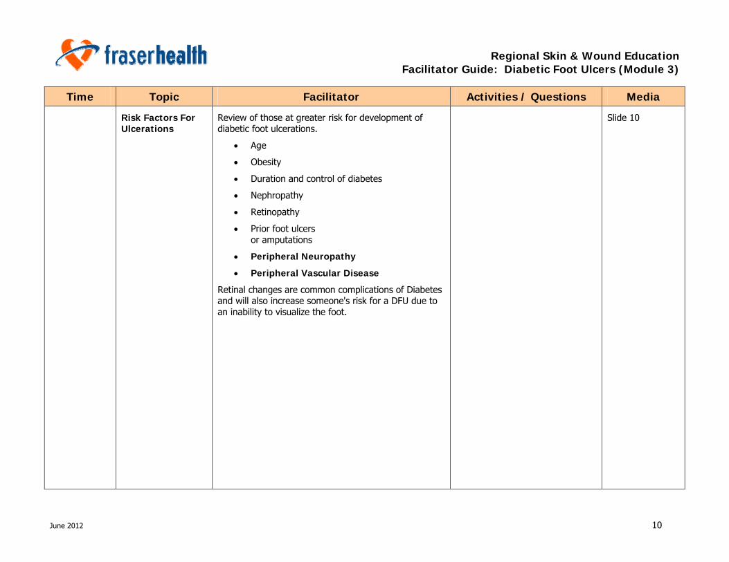
Risk Factors For Ulcerations
Review of those at greater risk for development of diabetic foot ulcerations.
• Age
• Obesity
• Duration and control of diabetes
• Nephropathy
• Retinopathy
• Prior foot ulcers or amputations
• Peripheral Neuropathy
• Peripheral Vascular Disease
Retinal changes are common complications of Diabetes and will also increase someone's risk for a DFU due to an inability to visualize the foot.
Slide 10
Regional Skin & Wound Education Facilitator Guide: Diabetic Foot Ulcers (Module 3)
June 2012 11
Time Topic Facilitator Activities / Questions Media
ABCs of Optimizing Diabetes
Define A1C:
The A1C test (also known as HbA1C, glycated hemoglobin or glycosylated hemoglobin) is a blood test done in the lab to provide a good general measure of diabetes care.
While conventional home glucose monitoring measures a person’s blood sugar at a given moment, the A1C test indicates a person’s average blood glucose level over the past few months.
AIC should be checked every 3 months
According to Dr Sibbald, a Canadian and International renowned Dermatologist specialized in wound care, A1C greater than 9% will affect wound healing & 7% will impair wound healing. Therefore, recommended A1C is less than 7%
Slide 11
Feet for L.I.F.E. Client Education crucial to promote adherence treatment plan and rapport with healthcare providers:
Lifestyle Choices
• Eat a healthy diet and maintain blood sugars within normal range
• Exercise daily
• If you have foot problems try a stationary bike or swimming
• Don’t smoke – IMPORTANT FOR ALL! – each cigarette decreases leg circulation for 30% for an hour or increase sympathetic tone for 8 hours
Slide 12
Regional Skin & Wound Education Facilitator Guide: Diabetic Foot Ulcers (Module 3)
June 2012 12
Time Topic Facilitator Activities / Questions Media
Inspect your feet and footwear
• Look at your feet daily to check for cuts, scratches or blisters
• If you cannot see your feet clearly, have a friend or family member check them or use a mirror
• Check your shoes and socks on a regular basis
• Always wear good fitting shoes and socks
• Check for foreign objects in shoes before putting them on
• Check for rough areas inside shoes
• Wear well fitting socks with no seams or darning
Slide 12
Find Professional Assistance
Your feet deserve the best professional care you can find
• Foot care professionals include MD, podiatrist or RN
• Orthotist
• Shoe fitter (pedorthotist)
Slide 12
Regional Skin & Wound Education Facilitator Guide: Diabetic Foot Ulcers (Module 3)
June 2012 13
Time Topic Facilitator Activities / Questions Media
Expect your feet to last a lifetime
• Wash and dry feet daily
o Dry gently between each toe
• Avoid extreme temperature changes
o Test water with a thermometer
• Do not use heating pads on your feet
• Do not soak feet
• Use unscented moisturizer for dry skin
• Do not use adhesive tape, wart treatments, corn plasters or strong antiseptics on your feet
• If you have an open area/crack on your feet, see your health care professional
Slide 12
Regional Skin & Wound Education Facilitator Guide: Diabetic Foot Ulcers (Module 3)
June 2012 14
Time Topic Facilitator Activities / Questions Media
Diabetic Educators can help
Therefore, need to emphasize to Sam that Foot Care is important
FH Diabetic Education Center
• If you have trouble seeing or reaching your feet - see a foot doctor or nurse to assist you. These may be covered by your private plan but are not covered under Fair Pharmacare
• Wash feet daily - in warm water with a mild soap.
• Pat dry with a soft towel and dry carefully between toes
• Always check the water temperature with your elbow or wrist. Hot water, hot pavement or heating pads can all cause severe burns.
• If skin is dry, apply a urea-based moisturizer. If you have very dry skin-use a product with urea 10-25%, which will pull moisture to the skin surface. Remember no lotion between toes and this makes the toe webs too wet and may encourage a fungus infection.
• Soak nails for 10 min to soften before cutting and use a nail clipper (not scissors). Cut straight across and file rough edges with an emery board ( not steel)
• Do not self treat corns and warts with chemicals, or sharp instruments- these can damage your feet or cause infection.
• Always protect your feet by wearing hard-soled slippers and shoes avoid flip-flops.
Slide 13

Regional Skin & Wound Education Facilitator Guide: Diabetic Foot Ulcers (Module 3)
June 2012 15
Time Topic Facilitator Activities / Questions Media • Wear seamless socks that don’t constrict. If
you have swelling ask your doctor about compression stockings available at pharmacies or from a foot care specialist.
• Wear foot shaped shoes with low heels and good support –
o go to specialty shoe store, that knows how to fit shoes for diabetic feet. Always get your feet measured or draw a tracing of your foot standing up. Cut it out and take it with you to insert into the shoe.
o Remember a numb foot cannot feel a tight shoe!
o Shop for shoes in the late afternoon when feet tend to swell and take the socks and orthotics you usually wear with you for fitting.
Be smoke-free
Client Education: Early Detection
Early detection is the second step in caring for your feet.
Look for signs of:
• corns, calluses, blisters, scrapes.
• of infection such as redness, swelling , heat, or discharge.
Wear white socks so you will see if there is blood or drainage from an injury, such as a torn nail or stubbed toe.
Slide 14
Regional Skin & Wound Education Facilitator Guide: Diabetic Foot Ulcers (Module 3)
June 2012 16
Time Topic Facilitator Activities / Questions Media
Client Education: Prompt Treatment
Could be an infected cut, sore, blister, callus or bruise
Do not try to treat your feet yourself.
Pain of an injury may not be felt due to loss of sensory related to DM changes
Remember most severe infections and amputations are a result of a minor injury or problem that was left unattended!
Don’t let this happen to you.
Always follow the treatment plan your doctor gives you and finish all the medication as prescribed.
• Antifungal creams must be used twice/day for 6-12 weeks even after the itch and burning has gone.
Slide 15

Regional Skin & Wound Education Facilitator Guide: Diabetic Foot Ulcers (Module 3)
June 2012 17
Time Topic Facilitator Activities / Questions Media
Diabetes & Healthy Feet
A CAWC expert advisory group, in collaboration with a patient focus group, has developed a self-assessment brochure and an interactive website in many languages to help patients in recognizing risk factors and identifying foot issues that they may have been previously unaware of.
The brochure and interactive website are available at www.cawc.net/diabetesandhealthyfeet (Botros et al 2010)
Review CAWC page by opening up hyperlink & review the information below
• Select personal self management questionnaire
• Select personal foot care plan.
• Handout Healthy feet checklist and personal plan.
• Select “ Your foot a closer look”
Slide 16
The brochure and interactive website at www.cawc.net/diabetesandhealthyfeet (Botros et al 2010)
Slide 17
Regional Skin & Wound Education Facilitator Guide: Diabetic Foot Ulcers (Module 3)
June 2012 18
Time Topic Facilitator Activities / Questions Media
0915 – 0945 SAM, LOPS, Monofilament, Self Management, Nutrition
Review the key points for Prevention and Treatment of DFU
Disclaimer: the term “Client” is used interchangeably with patient in acute care and residents in residential care, or any other settings
(Botros et al 2010)
Diabetic sensory neuropathy is the leading cause of foot ulcers.
It generally presents as a distal symmetric sensori-motor neuropathy and is believed to contribute to ulcers because the patient cannot feel harmful stimuli.
Peripheral neuropathy affects sensory, motor and autonomic nerves.
Emphasize Loss of protective sensation is the most significant predictor of diabetic foot ulceration.
People with diabetes are prone to serious injury from minor trauma because they cannot feel the injury to the foot as it occurs.
In addition to single injurious incidents, such as stepping on a needle, repetitive stress simply from walking can lead to tissue breakdown in the absence of protective sensation.
Slide 18

Regional Skin & Wound Education Facilitator Guide: Diabetic Foot Ulcers (Module 3)
June 2012 19
Time Topic Facilitator Activities / Questions Media
Components of Foot Assessment
What would you include under history?
• Reason for referral, general health, co morbidities, history of foot problems/ traumatic surgeries, characteristics of pain, work and leisure activities.
• Glycemic control
Foot appearance and structure:
• Assessing for bunions, callous, corns missing digits, Charcot( discuss later), hallgus limitus ( inflexible great toe)
• Alignment of foot when wt bearing, do the arches drop i.e. flat foot.
Gait:
• Look for lack of range of motion e.g. Shuffling gait indicating inability to dorsiflex.
• Muscle weakness, poor balance, uncoordination
Neuralgic: monofilament testing
Infection: bacterial, viral fungal
Footwear:
• Are they wearing protection in and out doors. What is the fit?
• Are they wearing socks?
Slide 19
Sixty Second Foot Exam
In summary here are guidelines for foot examinations of intact diabetic feet.
Slide 20

Regional Skin & Wound Education Facilitator Guide: Diabetic Foot Ulcers (Module 3)
June 2012 20
Time Topic Facilitator Activities / Questions Media
Risk Classification The IWGDF developed this straight forward risk classification system which it modified recently.
It quickly and accurately classifies patients and guides the clinician in predicating foot complications & guides in choosing the most appropriate therapeutic interventions.
Inform the learners that Neuropathy and LOPS will be further discussed later
Refer to handouts provided
Slide 21
Handouts
Peripheral Neuropathy
The presence of vascular disease and neuropathy, or a combination of both, are the most important risk factors in the development of diabetic neuropathy. (Salzeda et al. OWM 2003.)
While neuropathy is the most common reason for diabetic foot ulceration, peripheral vascular disease and infection can also be factors in skin breakdown.
Vascular disease also plays a role in diabetic foot ulcer development and is responsible for 15 to 20% of diabetic foot ulcers.
Diabetes is a risk factor in the development of arteriosclerosis.
Smoking, hypertension and hyperlipidemia are also risk factors in the development of peripheral vascular disease and these factors will add additional risks for the diabetic patient.
Slide 22

Regional Skin & Wound Education Facilitator Guide: Diabetic Foot Ulcers (Module 3)
June 2012 21
Time Topic Facilitator Activities / Questions Media
Diabetic neuropathy is thought to be metabolic in origin and related to an over stimulation of the polyol pathway in neural tissue.
Hyperglycemia is associated with uncontrolled diabetes.
Elevated blood glucose is metabolized by the enzyme aldose reductase which then produces sorbitol and polyol.
Sorbitol accumulates in the tissues and causes damage in many ways.
In the nerves, sorbitol is toxic and causes segmental demyelination which leads to lower conduction of speed in the peripheral nerves.
A demyelinating disease is any disease of the nervous system in which the myelin sheath of neurons is damaged.
This impairs the conduction of signals in the affected nerves, causing impairment in sensation, movement, cognition, or other functions depending on which nerves are involved.
Slide 22
Neuropathy Review the three different types of neuropathy and the major outcome of each type of neuropathy which is causative of problems with diabetic foot.
Sensory
Autonomic
Motor
Slide 23
Regional Skin & Wound Education Facilitator Guide: Diabetic Foot Ulcers (Module 3)
June 2012 22
Time Topic Facilitator Activities / Questions Media
Sensory Neuropathy (S.A.M.)
Hyperesthesia can lead to poor skin care practices (walking bare foot because they can not stand the feel of socks on the feet, or can not stand to have their feet touched or washed due to the increased sensitivity to touch).
Loss of protective sensation leads to diminished or absent pain sensation.
Chemical trauma from over the counter “wart” or callous remedies such as acids or other inventive chemical applications.
Mechanical trauma can be from improper foot wear, nail care practices or callous “care”
Thermal damage from heat or cold causing tissue damage.
Slide 24
Sensory Neuropathy & LOPS
Sensory Neuropathy results in Loss Of Protective Sensation (LOPS):
Photo shows a client who has no pain even with the toe caught on a piece of furniture.
Slide 25
Why Loss Of Protective Sensation causes ulceration
With no feeling, client cannot protect self from injury from chemical, mechanical and thermal damage.
“Bath room surgery”
Slide 26
Regional Skin & Wound Education Facilitator Guide: Diabetic Foot Ulcers (Module 3)
June 2012 23
Time Topic Facilitator Activities / Questions Media
Un-trimmed Nail Causing Pressure (S.A.M.)
Another example of poor nail hygiene due to lack of sensation (client does not sense that the nail is digging into the toe beside it), or that the toe nail is thickened and may be exerting pressure into the nail bed.
Ask participants questions as indicated
QUESTION: What do you assess in terms of infection and vascular supply here (V.I.P.)?
ANSWER: Toe is reddened Hair on knuckles of the toe indicate reason able blood flow
QUESTION: How else would you assess for vascular supply and infection?
ANSWER: Pedal pulses Colour of limb Temp of limb Palour or rubour
QUESTION: Who would you recommend referral of this client to ?
ANSWER: Podiatrist or nail care service
Slide 27

Regional Skin & Wound Education Facilitator Guide: Diabetic Foot Ulcers (Module 3)
June 2012 24
Time Topic Facilitator Activities / Questions Media
Monofilament Testing
Assessment for LOPS ins easily accomplished by the clinician, client or caregiver using a Semmes Weinstein monofilament.
The inability to perceive the forces applied by the monofilament is associated with clinically significant large fiber neuropathy.
Monofilaments are available in each office.
Client should have a monofilament test performed.
Refer to handout on Pixalere foot/Sensation assessment
Slide 28
Handout: Pixalere Foot / Sensation Assessment
Regional Skin & Wound Education Facilitator Guide: Diabetic Foot Ulcers (Module 3)
June 2012 25
Time Topic Facilitator Activities / Questions Media
5 Minutes Monofilament Testing Activity
Ask learner to take out the Monofilament testing & Pixalere foot/sensation assessment sheet from the handout (Reference: CDST procedure on Monofilament testing).
Before beginning, review the use of the monofilament and read through procedure together.
Ask participants to record findings on Pixalere foot assessment sheet, sites where the participant can feel are checked.
Avoid leading questions and cues when assessing with monofilaments
Interpretation:
If all sites are felt with the monofilament the score is 10/10
If the monofilament is not felt in an area on the foot, this indicates LOPS in that area and requires a referral to the wound clinician
Instruct participants to partner with buddy in groups of 2.
One person will perform a monofilament assessment; the other will be the receiver.
Slide 29
Autonomic Neuropathy (S.A.M.)
Dry skin is 2-3 times more likely to break down
Infection/cellulitis can be initiated with any loss of skin integrity no matter how small and seemingly insignificant esp. in a diabetic client who may not be responding with a full immune system compliment and less than robust inflammatory responses.
Example in this photo is of fungal infection from too much moisture
Slide 30
Regional Skin & Wound Education Facilitator Guide: Diabetic Foot Ulcers (Module 3)
June 2012 26
Time Topic Facilitator Activities / Questions Media
Autonomic Neuropathy (S.A.M.)
Examples of the severity of the dry skin related to autonomic neuropathy:
• Xerosis/Anhydrosis and
• Fissures
To correct the dryness of this skin suggest use of pumice stone and moisturizer, off loading and good local wound care.
Slide 31
Moisturizers to Protect Skin (S.A.M.)
Use animation, after discussing bullet 1, 2, 3, pause, and ask what a good moisturizer should be.
Click and review the answer
Water accounts for 60-80% of most commercial moisturizers, but externally applied moisture does not re-moisturize the skin.
The thin consistency of most commercial lotions provide some replacement of natural oils in the stratum corneum, but the effect is short lived due to the continued transepidural moisture loss.
Slide 32
Regional Skin & Wound Education Facilitator Guide: Diabetic Foot Ulcers (Module 3)
June 2012 27
Time Topic Facilitator Activities / Questions Media
Lotions have the most water, followed by cremes and then ointments.
Cremes have a higher oil content than lotions but do not provide total occlusion of the skin.
Ointments are near to 100% oil and are occlusive of the skin and generally not well tolerated for cosmetic reasons anyways.
Therefore choices for diabetic foot should be a creme (higher occlusive properties to it than a lotion), containing humectants. (Atractain)
Problem with total occlusion of the skin is that once the occlusive agent is removed water loss resumes to it’s pre-application level
Vaseline (petrolatum) is not totally occlusive and may be a reasonable alternative if costs for other cremes are prohibitive.
Occlusion – physical covering of the skin preventing water loss (total occlusion is not desirable)
Slide 32
Motor Neuropathy (S.A.M.)
Neuropathy of the innervating motor neurons of the lower extremities
Distal to proximal cell death pattern
Intrinsic muscles of the foot are primarily involved
Slide 33
Specifically an imbalance between flexors and extensors muscles of the toes
Intrinsic muscles are the muscles with in the foot. There main function is assisting the extrinsic (i.e. in the calf and shin) to flex and extend the toes.
Slide 34
Regional Skin & Wound Education Facilitator Guide: Diabetic Foot Ulcers (Module 3)
June 2012 28
Time Topic Facilitator Activities / Questions Media
Hammer toe is a permanently flexed digit usually the 2nd toe.
Claw toe is a hyperextension of the toes at the metatarsal head, may be claw foot as well, which is an excessively flexed arch of the foot.
Ask participants question indicated.
QUESTION: Where would ulcers occur in the toes seen here?
ANSWER: Tops of the toes from shoes rubbing or bottom of the toes from pressure
Slide 35
Claw Toe & Hammer Toe pictured. Slide 36
Fat Pad Migration (S.A.M.)
Animation : Click to animate the fat pad to move forward, then to move up the pressure
Slide 37
Reduces area to distribute pressure
Increases pressure on the front of the foot
If the client has reduced range of motion in their big toe (i.e. hallicus rigidus) the extra pressure moved to the forefoot frequently causes big toe and 1st metatarsal wounds
Slide 38
Regional Skin & Wound Education Facilitator Guide: Diabetic Foot Ulcers (Module 3)
June 2012 29
Time Topic Facilitator Activities / Questions Media
The transverse arch runs across the front of the foot, just before the toes. It’s much less pronounced then the long arch, but integrates to normal distribution of pressure in the fore foot.
As the arch collapses, the metatarsal head drops, creating new / unnatural pressure area.
1. Middle of the foot
2. Sides of the foot (i.e. big / little toes)
This new pressure area is not normal and the tissue in this area is not able to manage the increased load.
Slide 39
How Motor Deformity Contributed To Ulcer?
Use animation, after showing 1, 2, 3, pause and ask question.
Click to review the answer
Example of callous formation that may have gotten so thick that it impeded the blood flow to the underlying tissues and/or there may be loss of fat pads in this foot and the metatarsal heads may be very close to the surface of the skin, leading to ulcerations.
Hammer toe formations may cause this foot to ulcerate in this manner.
QUESTION: What would happen?
ANSWER: All adds up to pressure
Slide 40

Regional Skin & Wound Education Facilitator Guide: Diabetic Foot Ulcers (Module 3)
June 2012 30
Time Topic Facilitator Activities / Questions Media
Deformities in the foot
Bunion type deformity which may be related to diabetes or other reasons
Ask participants question indicated.
Click to reveal the 11 spots
QUESTION: Where is the ulcer most likely to occur in this foot?
ANSWER: There are at least 11 spots - boney prominences and at calloused area of foot - toe box of shoe - under great toe and second toe
Slide 41
Address Client Concerns Related to SAM
Treat the patient!
DM control
Diet, exercise, footwear, foot inspection, etc.
Consider pain (nociceptive neuropathic), activities of daily living, financial, social, emotional issues.
Slide 42

Regional Skin & Wound Education Facilitator Guide: Diabetic Foot Ulcers (Module 3)
June 2012 31
Time Topic Facilitator Activities / Questions Media
Self Management Remember Effective foot care can reduce amputations by 2/3 (Foster 2002 from Nursing Standard Vol 19, no 45 2005).
Review Diabetic Screening tool handout with participants
Awareness of personal risk factors
Importance of at least annual inspection of the feet by a health care professional
Daily self inspection of feet
Proper nail and skin care
Proper foot wear selection
Injury prevention and management of problems
When to seek help or specialized referral
Slide 43
Nutrition Optimal glycemic control
Adequate calories
Protein requirements 2-3 times normal
Supplement with multi-vitamins and minerals
Adequate hydration
Referral to dietitian
Slide 44

Regional Skin & Wound Education Facilitator Guide: Diabetic Foot Ulcers (Module 3)
June 2012 32
Time Topic Facilitator Activities / Questions Media
Role of Dietitian Macronutrients
There are three primary macronutrients defined as being the classes of chemical compounds humans consume in the largest quantities and which provide bulk energy.
These are protein, fat, and carbohydrate. This list shows the categorization of the most common food components by these macronutrients.
Macronutrients can also refer to the chemical elements humans consume in the largest quantities, see Nutrient.
Micronutrients:
Nutrients that are required by humans and other living things throughout life in small quantities to orchestrate a whole range of physiological functions, but which the organism itself cannot produce.
For people, they include dietary trace minerals in amounts generally less than 100 milligrams/day - as opposed to macrominerals which are required in larger quantities.
The microminerals or trace elements include at least iron, cobalt, chromium, copper, iodine, manganese, selenium, zinc and molybdenum.
Micronutrients also include vitamins, which are organic compounds required as nutrients in tiny amounts by an organism.
Slide 45
Regional Skin & Wound Education Facilitator Guide: Diabetic Foot Ulcers (Module 3)
June 2012 33
Time Topic Facilitator Activities / Questions Media
Dietitian Referral For ambulatory client’s in the community a referral can be made to a diabetes services “link on slide” takes to the referral form.
For acute and residential clients there are dietitians in each facility.
Other resources through healthlink BC
Slide 46
Regional Skin & Wound Education Facilitator Guide: Diabetic Foot Ulcers (Module 3)
June 2012 34
Time Topic Facilitator Activities / Questions Media
0945 – 1000 Charcot Foot Transition slide to Charcot Foot
Charcot foot is a particularly acute and devastating occurrence which can occur in a person with neuropathy, but is far more common in diabetics with neuropathy.
It is characterized by bony re-absorption and multiple spontaneous fractures which result from autonomic-neuropathy induced bone blood flow hyperemia.
Hypervascularity of the mid foot osseous structures results in decreased structural integrity of the bone significantly increasing risk of fracture.
These fractures may result from ADLs and not obvious trauma.
Clinical presentation includes dermal flush, redness, increased skin temperature, +/- deep bony pain, +/- local edema and bounding pulses.
The condition may mimic cellulitis or deep vein thrombosis.
X-ray and bone scan are used to assess and reconfirm re-ossification
Clients frequently do not experience pain due to their neuropathy
Charcot fracture results in catastrophic deformity often ignored by Clients.
Slide 47
Use Animation to add WCC/ET/WOCN to the team
Specialists include Foot Surgeon, Podiatrist, Endocrinologist
Slide 48
Regional Skin & Wound Education Facilitator Guide: Diabetic Foot Ulcers (Module 3)
June 2012 35
Time Topic Facilitator Activities / Questions Media
Risk Factors for Charcot Foot
• Peripheral sensory neuropathy
• Normal circulation
• Preceding trauma, often minor, e.g. sprains or contusions
• Foot deformities, prior amputations, joint infections or surgical trauma disease
Slide 49
This table is an overview of the Stages of Charcot foot compiled by CAWC in the 2010 BPR review
Question: Why is it important to recognize acute Charcot?
Answer: So you can prevent foot deformities ensuring that client is completely offloaded. Stage 1 is the is the most important stage for clinicians to recognized and where they can make the greatest difference in prevention. (Frykberg et al., 2006)
Slide 50

Regional Skin & Wound Education Facilitator Guide: Diabetic Foot Ulcers (Module 3)
June 2012 36
Time Topic Facilitator Activities / Questions Media
1000 – 1030 Transition to DFU Sam’s diabetes continues to be poorly managed.
Unfortunately, he developed foot ulcers on his soles
Approx. 9% of clients with diabetic neuropathy develop Charcot foot.
Early recognition & diagnosis of the acute Charcot is essential to prevent increased damage & prevent disastrous consequences including amputation.
Acute Charcot is a medical emergency.
The client must not bear weight and may benefit from medications which prevent bone re-absorption po or IV.
Temperature increases and decreases in the outside temp of the foot signal the amount of activity of bone absorption and is a key indicator of the internal processes of the foot.
2 degree C or 4 degrees F difference in skin temperature from contra lateral foot.
Dermal thermometers are being utilized in some centers and clients are being taught to self monitor for this condition as well as infective processes.
Diagnostics:
• Systemic symptoms, abnormal lab values are usually absent.
• Radiographic changes take time to develop and the initial finding can be normal but repeated xrays on patients who are not immobilized show abnormal findings.
• C reactive protein level is normal in Charcot.
Slide 51
Regional Skin & Wound Education Facilitator Guide: Diabetic Foot Ulcers (Module 3)
June 2012 37
Time Topic Facilitator Activities / Questions Media
Reference: American Family Physician June 1998. The Charcot foot in Diabetes: Six Key Points. Caputo, Cavanagh, Ulbrecht & Juliano
According to Caputo, Cavanagh et al 1998 approximately 9% of diabetics with neuropathy develop Charcot.
Slide 51
Diabetic Foot Ulcers (DFUs)
Unfortunately, Sam developed DFUs
Use Animation to Add Prosthetics to the team when amputation is needed for life threatening DFU
Specialists: include Foot Surgeon, Podiatrist, Endocrinologist
Prosthetics – if Amputation is needed
Slide 52
Pathway to Assessment & Treatment of Person with DFU
Draw attention to the best practice recommendations of CAWC 2006.
This algorithm is on pg 59 and highlight the importance of identification of cause, local wound care and client centered concerns being addressed.
Slide 53
Risk Factors Affecting DFU Healing
Don’t smoke – IMPORTANT FOR ALL! – each cigarette decreases leg circulation by 30% for an hour or increase sympathetic tone for 8 hours
Slide 54
DFU Assessment Important ‘take home” acronym when beginning to look at diabetic foot ulcers is Vascular, Infection, Pressure - VIP.
Each of these will be looked at in greater detail within the content of the presentation.
Slide 55
Regional Skin & Wound Education Facilitator Guide: Diabetic Foot Ulcers (Module 3)
June 2012 38
Time Topic Facilitator Activities / Questions Media
Vascular Supply
Infection
Pressure
Review with participants what features of the wound shown are indicative of vascular insufficiency (review of content from module #2)
• punched out
• over a boney prominence
• distal portion of foot/toes
• quality of granulation tissue
• hairless toes
• palour with elevation
• dependent rubour
• cool to touch
• capillary refill of greater than 4 secs.
• condition of nails and skin of limb
• often minimal exudate and edema
Slide 56
Vascular Supply Ask participants what features of vascular insufficiency they are seeing here?
• location of wounds
• punched out border
• quality of granulation, slough in wound
• previous toe amp?
• drainage
• edema present?
• condition of nails
Slide 57

Regional Skin & Wound Education Facilitator Guide: Diabetic Foot Ulcers (Module 3)
June 2012 39
Time Topic Facilitator Activities / Questions Media
Vascular Assessment
• Thin atrophied skin
• Loss of hair on the foot and ankle
• Temperature of skin, feet
• Thickened nails
• Decreased or absent DP and PT pulses
• Claudication
• Pallor with elevation
• Intolerance of elevation
• Dependent rubour
• Slow capillary re-fill (greater than 4 seconds)
Slide 58
A palpable dorsalis pedis or posterior tibial pulse may indicate a systolic pressure of greater than or equal to 80 mm/Hg with the potential to heal.
Intermittent claudication and rest pain normally associated with vascular disease may be absent in the person with diabetes with peripheral neuropathy
Slide 59
Vascular Supply Occlusion of blood vessels can progress rapidly due to:
• hyperlipidemia
• hypertension
• insulin resistance
• hyperglycemia
• increases in plaque formation and coagulation
Slide 60
Regional Skin & Wound Education Facilitator Guide: Diabetic Foot Ulcers (Module 3)
June 2012 40
Time Topic Facilitator Activities / Questions Media
Review Vascular Disease (VIP)
Use animation
Ask question
QUESTION: Ask learners for ideas to assess the foot and the limb
ANSWER: Thin atrophied skin
Loss of hair on the foot and ankle
Temperature of skin, feet
Thickened nails
Decreased or absent DP and PT pulses
Claudication
Pallor with elevation
Intolerance of elevation
Dependent rubour
Slow capillary re-fill (greater than 5 seconds)
Slide 61
Vascular Supply Infection Pressure
Review signs and symptoms of infection (content from module #1)
In persons with diabetes some or all of the symptoms of infection both acute and chronic may not be present or may be difficult to assess.
Slide 62

Regional Skin & Wound Education Facilitator Guide: Diabetic Foot Ulcers (Module 3)
June 2012 41
Time Topic Facilitator Activities / Questions Media
Clinical Signs & Symptoms of Wound Infection
Assess the wound for:
• Superficial critical colonization/Deep infection/Abnormal Persistent Inflammation (mnemonic NERDS)
• Deep infection (mnemonic STONEES)
• Persistent inflammation
Treat the wound for:
• Any 3 NERDS – treat topically: Non-healing, ↑Exudate, Red-friable tissue, Debris, Smell
• Any 3 STONEES – treat systemically: ↑Size, ↑Temperature, Os (bone exposure), New breakdown, ↑Exudate, ↑Erythema/Edema (cellulitis), Smell;
• Persistent inflammation (non-infectious) – Topical &/or systemic anti-inflammatories
Slide 63
Increased Bacterial Burden:
• Non healing
• Non-granulation
• Friable or hypergranulation
• Slough
• ↑Exudate
• Serous to purulent
• Odour after cleansing
Slide 64
Localized Infection
Wound deterioration: increased wound dimensions, development of sinus tracts, or satellite wounds
SCI – abbreviation for Spinal Cord Injury
Slide 65
Regional Skin & Wound Education Facilitator Guide: Diabetic Foot Ulcers (Module 3)
June 2012 42
Time Topic Facilitator Activities / Questions Media
Systemic Infection
Malaise, predominately in clients who are elderly, immuno-compromised and in children
Fever: may be muted in clients who are elderly or immuno-compromised
Cognition: especially in elderly clients
Slide 66
Vascular Supply Infection Pressure
In persons with diabetes some or all of the symptoms of infection both acute and chronic may not be present or may be difficult to assess.
Skin temperature devices may be used by persons with diabetes at home but this is not widespread practice as of yet.
Swabbing of the wound may not reliably predict presence or causative organism.
Infection involving deep tissue compartment will often cause erythematic that extends 2 cm beyond the wound margin.
Any wound that show sinus tracking formation or undermining must be probed.
Any contact with bone or ligament structure indicates osteomyelitis.
Signs of deep wound and systemic infection are potentially life and limb threatening and require immediate attention.
Slide 67
Blood sugar trends may reflect presence of infection
Pain in the insensate foot may likely indicate deep tissue infection or possible osteomyelitis
Slide 68
Regional Skin & Wound Education Facilitator Guide: Diabetic Foot Ulcers (Module 3)
June 2012 43
Time Topic Facilitator Activities / Questions Media
Vascular Supply Infection Pressure
Diabetic neuropathy is the primary cause of structural changes in the foot, leading to pressure.
Pressure is the primary cause of 85% of diabetic foot ulcers and pressures must be offloaded in order for healing to occur.
One step on an improperly offloaded foot, can delay healing by up to 3 days”
Ask question
QUESTION: What structural changes have contributed to these ulcers?
ANSWER: Pressure, then ultimately ulceration
Slide 69
Pressure Related Forces
Each cause cell wall rupture and cell death
Capillary closing pressure ~ 32mmHg (in healthy tissue)
Inverse relationship between time and pressure
High Pressure/Short Duration = tissue death
Lower Pressure/Long Duration = tissue death
ACTIVITY:
Compression: Poke their hand with finger to demonstrate perpendicular force
Friction: Rub palms together
Shear: Place hands under buttocks and feel for ITs while shifting weight front to back.
Slide 70
Braden Scale Assessment
Brief recap that the learners have already learned in Level One Education and, we hope, have been conducting in daily practice
Slide 71
Pressure Slide 72
Regional Skin & Wound Education Facilitator Guide: Diabetic Foot Ulcers (Module 3)
June 2012 44
Time Topic Facilitator Activities / Questions Media
Image A: Highlight the higher pressure (red / pink) under the 1st metatarsal, 2-3rd metatarsal, and the 5th metatarsal in image A.
Image B: The insole has reduced the pressure to only the heel and the 1-2nd metatarsals.
Image C: The pressure has been further reduced because the custom insole redistributed pressure over a larger area of the foot.
Note the new pressure (low) in the arch and mid foot.
Slide 73
Pressure and Callous
Slide 74
Footwear, Pressure
Slide 75
Key Criteria for Appropriate Footwear
For people with peripheral neuropathy, it is common for them to chose footwear that is too narrow or too small.
This is because deep pressure sensation can be the last sensation remaining and they can feel tight shoes.
This leads to…..
Pressure, then ultimately ulceration
Slide 76
Regional Skin & Wound Education Facilitator Guide: Diabetic Foot Ulcers (Module 3)
June 2012 45
Time Topic Facilitator Activities / Questions Media
Shoe Components
Heel counter
Toe box depth
Ask participants to identify the components of a shoe:
(Click mouse to show components)
1. Toe Box
2. Heel Counter
3. Laces
4. Arch Support
5. Seams / ridges
6. Insole / orthotic / foot bed
7. Grip / Sole
If you have diabetes, you cannot break in your shoes; your shoes will break down your feet
Slide 77
Regional Skin & Wound Education Facilitator Guide: Diabetic Foot Ulcers (Module 3)
June 2012 46
Time Topic Facilitator Activities / Questions Media
Foot Tracing
Examine clients footwear.
Check for wear and tear.
“Motor neuropathy produces common abnormal gait characteristics. JPO 2005 page 8. RNAO Criteria for appropriate footwear.
Demonstrate how to check for correct size of shoes being worn.
Ask client to step on a piece of paper in stocking feet (must be weight bearing through the foot).
Trace the client’s foot with a pen onto the paper.
Hold the paper up to the shoe.
Often if the shoe is too small or much to large (potential for causing shearing and friction in the foot) this will be obvious in the diagram created.
ACTIVITY: Foot Tracing
Slide 78
Recommendations for appropriate footwear
So you find a potential problem with the footwear….
Educate the client on who, where, and when to get appropriate footwear.
But, what is appropriate footwear?
Slide 79
Appropriate Footwear
Remember that a person with diabetes and S.A.M (Sensory, Autonomic, and Motor Dysfunction) are at the least at risk for pressure and skin break down
“Pressure downloading is the most effective and least expensive method of addressing the treatable risk factors and reducing the patient’s risk of ulceration and ultimately amputation” (Inlow, et al 1998)”
Slide 80
Regional Skin & Wound Education Facilitator Guide: Diabetic Foot Ulcers (Module 3)
June 2012 47
Time Topic Facilitator Activities / Questions Media
1030 – 1045 BREAK Let’s take a 15-min.
After our break, more on Pressure
When we come back, we will discuss the approaches to Local Wound Management
Slide 81
1045 – 1115 Footwear More on Footwear to Manage V.I.Pressure
See hand out given out with this slide.
Cross reference this slide with slide 21 (International Working Group on Diabetic Foot Modified Tool Risk Classification), which will be a handout
Slide 82
Handout
Managing Pressure
1. Or call downloading
2. Or call relief
Slide 83
OT Assistance Slide 84
Footwear / Offloading options
Slide 85
Total Contact Cast Total contact casting requires a trained professional to apply a full plaster cast to the client’s leg and foot.
The cast offloads or redistributes the pressure from the plantar surface of the foot up and over the cone shape of the calf and shin.
The casts can be worn for a few days or weeks, depending on how often the wound requires assessment and dressing changes.
Slide 86
Regional Skin & Wound Education Facilitator Guide: Diabetic Foot Ulcers (Module 3)
June 2012 48
Time Topic Facilitator Activities / Questions Media
Not used or provided currently within Fraser Health.
Becoming more and more rare in Canada.
Slide 87
Removable Cast Walkers
The names and designs vary depending on the manufacturer.
The offloading concept is the same as a total contact cast.
The pressure is redistributing from the foot to the calf / shin and extra padding is provided for the foot.
Depending on the model, typically an air bladder is inflated to “suspend” the foot.
Slide 88
Removable Air Cast / Cast Walkers
Shows decreased pressure to the plantar forefoot by using the Cast Walker.
Notice the increased pressure on the heel.
This would not be an good choice with a heal ulcer.
Slide 89
Removable Cast Walkers
Advantages & Disadvantages Slide 90
Half Shoes Forefoot – Darco Ortho-wedge
Rear Foot – Darco Heel wedge
Slide 91
Advantages & Disadvantages
Minimal therapeutic benefits
Slide 92
Regional Skin & Wound Education Facilitator Guide: Diabetic Foot Ulcers (Module 3)
June 2012 49
Time Topic Facilitator Activities / Questions Media
Surgical Shoes Often confused with healing sandals, but not the same function or amount of padding
Don’t get focused on the name brands, but more their benefits and drawbacks.
Slide 93
Advantages & Disadvantages Slide 94
Healing Sandals Technically an open toe sandal that includes a rocker bottom shoe, limited seams / ridges, and a custom or very thick padded insole / foot bed.
Typically available only from pedorthotist, orthotist, or specialized diabetic foot clinics.
Slide 95
Advantages & Disadvantages Slide 96
OTC Diabetic Orthopaedic Shoes
Variety of types, manufacturers, and features.
Typically over sized with large toe boxes, velcro or limited laces, soft felt like material, and limited seams and ridges.
Can include a basic foam foot bed
Much more common option client’s choose because of cost and appearance
Slide 97
Advantages & Disadvantages
Much more common option client’s choose because of cost and appearance.
Slide 98

Regional Skin & Wound Education Facilitator Guide: Diabetic Foot Ulcers (Module 3)
June 2012 50
Time Topic Facilitator Activities / Questions Media
Custom Made Shoes
Only made by pedorthotists or orthotists Slide 99
Advantages & Disadvantages Slide 100
Customizable Foot Beds
Slide 101
Advantages & Disadvantages Slide 102
Custom Made Orthotic Foot Beds / Orthotics
Typically made of a foam material called plastazote by pedorthotists and orthotists.
Can be fabricated in specialized diabetic foot clinics.
Vary from ¼ inch thickness up to multi layer and multi-density foams 1 inch thick.
They have two main functions or types, depending on what stage the wound / ulcer or risk of ulcer is at:
1. Healing – very soft and pressure reducing / offloading
2. Preventative – slightly firmer and typically includes custom fitted arch support and accommodation for any deformity
Slide 103
Advantages & Disadvantages Slide 104
Peak Pressure Reduction Graph Slide 105
Healed Ulcers & Healing # Days Graph
Slide 106
Regional Skin & Wound Education Facilitator Guide: Diabetic Foot Ulcers (Module 3)
June 2012 51
Time Topic Facilitator Activities / Questions Media
Wound Healing Effectiveness
Best results at the top, but must have high patient compliance, fit by a professional, and mobility concerns are addressed for all options.
7/8. Orthotics / foot beds and over the counter orthopedic shoes are more effective when combined together.
Slide 107
Other Pressure Related Concerns
Brief reference to Braden Scale Slide 108
Mobility Aides Standard walker, two wheeled walkers, 4 wheeled walkers, canes, quad canes, crutches, forearm crutches, wheelchairs, etc
All can help the client reduce or completely off load the affected leg / foot.
Only the two wheeled walker, wheel less walker, crutches, and wheelchair can full offload
Slide 109
Advantages & Disadvantages Slide 110
1115 - 1130 PT Assistance (Transition to PT on mobility and exercise)
While the areas that PT’s can help with prevention and treatments of wounds will vary depending on each INDIVIDUAL client/patient/resident, these are some of the possible tools PT’s can bring to the table to help the client/patient/resident and the team.
Therapeutic exercise
To improve foot strength & range of motion to reduce effects of MOTOR changes (SAM)
Slide 111
Regional Skin & Wound Education Facilitator Guide: Diabetic Foot Ulcers (Module 3)
June 2012 52
Time Topic Facilitator Activities / Questions Media Gait training
To improve gait stability and pattern
Focus on decreasing single stance time, reducing rear foot & forefoot pressures that have been found in client’s with diabetic neuropathy
Modalities (if indicated)
Evidence for use of E-stim (High-voltage pulsed current=HVPC) and Ultrasound on diabetic foot ulcers
Manual therapy (if indicated)
To improve mobility of small joints in the foot
Protective footwear
Work with interdisciplinary team to assess and recommend appropriate footwear for clients/patients/residents
Patient education
In topics such as activity/exercise, mobility aids, pain relief, sensation checks, prevention, self management tips etc.
Adapted from Reference: Dressendorfer, 2009
NOTE: Electrophysical modalities such as Electrical Stimulation should ONLY be applied by those who have received proper training and whose professional scope of practice allows (e.g./ PT’s or wound care nurses with special training)

Regional Skin & Wound Education Facilitator Guide: Diabetic Foot Ulcers (Module 3)
June 2012 53
Time Topic Facilitator Activities / Questions Media
When to Refer to PT
As per Diabetic and Neuropathic Ulcer Guideline
Slide 112
Client Centered Concerns
Educational intervention for improvements in foot-care knowledge and behaviour in the short-term for people with diabetes.
People with diabetes who are at higher risk for foot ulceration benefit from both diabetes and foot care education and regular reinforcement of that education.
People who receive formal diabetes education regarding treatment and prevention strategies have a lower risk of amputation than those who receive no formal education.
The clinician needs to develop a plan of care that takes into account the patient’s socioeconomic, cultural and psychosocial and other needs and beliefs.
A self-assessment tool is available to assist in patient education.
Assessment and treatment of pain is essential in wound management.
Persons with DFU often experience moderate to severe neuropathic pain characterized as sharp and burning pain that is often difficult to manage
Slide 113

Regional Skin & Wound Education Facilitator Guide: Diabetic Foot Ulcers (Module 3)
June 2012 54
Time Topic Facilitator Activities / Questions Media
Neuropathic Pain Pain – mainly neuropathic
• Nociceptive: Sharp, constant, throbbing, gnawing, aching (e.g. surgical, #bone, burns, metastasis )
• Neuropathic: Burning, shooting, tingling, electric, pins & needles, itchy, numb (e.g. herpes zoster, diabetic neuropathy, phantom limb pain)
Non-cyclic – acute wound pain i.e. debridement
Cyclic - i.e.: daily dressing changes, turning, position, mobilization, night-time vs. daytime
Chronic - i.e.: persistent – no apparent mediating factors
Slide 114
Pain Assessment
• Onset: gradual/sudden?
• Region or radiation – where is pain?
• Severity (use pain scale – numeric, visual)
• Quality: Nociceptive/Neuropathic
• Effect on ADL’s & Quality of life?
• Timing: periodic, intermittent, or persistent?
• Other symptoms: e.g. fever, weakness, parasthesia
Regional Skin & Wound Education Facilitator Guide: Diabetic Foot Ulcers (Module 3)
June 2012 55
Time Topic Facilitator Activities / Questions Media
Pain Management
a. Teach client that new onset or worsening pain is a sign of infection and requires immediate medical attention.
b. If client has wound pain or treatment-related pain, organize care to coordinate with analgesic administration allowing sufficient time for the analgesic to take effect.
c. Administer analgesic medication regularly and in the appropriate dose to control pain; refer the client to a physician /NP if pain is not well controlled.
d. Use appropriate medications to control neuropathic pain, if present.
e. Refer to wound care clinician or physician / NP to determine the need for topical analgesic (e.g. morphine) or anaesthetic (e.g. EMLA) if wound pain is not well controlled.
f. Encourage clients to request a “time-out” during painful procedures.
g. Use dressings that require less frequent changes and are less likely to cause pain and trauma on removal, e.g. non adherent dressings.
h. Encourage repositioning as a means to reduce pain; use pressure redistribution devices or surfaces to reduce pressure.
i. When appropriate, use reassurance, music, distraction, conversation, or guided imagery to reduce pain during dressing changes.
Regional Skin & Wound Education Facilitator Guide: Diabetic Foot Ulcers (Module 3)
June 2012 56
Time Topic Facilitator Activities / Questions Media j. Reassess pain at regular intervals and note any
increase in severity.
Sensory / Pain Characteristics
Slide 115
1120 – 1155 Management of Diabetic Foot Ulcer
Transition to Local Wound Bed Preparation DIM E
Use animation to Transition to DIM E: DFU Local Wound Bed Preparation
Reminder to look at all opportunities to heal the wound and look at the whole patient.
Slide 116
Local Wound Care DIM before DIME Slide 117
Pressure & Ulceration
Unresolved Pressure Leads to Ulcerations
Pressure between the spaces of the toes with exposed bone present
Sensory neuropathy over metatarsal head
Slide 118
Regional Skin & Wound Education Facilitator Guide: Diabetic Foot Ulcers (Module 3)
June 2012 57
Time Topic Facilitator Activities / Questions Media
Vascular Supply Infection Pressure
Ask participants how they would describe this wound and what the likely underlying cause may be.
Area at metatarsal head should be assessed for presence of exposed bone.
Percentage of Red versus yellow ~30% red and 70% yellow
Assess area between 6 o'clock and 9 o'clock for undermining
Is this person off loaded?
Calloused edges of wound suggest longer standing pressure issues.
This person may have decreased mobility of the great toe leading to increased friction and shear and pressure with walking
Slide 119
Examples of the severity of conditions that can result from dry skin related to autonomic neuropathy
Potential this client also has some vascular supply problems.
Presence of infection under skin or related to poor toe nail hygiene can precipitate loss of toes/limbs.
Slide 120
Limb Assessment & Ulcer Characteristics
Ask participants to identify the features of the limb and the ulcer they may expect to see and assess for.
Slide 121

Regional Skin & Wound Education Facilitator Guide: Diabetic Foot Ulcers (Module 3)
June 2012 58
Time Topic Facilitator Activities / Questions Media
M.E.A.S.U.R.E. Parameter / Clinical Indicator
Measure
Exudate
Appearance Document percentages of granulation tissue, slough, eschar and/or underlying structures.
Suffering
Undermining
Re-Evaluate Remember photos and measurements.
Edge
Slide 122
Assessment Definitions:
Maintenance wounds are divided into 2 categories, those that can be healed; however underlying factors such as offloading or patient adherence are still issues preventing the wound from moving forward
Non healable maintenance wounds are those that do not have sufficient arterial flow for healing, or client is at “end of life care”
For more information go to the palliative wound workshop
Slide 123
Regional Skin & Wound Education Facilitator Guide: Diabetic Foot Ulcers (Module 3)
June 2012 59
Time Topic Facilitator Activities / Questions Media
Debridement Refer to CDST.
It depends on if the wound is dry or wet whether or not debriding is commenced.
If wound is wet, it depends on if there is sufficient vascular flow for healing how much debridement should be done.
Slide 124
Dry Stable Eschar Teach client/family to paint daily or every other day to ensure wound remains dry.
Protective dry dressing not required if not draining. Source Provincial CDST on neuropathic ulcers
Povidone Iodine 10%
• If allergic to iodine, use Baxidine solution (Catherine will add details)
Ideally done daily – minimally twice a week
Slide 125
Moist Healable Ulcer
Delay debridement until there are activity modifications/offloading/ appropriate footware.
Quick debridement in diabetes is often preferable due to the risk of infection with necrotic tissue; ONLY IF YOU KNOW THAT WOUND IS HEALABLE.
Slide 126
Regional Skin & Wound Education Facilitator Guide: Diabetic Foot Ulcers (Module 3)
June 2012 60
Time Topic Facilitator Activities / Questions Media
Non-Healable Wet Ulcer
Source Provincial CDST on neuropathic ulcers.
Non healable ulcers are those that do not have sufficient vascular flow for healing OR the client is non adherent to treatment recommendations.
ABI of 0.4 indicates critical ischemia with a very low probability for healing.
Requires immediate Referral to WCC/physician or NP
WCC needs to assess and decide on conservative debridement such as iodosorb to reduce bioburden but not completely remove slough, Inadine, which is a povidone impregnated non-adherent gauze.
Keep the wound dry and antiseptic.
Recommended Cleansing Solutions: Iodine 10% and Baxedin 0.05% with no alcohol.
Possible Topical Antiseptics: AMD, Bactigras,
Possible Topical Antimicrobials: Inadine (Low to Moderate Exudate), Iodosorb (Moderate to Large Exudate)
Slide 127

Regional Skin & Wound Education Facilitator Guide: Diabetic Foot Ulcers (Module 3)
June 2012 61
Time Topic Facilitator Activities / Questions Media
Choices for Debridement
Surgical debridement is not within the scope of nursing practice
Conservative Sharps wound debridement: CRNBC has placed limits and conditions on Registered Nurses that only those nurses who have advanced education such as CAET, IIWCC, WOCN with mentorship and competency assessment can perform this skill
Autolytic: In the presence of moisture the body’s own mechanism breaks down and liquefies devitalized tissue.
Enzymatic: Santyl Collagenase is available by prescription & covered by fair pharmacare for HH and residential care client’s
Mechanical: Safe mechanical debridement using a 30 cc syringe & wound irrigation tip with at least 100cc of saline used. Wiping slough with dry gauze
Biological: Medical Maggots, sterile, grown in a laboratory setting under strict conditions
Slide 128
Regional Skin & Wound Education Facilitator Guide: Diabetic Foot Ulcers (Module 3)
June 2012 62
Time Topic Facilitator Activities / Questions Media
Animation
Determine the type of debridement is within the nurse’s scope of practice in the process the role of home care nurses.
Autolytic Debridement - Utilizes the body’s enzymes to soften and break down devitalized tissue.
• To support autolytic debridement, keep the wound moist with occlusive or semi-occlusive moisture retentive dressings. (such as?)
• Debridement may be speeded up by scoring eschar, however you must receive special training to perform this skill. [1]
• Autolytic debridement with occlusive dressings is contraindicated for infected wounds.
• All wounds undergoing autolytic debridement must be monitored closely for the onset of infection.
• Dressings that support autolytic debridement should not be left in place for longer than 3 days.
Slide 129

Regional Skin & Wound Education Facilitator Guide: Diabetic Foot Ulcers (Module 3)
June 2012 63
Time Topic Facilitator Activities / Questions Media
Mechanical Debridement - Physically removes debris from the wound.
• Irrigation is considered mechanical debridement when it is done with a 30 – 35 mL syringe and an irrigation tip catheter or an 18 - 19 gauge device.
• Whirlpool therapy provides mechanical debridement but is contraindicated in clients with diabetic ulcers. ?? infection control concerns
• Using gauze or a Q-tip to create friction over the wound surface to remove biofilm (an accumulation of micro-organisms on a surface).
• Wet to dry dressings must not be used on wounds as they are painful and non selective when removed.
Enzymatic Debridement - Utilizes a naturally occurring enzyme, collagenase [2], which is applied to the wound surface to degrade necrotic tissue in the wound. A physician / NP or wound care clinician order may be required for collagenase.
• Debridement may be accelerated up by scoring eschar. [3]
• Enzyme use can cause excessive exudate, irritation to peri-wound skin and possible infection.
• A moist wound environment must be maintained when using collagenase.

Regional Skin & Wound Education Facilitator Guide: Diabetic Foot Ulcers (Module 3)
June 2012 64
Time Topic Facilitator Activities / Questions Media
Biodebridement (Link to Maggot Debridement Therapy DST)
• Is very selective; removes only dead tissue.
• Can be used with infected wounds.
Conservative Sharp Wound Debridement (Link to CSWD DST) - Removes devitalized tissue down to the level of viable tissue using a sterile scalpel, scissors or curette.
• Less invasive than surgical debridement as it does not cause pain or bleeding.
• Must be done by a physician / NP
• Registered nurses must follow established decision support tools and must successfully complete additional education before carrying out CSWD.
• Today, FH does not have a CSWD CDST/Policy to support practice and FH does not have a education module to support the nurses to acquire the required competency to perform CSWD
Surgical Debridement
• Must be done by a physician.
• Out of nurse’s scope of practice
Regional Skin & Wound Education Facilitator Guide: Diabetic Foot Ulcers (Module 3)
June 2012 65
Time Topic Facilitator Activities / Questions Media
Healed Hole Beware of ulcers that “heal” over with calloused material.
Beware of new calluses that may be covering up for ulcers underlying especially “blood blisters or blood that forms under calluses”
Slide 130
Non-viable Debridement of Peri-wound Callous
This is in the skillset of an RN, but none in FH have been trained how to do this yet.
Type of debridement is in the process the role of home care nurses. MARINE Please clarify
Slide 131
Maggot Therapy There is a video on CL’ck
Review of medical maggots from module #1
Slide 132
Infection In persons with diabetes some or all of the symptoms of infection both acute and chronic may not be present or may be difficult to assess.
Skin temperature devises may be used by person with diabetes at home but his is not wide spread practice as of yet.
Swabbing of the wound may not reliably predict presence or causative organism.
(85% positive rates for osteo)
Slide 133

Regional Skin & Wound Education Facilitator Guide: Diabetic Foot Ulcers (Module 3)
June 2012 66
Time Topic Facilitator Activities / Questions Media
Animation
CAWC best practice guidelines advocate for probing wounds.
Make sure that if there are two wounds in close proximity that they are not in fact one wound with deeper tissue involvements than may be seen from the surface of the skin.
Slide 134
Eliminate Infection
Animation
Baxedin 0.05% is a chlorhexidine solution – For use on non-healable maintenance wounds to decrease bacterial burden and prevent infection (will this sting?).
Povidone 7-10% solution – not the 2% solutions over the counter- ask Pharmacists
Indications
An antiseptic solution used on dry eschar to maintain an intact covering of a wound where to goal of healing has been determined by the Wound Care Clinician as maintenance.
Precautions
1. Use with caution in patients with known sensitivity to iodine
2. Do not use on irritated or broken skin
Slide 135
Regional Skin & Wound Education Facilitator Guide: Diabetic Foot Ulcers (Module 3)
June 2012 67
Time Topic Facilitator Activities / Questions Media
If wound is still open and draining – options to dry out:
1. INADINE - Antimicrobial: Povidone Iodine Impregnated Gauze
Key Points
• Broad spectrum topical antimicrobial dressing
• A non-adherent viscose sheet impregnated with a polyethylene glycol base containing 10% povidone-iodine; equivalent to 1% available iodine
Indications
• For shallow wounds which show signs and symptoms (S&S) of local wound infection
• For maintenance/nonhealing shallow wounds
Contraindications
• Do not use with clients with known iodine sensitivity or allergy
• Do not use before and after the use of radio-iodine therapy (until permanent healing)
• Do not use for new-born babies and infants less than 6 months old
• Do not use for pregnant or breast feeding women

Regional Skin & Wound Education Facilitator Guide: Diabetic Foot Ulcers (Module 3)
June 2012 68
Time Topic Facilitator Activities / Questions Media • Do not use for clients with any type of
thyroid disease/history of thyroid disease or for clients with renal insufficiency as these clients are more susceptible to alternation in thyroid function
• Do not use in cases of Duhring’s herpetiform dermatitis (a rare skin disease)
2. IODOSORB - Antimicrobial: Cadexomer Iodine
Key Points
• Broad spectrum topical antimicrobial; dark brown ointment/paste consisting of cadexomer, polyethylene glycol and iodine
Indications
• For ‘sloughy’ moist wounds which show signs and symptoms of local wound infection
Precautions
• If used for client who is on Lithium, monitor Lithium blood work on a regular basis
• Should be used with caution in clients with severely impaired renal function or a past history of any thyroid disorder as they are more susceptible to alterations in thyroid metabolism
• Use no more than 50gm of Iodosorb per dressing and no more than 150gm per week
Regional Skin & Wound Education Facilitator Guide: Diabetic Foot Ulcers (Module 3)
June 2012 69
Time Topic Facilitator Activities / Questions Media Contraindications
• Do not use for client with known sensitivity or allergy to iodine
• Do not use on client who are breast feeding or pregnant
• Do not use on children between 0-18 years old
Antimicrobials Animation
There are now at least 5 classes of antimicrobial dressings and some miscellaneous products for use in chronic wounds with critical colonization as defined by any 3 of the NERDS criteria.
Hand out product info sheets re products and the clwk.ca
Discuss options with WCC WOCN ET
Select a dressing to match the appropriate wound and individual person characteristics:
Healable wounds: Autolytic debridement: alginates, hydrogels, hydrocolloids, acrylics
Critical colonization: silver, iodides, PHMB, honey
Persistent inflammation: anti-inflammatory dressings
Moisture balance: foams, hydrofibers, alginates, hydrocolloids, films, acrylics
Non-healable, Maintenance Wounds: chlorhexidine, Povidone-iodine
Slide 136
Handouts: Product Information Sheets

Regional Skin & Wound Education Facilitator Guide: Diabetic Foot Ulcers (Module 3)
June 2012 70
Time Topic Facilitator Activities / Questions Media
Discuss broad categories:
A. Silver products
Acticoat: silver in a flexible mesh sheet format which has anti-inflammatory properties
Indications:
For wounds and donor/graft sites which show signs & symptoms (S&S) of local wound infection at risk for developing a local wound infection
Can be used with Negative Pressure Wound Therapy (NPWT) as the small “mesh” allows exudate to move through the dressing
Can be used when client is undergoing Hyperbaric Oxygen therapy or CT Scan
Can be used on pregnant or nursing women
Precautions
Should only be used on premature infants (less than 37 weeks gestation) when clinical benefits outweigh potential risks.
Transient pain may be experienced on application; this can be minimized by carefully following application procedure. Should continuous pain be experienced after application, remove the dressing and discontinue use (inform Wound Clinician, NP or Physician)
Avoid putting electrodes or conductive gels in contact with silver products.
Upon removal for its package, the dressing must be uniform in colour on both sides (no discolouration)
Regional Skin & Wound Education Facilitator Guide: Diabetic Foot Ulcers (Module 3)
June 2012 71
Time Topic Facilitator Activities / Questions Media
Contraindications
Do no use for clients with a known sensitivity or allergy to silver or polyester
Do not apply dressing to exposed internal organs
Do not use saline or saline based gels to moisten or cover product
Do not use silver products in combination with oil-based products such as petrolatum or paraffin
Do not use silver products when client is undergoing MRI examination or during radiation therapy (dressing can be replaced after MRI or radiation treatment is completed)
Regional Skin & Wound Education Facilitator Guide: Diabetic Foot Ulcers (Module 3)
June 2012 72
Time Topic Facilitator Activities / Questions Media
Silvercel: This product is a combination of silver, alginate and carboxymethyl cellulose sandwiched between non-adherent film layers to help prevent sticking to wounds or shedding fibres
Indications:
Wounds with moderate to large amounts of exudate which show signs and symptoms (S&S) of local wound infection
Contraindications
Do not use for clients with known sensitivity or allergy to silver, alginates or ethylene methylacrylate (EMA)
Do not use for pregnant or lactating women due to absence of specific information
Do not use silver products in combination with oil-based products such as petrolatum or paraffin
Do not put electrodes or conductive gels in contact with silver products
Do not use silver products when client is undergoing MRI examination or during radiation therapy (dressing can be replaced after MRI or radiation treatment is completed)
Regional Skin & Wound Education Facilitator Guide: Diabetic Foot Ulcers (Module 3)
June 2012 73
Time Topic Facilitator Activities / Questions Media
Aquacel Ag - Ionic silver in a hydrofiber format. Ribbon is stitched with strengthening fibre for extra strength when saturated.
Indications
Wounds with moderate to heavy drainage which show signs and symptoms (S&S) of local wound infection or that are at increased risk for infection
Contraindications:
Do not use for client with known sensitivity to silver & sodium carboxymethlycellulose
Do not use silver products in combination with oil-based products such as petrolatum or paraffin
Do not put electrodes or conductive gels in contact with silver products
Do not use silver products when client is undergoing MRI examination or during radiation therapy (dressing can be replaced after MRI or radiation treatment is completed

Regional Skin & Wound Education Facilitator Guide: Diabetic Foot Ulcers (Module 3)
June 2012 74
Time Topic Facilitator Activities / Questions Media
Silvasorb - Ionic silver suspended in a hydrogel base. Allows for slow release of silver over 3 days
Indications:
For wounds with small amount of exudate which show signs and symptoms (S&S) of local wound infection
Precautions:
Avoid putting electrodes or conductive gels in contact with silver products
Contraindications:
Do not use for clients with a known sensitivity or allergy to silver
Do not use for wounds with moderate to large amounts of exudate
Do not use silver products in combination with oil-based products such as petrolatum or paraffin
Do not use silver products when client is undergoing MRI examination or during radiation therapy (dressing can be replaced after MRI or radiation treatment is completed)
Regional Skin & Wound Education Facilitator Guide: Diabetic Foot Ulcers (Module 3)
June 2012 75
Time Topic Facilitator Activities / Questions Media
B. Honey products:
Product has an antimicrobial effect due to low pH (3.2-4.5) and high osmolarity.
Medihoney Calcium Alginate - Calcium alginate with medical grade Manuka honey (leptospermum). For wounds with moderate to large amounts of exudate
Medihoney Gel - Medical grade Manuka honey (leptospermum). For wounds with a small amount of exudate or ‘sloughy’ wounds needing autolytic debridement.
For both Gel and Alginate:
Indications:
For wounds which show signs and symptoms(S&S) of local wound infection
Precautions:
Low pH may cause transient stinging; discontinue if stinging persists
Contraindications:
Known sensitivity or allergy to honey
C. Iodides
Cadexamer iodine – iodosorb (already discussed in earlier slide)
Inadine – Povidone impregnated gauze (already discussed in earlier slide)
Regional Skin & Wound Education Facilitator Guide: Diabetic Foot Ulcers (Module 3)
June 2012 76
Time Topic Facilitator Activities / Questions Media
D. AMD gauze
Broad spectrum topical antiseptic dressing effective against gram negative; gram positive bacteria, (MRSA,VRE, Pseudomonas) fungi, yeast. High tensile strength woven cotton packing strips impregnated with 0.2% PHMB (Polyhexamethylene Biquanide) which remains effective in the presence of blood and/or proteins.
Indications:
For wounds which show signs and symptoms (S•&S) of local wound infection
Contraindications:
Do not use for clients with known sensitivity or allergy to PHMB
Do not use with Dakin’s Solution or bleach solutions as these solutions will deactivate PHMB
The treatment of critical colonization often takes 2 to 4 weeks in a healable wound where the cause has been corrected and patient-centered concerns have been addressed.
If the wound is in bacterial balance, antibacterial dressings are not needed for the re-epitheliazation stage of wound healing, unless they also provide anti-inflammatory activity.
They also are not efficacious in the treatment of deep and surrounding tissue infection that requires the use of systemic agents.
Regional Skin & Wound Education Facilitator Guide: Diabetic Foot Ulcers (Module 3)
June 2012 77
Time Topic Facilitator Activities / Questions Media The use of antimicrobial dressings should be reviewed at frequent and regular intervals every 1 to 2 weeks and discontinued if critical colonization has been corrected or if they do not demonstrate a beneficial effect after 2 to 4 weeks.
There is currently a great tendency to overuse antimicrobial dressings, creating a cost-inefficient use of these useful devices.
References:
WBP, 2011
Moist Wound Healing
Reminder to learners not to use occlusive dressings (i.e. hydrocolloid) if the wound is infected or if you suspect the wound to be infected.
Consider:
• Foams for absorbency
• Calcium Alginates for absorbency & hemostasis
• Hydrogels for added moisture
• Hydrofibers for absorbency & control of maceration
• Iodosorb for signs of heavy colonization/infection
• Silvers for signs of heavy colonization/infection
Add picture of the category of the wound care product
Slide 137

Regional Skin & Wound Education Facilitator Guide: Diabetic Foot Ulcers (Module 3)
June 2012 78
Time Topic Facilitator Activities / Questions Media
Moderate High Absorbency Dressings
Allevyn Gentle – Foam with silicone for moderate to large amounts of exudate
All Dress - A multilayer, absorbent, vapour-permeable dressing with border; border is coated with a water-based solvent-free polyacrylate adhesive. For small to moderate amounts of exudate
ABD Pad
Mesorb – highly absorbant composite dressing for wounds exuding moderate to large amounts and require daily dressing changes
Versiva XC – hydrofibre dressing which turns to gel on contact with exudate. For use with small amounts of exudate. In combination with other wound care products it can be used for wounds with moderate to large amounts of exudate
Combiderm ACD – Absorbant pad with hydrocolloid adhesive border. Can be used on wounds with moderate to large amounts of exudate.
Eclypse – Composite, high capacity wound exudate management dressing. Works by combining rapid wicking action with moisture locking system. For use on wounds with large amounts of exudate
Non-sterile absorbant pad ‘supersoaker’ - For chronic non-healable wounds with copious drainage where a sterile product is not required. Can be used in combination with other wound care products.
Slide 138

Regional Skin & Wound Education Facilitator Guide: Diabetic Foot Ulcers (Module 3)
June 2012 79
Time Topic Facilitator Activities / Questions Media
Non-Adherence Dressings
Indications:
1. To prevent skin strip during dressing removal especially when wounds have small exudates
2. To reduce pain during dressing removal, especially for clients experiencing moderate to severe wound pain
These dressings are also the Recommended Dressing Selection Specific to Skin Tears (LeBlanc et al 2008)
Adaptic - Petrolatum impregnated cellulose acetate dressing.
For prevention of wound bed trauma by decreasing adherence of the secondary dressing.
Avoid using this product with silicone-type products as they have the same functionality.
Slide 139
Allevyn Gentle/Allevyn Gentle Lite - Cover Dressing: Foam with Silicone. Combines an absorbent hydrocellular pad sandwiched between a soft gel layer
Coated with silicone adhesive layer for atraumatic dressing removal
Outer pink side is shower-proof, bacteria proof and prevents strike-through of exudate
Coated with silicone adhesive layer for atraumatic dressing removal.

Regional Skin & Wound Education Facilitator Guide: Diabetic Foot Ulcers (Module 3)
June 2012 80
Time Topic Facilitator Activities / Questions Media
Allevyn Gentle: For wounds with moderate to large amount of exudate.
Allevyn Lite: For wounds with small exudate.
Contraindication:
Do not use with oxidizing agents such as hypochlorite solutions (Eusol) or hydrogen peroxide.
Do not use if redness or sensitivity occur.
Mepitel/Mepitel One - Silicone contact layer applied directly on the wound bed underneath secondary dressing for preventing wound bed trauma by decreasing adherence of the secondary dressing.
Precaution:
Do not use with skin barrier or sealant
When used on burns treated with meshed grafts or after facial resurfacing, imprints can occur if too much pressure is applied
Mepilex/Mepilex Lite/ Mepilex Transfer – Foam (adhesive border or no border).
Indications:
To be used as a cover dressing
May be used in combination with other wound care products
May be used for shallow and cavity wounds with moderate to heavy drainage
May be used on friable wounds or fragile skin

Regional Skin & Wound Education Facilitator Guide: Diabetic Foot Ulcers (Module 3)
June 2012 81
Time Topic Facilitator Activities / Questions Media Precautions:
Do not use with skin sealants
Do not use for dry wounds
Does not provide pressure reduction or relief
Do not use during Hyperbaric Oxygen therapy procedure
Tegaderm Acrylic - A semi-occlusive, transparent, moisture retentive primary dressing.
Wear time up to 21 days.
One of the recommended dressing for skin tears, especially for older adults.
For wounds with small to moderate amount of exudate.
Do not use on untreated clinically-infected wounds.
Do not use under compression as compression wrap will not allow for exudate to evaporate
Inadine
Thermal Insulation
Research has shown that wounds which are kept at ideal body temp heal faster than wounds that are cooled and left to warm up to body temp.
Studies using “wound warmers” have demonstrated the importance of thermal insulation to the processes of wound healing.
Slide 140
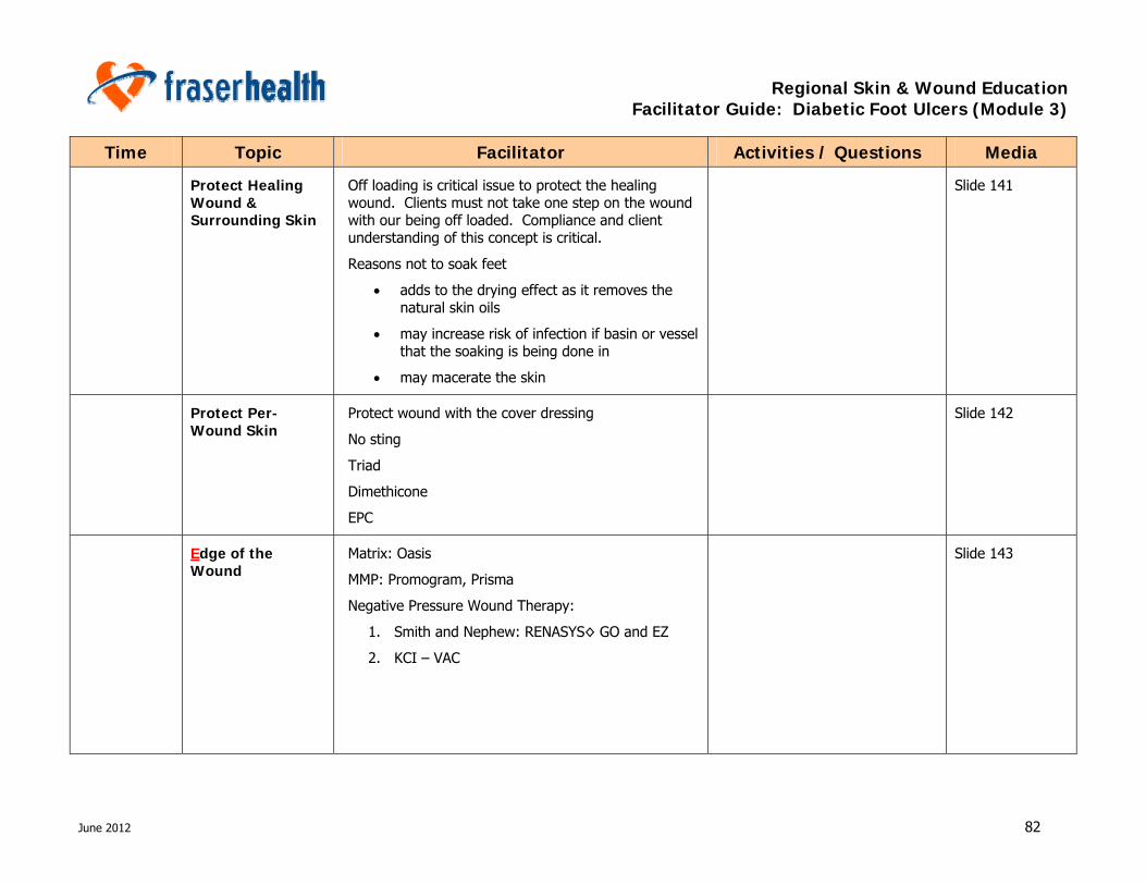
Regional Skin & Wound Education Facilitator Guide: Diabetic Foot Ulcers (Module 3)
June 2012 82
Time Topic Facilitator Activities / Questions Media
Protect Healing Wound & Surrounding Skin
Off loading is critical issue to protect the healing wound. Clients must not take one step on the wound with our being off loaded. Compliance and client understanding of this concept is critical.
Reasons not to soak feet
• adds to the drying effect as it removes the natural skin oils
• may increase risk of infection if basin or vessel that the soaking is being done in
• may macerate the skin
Slide 141
Protect Per-Wound Skin
Protect wound with the cover dressing
No sting
Triad
Dimethicone
EPC
Slide 142
Edge of the Wound
Matrix: Oasis
MMP: Promogram, Prisma
Negative Pressure Wound Therapy:
1. Smith and Nephew: RENASYS◊ GO and EZ
2. KCI – VAC
Slide 143

Regional Skin & Wound Education Facilitator Guide: Diabetic Foot Ulcers (Module 3)
June 2012 83
Time Topic Facilitator Activities / Questions Media
1155 – 1200 Summary Refer participates to CDST guideline and review content.
This tool is to assist the HCP to review and ensure that all aspects have been covered in DFU management.
Slide 144
CDST
Interprofessional Team
Transition to address patient concern – brief discussion on common issues that the clients will be experience, particularly those affecting the adherence to the treatment plan
• Consider pain (nociceptive neuropathic)
• Treatment plan determined by healability, cost-effectiveness / burden of treatment
• Educational and addressing person-centered concerns increase adherence to treatment plan, which in turns affects healability
• Adherence to compression for life – comfort, ability to put on/remove daily, daily wash, new pairs every 6 months if wearing I pairs or every 12 if wearing 2 pairs simultaneously
• Affordability of compression stockings
Slide 145
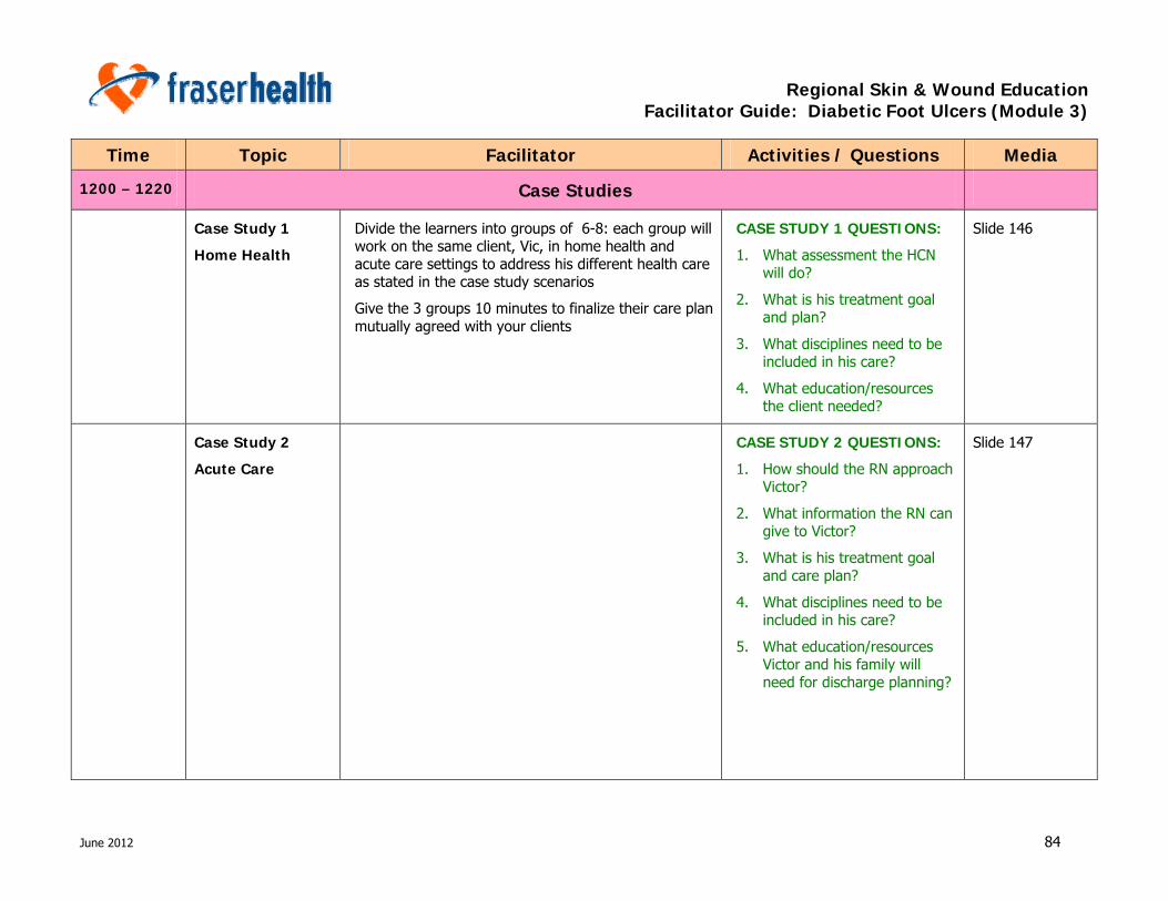
Regional Skin & Wound Education Facilitator Guide: Diabetic Foot Ulcers (Module 3)
June 2012 84
Time Topic Facilitator Activities / Questions Media
1200 – 1220 Case Studies
Case Study 1
Home Health
Divide the learners into groups of 6-8: each group will work on the same client, Vic, in home health and acute care settings to address his different health care as stated in the case study scenarios
Give the 3 groups 10 minutes to finalize their care plan mutually agreed with your clients
CASE STUDY 1 QUESTIONS:
1. What assessment the HCN will do?
2. What is his treatment goal and plan?
3. What disciplines need to be included in his care?
4. What education/resources the client needed?
Slide 146
Case Study 2
Acute Care
CASE STUDY 2 QUESTIONS:
1. How should the RN approach Victor?
2. What information the RN can give to Victor?
3. What is his treatment goal and care plan?
4. What disciplines need to be included in his care?
5. What education/resources Victor and his family will need for discharge planning?
Slide 147

Regional Skin & Wound Education Facilitator Guide: Diabetic Foot Ulcers (Module 3)
June 2012 85
Time Topic Facilitator Activities / Questions Media
Case Study 3
Residential Care
CASE STUDY 3 QUESTIONS:
1. How should the nurse approach Vic?
2. What information the nurse can give to Vic?
3. What is his treatment goal and care plan?
4. What disciplines need to be included in his care?
5. What education/resources Victor and his family will need to support him settling in residential care living?
Slide 148
SWT DFU Education Module SWT Members
Slide 149
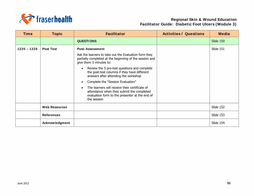
Regional Skin & Wound Education Facilitator Guide: Diabetic Foot Ulcers (Module 3)
June 2012 86
Time Topic Facilitator Activities / Questions Media
QUESTIONS Slide 150
1220 – 1225 Post Test Post-Assessment
Ask the learners to take out the Evaluation form they partially completed at the beginning of the session and give them 5 minutes to:
• Review the 5 pre-test questions and complete the post-test columns if they have different answers after attending the workshop
• Complete the “Session Evaluation”
• The learners will receive their certificate of attendance when they submit the completed evaluation form to the presenter at the end of the session
Slide 151
Web Resources Slide 152
References Slide 153
Acknowledgment Slide 154


![Skin integrity and wound care [autosaved] (2)](https://img.pdfslide.us/doc/110x75/554b4695b4c905ff268b5052/skin-integrity-and-wound-care-autosaved-2.jpg)


![Skin integrity and wound care [autosaved]](https://img.pdfslide.us/doc/110x75/554b5a28b4c9051b458b4cd6/skin-integrity-and-wound-care-autosaved.jpg)