Embed Size (px)
Citation preview
CASE REPORT
Copyright © 2011, the Korean Surgical Society
J Korean Surg Soc 2011;81:S25-29http://dx.doi.org/10.4174/jkss.2011.81.Suppl1.S25
JKSSJournal of the Korean Surgical Society
pISSN 2233-7903ㆍeISSN 2093-0488
Received January 19, 2011, Revised March 11, 2011, Accepted March 18, 2011
Correspondence to: Dong Yi KimDivision of Gastroenterologic Surgery, Department of Surgery, Chonnam National University Medical School, 8 Hak-dong, Dong-gu, Gwangju 501-757, KoreaTel: +82-62-220-6470, Fax: +82-62-227-1635, E-mail: [email protected]
cc Journal of the Korean Surgical Society is an Open Access Journal. All articles are distributed under the terms of the Creative Commons Attribution Non-Commercial License (http://creativecommons.org/licenses/by-nc/3.0/) which permits unrestricted non-commercial use, distribution, and reproduction in any medium, provided the original work is properly cited.
Recurring gastrointestinal stromal tumor with splenic metastasis
Ho Gun Kim, Seong Yeob Ryu, Jae Kyoon Joo, Hyo Kang, Jae Hyuk Lee1, Dong Yi Kim
Division of Gastroenterologic Surgery, Departments of Surgery and 1Pathology, Chonnam National University Medical School, Gwangju, Korea
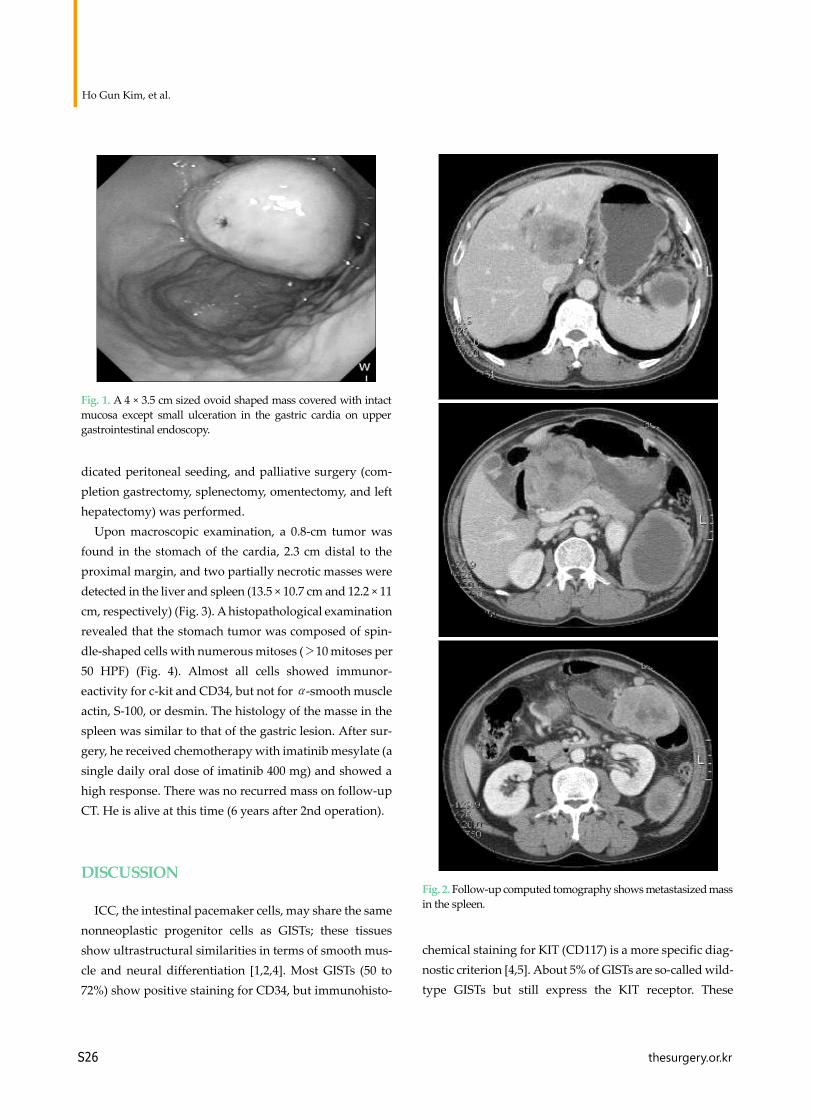
Malignant gastrointestinal stromal tumors (GISTs) are rare non-epithelial, mesenchymal neoplasms of the gastrointestinal tract that metastasize or recur in 30% of patients who undergo surgical resection with curative intent. A 59-year-old man vis-ited our hospital for an examination of a palpable mass in the left abdomen. Fourteen months prior to his visit, the patient un-derwent gastric wedge resection to remove a GIST of the gastric cardia. At the time of surgery, no evidence of metastatic dis-ease was observed and the pathological interpretation was a high-risk GIST. A follow-up computed tomography scan of the abdomen revealed a partially necrotic solid mass (9.8 × 7.6 cm) and enhancing mass in the spleen (2.3 cm). On exploration, multiple masses were found in the liver, greater omentum, and mesentery. Here, we report a case of recurring GIST of the stomach that metastasized to the spleen. To the best of our knowledge, few reports of metastasis to the spleen exist.
Key Words: Gastrointestinal stromal tumors, Neoplasm metastasis, Spleen
INTRODUCTION
Gastrointestinal stromal tumors (GISTs) are thought to arise from the interstitial cells of Cajal (ICC), which func-tion as gastrointestinal pacemaker cells [1]. GISTs exhibit a wide clinical spectrum, from benign to frankly malignant, and more than 30% of the patients with malignant tumors develop local recurrence and distant metastases who un-dergo resection with curative intent [2]. The most common metastatic site is the liver (65%) [3]. To the best of our knowledge, rare reports of metastasis to the spleen exist. Here, we document a case of recurring GIST of the stom-ach that metastasized to the spleen.
CASE REPORT
A 59-year-old man visited our hospital for an examina-tion of a palpable mass in the left abdomen. Fourteen months prior to his visit, the patient underwent gastric wedge resection to remove a GIST of the gastric cardia (Fig. 1). At the time of surgery, no evidence of metastatic disease was observed and the pathological interpretation was a high-risk GIST. A follow-up computed tomography (CT) scan of the abdomen revealed a partially necrotic sol-id mass (9.8 × 7.6 cm) and enhancing mass in the spleen (2.3 cm) (Fig. 2). Two masses (8.3 × 5.5 cm, 10.7 × 8.5 cm) were also found in the liver. Laparoscopic examination in-
Ho Gun Kim, et al.
S26 thesurgery.or.kr
Fig. 1. A 4 × 3.5 cm sized ovoid shaped mass covered with intact mucosa except small ulceration in the gastric cardia on upper gastrointestinal endoscopy.
Fig. 2. Follow-up computed tomography shows metastasized mass in the spleen.
dicated peritoneal seeding, and palliative surgery (com-pletion gastrectomy, splenectomy, omentectomy, and left hepatectomy) was performed.
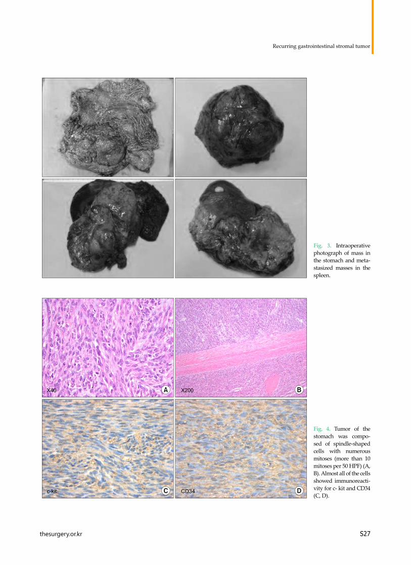
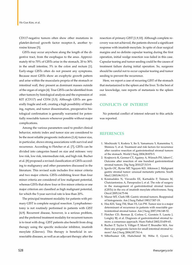
Upon macroscopic examination, a 0.8-cm tumor was found in the stomach of the cardia, 2.3 cm distal to the proximal margin, and two partially necrotic masses were detected in the liver and spleen (13.5 × 10.7 cm and 12.2 × 11 cm, respectively) (Fig. 3). A histopathological examination revealed that the stomach tumor was composed of spin-dle-shaped cells with numerous mitoses (>10 mitoses per 50 HPF) (Fig. 4). Almost all cells showed immunor-eactivity for c-kit and CD34, but not for α-smooth muscle actin, S-100, or desmin. The histology of the masse in the spleen was similar to that of the gastric lesion. After sur-gery, he received chemotherapy with imatinib mesylate (a single daily oral dose of imatinib 400 mg) and showed a high response. There was no recurred mass on follow-up CT. He is alive at this time (6 years after 2nd operation).
DISCUSSION
ICC, the intestinal pacemaker cells, may share the same nonneoplastic progenitor cells as GISTs; these tissues show ultrastructural similarities in terms of smooth mus-cle and neural differentiation [1,2,4]. Most GISTs (50 to 72%) show positive staining for CD34, but immunohisto-
chemical staining for KIT (CD117) is a more specific diag-nostic criterion [4,5]. About 5% of GISTs are so-called wild-type GISTs but still express the KIT receptor. These
Recurring gastrointestinal stromal tumor
thesurgery.or.kr S27
Fig. 3. Intraoperative photograph of mass in the stomach and meta-stasized masses in the spleen.
Fig. 4. Tumor of the stomach was compo-sed of spindle-shaped cells with numerous mitoses (more than 10 mitoses per 50 HPF) (A, B). Almost all of the cells showed immunoreacti-vity for c- kit and CD34 (C, D).
Ho Gun Kim, et al.
S28 thesurgery.or.kr
CD117-negative tumors often show other mutations in platelet-derived growth factor receptor-A, another ty-rosine kinase [5].
GISTs may occur anywhere along the length of the di-gestive tract, from the esophagus to the anus. Approxi-mately 60 to 70% of GISTs arise in the stomach, 20 to 30% in the small intestine, 5% in the colon and rectum [1]. Early-stage GISTs often do not present any symptoms. Because most GISTs show an exophytic growth pattern and arise within the muscularis propria of the stomach or intestinal wall, they present as dominant masses outside of the organ of origin [4]. True GISTs can be identified from other tumors by histological analysis and the expression of KIT (CD117) and CD34 [3,5]. Although GISTs are gen-erally fragile and soft, creating a high possibility of bleed-ing, rupture, and tumor dissemination, preoperative his-tological confirmation is generally warranted for poten-tially resectable tumors whenever possible without major complications.
Among the various parameters used to predict clinical behavior, mitotic index and tumor size are considered to be the most reliable prognostic indicators [1,6]; tumor size, in particular, shows strong associations with survival and recurrence. According to Fletcher et al. [7], GISTs can be divided into categories based on size and mitoses: very low risk, low risk, intermediate risk, and high risk. Bucher et al. [8] proposed a revised classification of GISTs accord-ing to malignancy and other parameters discussed in the literature. This revised scale includes five minor criteria and two major criteria. GISTs exhibiting fewer than four minor criteria are considered of low malignant potential, whereas GISTs that show four or five minor criteria or one major criterion are classified as high malignant potential, for which the 5-year survival rate is less than 20%.
The principal treatment modality for patients with pri-mary GIST is complete surgical resection. Lymphadenec-tomy is not routinely performed in patients with GIST [4,9]. Recurrent disease, however, is a serious problem, and the preferred treatment modality for recurrent tumors is to treat with drug. GIST patients also benefit from drug therapy using the specific molecular inhibitor, imatinib mesylate (Gleevec). This therapy is beneficial in un-resectable diseases, as well as an adjuvant therapy after the
resection of primary GIST [1,9,10]. Although complete re-covery was not achieved, the patients showed a significant response with imatinib mesylate. In spite of clear surgical margins and no definite capsular tearing during the first operation, initial wedge resection was failed in this case. Capsular tearing and tumor seeding could be the causes of treatment failure during initial operation. So, surgeons should be careful not to occur capsular tearing and tumor seeding to prevent the recurrence.
Here, we report a case of recurring GIST of the stomach that metastasized to the spleen and the liver. To the best of our knowledge, rare reports of metastasis to the spleen exist.
CONFLICTS OF INTEREST
No potential conflict of interest relevant to this article was reported.
REFERENCES
1. Mochizuki Y, Kodera Y, Ito S, Yamamura Y, Kanemitsu Y, Shimizu Y, et al. Treatment and risk factors for recurrence after curative resection of gastrointestinal stromal tumors of the stomach. World J Surg 2004;28:870-5.
2. Krajinovic K, Germer CT, Agaimy A, Wünsch PH, Isbert C. Outcome after resection of one hundred gastrointestinal stromal tumors. Dig Surg 2010;27:313-9.
3. Igwilo OC, Byrne MP, Nguyen KD, Atkinson J. Malignant gastric stromal tumor: unusual metastatic patterns. South Med J 2003;96:512-5.
4. Kosmadakis N, Visvardis EE, Kartsaklis P, Tsimara M, Chatziantoniou A, Panopoulos I, et al. The role of surgery in the management of gastrointestinal stromal tumors (GISTs) in the era of imatinib mesylate effectiveness. Surg Oncol 2005;14:75-84.
5. Mazur MT, Clark HB. Gastric stromal tumors. Reappraisal of histogenesis. Am J Surg Pathol 1983;7:507-19.
6. Hsu KH, Yang TM, Shan YS, Lin PW. Tumor size is a major determinant of recurrence in patients with resectable gas-trointestinal stromal tumor. Am J Surg 2007;194:148-52.
7. Fletcher CD, Berman JJ, Corless C, Gorstein F, Lasota J, Longley BJ, et al. Diagnosis of gastrointestinal stromal tu-mors: a consensus approach. Hum Pathol 2002;33:459-65.
8. Bucher P, Taylor S, Villiger P, Morel P, Brundler MA. Are there any prognostic factors for small intestinal stromal tu-mors? Am J Surg 2004;187:761-6.
9. Silberhumer GR, Hufschmid M, Wrba F, Gyoeri G,
Recurring gastrointestinal stromal tumor
thesurgery.or.kr S29
Schoppmann S, Tribl B, et al. Surgery for gastrointestinal stromal tumors of the stomach. J Gastrointest Surg 2009;13: 1213-9.
10. Samelis GF, Ekmektzoglou KA, Zografos GC. Gastroin-
testinal stromal tumours: clinical overview, surgery and re-cent advances in imatinib mesylate therapy. Eur J Surg Oncol 2007;33:942-50.