Embed Size (px)
Citation preview

Recurrent Renal Pheochromocytoma in the
Retroperitoneum
Presented By:
Sai Swarupa, Shiguang, A. Singareddy, Isiri Kadambi,K.Mohamed and
Jayanthraj
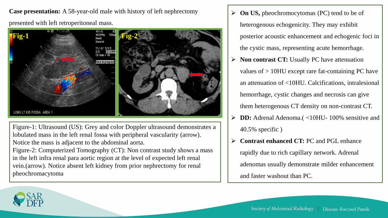
Case presentation: A 58-year-old male with history of left nephrectomy
presented with left retroperitoneal mass.
Fig-1 Fig-2
➢ On US, pheochromocytomas (PC) tend to be of
heterogenous echogenicity. They may exhibit
posterior acoustic enhancement and echogenic foci in
the cystic mass, representing acute hemorrhage.
➢ Non contrast CT: Usually PC have attenuation
values of > 10HU except rare fat-containing PC have
an attenuation of <10HU. Calcifications, intralesional
hemorrhage, cystic changes and necrosis can give
them heterogenous CT density on non-contrast CT.
➢ DD: Adrenal Adenoma.( <10HU- 100% sensitive and
40.5% specific )
➢ Contrast enhanced CT: PC and PGL enhance
rapidly due to rich capillary network. Adrenal
adenomas usually demonstrate milder enhancement
and faster washout than PC.
Figure-1: Ultrasound (US): Grey and color Doppler ultrasound demonstrates a
lobulated mass in the left renal fossa with peripheral vascularity (arrow).
Notice the mass is adjacent to the abdominal aorta.
Figure-2: Computerized Tomography (CT): Non contrast study shows a mass
in the left infra renal para aortic region at the level of expected left renal
vein.(arrow). Notice absent left kidney from prior nephrectomy for renal
pheochromacytoma
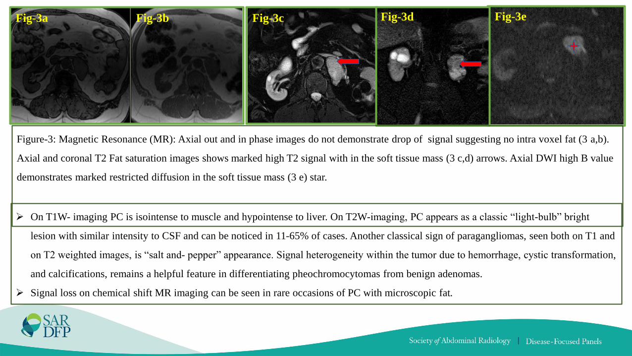
Fig-3a Fig-3b Fig-3c Fig-3d Fig-3e
Figure-3: Magnetic Resonance (MR): Axial out and in phase images do not demonstrate drop of signal suggesting no intra voxel fat (3 a,b).
Axial and coronal T2 Fat saturation images shows marked high T2 signal with in the soft tissue mass (3 c,d) arrows. Axial DWI high B value
demonstrates marked restricted diffusion in the soft tissue mass (3 e) star.
➢ On T1W- imaging PC is isointense to muscle and hypointense to liver. On T2W-imaging, PC appears as a classic “light-bulb” bright
lesion with similar intensity to CSF and can be noticed in 11-65% of cases. Another classical sign of paragangliomas, seen both on T1 and
on T2 weighted images, is “salt and- pepper” appearance. Signal heterogeneity within the tumor due to hemorrhage, cystic transformation,
and calcifications, remains a helpful feature in differentiating pheochromocytomas from benign adenomas.
➢ Signal loss on chemical shift MR imaging can be seen in rare occasions of PC with microscopic fat.
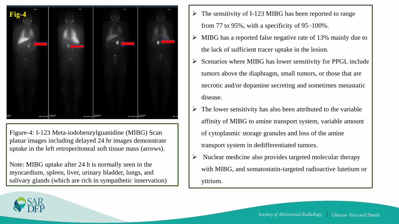
Fig-4 ➢ The sensitivity of I-123 MIBG has been reported to range
from 77 to 95%, with a specificity of 95–100%.
➢ MIBG has a reported false negative rate of 13% mainly due to
the lack of sufficient tracer uptake in the lesion.
➢ Scenarios where MIBG has lower sensitivity for PPGL include
tumors above the diaphragm, small tumors, or those that are
necrotic and/or dopamine secreting and sometimes metastatic
disease.
➢ The lower sensitivity has also been attributed to the variable
affinity of MIBG to amine transport system, variable amount
of cytoplasmic storage granules and loss of the amine
transport system in dedifferentiated tumors.
➢ Nuclear medicine also provides targeted molecular therapy
with MIBG, and somatostatin-targeted radioactive lutetium or
yttrium.
Figure-4: I-123 Meta-iodobenzylguanidine (MIBG) Scan
planar images including delayed 24 hr images demonstrate
uptake in the left retroperitoneal soft tissue mass (arrows).
Note: MIBG uptake after 24 h is normally seen in the
myocardium, spleen, liver, urinary bladder, lungs, and
salivary glands (which are rich in sympathetic innervation)
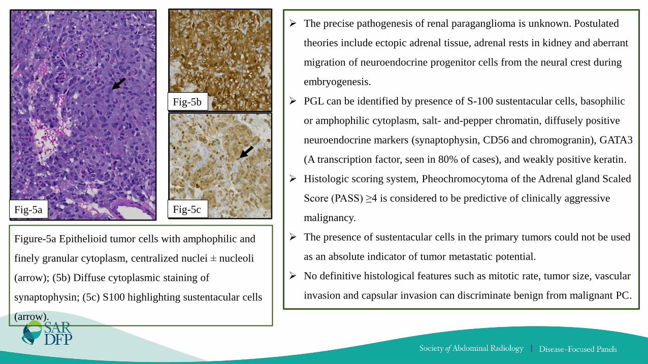
Fig-5b
Fig-5a Fig-5c
Figure-5a Epithelioid tumor cells with amphophilic and
finely granular cytoplasm, centralized nuclei ± nucleoli
(arrow); (5b) Diffuse cytoplasmic staining of
synaptophysin; (5c) S100 highlighting sustentacular cells
(arrow).
➢ The precise pathogenesis of renal paraganglioma is unknown. Postulated
theories include ectopic adrenal tissue, adrenal rests in kidney and aberrant
migration of neuroendocrine progenitor cells from the neural crest during
embryogenesis.
➢ PGL can be identified by presence of S-100 sustentacular cells, basophilic
or amphophilic cytoplasm, salt- and-pepper chromatin, diffusely positive
neuroendocrine markers (synaptophysin, CD56 and chromogranin), GATA3
(A transcription factor, seen in 80% of cases), and weakly positive keratin.
➢ Histologic scoring system, Pheochromocytoma of the Adrenal gland Scaled
Score (PASS) ≥4 is considered to be predictive of clinically aggressive
malignancy.
➢ The presence of sustentacular cells in the primary tumors could not be used
as an absolute indicator of tumor metastatic potential.
➢ No definitive histological features such as mitotic rate, tumor size, vascular
invasion and capsular invasion can discriminate benign from malignant PC.
Discussion
➢ Pheochromocytomas are catecholamine secreting tumors originating from adrenal chromaffin cells. They are termed as paragangliomas
(PGL) when originate at extra-adrenal sites.
➢ PGL comprise 10% of PC. Estimated prevalence of 1 in 2000 to 1 in 6500.
➢ Most common site of PGL- Organ of Zuckerkandl (between the origin of the inferior mesenteric artery and the aortic bifurcation)
➢ Unusual extra-adrenal sites of origin include kidney, uterus and prostate.
➢ Associated with familial syndromes:
Multiple endocrine neoplasia type 2 (MEN2)
Neurofibromatosis type 1 or von Recklinghausen’s disease
Von Hippel–Lindau (VHL) disease
Hereditary pheochromocytoma– PGLs syndrome (succinate dehydrogenase (SDH) mutations)
➢ Although CT and MRI have greater sensitivity than MIBG in detecting PGL, they are limited by lower specificity.
➢ Renal paraganglioma is rare and is difficult to distinguish from
renal cell carcinoma, clinically and pathologically. To date, <20
cases of renal PGL are reported in the literature.
➢ PGL are usually benign and up to 10% of patients encounter
malignant transformation.
➢ Due to difficulty in characterizing benign, and malignant tumors on
imaging and histopathology, all PGL are considered malignant and
treated by surgical resection.
➢ Recurrence rate of PGL ranges from 6.5- 16.5%, with large tumor
size (>5 cm), age, tumor location (right-sided and extra-adrenal),
and familial disease pattern (MEN2, VHL, NF1) being the critical
risk factors.
➢ Treatment of recurrent tumor is unlikely to result in cure and hence
MIBG can be considered as a palliative management. MIBG have
shown symptom response in 75-90% and tumor response in 30-
47% of patients.
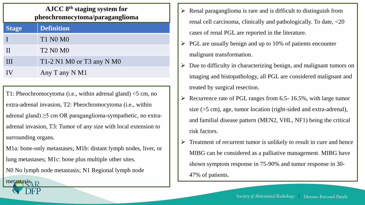
Stage Definition
I T1 N0 M0
II T2 N0 M0
III T1-2 N1 M0 or T3 any N M0
IV Any T any N M1
AJCC 8th staging system for
pheochromocytoma/paraganglioma
T1: Pheochromocytoma (i.e., within adrenal gland) <5 cm, no
extra-adrenal invasion, T2: Pheochromocytoma (i.e., within
adrenal gland) ≥5 cm OR paraganglioma-sympathetic, no extra-
adrenal invasion, T3: Tumor of any size with local extension to
surrounding organs.
M1a: bone-only metastases; M1b: distant lymph nodes, liver, or
lung metastases; M1c: bone plus multiple other sites.
N0 No lymph node metastasis; N1 Regional lymph node
metastasis
References
➢ Venugopal S, Chhabria M, Quartuccio M. Recurrence of Pheochromocytoma With Metastases After Resection of Primary
Tumor. Cureus. 2020;12(5):e8328. Published 2020 May 28. doi:10.7759/cureus.8328
➢ Press D, Akyuz M, Dural C, et al. Predictors of recurrence in pheochromocytoma. Surgery. 2014;156(6):1523-1528.
doi:10.1016/j.surg.2014.08.044
➢ Leung K, Stamm M, Raja A, Low G. Pheochromocytoma: the range of appearances on ultrasound, CT, MRI, and functional imaging
[published correction appears in AJR Am J Roentgenol. 2013 Mar;200(3):705]. AJR Am J Roentgenol. 2013;200(2):370-378.
doi:10.2214/AJR.12.9126
➢ Abdel-Rahman O. Assessment of the AJCC staging system of pheochromocytomas/paragangliomas [published online ahead of print,
2021 Aug 27]. Endocrine. 2021;10.1007/s12020-021-02854-3. doi:10.1007/s12020-021-02854-3
➢ Abdel-Rahman O. Assessment of the AJCC staging system of pheochromocytomas/paragangliomas [published online ahead of print,
2021 Aug 27]. Endocrine. 2021;10.1007/s12020-021-02854-3. doi:10.1007/s12020-021-02854-3
Thank You