Embed Size (px)
Citation preview

En
do
crin
e-R
ela
ted
Can
cer
Thematic ReviewT Else History of pheochromocytoma
and paraganglioma22 :4 T147–T159
15 YEARS OF PARAGANGLIOMA
Pheochromocytoma, paragangliomaand genetic syndromes: a historicalperspective
Tobias Else
Metabolism, Endocrinology and Diabetes, Department of Internal Medicine, University of Michigan,
2560E MSRB2, 1150 West Medical Center Drive, Ann Arbor, Michigan 48109-5674, USA
http://erc.endocrinology-journals.org q 2015 Society for EndocrinologyDOI: 10.1530/ERC-15-0221 Printed in Great Britain
Published by Bioscientifica Ltd.
This paper is part of a thematic review sectioand Pheochromocytoma. The Guest Editorsand Hartmut Neumann. Downloa
Correspondence
should be addressed
to T Else
Abstract
The last decades have elucidated the genetic basis of pheochromocytoma (PC) and
paraganglioma (PGL) (PCPGL)-associated hereditary syndromes. However, the history of
these syndromes dates back at least another 150 years. Detailed descriptions by clinicians and
pathologists in the 19th and 20th centuries led to the recognition of the PCPGL-associated
syndromes von Hippel-Lindau disease, neurofibromatosis type 1, and multiple endocrine
neoplasia type 2. In the beginning of the current millennium the molecular basis of the
hereditary PGL syndrome was elucidated by the discovery of mutations in genes encoding
enzymes of the Krebs cycle, such as succinate dehydrogenase genes (SDHx) and other
mutations, causing ‘pseudo-hypoxia’ signaling. These recent developments also marked
a paradigm shift. It reversed the traditional order of genetic research that historically aimed
to define the genetic basis of a known hereditary syndrome but now is challenged with
defining the full clinical phenotype associated with a newly defined genetic basis.
This challenge underscores the importance to learn from medical history, continue providing
support for clinical research, and train physicians with regards to their skills to identify
patients with PCPGL-associated syndromes to extend our knowledge of the associated
phenotype. This historical overview provides details on the history of the paraganglial
system and PCPGL-associated syndromes. As such, it hopefully will not only be an interesting
reading for the physician with a historical interest but also emphasize the necessity of
ongoing astute individual clinical observations and clinical registries to increase our
knowledge regarding the full phenotypic spectrum of these conditions.
Key Words
" medical history
" pheochromocytoma
" paraganglioma
" neurofibromatosis type 1
" multiple endocrineneoplasia type 2
" von Hippel-Lindau disease
" hereditary paraganglioma
syndrome
n ofor
ded
Endocrine-Related Cancer
(2015) 22, T147–T159
Introduction
The last 15 years have brought significant advances in our
understanding of the genetic basis of hereditary syn-
dromes with a predisposition to pheochromocytoma (PC)
and paraganglioma (PGL) (PCPGL) development. In the
1990s the genetic causes for three classic cancer
syndromes for which PC is a well-recognized clinical
feature were identified: neurofibromatosis type 1 (NF1),
von Hippel-Lindau (VHL) disease, and multiple endocrine
neoplasia type 2 (RET) (Cawthon et al. 1990, Xu et al. 1990,
Latif et al. 1993, Mulligan et al. 1993, Crossey et al. 1994).
n 15th Anniversary of Paragangliomathis section were Wouter de Herder
from Bioscientifica.com at 09/17/2018 11:06:59AMvia free access

Figure 1
Illustration (Tabula Secunda) from Bartolomeo Eustachi’s first description
of the adrenal gland (Eustachi 1714).
En
do
crin
e-R
ela
ted
Can
cer
Thematic Review T Else History of pheochromocytomaand paraganglioma
22 :4 T148
In the early 2000s mutations in succinate dehydrogenase
subunits (SDHA, SDHB, SDHC, SDHD) and related genes
(SDHAF2) were discovered to cause hereditary PGL
syndromes (PGL1–5) (Baysal et al. 2000, Niemann &
Muller 2000, Astuti et al. 2001, Hao et al. 2009, Burnichon
et al. 2010). More recently, PCPGLs have been suggested to
be part of other syndromes, such as hereditary leiomyo-
matosis and renal cell cancer (HLRCC) caused by
mutations in fumarate hydratase (FH) (Clark et al. 2014).
For some gene mutations, PCPGL is currently the only
known phenotypic expression (e.g., TMEM127 or MAX)
(Qin et al. 2010, Comino-Mendez et al. 2011). Lastly, PCs
also occur in patients with mosaicism for activating HIF2a
mutations (Zhuang et al. 2012).
However, the history of uncovering the hereditary
basis of PCPGL dates back at least another 150 years. Our
current knowledge was only made possible through the
observations of astute clinicians in realizing the associ-
ation of PCPGL with other syndromic features. There is
still an ongoing need for clinical observations to further
define PCPGL syndrome associations; therefore, it is
important to understand the historic advances that led
to our current knowledge of PCPGL-associated syndromes.
This review will not only highlight historic exciting events
but also hopefully spark the interest of physicians and
scientists to keep detailed clinical observations of patients
with PCPGL to better define the associated syndromes to
improve patient care.
From the discovery of the adrenal gland to thedetailed understanding of the paraganglialsystem
It is still a matter of debate whether Galen actually
encountered the adrenal gland in his studies. It is safe to
say that he certainly did not appreciate it as a distinct
organ. The only suggestion of Galen finding the adrenal
gland is the comment on some ‘loose flesh’ (‘lockeres
Fleisch,’ translation by Simon) at the end of a blood vessel
originating from the left renal vein, likely representing the
left adrenal vein (Simon 1906). On the contralateral side,
he appreciated small venous blood vessels, but no distinct
organ. The first definitive description of the adrenal glands
was by Eustachius in 1563, and he termed them ‘glandulae
renibus incumbentes’ (glands adjacent to the kidney;
Eustachi 1714) (Fig. 1). For the next two centuries there
was the common notion that the adrenal glands were
hollow organs with diverse proposed functions, such as
the secretion of a lithotryptic agent into the kidneys or
fetal meconium into the gut or filtration of the intestinal
http://erc.endocrinology-journals.org q 2015 Society for EndocrinologyDOI: 10.1530/ERC-15-0221 Printed in Great Britain
lymphatic fluids (Carmichael & Rochester 1989). Some
authors even reported on excretory ducts of the adrenal
glands. The central cavity, which was observed by
anatomists in the early ages of modern medical sciences,
was likely caused by the large central vein and the post-
mortem autolytic fragility of the organ. Nagel (1836) was
first to appreciate the two parts of the adrenal gland using
the term ‘medulla’ (Mark) and ‘cortex’ (Rinde). He
described the adrenal glands of multiple species in detail,
including the less distinct separation of cortical and
medullary cells in birds and reptiles.
The distinct color reaction of the adrenal medulla when
exposed to chromium salts was first described by Werner
(1857) and further explored by Henle (1865) (Carmichael
1989). Later, Kohn employed the chromium staining
method to systematically explore the developmental
distribution of what he called ‘chromaffin tissue.’Kohn
(1903) published a detailed report on the abdominal
paraganglial tissue, which he found closely related to the
adrenal medulla and sympathetic nervous system (Fig. 2).
A main focus of Kohn’s manuscript is on the embryological
description of the ‘organ of Zuckerkandl.’ This paired organ
had been initially described by the anatomist Zuckerkandl
in 1898 as part of the abdominal sympathetic nervous
system that was particularly prominent in fetuses and
newborn children (Fig. 3) (Zuckerkandl 1901). Kohn also
gives the first detailed insight into the embryology of the
carotid body (‘Carotisdruese’), which had originally been
described in 1743 by Taubeand Berckelmann, both mentees
Published by Bioscientifica Ltd.
Downloaded from Bioscientifica.com at 09/17/2018 11:06:59AMvia free access
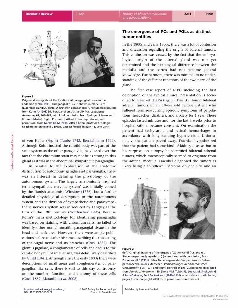
Figure 2
Original drawing about the locations of paraganglial tissue in the
abdomen (Kohn 1903). Paraganglial tissue is shown in black. Left:
N, adrenal gland; A, aorta; U, ureter; P, paraganglia; R, rectum (reproduced
from Kohn A (1903) Die Paraganglien, Archiv fur Mikroskopische
Anatomie, 62, 263–367, with kind permission from Springer Science and
Business Media). Right: Portrait of Alfred Kohn (reproduced, with
permission, from Nanka OGM (2008) Alfred Kohn, profesor histologie
na Nemecke univerzite v praze. Casopıs lekaru ceskych 147 240–244).
Figure 3
(left) Original drawing of the organs of Zuckerkandl (n.r. and n.l.
‘Nebenorgan des Sympathicus’) (reproduced, with permission, from
Zuckerkandl E (1901) Ueber Nebenorgane des Sympathicus im Retro-
peritonaealraum des Menschen. Verhandlungen der Anatomischen
Gesellschaft 14 95–107), and (right) portrait of Emil Zuckerkandl (reprinted
from Annals of Anatomy; 190; Shoja MM, Tubbs RS, Loukas M, Shokouhi G
& Jerry Oakes W; Emil Zuckerkandl (1849–1910): anatomist and pathologist;
pages 33–36; Copyright 2008, with permission from Elsevier).
En
do
crin
e-R
ela
ted
Can
cer
Thematic Review T Else History of pheochromocytomaand paraganglioma
22 :4 T149
of von Haller (Fig. 4) (Taube 1743, Berckelmann 1744).
Although Kohn insisted the carotid body was part of the
same system as the other paraganglia, he glossed over the
fact that the chromium stain may not be as strong in this
gland as it was in the abdominal sympathetic paraganglia.
In parallel to the exploration of the anatomic
distribution of autonomic ganglia and paraganglia, there
was an interest in defining the physiology of the
autonomous system. The largely anatomically inspired
term ‘sympathetic nervous system’ was initially coined
by the Danish anatomist Winslow (1776), but a further
detailed physiological description of the autonomous
system and the division of sympathetic and parasympa-
thetic nervous system was introduced by Langley at the
turn of the 19th century (Nozdrachev 1995). Because
Kohn’s main methodology for identifying paraganglia
was based on staining with chromium salts, he failed to
identify other non-chromaffin paragangial tissue in the
head and neck area. However, there were ample publi-
cations before and after his time describing the thickening
of the vagal nerve and its branches (Cock 1837). The
glomus jugulare, a conglomerate of cells analogous to the
carotid body but of smaller size, was definitively described
by Guild (1941). Although since the early 1800s there were
descriptions of small areas and conglomerates of para-
ganglion-like cells, there is still to this day controversy
on the number, function, and anatomy of these cells
(Cock 1837, Maranillo et al. 2008).
http://erc.endocrinology-journals.org q 2015 Society for EndocrinologyDOI: 10.1530/ERC-15-0221 Printed in Great Britain
The emergence of PCs and PGLs as distincttumor entities
In the 1800s and early 1900s, there was a lot of confusion
and discussion regarding the origin of adrenal tumors.
This confusion was caused by the fact that the embryo-
logical origin of the adrenal gland was not yet
determined and the histological difference between the
medulla and the cortex had not become general
knowledge. Furthermore, there was minimal to no under-
standing of the different functions of the two parts of the
gland.
The first case report of a PC including the first
description of the typical clinical presentation is accre-
dited to Fraenkel (1886) (Fig. 5). Fraenkel found bilateral
adrenal tumors in an 18-year-old female patient who
suffered from reoccurring episodic symptoms of palpita-
tions, headaches, dizziness, and anxiety for 1 year. These
episodes lasted minutes and, for the last 4 weeks prior to
hospitalization, became constant. On examination the
patient had tachycardia and retinal hemorrhages in
accordance with long-standing hypertension. Unfortu-
nately, the patient passed away. Fraenkel hypothesized
that the patient had some kind of kidney disease, but to
his surprise, on autopsy he identified bilateral adrenal
tumors, which microscopically seemed to originate from
the adrenal medulla. Fraenkel diagnosed the tumors as
likely being a spindle-cell sarcoma on one side and an
Published by Bioscientifica Ltd.
Downloaded from Bioscientifica.com at 09/17/2018 11:06:59AMvia free access

Figure 4
Front page of original dissertation by Hartwig Taube (Taube 1743), mentee
of Albrecht von Haller (portrait reproduced, with permission, from Peiper HJ
(2008) Albrecht von Haller. An illustrious ancestor. Chirurg 79 486–493, with
kind permission from Springer Science and Business Media).
Figure 5
Title of Fraenkel’s initial description of bilateral pheochromocytoma (with
kind permission from Springer ScienceCBusiness Media: Virchows Archiv.
A, Pathological Anatomy and Histology, Ein Fall von doppelseitigem voellig
latent verlaufenden Nebennierentumor und gleichzeitiger Nephritis mit
Veraenedungen am Circulationsapparat und Retinitis, 103, 1886, 244–263,
Fraenkel F). Particularly remarkable is the clinical description: ‘One night at
9 o’clock the patient had severe palpitations, accompanied by anxiety,
dizziness and headaches. This spell lasted several minutes. The patient felt
back to normal afterwards and reports to have pursued her work on the
farmland. About 4 months later she had a second episode, and a third one
later that summer. Since then symptoms worsened: she suffered from
headaches and vomiting’.
En
do
crin
e-R
ela
ted
Can
cer
Thematic Review T Else History of pheochromocytomaand paraganglioma
22 :4 T150
angiosarcoma on the other side. Therefore, although
Fraenkel is accredited with the first description of a PC,
he did not identify it as a unique tumor entity. It is
noteworthy that the discussions on adrenal tumors in the
literature for the following decades fail to mention or cite
Fraenkel’s observation, suggesting that subsequent
researchers were not aware of his initial description of
adrenal tumors.
Manasse (1893) reported on tumorous and hyperplas-
tic changes in the adrenal medulla. He mentioned four
cases of adrenocortical tumors, likely adenomas, which he
referred to as ‘struma suprarenalis.’ He also described
adrenal medullary tumors seen on autopsy of a female
patient with the incidental finding of a chicken egg-sized
encapsulated brown–black medullary tumor, which was
not reported to cause any symptoms at any time of her life.
The adrenal cortex in this case was completely pushed to
the periphery of this tumor. Another largely cystic
medullary tumor was described by Hedinger (1911) as
‘Struma medullaris cystica.’ The term ‘PC’ was finally
introduced by Pick in 1912 in his manuscript ‘Das
Ganglioma embryonale sympathicum (Sympathoma
embryonale).’ The main focus was dedicated to tumors,
which are now called neuroblastomas, but he also
acknowledged a class of more differentiated adrenal
tumors, either originating from the chromaffin cells of
the medulla or consisting of tumor cells that differentiate
to the stage of chromaffin or ‘phaeochrome’ cells. To
describe these tumors, he coined the term ‘PC’ (Pick
1912a,b).
Carotid body tumors were well described prior to
Kohn’s detailed description of the association of the
http://erc.endocrinology-journals.org q 2015 Society for EndocrinologyDOI: 10.1530/ERC-15-0221 Printed in Great Britain
carotid body/carotid gland with the sympathetic nervous
system. Possibly the first such tumor was mentioned by
Morgagni (Fig. 6) in 1761 when he described two cases:
one encountered by himself, another one seen with his
mentor Valsalva.
‘A woman of about 50 years of age had been suffering for
3 months from a tumor in the right side of her neck that
was hard, oblong, and equal to a turkey egg in size; its base
was on the carotid artery in the same side (of the neck),
from which it extended all the way to the division of that
artery. Sometimes this would hurt, sometimes it brought
no pain whatsoever. Ultimately, about twenty days before
her death, it began to bother her more frequently,
especially in the region of the larynx, such that it forced
her tobreathewithacertainpeculiar stertor, accompanied
by a particular sensation of heat in her throat. And so it
killed the woman’ (Morgagni 1779)
Although no further histological or anatomical
description is available, the typical localization and
appearance are highly suggestive of a carotid body
tumor. In 1891 two seminal manuscripts appeared
discussing the origin and existence of tumors of the
carotid body. In the first manuscript, Paltauf (1891) gives a
detailed description of four cases of carotid body tumors
(Fig. 7). He was startled by their histological appearance
and therefore suggested that the tumors must have arisen
from the carotid body itself and termed them ‘endo-(peri-)
thelioma of the glandula intercarotica.’ In the same year
Marchand (1891) also described a carotid body tumor. The
Published by Bioscientifica Ltd.
Downloaded from Bioscientifica.com at 09/17/2018 11:06:59AMvia free access
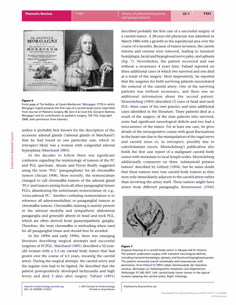
Figure 7
Original drawing of a carotid body tumor in 28-year-old Dr Ferencz.
The patient underwent surgery with transient neurological deficits,
including transient hemiplegia, aphasia, and facial and hypoglossal paresis.
The patient recovered overall remarkably well (reproduced, with
permission, from Paltauf R (1891) Ueber Geschwuelste der Glandula
carotica. Beitraege zur Pathologischen Anatomie und Allgemeinen
Pathologie 11 260–301). Left: carotid body tumor shown in the typical
location splaying the carotid artery. Right: histology.
Figure 6
Front page of ‘De Sedibus, et Causis Morborum’ (Morgagni 1779) in which
Morgagni (right) presents the first case of a carotid body tumor (reprinted
from Journal of Pediatric Surgery, 43, Zani A & Cozzi DA, Giovanni Battista
Morgagni and his contribution to pediatric surgery, 729–733, Copyright
2008, with permission from Elsevier).
En
do
crin
e-R
ela
ted
Can
cer
Thematic Review T Else History of pheochromocytomaand paraganglioma
22 :4 T151
author is probably best known for the description of the
accessory adrenal glands (‘adrenal glands of Marchand’)
that he had found in one particular case, which in
retrospect likely was a woman with congenital adrenal
hyperplasia (Marchand 1883).
In the decades to follow there was significant
confusion regarding the terminology of tumors of the PC
and PGL spectrum. Alezais and Pyron finally suggested
using the term ‘PGL’ (paragangliome) for all chromaffin
tumors (Alezais 1908). More recently, the nomenclature
changed to call chromaffin tumors of the adrenal gland
‘PCs’ and tumors arising from all other paraganglial tissues
PGLs, abandoning the unfortunate nomenclature of, e.g.,
‘extra-adrenal PC.’ Another confusing nomenclature is to
reference all adrenomedullary or paraganglial tumors as
chromaffin tumors. Chromaffin staining is mainly present
in the adrenal medulla and sympathetic abdominal
paraganglia and generally absent in head and neck PGL,
which are often derived from parasympathetic ganglia.
Therefore, the term chromaffin is misleading when used
for all paraganglial tissue and should best be avoided.
In the 1890s and early 1900s, there was emerging
literature describing surgical attempts and successful
surgeries of PCPGL. Marchand (1891) described a 32-year-
old woman with a 5.5 cm carotid body tumor that had
grown over the course of 4.5 years, encasing the carotid
artery. During the surgical attempt, the carotid artery and
the jugular vein had to be ligated. He described that the
patient postoperatively developed tachycardia and high
fevers and died 3 days after surgery. Paltauf (1891)
http://erc.endocrinology-journals.org q 2015 Society for EndocrinologyDOI: 10.1530/ERC-15-0221 Printed in Great Britain
described probably the first case of a successful surgery of
a carotid tumor. A 28-year-old physician was admitted in
March 1886 with a growth in the suprahyoid area over the
course of 6 months. Because of tumor invasion, the carotis
interna and externa were removed, leading to transient
hemiplegia, facial and hypoglossal nerve palsy, and aphasia
(Fig. 7). Nevertheless, the patient recovered and was
without a recurrence 4 years later. Paltauf reported on
three additional cases of which two survived and one died
as a result of the surgery. Most impressively, he reported
that the surgeries for both surviving patients necessitated
the removal of the carotid artery. One of the surviving
patients was without recurrence, and there was no
additional information about the second patient.
Moenckeberg (1905) described 12 cases of head and neck
PGL: three cases of his own practice and nine additional
cases identified in the literature. Three patients died as a
result of the surgery; of the nine patients who survived,
some had significant neurological deficits and two had a
reoccurrence of the tumor. For at least one case, he gives
details of the intraoperative course with great fluctuations
in the heart rate due to the manipulation of the vagal nerve
and carotid sinus or, in retrospect, possibly due to
catecholamine excess. Moenckeberg’s publication also
holds the first case report of a malignant carotid body
tumor with metastasis to local lymph nodes. Moenckeberg
additionally comments on three ‘submastoid potatoe
tumors’ described by Gilford (1904), but he raises doubt
that these tumors were true carotid body tumors as they
were only immediately adjacent to the carotid artery rather
than involving the artery itself. These tumors might have
arisen from different paraganglia. Rosenwasser (1945)
Published by Bioscientifica Ltd.
Downloaded from Bioscientifica.com at 09/17/2018 11:06:59AMvia free access

Figure 8
Anton Freiherr von Eiselsberg (left) performed the first surgery for an
abdominal paraganglioma as reported by Stangl (1903). Both Cesar Roux
(middle) and Charles Horace Mayo (right) conducted the first surgeries for a
pheochromocytoma on European and American continents respectively
(left: reproduced from the Wellcome Library, London, UK. Published under
the terms of the CC BY 4.0 licence; middle: reproduced, with permission,
from Dhayat S, Renggli JC, Dhayat N & Merlini M (2007) On the 150th
birthday of Cesar Roux (1857–1918). Memories of the life and work of an
important pupil of Kocher, Chirurg 78 155–160, with kind permission from
Springer Science and Business Media; right: reproduced, with permission,
from Mayo CH (1982) Charles Horace Mayo (1865–1939). Diseases of the
Colon & Rectum 25 734–739, with kind permission from Springer Science
and Business Media. Photograph courtesy of the Mayo Clinic).
En
do
crin
e-R
ela
ted
Can
cer
Thematic Review T Else History of pheochromocytomaand paraganglioma
22 :4 T152
reports on the successful surgery of a 36-year-old man with
a carotid body-like tumor of the middle ear, and he
concludes that this tumor likely arose from the glomus
jugulare, which had been recently described by
Guild (1941). There were reports prior to Rosenwasser’s
publications describing what in retrospect likely were
glomus jugulare tumors, such as the report by Goekopp
(1932) (see below).
Surgical reports from the early 1900s describe attempts
to remove adrenal PCs and extra-adrenal PGLs. The first
documented surgery for an abdominal PGL of the organ of
Zuckerkandl was reported by Stangl (1903) (Fig. 8). A
32-year-old farm worker was found to have a walnut-sized
tumor on palpation in the umbilical area. The patient
underwent laparotomy by von Eiselsberg, and a tumor
located at the bifurcation of the aorta, likely arising from
the organ of Zuckerkandl, was removed. The patient
recovered well and left the hospital 3 weeks later. The
history of the surgery for PCs was reviewed in detail by
Welbourn (1987). The first successful surgeries were carried
out by Roux in Switzerland and shortly thereafter by Mayo
in Rochester, Minnesota in 1926 (Mayo 1927, Welbourn
1987). Interestingly, the first description of unexpected,
deleterious effects of anesthetic agents precedes the reports
on surgery for PCs. In 1910 Kolisko described a case of a
43-year-old patient undergoing tooth extraction with the
use of cocaine as a local anesthetic who died a couple of
hours after the procedure (Neusser 1910). The autopsy
http://erc.endocrinology-journals.org q 2015 Society for EndocrinologyDOI: 10.1530/ERC-15-0221 Printed in Great Britain
shows cardiac hypertrophy and a cystic adrenal tumor. The
tumor was proven to consist of chromaffin tissue, and the
aspirate from the cystic part was shown to induce a blood
pressure surge in animals (Hedinger 1911).
Association of PC and PGL with hereditarysyndromes
Although it took several decades from the early descriptions
of PCPGL to connect PCPGL to the now known predisposi-
tion syndromes, there were some characteristics suggestive
for a hereditary predisposition even in the late 1890s and
early 1900s. Most of the initially described patients were
fairly young (20–40 years) when afflicted with these tumors,
which we now know often suggests an inherited syndrome.
Some reports suggested a possible hereditary component
such as the case described by Marchand (1891) of a 20-year-
old woman with an adrenal tumor and a family history of a
large retroperitoneal tumor in her mother. The knowledge
of an association of PCs with classic syndromes – NF1, VHL
disease, and multiple endocrine neoplasia type 2 (MEN2) –
emerged slowly from detailed case reports starting in the
early 1900s and was fully defined by several systematic
reviews of the literature and case series in the 1940s to 1960s.
Althoughnot per se a genetic predisposition, there is the
peculiar finding of an association of PCPGL development
in chronic hypoxic states. This represents a clinical link to
other often genetic conditions, such as cyanotic congenital
heart disease, and a biological link, as there is a close
connection between hypoxia-like signaling changes caused
by genetic mutations in VHL- and SDHx-associated tumors.
An association of PCPGL and hypoxia as well as mitochon-
drial abnormalities was first suggested in the 1970s (Cornog
et al. 1970, Arias-Stella & Valcarcel 1973, Watanabe et al.
1976). Saldana et al. (1973) showed a higher prevalence of
carotid body tumors in Peruvians who live at high altitudes
than those living at sea level. Of note, most patients in this
publication also had lung or heart disease, aggravating the
hypoxia. One might also criticize that there is a possibility of
a genetic influence because these patients are likely from
different populations and gene pools. However, there is
good evidence that chronic hypoxia due to medical
conditions isagrowthstimulus for thecarotidbody.Patients
with either cystic fibrosis or cyanotic heart disease have
significantly enlarged carotid bodies (Lack 1978).
In the following sections, there will be a short discussion
of the history of PCPGL-associated syndromes. Rather than
describing every syndrome in minute detail, there will be a
focusonhistoricalpertinentevents,particularlyas itpertains
to the association of PCs with these syndromes (Table 1).
Published by Bioscientifica Ltd.
Downloaded from Bioscientifica.com at 09/17/2018 11:06:59AMvia free access

Tab
le1
Tim
eli
ne
of
dis
cove
ries
an
deve
nts
reg
ard
ing
the
ad
ren
al
gla
nd
an
dp
heo
chro
mo
cyto
mas
an
dp
ara
gan
gli
om
as
Ph
eo
chro
mo
cyto
ma
/
ad
ren
al
bla
nd
Pa
rag
an
gli
om
a/
Pa
rag
an
gli
a
Ne
uro
fib
rom
ato
sis
typ
e1
vo
nH
ipp
el-
Lin
da
u
dis
ea
se
Mu
ltip
lee
nd
ocr
ine
ne
op
lasi
aty
pe
2
He
red
ita
ryp
ara
ga
ng
lio
ma
syn
dro
me
wA
D200
Gale
nd
esc
rib
es
‘lo
ose
flesh
,’li
kely
the
rig
ht
ad
ren
al
gla
nd
w1300
Dep
icti
on
sre
sem
bli
ng
neu
rofi
bro
mato
sis
1563
Eu
stach
id
isco
vers
the
ad
ren
al
gla
nd
1743
von
Hall
er
dis
cove
rsth
eca
roti
db
od
y1761
Mo
rgag
ni
desc
rib
es
atu
mo
rat
the
caro
tid
bif
urc
ati
on
1768
Aken
sid
eco
m-
men
tso
nfa
mil
ial
natu
reo
fN
F11857
Wen
er
desc
rib
es
chro
miu
msa
ltst
ain
ing
1886
Fraen
kel
desc
rib
es
bil
ate
ral
ph
eo
chro
-m
ocy
tom
a
1882
von
Reck
lin
gh
au
-se
nd
esc
rib
es
NF1
ind
eta
il1891
Firs
td
eta
iled
desc
rip
-ti
on
so
fca
roti
db
od
ytu
mo
rsb
yPalt
au
fan
dM
arc
han
d
1894
Co
llin
sd
esc
rib
es
reti
nalan
gio
mato
sis
intw
osi
bli
ng
s
1886
Retr
osp
ect
ively
,fi
rst
pati
en
tw
ith
MEN
2
1898
Desc
rip
tio
no
fth
eo
rgan
of
Zu
ckerk
an
dl
1895
von
Hip
pel’s
firs
td
esc
rip
tio
no
fre
tin
al
an
gio
mato
sis
1903
Deta
iled
desc
rip
tio
no
fth
ep
ara
gan
gli
al
syst
em
by
Ko
hn
1904
Stan
gl
desc
rib
es
firs
tsu
rgery
for
an
ab
do
min
al
para
gan
gli
om
a1912
Pic
kin
tro
du
ces
the
term
‘ph
eo
chro
mo
cyto
ma’
1910
Suzu
ki
desc
rib
es
firs
tp
ati
en
tw
ith
aPC
an
dN
F1
1915
Firs
td
esc
rip
tio
no
fa
pati
en
tw
ith
class
ical
visc
era
lm
an
ifest
ati
on
s1926
Firs
tsu
ccess
ful
surg
eri
es
for
ph
eo
chro
mo
cyto
ma
by
Ro
ux
an
dM
ayo
1926
Au
toso
mal
do
mi-
nan
tin
heri
tan
ceo
fV
HL
1927
Lin
dau
desc
rib
es
the
syn
dro
me
ind
eta
il1937
Go
eko
pp
desc
rib
es
thre
esi
bli
ng
sw
ith
glo
mu
sju
gu
lare
tum
ors
En
do
crin
e-R
ela
ted
Can
cer
Thematic Review T Else History of pheochromocytomaand paraganglioma
22 :4 T153
http://erc.endocrinology-journals.org q 2015 Society for EndocrinologyDOI: 10.1530/ERC-15-0221 Printed in Great Britain
Published by Bioscientifica Ltd.
Downloaded from Bioscientifica.com at 09/17/2018 11:06:59AMvia free access
Tab
le1
Co
nti
nu
ed
Ph
eo
chro
mo
cyto
ma
/
ad
ren
al
bla
nd
Pa
rag
an
gli
om
a/
Pa
rag
an
gli
a
Ne
uro
fib
rom
ato
sis
typ
e1
vo
nH
ipp
el-
Lin
da
u
dis
ea
se
Mu
ltip
lee
nd
ocr
ine
ne
op
lasi
aty
pe
2
He
red
ita
ryp
ara
ga
ng
lio
ma
syn
dro
me
1941
Desc
rip
tio
no
fth
eg
lom
us
jug
ula
re1945
Firs
tsu
rgery
an
did
en
tifi
cati
on
of
glo
mu
sju
gu
lare
tum
ors
1961
Init
ial
desc
rip
tio
no
fM
EN
2(S
ipp
lesy
n-
dro
me)
1962
Reco
gn
itio
no
fau
toso
mal
do
min
an
tin
heri
tan
ce1990
Clo
nin
go
fN
F11993
Clo
nin
go
fV
HL
1993
Iden
tifi
cati
on
so
fm
uta
tio
ns
inR
ET
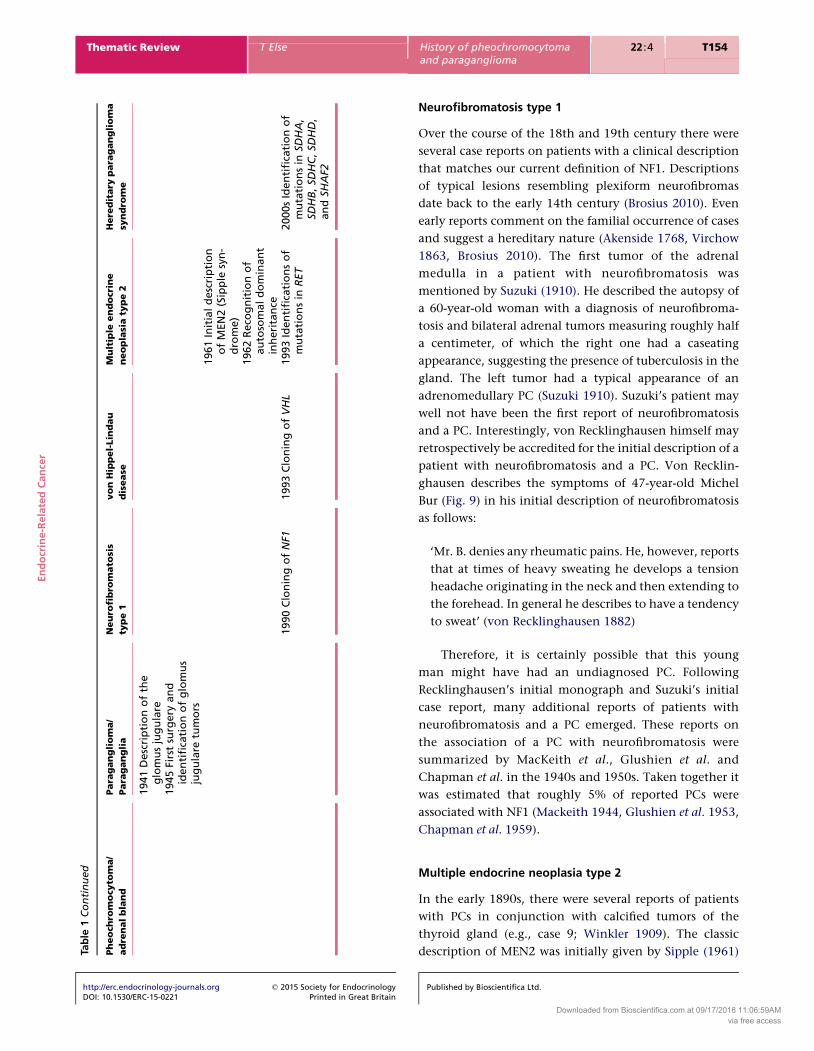
2000s
Iden
tifi
cati
on
of
mu
tati
on
sin
SDH
A,
SDH
B,
SDH
C,
SDH
D,
an
dSH
AF2
En
do
crin
e-R
ela
ted
Can
cer
Thematic Review T Else History of pheochromocytomaand paraganglioma
22 :4 T154
http://erc.endocrinology-journals.org q 2015 Society for EndocrinologyDOI: 10.1530/ERC-15-0221 Printed in Great Britain
Neurofibromatosis type 1
Over the course of the 18th and 19th century there were
several case reports on patients with a clinical description
that matches our current definition of NF1. Descriptions
of typical lesions resembling plexiform neurofibromas
date back to the early 14th century (Brosius 2010). Even
early reports comment on the familial occurrence of cases
and suggest a hereditary nature (Akenside 1768, Virchow
1863, Brosius 2010). The first tumor of the adrenal
medulla in a patient with neurofibromatosis was
mentioned by Suzuki (1910). He described the autopsy of
a 60-year-old woman with a diagnosis of neurofibroma-
tosis and bilateral adrenal tumors measuring roughly half
a centimeter, of which the right one had a caseating
appearance, suggesting the presence of tuberculosis in the
gland. The left tumor had a typical appearance of an
adrenomedullary PC (Suzuki 1910). Suzuki’s patient may
well not have been the first report of neurofibromatosis
and a PC. Interestingly, von Recklinghausen himself may
retrospectively be accredited for the initial description of a
patient with neurofibromatosis and a PC. Von Recklin-
ghausen describes the symptoms of 47-year-old Michel
Bur (Fig. 9) in his initial description of neurofibromatosis
as follows:
‘Mr. B. denies any rheumatic pains. He, however, reports
that at times of heavy sweating he develops a tension
headache originating in the neck and then extending to
the forehead. In general he describes to have a tendency
to sweat’ (von Recklinghausen 1882)
Therefore, it is certainly possible that this young
man might have had an undiagnosed PC. Following
Recklinghausen’s initial monograph and Suzuki’s initial
case report, many additional reports of patients with
neurofibromatosis and a PC emerged. These reports on
the association of a PC with neurofibromatosis were
summarized by MacKeith et al., Glushien et al. and
Chapman et al. in the 1940s and 1950s. Taken together it
was estimated that roughly 5% of reported PCs were
associated with NF1 (Mackeith 1944, Glushien et al. 1953,
Chapman et al. 1959).
Multiple endocrine neoplasia type 2
In the early 1890s, there were several reports of patients
with PCs in conjunction with calcified tumors of the
thyroid gland (e.g., case 9; Winkler 1909). The classic
description of MEN2 was initially given by Sipple (1961)
Published by Bioscientifica Ltd.
Downloaded from Bioscientifica.com at 09/17/2018 11:06:59AMvia free access

Figure 9
Michel Bur might have had a pheochromocytoma. He was one of
von Recklinghausen’s initial neurofibromatosis patients who reported
headaches and heavy perspiration (reproduced, with permission, from
von Recklinghausen F (1882) Ueber die multiplen Fibrome der Haut und
ihre Beziehung zu den multiplen Neuromen, pp138. Berlin, Germany:
A. Hirschfeld).
En
do
crin
e-R
ela
ted
Can
cer
Thematic Review T Else History of pheochromocytomaand paraganglioma
22 :4 T155
and is still occasionally termed ‘Sipple’s disease.’ Sipple
described a patient affected with all core tumors of MEN2.
The 33-year-old patient had bilateral adrenal tumors, a
parathyroid adenoma, and a thyroid tumor. Sipple also
cited another six cases of PCs with thyroid adenocarcino-
mas. The distinct pathological entity of medullary thyroid
cancer was described in the year following his initial
publication (Hazard et al. 1959). Williams reviewed several
cases of PCs with thyroid cancer and he was able to clarify
the medullary histology of the thyroid cancers in all
reviewed cases (Williams 1965). The hereditary nature of
MEN2 became evident shortly after Sipple’s initial
publication, when several families with MEN2-associated
manifestations were reported (Cushman 1962). Of note,
the initial patient with bilateral adrenal tumors described
by Fraenkel (1886) had an enlarged thyroid as well, and
interestingly, it turned out that she was part of a family
that was later diagnosed with MEN2 based on having a
RET p.Cys634Trp mutation (Neumann et al. 2007). The
term multiple endocrine neoplasia type 2 was introduced
in 1968 to differentiate this syndrome from MEN1, which
was until that point largely known as ‘multiple endocrine
adenomatosis’ (Steiner et al. 1968).
http://erc.endocrinology-journals.org q 2015 Society for EndocrinologyDOI: 10.1530/ERC-15-0221 Printed in Great Britain
VHL disease
Collins (1894) published a manuscript that ultimately
received little attention in the medical community,
describing a pair of siblings affected with angiomatous
tumors of the retina. A patient described by Harbitz (1915)
is likely the first patient in which the visceral manifes-
tations of VHL disease were present. At autopsy of the
patient who had died at age 47 due to a large kidney
tumor, a chromaffin tumor of the adrenal medulla, cystic
pancreas changes, and kidney tumors were found. In 1885,
von Hippel first reported on a case of the clinical
syndrome, which later took his name. He presented a
patient with unusual retinal findings in 1895 and later in
1904 published a follow-up report, ‘Ueber eine sehr seltene
Erkrankung der Netzhaut’ (‘About a very rare disease of the
retina’), on two cases of retinal angiomas (von Hippel
1895, 1904). Von Hippel believed the physical findings
were likely due to an infectious agent. One year later
Czermak (1905) was able to identify the lesions on a
surgical specimen as retinal angiomas. Von Hippel (1911)
was able to confirm the pathology on one of his own
initial cases after enucleation of the eye. The Swedish
pathologist Arvid Lindau was the first one to analyze
several patients with VHL disease systematically (Lindau
1926). He put the angiomatous changes of the retina in
relation to the often observed cystic changes of the
cerebellum. In addition he describes almost all of the
currently known manifestations of VHL disease. Lindau
reviewed 15 cases, nine of which were his own. He found
angiomas of the retina in four cases, the cerebellum in
nine, the medulla oblongata in five, the spinal cord in
four, and the skin in two. Eight patients had cystic
pancreatic changes, six patients had renal cell cancer,
and two patients had adrenal tumors. Glushien et al.
(1953) gave a thorough review of the literature providing
evidence for an association of VHL disease and PCs. The
hereditary nature of VHL disease could have already been
suspected from the description of affected siblings by
Collins and two affected brothers described by Seidel –
interestingly, both professional tightrope dancers with
repeated falls, possibly due to retinal and cerebral
angiomas (Collins 1894, Seidel 1912). The focus on
patient family pedigrees and the finding of autosomal
dominant inheritance were proposed in two publications
at the end of the 1920s by Rochat (1927) and Moeller
(1929). Interestingly, several family members of von
Hippel’s initial proband had been described indepen-
dently in the medical literature. The whole pedigree
and the clinical history of affected family members were
Published by Bioscientifica Ltd.
Downloaded from Bioscientifica.com at 09/17/2018 11:06:59AMvia free access

En
do
crin
e-R
ela
ted
Can
cer
Thematic Review T Else History of pheochromocytomaand paraganglioma
22 :4 T156
finally summarized in a publication by Piotrowski and
Rohrborn (1965).
Hereditary PGL syndrome
Although for the previously discussed syndromes PCs were
recognized after the initial description of other syndromic
core manifestations, elucidation of the genetic and clinical
basis of hereditary PGL syndromes (PGL1–5) was exclu-
sively based on the familial occurrence of PCPGL. Only
after the genetic basis was determined, pituitary tumors,
gastrointestinal stroma tumors, and renal cell cancer were
recognized as part of the full tumor spectrum, albeit with
lower penetrance than PCPGL. The first possible
descriptions of familial cases of PCPGL were by Marchand
(1891) of a patient with a PC and a family history of a
mother with a large retroperitoneal tumor. In the 1930s to
1950s more detailed reports by Chase on familial carotid
body tumors and by Cook et al. on familial malignant
PGLs of the organ of Zuckerkandl followed (Chase 1933,
Cook et al. 1960). In 1932 Goekopp reports on three sisters
with ‘fibro-hemangiomas of the middle ear and petrosal
bone,’ likely representing glomus jugulare tumors as
described 8 years later by Rosenwasser (Goekoop 1932,
Rosenwasser 1945).
Since defining the genetic basis of hereditary PGL
syndromes in the early 2000s, a growing number of
families with mutations in the succinate dehydrogenase
subunits (SDHx) and associated genes have been identified
(Baysal et al. 2000, Niemann & Muller 2000, Astuti et al.
2001, Hao et al. 2009, Burnichon et al. 2010). Importantly,
there is the common notion that the syndrome is not
entirely understood and that there are likely other
associations, such as a relative risk increase for other rare
tumors and possibly other non-neoplastic manifestations.
Therefore the SDHx-associated syndromes still provide
ample opportunity for systematic clinical research to
complete our understanding of risk associations, which
is necessary to provide the right genetic counseling and
offer the correct and most cost-effective surveillance to
these patients.
Recently there have been some descriptions of
mutations in FH, another enzyme of the Krebs cycle
known to cause hereditary leimyomatosis and renal cell
cancer, in patients with PC (Clark et al. 2014). There is
accumulating evidence that mutations in Krebs cycle
enzymes as well as VHL mutations cause hypoxia-
signaling (‘pseudo-hypoxia’) in adrenomedullary and
paraganglial cells, contributing to tumor development
(Favier & Gimenez-Roqueplo 2010). In addition, in rare
http://erc.endocrinology-journals.org q 2015 Society for EndocrinologyDOI: 10.1530/ERC-15-0221 Printed in Great Britain
instances, tumors of the paraganglial system (as well as
erythrocytosis) may also arise in patients with germline
mutations in EGLN1 or somatic gain of function
mutations in HIF2A, further underscoring the importance
of the hypoxia signaling pathway (Ladroue et al. 2008,
Zhuang et al. 2012).
Lessons learned from history
Over the last 150 years our knowledge about PCPGL and
associated hereditary conditions has significantly broa-
dened. This acquisition of knowledge started with the
definition of the origins of the adrenal medulla and its
connection to other chromaffin tissues, followed by the
description of the paraganglial tissue as its own tissue entity
and finally led to the definition of the genetic basis of
hereditary syndromes associated with PCPGL. The general
principle of this research historically always used the same
approach: initially, there were some emerging case reports
and, finally, a systematic analysis identifying a new clinical
syndrome. Often the discovery of a syndrome is accredited
to the first researcher taking the systematic approach in
combining cases from the literature with the researcher’s
own observations. But it is important to note that the final
‘syndrome discoveries’ were generally based on accumulat-
ing bitsof small data emerging inmedical sciences, which in
itself represented important steps forward. Along the way
astute physicians and researchers detailed the clinical
observations and continued to further define these clinical
phenotypes, including associations with other tumors, and
described the epidemiology of these conditions.
Over the last two decades, however, there has been
a paradigm shift. The initial recognition of rare genetic
syndromes nowadays is often preceded by genetic findings
lacking a complete knowledge of the clinical phenotype.
Nowadays, molecular genetic findings are often evident prior
to a fully clinically defined phenotype. It seems almost that
thorough clinical descriptions are often ignored. Therefore,
continuation (or maybe resurrection) of the ability to obtain
detailed clinical observations has become more important
than ever before. It is imperative to continue teaching and
improving the clinical skills of future generations to provide
the data for describing the clinical phenotype for genetic
syndromes. In addition, systematic clinical efforts are needed
to completeour knowledgeabout theentire associated tumor
spectrum, the age of onset of these tumors, and the
penetrance of phenotypes in hereditary syndromes,
especially those associated with PCPGL. On the molecular
level, attention to clinical detail will also allow for a
streamlined approach to identify modifying circumstances,
Published by Bioscientifica Ltd.
Downloaded from Bioscientifica.com at 09/17/2018 11:06:59AMvia free access

En
do
crin
e-R
ela
ted
Can
cer
Thematic Review T Else History of pheochromocytomaand paraganglioma
22 :4 T157
such as environmental exposures or modifier genes. This
knowledge will be necessary to make the right decisions in
the clinical care of affected patients. For example, there are
still pertinent questions that remain unanswered, such as to
what is the right imaging frequency for SDHx mutation
carriers and whether the risk is truly the same over the course
of the lifetime of a patient. Our current assumptions
regarding penetrance of the SDHx-associated phenotypes is
still strongly biased by the analysis of the initial descriptions
of high penetrance families. The actual absolute risk for
PCPGL development, therefore, remains undetermined.
With regards to the risk of tumor development over a
mutation carrier’s lifetime, it is interesting to note that most
patients with head and neck PGLs in historical descriptions
were fairly young at diagnosis and often described a time of
acceleratedtumor growth followedbyminimalornogrowth.
This suggests that the very initial events of tumorigenesis
might fall into fetal life, childhood, or adolescence, and there
may be less risk of tumor development in adulthood. Only
the definition of age-associated risks will allow for establish-
ing evidence-based screening guidelines. Furthermore, it will
be important to understand the full clinical spectrum of
predisposition caused by mutations in genes that were only
recently described, such as TMEM127 and MAX, possibly
identifying other associated tumors. It also remains necessary
to analyze larger cohorts to prove the causative relationship
between PCPGL and genetic mutations that have so far only
been found in a singlepatient or family.Therefore, there isno
doubt that future clinical and genetic findings will be as
exciting and unexpected as the seminal findings over the last
centuries and will most importantly improve preventive care
in gene mutation carriers.
Declaration of interest
The authors declare that there is no conflict of interest that could be
perceived as prejudicing the impartiality of this review.
Funding
T Else is funded by the American Heart Association (AHA14SDG17990000).
Acknowledgements
I would like to thank Benjamin Fortson for the Latin-English translation of
the manuscripts by Haller and Morgagni. I thank Lauren Fishbein for the
careful reading of this manuscript. I am particularly indebted to the current
efforts of many libraries and enterprises to make historic manuscripts
available to the public (e.g., Google, Hathi Trust). Without these resources a
meaningful research on the discussed topic would not have been possible.
For further reading, I would like to hint the reader to some detailed and
more in-depth reading on the exciting history of the adrenal medulla and
neurofibromatosis: Brosius (2010), Carmichael (1989).
http://erc.endocrinology-journals.org q 2015 Society for EndocrinologyDOI: 10.1530/ERC-15-0221 Printed in Great Britain
References
Akenside M 1768 Observations on cancers. Medical Transactions Royal
College of Physicians of London 1 64–92.
Alezais HP 1908 Un groupe nouveau de tumeurs epitheliliale: les para-
gangliomes. Comptes Rendus des Seances de la Societe de Biologie 65 745–747.
Arias-Stella J & Valcarcel J 1973 The human carotid body at high altitudes.
Pathologia et Microbiologia 39 292–297.
Astuti D, Latif F, Dallol A, Dahia PL, Douglas F, George E, Skoldberg F,
Husebye ES, Eng C & Maher ER 2001 Gene mutations in the succinate
dehydrogenase subunit SDHB cause susceptibility to familial
pheochromocytoma and to familial paraganglioma. American Journal of
Human Genetics 69 49–54. (doi:10.1086/321282)
Baysal BE, Ferrell RE, Willett-Brozick JE, Lawrence EC, Myssiorek D, Bosch A,
van der Mey A, Taschner PE, Rubinstein WS, Myers EN et al. 2000
Mutations in SDHD, a mitochondrial complex II gene, in hereditary
paraganglioma.Science 287 848–851. (doi:10.1126/science.287.5454.848)
Berckelmann MLR 1744 De Nervorun in Arterias Imperio. Goettingen,
Germany: Abram Vandenhoeck.
Brosius S 2010 A history of von Recklinghausen’s NF1. Journal of the History
of the Neurosciences 19 333–348. (doi:10.1080/09647041003642885)
Burnichon N, Briere JJ, Libe R, Vescovo L, Riviere J, Tissier F, Jouanno E,
Jeunemaitre X, Benit P, Tzagoloff A et al. 2010 SDHA is a tumor
suppressor gene causing paraganglioma. Human Molecular Genetics 19
3011–3020. (doi:10.1093/hmg/ddq206)
Carmichael SW 1989 The history of the adrenal medulla. Reviews in the
Neurosciences 2 83–100. (doi:10.1515/REVNEURO.1989.2.2.83)
Cawthon RM, Weiss R, Xu GF, Viskochil D, Culver M, Stevens J, Robertson M,
Dunn D, Gesteland R, O’Connell P et al. 1990 A major segment of the
neurofibromatosis type 1 gene: cDNA sequence, genomic structure, and
point mutations. Cell 62 193–201. (doi:10.1016/0092-8674(90)90253-B)
Chapman RC, Kemp VE & Taliaferro I 1959 Pheochromocytoma associated
with multiple neurofibromatosis and intracranial hemangioma. American
Journal of Medicine 26 883–890. (doi:10.1016/0002-9343(59)90210-4)
Chase WH 1933 Familial and bilateral tumours of the carotid body.
Journal of Pathology and Bacteriology 36 1–12. (doi:10.1002/path.
1700360102)
Clark GR, Sciacovelli M, Gaude E, Walsh DM, Kirby G, Simpson MA,
Trembath RC, Berg JN, Woodward ER, Kinning E et al. 2014 Germline FH
mutations presenting with pheochromocytoma. Journal of Clinical
Endocrinology and Metabolism99 E2046–E2050. (doi:10.1210/jc.2014-1659)
Cock E 1837 Observations on the ganglionic enlargement of the pneumo-
gastric nerve. Guy’s Hospital Reports 2 311–317.
Collins ET 1894 Intra-ocular growth (two cases, brother and sister, with
peculiar vascular new growth, probably retinal, affecting both eyes).
Transactions of the Ophthalmological Societies of the United Kingdom 14
141–149.
Comino-Mendez I, Gracia-Aznarez FJ, Schiavi F, Landa I, Leandro-Garcia LJ,
Leton R, Honrado E, Ramos-Medina R, Caronia D, Pita G et al. 2011
Exome sequencing identifies MAX mutations as a cause of hereditary
pheochromocytoma. Nature Genetics 43 663–667. (doi:10.1038/ng.861)
Cook JE, Urich RW, Sample HG Jr & Fawcett NW 1960 Peculiar familial and
malignant pheochromocytomas of the organs of Zuckerkandl. Annals of
Internal Medicine 52 126–133. (doi:10.7326/0003-4819-52-1-126)
Cornog JL, Wilkinson JH, Arvan DA, Freed RM, Sellers AM & Barker C 1970
Extra-adrenal pheochromocytoma. Some electron microscopic and
biochemical studies. American Journal of Medicine 48 654–660.
(doi:10.1016/0002-9343(70)90018-5)
Crossey PA, Richards FM, Foster K, Green JS, Prowse A, Latif F, Lerman MI,
Zbar B, Affara NA, Ferguson-Smith MA et al. 1994 Identification of
intragenic mutations in the von Hippel-Lindau disease tumour
suppressor gene and correlation with disease phenotype. Human
Molecular Genetics 3 1303–1308. (doi:10.1093/hmg/3.8.1303)
Cushman P Jr 1962 Familial endocrine tumors; report of two unrelated
kindred affected with pheochromocytomas, one also with multiple
Published by Bioscientifica Ltd.
Downloaded from Bioscientifica.com at 09/17/2018 11:06:59AMvia free access

En
do
crin
e-R
ela
ted
Can
cer
Thematic Review T Else History of pheochromocytomaand paraganglioma
22 :4 T158
thyroid carcinomas. American Journal of Medicine 32 352–360.
(doi:10.1016/0002-9343(62)90126-2)
Czermak W 1905 Pathologisch-anatomischer Befund bei der von
E.v.Hippel beschriebenen sehr seltene Netzhauterkrankung. Bericht
ueber die Versammlung der Ophtalmologischen Gesellschaft 32 184–195.
Dhayat S, Renggli JC, Dhayat N & Merlini M 2007 On the 150th birthday
of Cesar Roux (1857–1918). Memories of the life and work of an
important pupil of Kocher. Chirurg 78 155–160. (doi:10.1007/
s00104-006-1267-3)
Eustachi B 1714 Tabulae Anatomicae Clarissimi Viri Bartholomaei Eustachii.
Rome, Italy: Francisi Gonzagae.
Favier J & Gimenez-Roqueplo AP 2010 Pheochromocytomas: the (pseudo)-
hypoxia hypothesis. Best Practice & Research. Clinical Endocrinology &
Metabolism 24 957–968. (doi:10.1016/j.beem.2010.10.004)
Fraenkel F 1886 Ein Fall von doppelseitigem voellig latent verlaufenden
Nebennierentumor und gleichzeitiger Nephritis mit Veraenedungen
am Circulationsapparat und Retinitis. Virchows Archiv. A, Pathological
Anatomy and Histology 103 244–263.
Gilford H 1904 "Potato" tumours of the neck and their origin as
endotheliomas of the carotid body, with an account of three cases.
Practitioner 73 729–739.
Glushien AS, Mansuy MM & Littman DS 1953 Pheochromocytoma; its
relationship to the neurocutaneous syndromes. American Journal of
Medicine 14 318–327. (doi:10.1016/0002-9343(53)90043-6)
Goekoop C 1932 Fibro-Haemangiom des Felsenbeins und des Mittelohres
bei drei Schwestern. Acta Oto-Laryngologica 18 153–162. (doi:10.3109/
00016483309134897)
Guild SR 1941 A hitherto Unrecognized Structure, the Glomus Jugularis, in
man. American Association of Anatomists 57 28 (abstract 72).
Hao HX, Khalimonchuk O, Schraders M, Dephoure N, Bayley JP, Kunst H,
Devilee P, Cremers CW, Schiffman JD, Bentz BG et al. 2009 SDH5, a
gene required for flavination of succinate dehydrogenase, is mutated in
paraganglioma. Science 325 1139–1142. (doi:10.1126/science.1175689)
Harbitz F 1915 Tumors of the sympathetic nervous system and the adrenal
medulla of the adrenal glands, especially malignant neuroblastoma.
Archives of Internal Medicine 15 341–341.
Hazard JB, Hawk WA & Crile G Jr 1959 Medullary (solid) carcinoma of the
thyroid; a clinicopathologic entity. Journal of Clinical Endocrinology and
Metabolism 19 152–161. (doi:10.1210/jcem-19-1-152)
Hedinger E 1911 Struma medullaris cystica. Frankfurter Zeitschrift fur
Pathologie 7 112–126.
Henle J 1865 Ueber das Gewebe der Nebenniere und der Hypophyse.
Zeitschrift fur Rationelle Medicin 24 143–152.
von Hippel E 1895 Vorstellung eines Patienten mit einer sehr ungewohn-
lichen Netzhaut. Bericht ueber die Versammlung der Ophtalmologischen
Gesellschaft 24 269.
von Hippel E 1904 Ueber eine sehr seltene Erkrankung der Netzhaut.
Graefe’s Archive for Clinical and Experimental Ophthalmology 59 83–106.
(doi:10.1007/BF01994821)
von Hippel E 1911 Die anatomische Grundlage der von mir beschriebenen
‘sehr seltenen Erkrankung der Netzhaut’. Graefe’s Archives of
Ophthalmology 59 83–86. (doi:10.1007/BF01994821)
Kohn A 1903 Die Paraganglien. Archiv fur mikroskopische Anatomie 62
263–367.
Lack EE 1978 Hyperplasia of vagal and carotid body paraganglia in patients
with chronic hypoxemia. American Journal of Pathology 91 497–516.
Ladroue C, Carcenac R, Leporrier M, Gad S, Le Hello C, Galateau-Salle F,
Feunteun J, Pouyssegur J, Richard S & Gardie B 2008 PHD2
mutation and congenital erythrocytosis with paraganglioma.
New England Journal of Medicine 359 2685–2692. (doi:10.1056/
NEJMoa0806277)
Latif F, Tory K, Gnarra J, Yao M, Duh FM, Orcutt ML, Stackhouse T,
Kuzmin I, Modi W, Geil L et al. 1993 Identification of the von
Hippel-Lindau disease tumor suppressor gene. Science 260 1317–1320.
(doi:10.1126/science.8493574)
http://erc.endocrinology-journals.org q 2015 Society for EndocrinologyDOI: 10.1530/ERC-15-0221 Printed in Great Britain
Lindau A 1926 Studien ueber Kleinhirncysten. Bau, Pathogenese und
Beziehungen zur Angiomatosis Retinae. Acta Pathologica et Microbiolo-
gica Scandinavica 3 1–128.
Mackeith R 1944 Adrenal-sympathetic syndrome: chromaffin tissue
tumour with paroxysmal hypertension. British Heart Journal 6 1–12.
(doi:10.1136/hrt.6.1.1)
Manasse P 1893 Ueber die hyperplastischen Tumoren der Nebennieren.
Archiv fur pathologische Anatomie und Physiologie und fur klinische Medicin
133 391–404.
Maranillo E, Vazquez T, Ibanez M, Hurtado M, Pascual-Font A,
McHanwell S, Valderrama-Canales F & Sanudo J 2008 Anatomic study
of human laryngeal ganglia: number and distribution. Clinical Anatomy
21 641–646. (doi:10.1002/ca.20699)
Marchand F 1883 Ueber die accessorischen Nebennieren im Ligamentum
latum. Virchows Archiv fur pathologische Anatomie und Physiologie und fur
klinische Medizin 126 11–19.
Marchand F 1891 Beitraege zur Kenntnis der normalen und pathologischen
Anatomie der Glandula carotica und der Nebennieren. Internationale
Beitrage zur wissenschaftlichen Medizin. Festschrift fur Rudolf Virchow 1
537–581.
Mayo CH 1927 Paroxysmal hypertension with tumor of retroperitoneal
nerve. Journal of the American Medical Association 89 1047–1050.
(doi:10.1001/jama.1927.02690130035013)
Mayo CH 1982 Charles Horace Mayo (1865–1939). Diseases of the Colon &
Rectum 25 734–739. (doi:10.1007/BF02629554)
Moeller HU 1929 Familial angiomatosis retinæ et cerebelli – Lindau’s
disease. Acta Ophthalmologica 7 244–260. (doi:10.1111/j.1755-3768.
1929.tb07929.x)
Moenckeberg IG 1905 Die Tumoren der Glandula Carotica. Beitraege zur
Pathologischen Anatomie und Allgemeinen Pathologie 38 1–66.
Morgagni JP 1779 De sedibus et causis morborum per anatomen indagatis.
Ebroduni in Helvetia: Tissot, S. A. D.
Mulligan LM, Kwok JB, Healey CS, Elsdon MJ, Eng C, Gardner E, Love DR,
Mole SE, Moore JK, Papi L et al. 1993 Germ-line mutations of the RET
proto-oncogene in multiple endocrine neoplasia type 2A. Nature 363
458–460. (doi:10.1038/363458a0)
Nagel M 1836 Ueber die Structur der Nebennieren. Archiv fur Anatomie,
Physiologie und Wissenschaftliche Medicin 365–383.
Nanka OGM 2008 Alfred Kohn, profesor histologie na Nemecke univerzite
v praze. Casopıs lekaru ceskych 147 240–244.
Neumann HP, Vortmeyer A, Schmidt D, Werner M, Erlic Z, Cascon A,
Bausch B, Januszewicz A & Eng C 2007 Evidence of MEN-2 in the
original description of classic pheochromocytoma. New England
Journal of Medicine 357 1311–1315. (doi:10.1056/NEJMoa071407)
Neusser EWJ 1910 Erkrankungen der Nebennieren. Wien und Leipzig,
Germany: A. Hoelder.
Niemann S & Muller U 2000 Mutations in SDHC cause autosomal
dominant paraganglioma, type 3. Nature Genetics 26 268–270.
(doi:10.1038/81551)
Nozdrachev AD 1995 [The 90th anniversary of the creation by J.
Newport Langley of the theory of autonomic nervous system structure
(historical essay)]. Fiziologicheskii zhurnal imeni I.M. Sechenova 81
175–184.
Paltauf R 1891 Ueber Geschwuelste der Glandula carotica. Beitraege zur
Pathologischen Anatomie und Allgemeinen Pathologie 11 260–301.
Peiper HJ 2008 Albrecht von Haller. An illustrious ancestor. Chirurg 79
486–493. (doi:10.1007/s00104-008-1495-9)
Pick L 1912a Das Ganglioma embryonale sympathicum (Sympathoma
embryonale). Berliner Klinische Wochenschrift 49 16–22.
Pick L 1912b Das Ganglioma embryonale sympathicum (sympathoma
embryonale), eine typische bosartige geschwuestform des sym-
pathischen nervensystems. Berliner Klinische Wochenschrift 49 67–72.
Piotrowski W & Rohrborn G 1965 Family study of a classic case of
von Hippellindau syndrome. Langenbecks Archiv fur Chirurgie 311
310–322.
Published by Bioscientifica Ltd.
Downloaded from Bioscientifica.com at 09/17/2018 11:06:59AMvia free access

En
do
crin
e-R
ela
ted
Can
cer
Thematic Review T Else History of pheochromocytomaand paraganglioma
22 :4 T159
Qin Y, Yao L, King EE, Buddavarapu K, Lenci RE, Chocron ES, Lechleiter JD,
Sass M, Aronin N, Schiavi F et al. 2010 Germline mutations in
TMEM127 confer susceptibility to pheochromocytoma. Nature Genetics
42 229–233. (doi:10.1038/ng.533)
von Recklinghausen F 1882 Ueber die multiplen Fibrome der Haut und ihre
Beziehung zu den multiplen Neuromen, pp138. Berlin, Germany:
A. Hirschfeld.
Rochat G 1927 Familiaere Angiomatosis retinae und Kleinhirnangiom.
Klin Mbl Augenheilk 78 601.
Rosenwasser H 1945 Carotid body like tumor involving the middle ear and
mastoid bone. Archives of Otolaryngology 41 64–67. (doi:10.1001/
archotol.1945.00680030087006)
Saldana MJ, Salem LE & Travezan R 1973 High altitude hypoxia and
chemodectomas. Human Pathology 4 251–263. (doi:10.1016/S0046-
8177(73)80012-7)
Seidel 1912 Ueber ein Angiom der Netzhaut. Bericht ueber die Versammlung
der Ophtalmologischen Gesellschaft 38 335–339.
Shoja MM, Tubbs RS, Loukas M, Shokouhi G & Jerry Oakes W 2008 Emil
Zuckerkandl (1849–1910): anatomist and pathologist. Annals of
Anatomy 190 33–36. (doi:10.1016/j.aanat.2007.09.001)
Simon M 1906 Sieben Buecher Anatomie des Galen. Leipzig: J. C.
Heinrichs’sche Buchhandlung.
Sipple JH 1961 The Association of Pheochromocytoma with carcinoma of
the thyroid. Journal of the American Medical Association 31 163–166.
Stangl E 1903 Zur Pathologie der Nebnorgane des Sympathicus.
Verhandlungen der Deutschen Pathologischen Gesellschaft 5 250–254.
Steiner AL, Goodman AD & Powers SR 1968 Study of a kindred with
pheochromocytoma, medullary thyroid carcinoma, hyperparathyr-
oidism and Cushing’s disease: multiple endocrine neoplasia, type 2.
Medicine 47 371–409. (doi:10.1097/00005792-196809000-00001)
Suzuki S 1910 Ueber zwei Tumoren aus Nebennierenmarkgewebe. Berliner
Klinische Wochenschrift 47 1623–1625.
http://erc.endocrinology-journals.org q 2015 Society for EndocrinologyDOI: 10.1530/ERC-15-0221 Printed in Great Britain
Taube HWL 1743 De Vera Nervi Intercostali Origine. Goettingen, Germany:
Abram Vandenhoeck.
Virchow R 1863 Die krankhaften Geschwuelste. Berlin: Hirschwald.
Watanabe H, Burnstock G, Jarrott B & Louis WJ 1976 Mitochondrial
abnormalities in human phaeochromocytoma. Cell and Tissue Research
172 281–288. (doi:10.1007/BF00226032)
Welbourn RB 1987 Early surgical history of phaeochromocytoma. British
Journal of Surgery 74 594–596. (doi:10.1002/bjs.1800740717)
Werner B 1857 De Capsulis Suprarenalibus. Dorpat, Estonia:
J. C. Schuemann, C. Mattiesen.
Williams ED 1965 A review of 17 cases of carcinoma of the thyroid and
phaeochromocytoma. Journal of Clinical Pathology 18 288–292.
(doi:10.1136/jcp.18.3.288)
Winkler C 1909 Die Gewaechse der Nebenniere. Jena, Germany:
Gustav Fischer.
Winslow JB 1776 Exposition Anatomique de la Structure du Corps Humain.
Paris, France: G. Desprez, and J. Desessartz.
Xu GF, O’Connell P, Viskochil D, Cawthon R, Robertson M, Culver M,
Dunn D, Stevens J, Gesteland R, White R et al. 1990 The neurofi-
bromatosis type 1 gene encodes a protein related to GAP. Cell 62
599–608. (doi:10.1016/0092-8674(90)90024-9)
Zani A & Cozzi DA 2008 Giovanni Battista Morgagni and his contribution
to pediatric surgery. Journal of Pediatric Surgery 43 729–733.
(doi:10.1016/j.jpedsurg.2007.12.065)
Zhuang Z, Yang C, Lorenzo F, Merino M, Fojo T, Kebebew E, Popovic V,
Stratakis CA, Prchal JT & Pacak K 2012 Somatic HIF2A gain-
of-function mutations in paraganglioma with polycythemia.
New England Journal of Medicine 367 922–930. (doi:10.1056/
NEJMoa1205119)
Zuckerkandl E 1901 Ueber Nebenorgane des Sympathicus im Retro-
peritonaealraum des Menschen. Verhandlungen der Anatomischen
Gesellschaft 14 95–107.
Received in final form 9 June 2015Accepted 18 June 2015Made available online as an Accepted Preprint25 June 2015
Published by Bioscientifica Ltd.
Downloaded from Bioscientifica.com at 09/17/2018 11:06:59AMvia free access