Embed Size (px)
Citation preview
Recruitment of Matrix Metalloproteinase-9 (MMP-9) to theFibroblast Cell Surface by Lysyl Hydroxylase 3 (LH3) TriggersTransforming Growth Factor-� (TGF-�) Activation andFibroblast Differentiation*
Received for publication, October 28, 2014, and in revised form, March 30, 2015 Published, JBC Papers in Press, March 30, 2015, DOI 10.1074/jbc.M114.622274
Cynthia Dayer and Ivan Stamenkovic1
From the Division of Experimental Pathology, Institute of Pathology, CHUV, Faculty of Biology and Medicine, University ofLausanne, Rue du Bugnon 25, Lausanne CH1011, Switzerland
Background: MMP-9 cell surface activity promotes tissue remodeling.Results: Fibroblast cell surface recruitment of MMP-9 via its fibronectin-like domain (FN) by lysyl hydroxylase 3 (LH3) inducesTGF-� activation and myofibroblast differentiation.Conclusion: We identify a novel mechanism of MMP-9 recruitment to stromal cells that can be modulated by recombinant FN.Significance: Recombinant FN may allow selective MMP-9 blockade in tumor-associated tissue remodeling.
Solid tumor growth triggers a wound healing response. Simi-lar to wound healing, fibroblasts in the tumor stroma differen-tiate into myofibroblasts (also referred to as cancer-associatedfibroblasts) primarily, but not exclusively, in response to trans-forming growth factor-� (TGF-�). Myofibroblasts in turnenhance tumor progression by remodeling the stroma. Amongproteases implicated in stroma remodeling, matrix metallopro-teinases (MMPs), including MMP-9, play a prominent role.Recent evidence indicates that MMP-9 recruitment to thetumor cell surface enhances tumor growth and invasion. In thepresent work, we addressed the potential relevance of MMP-9recruitment to and activity at the surface of fibroblasts. We showthat recruitment of MMP-9 to the fibroblast cell surface occursthrough its fibronectin-like (FN) domain and that the moleculeresponsible for the recruitment is lysyl hydroxylase 3 (LH3).Functional assays suggest that both pro- and active MMP-9 trig-ger �-smooth muscle actin expression in cultured fibroblasts,reflecting myofibroblast differentiation, possibly as a result ofTGF-� activation. Moreover, the recombinant FN domaininhibited both MMP-9-induced TGF-� activation and�-smooth muscle actin expression by displacing MMP-9 fromthe fibroblast cell surface. Together our results uncover LH3 as anew docking receptor of MMP-9 on the fibroblast cell surfaceand demonstrate that the MMP-9 FN domain is essential for theinteraction. They also show that the recombinant FN domaininhibits MMP-9-induced TGF-� activation and fibroblast dif-ferentiation, providing a potentially attractive therapeutic re-agent toward attenuating tumor progression where MMP-9activity is strongly implicated.
Tumor cell interactions with host tissue stroma play a keyrole in determining tumor progression that culminates in met-
astatic growth. It is well established that malignant tumorgrowth initiates a wound healing response that maintains astate of tissue remodeling, which favors tumor survival, inva-sion, and dissemination. Orchestration of tumor-associated tis-sue remodeling is mediated in part by tumor cells and in part bya variety of recruited host tissue cells, including various leuko-cyte subsets and mesenchymal cell subtypes ranging from mes-enchymal stem cells to myofibroblasts (1–3). Most of these cellsparticipate in generating soluble mediators that include a pleth-ora of cytokines, chemokines, growth factors, and enzymes.Among the proteolytic enzymes implicated in tumor-hostcross-talk are matrix metalloproteinases (MMPs),2 a large fam-ily of zinc-dependent extracellular matrix (ECM)-degradingendopeptidases that play a key role in tissue remodeling duringdevelopment and repair (4). The majority of MMPs aresecreted, but at least a fraction of their proteolytic activity isobserved at the cell surface where they can be anchored by avariety of cell surface receptors to provide controlled degrada-tion of the ECM and activation of a variety of latent growthfactors (5). Whether or not cell surface anchoring of secretedMMPs occurs exclusively in autocrine fashion in the context oftissue remodeling or whether it may also occur in paracrinefashion whereby MMP-anchoring cells are distinct from MMP-secreting cells remains to be clarified.
MMP-9, also known as gelatinase B, has been shown to play aprominent role in the progression of numerous tumor types bypromoting tumor cell invasion and angiogenesis (6, 7). Similarto other MMPs, MMP-9 is synthesized as an inactive zymogen,or pro-MMP-9, composed of a propeptide, a catalytic domaincontaining fibronectin-like (FN) repeats, a linker region orhinge domain, and a C-terminal hemopexin-like (HEX) domainthought to be necessary for substrate recognition (4). The FN
* This work was supported by Swiss National Science Foundation (FNS) Grant310030_150024 and a grant from the Swiss Institute for Experimental Can-cer Research (ISREC) Foundation.
1 To whom correspondence should be addressed. Tel.: 41-21-314-7136; Fax:41-21-314-7110; E-mail: [email protected].
2 The abbreviations used are: MMP, matrix metalloproteinase; LH3, lysylhydroxylase-3; FN, fibronectin-like; �-SMA, �-smooth muscle actin; ECM,extracellular matrix; HEX, hemopexin-like; TMLC, transformed mink lungepithelial cells; HSF, human skin fibroblasts; Ni-NTA, nickel-nitrilotriaceticacid; PLA, proximity ligation assay; PLOD3, procollagen-lysine, 2-oxogl-utarate 5-dioxygenase 3.
THE JOURNAL OF BIOLOGICAL CHEMISTRY VOL. 290, NO. 22, pp. 13763–13778, May 29, 2015© 2015 by The American Society for Biochemistry and Molecular Biology, Inc. Published in the U.S.A.
MAY 29, 2015 • VOLUME 290 • NUMBER 22 JOURNAL OF BIOLOGICAL CHEMISTRY 13763
by guest on June 18, 2018http://w
ww
.jbc.org/D
ownloaded from
domain, which is found only in MMP-9 and MMP-2, is com-posed of three tandem fibronectin type II-like motifs that forma collagen-binding domain critical for the positioning of sub-strates for subsequent cleavage (8). The collagen-bindingdomain of MMP-9 has been shown to bind gelatin (9), elastin,and both native and denatured types I, II, III, IV, and V collagen(8, 10). Each fibronectin type II-like module displays bindingspecialization, which generates exosites specific for otherligands degraded by the protease (10). Cooperation among col-lagen binding sites within these three modules increases sub-strate specificity and thereby has the potential to localize theenzyme to collagen either in the extracellular matrix or on thecell surface (11).
MMP-9 expression is low or absent in normal quiescent tis-sues but is strongly induced under conditions that trigger tissueremodeling, including development, wound healing, and tumorinvasion. MMP-9 is produced by tumor-associated host tissuecells, including endothelial cells, various leukocytes, and tumorcells themselves, and is thought to promote tumor growth andmetastasis (4, 6, 12–14). Several secreted MMPs, including pro-MMP-9, can at least transiently be anchored to the cell surface,which directs their proteolytic activity toward pericellular sub-strates and may provide protection from natural inhibitors.However, the mechanisms that underlie their association withthe cell membrane appear to be diverse and remain to be fullyexplored (5, 15, 16). Thus far, MMP-9 has been shown to useamong others the cell surface hyaluronan receptor CD44 as adocking molecule in certain tumor cell types and keratinocytes(17). This association stabilizes MMP-9 proteolytic activity atthe cell surface to facilitate controlled collagen IV degradationand to promote invasion (17). In addition, CD44-associatedMMP-9 as well as MMP-2 can cleave and activate latentTGF-�1 and -2 (18). Thus, coordination of CD44, MMP-9, andTGF-� function may provide a physiological mechanism of tis-sue remodeling that can be adopted by malignant cells to pro-mote their own growth and dissemination (18, 19). As key reg-ulators of ECM turnover, fibroblasts may include MMP-9 intheir arsenal of tissue remodeling reagents. However, mecha-nisms that govern putative MMP-9 association with the surfaceof normal stromal cells, including fibroblasts, remain to be elu-cidated. Fibroblasts produce low amounts of MMP-9, suggest-ing that they may recruit tumor cell or leukocyte-derivedMMP-9 to their own cell surface to promote ECM remodelingby harnessing its proteolytic activity.
In the present work, we show that MMP-9 produced bytumor cells is recruited to the fibroblast surface and thatrecruitment requires the FNII repeats or collagen-bindingdomain of MMP-9. We demonstrate that the structure thatmediates MMP-9 docking to the fibroblast surface is providedby lysyl hydroxylase 3 (LH3), which displays lysyl hydroxylase aswell as galactosyl- and glucosyltransferase activity (20). LH3 isexpressed in the endoplasmic reticulum but is also associatedwith the plasma membrane via collagenous proteins (21). Weshow that LH3-mediated MMP-9 recruitment contributes toTGF-� activation, which stimulates fibroblast differentiationinto myofibroblasts. Tumor cells and leukocytes may thus pro-vide a source of MMP-9 that fibroblasts can recruit and use toactivate TGF-� and stimulate their own differentiation.
Experimental Procedures
Cell Lines—Human embryonic kidney (HEK) 293T, fibrosar-coma (HT1080), transformed mink lung epithelial cells(TMLC), glioblastoma (U251), osteosarcoma (U2OS), breastadenocarcinoma (MDA-MB231), human skin fibroblasts(HSF), human lung embryonic fibroblasts (MRC-5), and Chi-nese hamster ovary (CHO) cells were cultures in DMEM sup-plemented with 10% fetal bovine serum.
Chemical Compounds—Chemical compounds used included:4-aminophenylmercuric acetate (164610, Calbiochem), Com-plete Mini EDTA-free protease inhibitors (11836170001,Roche Applied Science), FcR blocking reagent (130-059-901,Miltenyi Biotec), FuGENE 6 Transfection Reagent (E2692, Pro-mega), Interferin (409-01, Polyplus Transfection), Sulfo-SBEDBiotin Label Transfer Reagent (33034, Pierce), SuperSignalWest Pico Chemiluminescent Substrate (34080, Thermo Sci-entific Pierce), human TGF-�1 (100-B-001, R&D Systems),Duolink II PLA Probe Anti-Mouse PLUS (DUO92001, Sigma-Aldrich), Duolink II PLA Probe Anti-Rabbit MINUS(DUO92005, Sigma-Aldrich), Duolink In Situ DetectionReagents Red (DUO92008, Sigma-Aldrich), and procollagen-lysine, 2-oxoglutarate 5-dioxygenase 3 (PLOD3) (human; threeunique 27-mer siRNA duplexes) (SR305927, Origene).
Antibodies—Antibodies used were as follows: anti-HA-aga-rose matrix (11 815 016 001, Roche Applied Science), anti-LH3 (11027-1-AP, ProteinTech), anti-MMP-9 (MS-817-P0,Thermo Scientific), anti-�-SMA (A2547, Sigma), anti-tubulin(CP06, Calbiochem), anti-transferrin receptor (13-6800, Invit-rogen), anti-TGF-�1,2,3 (MAB1835, R&D Systems), anti-v5(R960-25, Invitrogen), donkey anti-mouse Alexa Fluor 488(A21202, Invitrogen), Ni-NTA-agarose beads (30210, Qiagen),streptavidin-agarose beads (DAM1467561, Millipore), anti-v5-agarose beads (A7345, Sigma), horseradish peroxidase (HRP)-conjugated sheep anti-mouse (NA931V, GE Healthcare), andgoat anti-rabbit (P0448, Dako).
Expression Constructs—Wild type (WT) pro-MMP-9 and thedifferent MMP-9 constructs, including the catalytically deadprotein containing the E402Q mutation, the FN domain com-posed of the fibronectin type II-like motifs (FN223–389), thehemopexin homology domain (HEX520 –707), the �FN orMMP-9�223–389 mutant lacking the FN domain, the �HEX orMMP-9�520 –707 mutant lacking the hemopexin homologydomain, and CD5, were inserted into the pLIVC vector, derivedfrom the pLVTHM lentiviral vector by the removal of theshRNA cassette and GFP gene and insertion of a phosphoglyc-erate kinase-puromycin cassette. All constructs were 3� taggedwith sequences encoding 6 histidines and the v5 peptide.
Virus Production—60% confluent HEK293T cells in a100-mm dish were transfected with 1.25 �g of pMD2G (enve-lope plasmid), 3.75 �g of pCMVs (packaging plasmid), and 5 �gof pLIVC (transfer vector) containing MMP-9 or the differentmutants using FuGENE 6 Transfection Reagent at a ratio of 1:3and incubated at 37 °C. Lentiviruses were collected after 48 h,filtered through 0.45-�m filters, and concentrated byultracentrifugation.
Retroviral Infection—Target cells (CHO, U2OS, HT1080,and MRC-5) at 40% confluence in 6 wells were washed with PBS
MMP-9 Recruitment to the Cell Surface by LH3
13764 JOURNAL OF BIOLOGICAL CHEMISTRY VOLUME 290 • NUMBER 22 • MAY 29, 2015
by guest on June 18, 2018http://w
ww
.jbc.org/D
ownloaded from
and infected in two rounds of 8-h intervals with lentivirusesusing Polybrene (1:1000) overnight at 37 °C. Cells were thenwashed with PBS and transferred to a 100-mm dish with freshmedium. On the following day, cells were selected with puro-mycin (1 �g/�l for CHO and U2OS and 2 �g/�l for MRC-5).
His Tag Purification—Stable transfectants of each His-taggedconstruct were established in U2OS and CHO cells. Purifica-tion was performed using the histidine tag and high affinitynickel beads as follows. The supernatant of CHO cells providedby Evitria (Zürich, Switzerland) was incubated with Ni-NTA-agarose beads (2 ml of beads for 1 liter of sample), which werethen washed with PBS and in washing solution (5 mM imidaz-ole, 20 mM Tris-HCl, pH 7.5, and 200 mM NaCl). Purified pro-teins were eluted in 20 mM and 200 mM imidazole, and fractionswere concentrated with Amicon centrifugal filters (Millipore)depending on the molecular weight (50,000 nominal molecularweight limit for pro-MMP-9 and �FN and 3000 nominalmolecular weight limit for FN). Protein concentration wasdetermined by densitometry using ImageJ.
Pro-MMP-9 Activation—Activation of pro-MMP-9 was per-formed directly on nickel beads using 4-aminophenylmercuricacetate. 35 mg of 4-aminophenylmercuric acetate was dissolvedin 10 ml of 0.1 M NaOH and diluted in TTC reaction buffer (50mM Tris-HCl, pH 7.5, 1 mM CaCl2, and 0.05% Triton X-100) toobtain a 2.5 mM solution. Pro-MMP-9 bound to Ni-NTA-aga-rose beads was incubated with this solution at 37 °C for 3 h andeluted as described before.
Recruitment Assay—Tumor cell lines and fibroblasts wereincubated overnight at 37 °C with filtered conditioned mediumfrom U2OS cells stably expressing recombinant MMP-9 or itsdifferent mutants or with 0.5 �g/ml purified peptides. The fol-lowing day, cells were lysed using lysis buffer (10 mM Tris-HCl,pH 7.5, 150 mM NaCl, and 1% Triton X-100) containing Com-plete Mini EDTA-free protease inhibitors. Immunoblotting ofconditioned medium and cell lysates was performed usinganti-v5 antibody, and the ImageJ program was used for recruit-ment quantification.
Cell Fractionation—Cells grown in 2 � 150-mm dishes until60 –70% confluent were washed and scraped in cold PBS andcentrifuged for 5 min at 300 � g at 4 °C. Membranes were sen-sitized by resuspending cell pellets in 1 ml of homogenizationbuffer (250 mM sucrose, 3 mM imidazole, and phosphatase andprotease inhibitor mixtures, pH 7.4). Postnuclear supernatantwas obtained by mechanical disruption of cells with a 22-gaugeneedle and centrifugation for 10 min at 600 � g at 4 °C. Post-nuclear supernatant was subjected to ultracentrifugation for 45min at 100,000 � g at 4 °C to separate cytosol (supernatant)from membrane (pellet) fractions. Membranes were washedtwice with homogenization buffer and solubilized using lysisbuffer containing Complete Mini EDTA-free proteaseinhibitors.
Western Blot—Western blotting was performed according tostandard procedures. The following antibody concentrationswere used: anti-v5, 1:5000; anti-transferrin receptor, 1:1000;anti-LH3, 1:500; anti-�-SMA, 1:5000; anti-tubulin, 1:4000;anti-MMP-9, 1:200; HRP-conjugated sheep anti-mouse,1:20,000; and goat anti-rabbit, 1:20,000. ECL was revealed usingSuperSignal West Pico Chemiluminescent Substrate.
Live Immunofluorescence—MRC-5 fibroblasts were grownon glass coverslips until they reached confluence. Cells weretreated with pro-MMP-9, FN, E402Q, �FN, and CD5 and incu-bated with anti-v5 antibody (1:1500) for 1 h at 4 °C, washed withPBS, and further incubated with secondary anti-mouse AlexaFluor 488 antibody (1:1500) for 1 h at 4 °C. Antibodies werediluted in blocking buffer (PBS and 10% FBS). Cells were thenfixed with 4% paraformaldehyde for 20 min at room tempera-ture, washed with PBS, and mounted using Immuno-Mount.DAPI (Roche Applied Science) was used to visualize the nuclei.Images were acquired with a Leica SP5 AOBS confocalmicroscope.
Mass Spectrometry—Confluent MRC-5 cells in square plates(Nunc) were treated with 50 �g of Sulfo-SBED Biotin LabelTransfer Reagent-labeled MMP-9, FN, and �FN at 37 °C for4 h. Cells were washed in the dark and cross-linked applying UVlight at 365 nm for 8 min before lysis. Finally, cell lysates wereimmunoprecipitated using v5-agarose beads and subjected tomass spectrometry analysis at the Protein Analysis Facility(Lausanne, Switzerland).
Luciferase Assay—The luciferase assay system (E1501, Pro-mega) was used according to the manufacturer’s instructions.Briefly, TMLC transfected with the plasminogen activatorinhibitor-1 promoter responsive to TGF-� and linked to a lucif-erase reporter system were plated at 3 � 105 cells/ml in 24 wellsfor 6 h. MRC-5-conditioned medium collected after 3 days wasincubated with TMLC at 37 °C for 20 h. Cells were then washedwith PBS and lysed with 1� lysis buffer for 20 min on ice. 20 �lof cell lysates was mixed with 90 �l of luciferase substrate.Luminescence was read at 570 nm using a Synergy MX lumi-nometer for 2 s with autosensitivity.
Immunoprecipitation—Confluent MRC-5 cells in a 25-cmdish were treated with 13 �g of Sulfo-SBED-labeled v5-taggedMMP-9, FN, and �FN overnight at 37 °C. The interaction wascross-linked with UV light at 365 nm for 8 min after whichMRC-5 cells were lysed with lysis buffer. 4 mg of cell lysates wasprecleared with HA-agarose matrix for 1 h at 4 °C and thenimmunoprecipitated with anti-v5-agarose beads overnight at4 °C. Beads were washed seven times with lysis buffer and a finalwash with PBS, and proteins were eluted by boiling the beadsfor 5 min in sample buffer. Purified complexes were analyzed byWestern blotting using anti-LH3 antibody.
LH3 Knockdown—MRC-5 cells in 6-well plates at 30% con-fluence were transfected with 1 nM siRNA pool against LH3.After 48 –72 h, 0.5 �g/ml purified v5-tagged MMP-9, FN, or�FN was incubated with control and LH3-depleted MRC-5cells overnight at 37 °C.
Proximity Ligation Assay (PLA)—MRC-5 cells at 80% conflu-ence in 6-well plates containing 8-mm coverslips were incu-bated with primary antibodies mouse anti-MMP-9 (1:300), rab-bit anti-LH3 (1:50), and mouse anti-v5 (1:1500) for 1 h at roomtemperature and then fixed with 4% paraformaldehyde inPIPES buffer, pH 6.8 for 12 min at room temperature. PLAamplification was labeled with Alexa Fluor 594. Coverslips werecounterstained with DAPI, mounted, and imaged using a ZeissLSM710 confocal fluorescence microscope with a 40� oilimmersion objective. The resulting images were analyzed usinga script written in ImageJ macro language.
MMP-9 Recruitment to the Cell Surface by LH3
MAY 29, 2015 • VOLUME 290 • NUMBER 22 JOURNAL OF BIOLOGICAL CHEMISTRY 13765
by guest on June 18, 2018http://w
ww
.jbc.org/D
ownloaded from
MMP-9 Recruitment to the Cell Surface by LH3
13766 JOURNAL OF BIOLOGICAL CHEMISTRY VOLUME 290 • NUMBER 22 • MAY 29, 2015
by guest on June 18, 2018http://w
ww
.jbc.org/D
ownloaded from
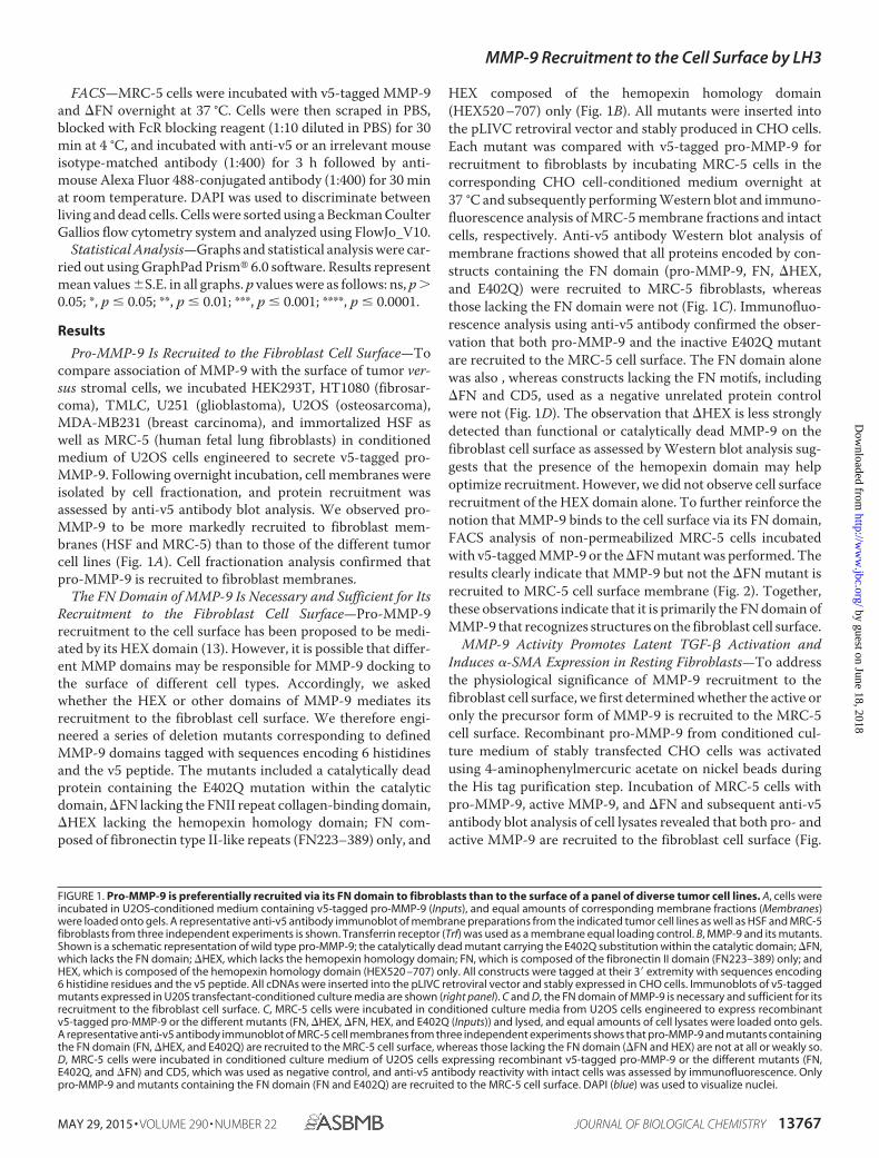
FACS—MRC-5 cells were incubated with v5-tagged MMP-9and �FN overnight at 37 °C. Cells were then scraped in PBS,blocked with FcR blocking reagent (1:10 diluted in PBS) for 30min at 4 °C, and incubated with anti-v5 or an irrelevant mouseisotype-matched antibody (1:400) for 3 h followed by anti-mouse Alexa Fluor 488-conjugated antibody (1:400) for 30 minat room temperature. DAPI was used to discriminate betweenliving and dead cells. Cells were sorted using a Beckman CoulterGallios flow cytometry system and analyzed using FlowJo_V10.
Statistical Analysis—Graphs and statistical analysis were car-ried out using GraphPad Prism� 6.0 software. Results representmean values �S.E. in all graphs. p values were as follows: ns, p �0.05; *, p � 0.05; **, p � 0.01; ***, p � 0.001; ****, p � 0.0001.
Results
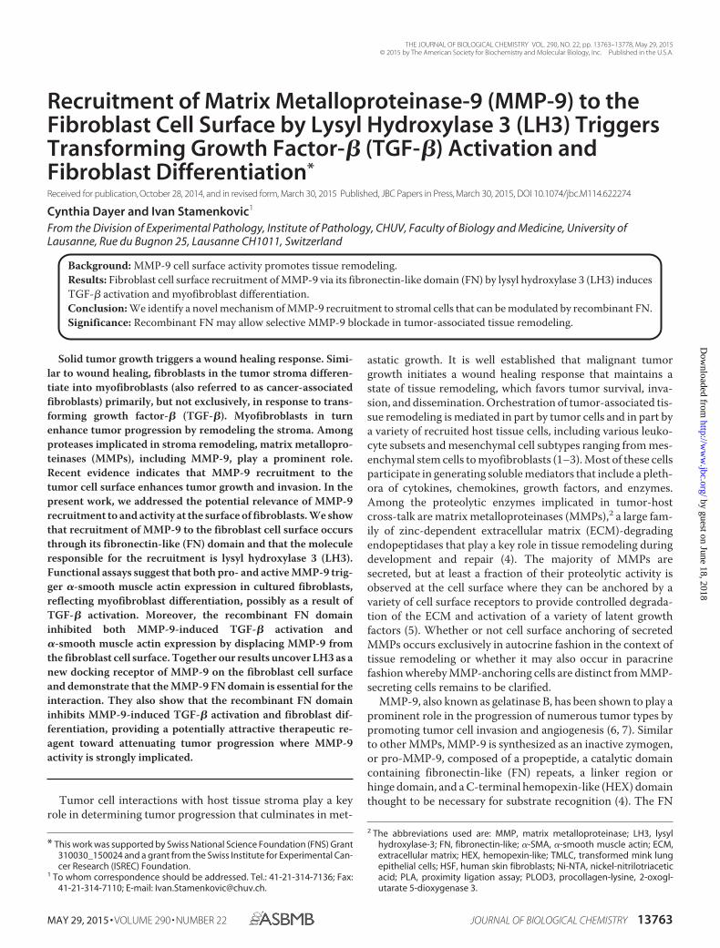
Pro-MMP-9 Is Recruited to the Fibroblast Cell Surface—Tocompare association of MMP-9 with the surface of tumor ver-sus stromal cells, we incubated HEK293T, HT1080 (fibrosar-coma), TMLC, U251 (glioblastoma), U2OS (osteosarcoma),MDA-MB231 (breast carcinoma), and immortalized HSF aswell as MRC-5 (human fetal lung fibroblasts) in conditionedmedium of U2OS cells engineered to secrete v5-tagged pro-MMP-9. Following overnight incubation, cell membranes wereisolated by cell fractionation, and protein recruitment wasassessed by anti-v5 antibody blot analysis. We observed pro-MMP-9 to be more markedly recruited to fibroblast mem-branes (HSF and MRC-5) than to those of the different tumorcell lines (Fig. 1A). Cell fractionation analysis confirmed thatpro-MMP-9 is recruited to fibroblast membranes.
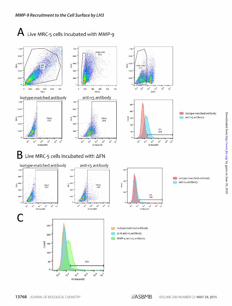
The FN Domain of MMP-9 Is Necessary and Sufficient for ItsRecruitment to the Fibroblast Cell Surface—Pro-MMP-9recruitment to the cell surface has been proposed to be medi-ated by its HEX domain (13). However, it is possible that differ-ent MMP domains may be responsible for MMP-9 docking tothe surface of different cell types. Accordingly, we askedwhether the HEX or other domains of MMP-9 mediates itsrecruitment to the fibroblast cell surface. We therefore engi-neered a series of deletion mutants corresponding to definedMMP-9 domains tagged with sequences encoding 6 histidinesand the v5 peptide. The mutants included a catalytically deadprotein containing the E402Q mutation within the catalyticdomain, �FN lacking the FNII repeat collagen-binding domain,�HEX lacking the hemopexin homology domain; FN com-posed of fibronectin type II-like repeats (FN223–389) only, and
HEX composed of the hemopexin homology domain(HEX520 –707) only (Fig. 1B). All mutants were inserted intothe pLIVC retroviral vector and stably produced in CHO cells.Each mutant was compared with v5-tagged pro-MMP-9 forrecruitment to fibroblasts by incubating MRC-5 cells in thecorresponding CHO cell-conditioned medium overnight at37 °C and subsequently performing Western blot and immuno-fluorescence analysis of MRC-5 membrane fractions and intactcells, respectively. Anti-v5 antibody Western blot analysis ofmembrane fractions showed that all proteins encoded by con-structs containing the FN domain (pro-MMP-9, FN, �HEX,and E402Q) were recruited to MRC-5 fibroblasts, whereasthose lacking the FN domain were not (Fig. 1C). Immunofluo-rescence analysis using anti-v5 antibody confirmed the obser-vation that both pro-MMP-9 and the inactive E402Q mutantare recruited to the MRC-5 cell surface. The FN domain alonewas also , whereas constructs lacking the FN motifs, including�FN and CD5, used as a negative unrelated protein controlwere not (Fig. 1D). The observation that �HEX is less stronglydetected than functional or catalytically dead MMP-9 on thefibroblast cell surface as assessed by Western blot analysis sug-gests that the presence of the hemopexin domain may helpoptimize recruitment. However, we did not observe cell surfacerecruitment of the HEX domain alone. To further reinforce thenotion that MMP-9 binds to the cell surface via its FN domain,FACS analysis of non-permeabilized MRC-5 cells incubatedwith v5-tagged MMP-9 or the �FN mutant was performed. Theresults clearly indicate that MMP-9 but not the �FN mutant isrecruited to MRC-5 cell surface membrane (Fig. 2). Together,these observations indicate that it is primarily the FN domain ofMMP-9 that recognizes structures on the fibroblast cell surface.
MMP-9 Activity Promotes Latent TGF-� Activation andInduces �-SMA Expression in Resting Fibroblasts—To addressthe physiological significance of MMP-9 recruitment to thefibroblast cell surface, we first determined whether the active oronly the precursor form of MMP-9 is recruited to the MRC-5cell surface. Recombinant pro-MMP-9 from conditioned cul-ture medium of stably transfected CHO cells was activatedusing 4-aminophenylmercuric acetate on nickel beads duringthe His tag purification step. Incubation of MRC-5 cells withpro-MMP-9, active MMP-9, and �FN and subsequent anti-v5antibody blot analysis of cell lysates revealed that both pro- andactive MMP-9 are recruited to the fibroblast cell surface (Fig.
FIGURE 1. Pro-MMP-9 is preferentially recruited via its FN domain to fibroblasts than to the surface of a panel of diverse tumor cell lines. A, cells wereincubated in U2OS-conditioned medium containing v5-tagged pro-MMP-9 (Inputs), and equal amounts of corresponding membrane fractions (Membranes)were loaded onto gels. A representative anti-v5 antibody immunoblot of membrane preparations from the indicated tumor cell lines as well as HSF and MRC-5fibroblasts from three independent experiments is shown. Transferrin receptor (Trf) was used as a membrane equal loading control. B, MMP-9 and its mutants.Shown is a schematic representation of wild type pro-MMP-9; the catalytically dead mutant carrying the E402Q substitution within the catalytic domain; �FN,which lacks the FN domain; �HEX, which lacks the hemopexin homology domain; FN, which is composed of the fibronectin II domain (FN223–389) only; andHEX, which is composed of the hemopexin homology domain (HEX520 –707) only. All constructs were tagged at their 3� extremity with sequences encoding6 histidine residues and the v5 peptide. All cDNAs were inserted into the pLIVC retroviral vector and stably expressed in CHO cells. Immunoblots of v5-taggedmutants expressed in U20S transfectant-conditioned culture media are shown (right panel). C and D, the FN domain of MMP-9 is necessary and sufficient for itsrecruitment to the fibroblast cell surface. C, MRC-5 cells were incubated in conditioned culture media from U2OS cells engineered to express recombinantv5-tagged pro-MMP-9 or the different mutants (FN, �HEX, �FN, HEX, and E402Q (Inputs)) and lysed, and equal amounts of cell lysates were loaded onto gels.A representative anti-v5 antibody immunoblot of MRC-5 cell membranes from three independent experiments shows that pro-MMP-9 and mutants containingthe FN domain (FN, �HEX, and E402Q) are recruited to the MRC-5 cell surface, whereas those lacking the FN domain (�FN and HEX) are not at all or weakly so.D, MRC-5 cells were incubated in conditioned culture medium of U2OS cells expressing recombinant v5-tagged pro-MMP-9 or the different mutants (FN,E402Q, and �FN) and CD5, which was used as negative control, and anti-v5 antibody reactivity with intact cells was assessed by immunofluorescence. Onlypro-MMP-9 and mutants containing the FN domain (FN and E402Q) are recruited to the MRC-5 cell surface. DAPI (blue) was used to visualize nuclei.
MMP-9 Recruitment to the Cell Surface by LH3
MAY 29, 2015 • VOLUME 290 • NUMBER 22 JOURNAL OF BIOLOGICAL CHEMISTRY 13767
by guest on June 18, 2018http://w
ww
.jbc.org/D
ownloaded from
MMP-9 Recruitment to the Cell Surface by LH3
13768 JOURNAL OF BIOLOGICAL CHEMISTRY VOLUME 290 • NUMBER 22 • MAY 29, 2015
by guest on June 18, 2018http://w
ww
.jbc.org/D
ownloaded from
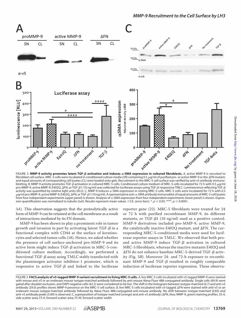
3A). This observation suggests that the proteolytically activeform of MMP-9 can be retained at the cell membrane as a resultof interactions mediated by its FN domain.
MMP-9 has been shown to play a prominent role in tumorgrowth and invasion in part by activating latent TGF-� in afunctional complex with CD44 at the surface of keratino-cytes and selected tumor cells (18). Hence, we asked whetherthe presence of cell surface-anchored pro-MMP-9 and itsactive form might induce TGF-� activation in MRC-5-con-ditioned culture medium. Accordingly, we performed afunctional TGF-� assay using TMLC stably transfected withthe plasminogen activator inhibitor-1 promoter, which isresponsive to active TGF-� and linked to the luciferase
reporter gene (22). MRC-5 fibroblasts were treated for 24or 72 h with purified recombinant MMP-9, its differentmutants, or TGF-�1 (10 ng/ml) used as a positive control.MMP-9 derivatives included pro-MMP-9, active MMP-9,the catalytically inactive E402Q mutant, and �FN. The cor-responding MRC-5-conditioned media were used for lucif-erase reporter assays in TMLC. We observed that both pro-and active MMP-9 induce TGF-� activation in culturedMRC-5 fibroblasts, whereas the inactive mutants E402Q and�FN do not enhance baseline MRC-5-derived TGF-� activ-ity (Fig. 3B). Moreover 24- and 72-h exposure to recombi-nant MMP-9 and TGF-� resulted in roughly comparableinduction of luciferase reporter expression. These observa-
FIGURE 2. FACS analysis of v5-tagged MMP-9 variant recruitment to living MRC-5 cells. A, live MRC-5 cells incubated with v5-tagged MMP-9 were stainedwith mouse anti-v5 or an irrelevant mouse isotype-matched antibody followed by anti-mouse Alexa Fluor 488-conjugated antibody. Single cells (69.9) weregated after doublet exclusion, and DAPI-negative cells (42.5) were considered to be live. The shift in the histogram between isotype-matched (0.7) and anti-v5antibody (29.0) profiles shows MMP-9 presence on the MRC-5 cell surface. B, live MRC-5 cells incubated with v5-tagged �FN were stained with anti-v5 or anirrelevant mouse isotype-matched antibody followed by Alexa Fluor 488-conjugated anti-mouse antibody. No shift between isotype-matched (0.83) andanti-v5 antibody peaks (5.89) is observed. C, superposition of isotype-matched (orange) and anti-v5 antibody (�FN, blue; MMP-9, green) staining profiles. SS-A,side scatter area; FS-A, forward scatter area; FS-W, forward scatter width.
FIGURE 3. MMP-9 activity promotes latent TGF-� activation and induces �-SMA expression in cultured fibroblasts. A, active MMP-9 is recruited tofibroblast cell surface. MRC-5 cells were incubated in conditioned culture media (SN) containing 0.5 �g/ml of purified pro- or active MMP-9 or the �FN mutant,and equal amounts of corresponding cell lysates (CL) were loaded onto gels. Recruitment to the MRC-5 cell surface was verified by anti-v5 antibody immuno-blotting. B, MMP-9 activity promotes TGF-� activation in cultured MRC-5 cells. Conditioned culture medium of MRC-5 cells incubated for 72 h with 0.5 �g/mlpro-MMP-9, active MMP-9, E402Q, �FN, or TGF-�1 (10 ng/ml) was collected for luciferase assays using TGF-�-responsive TMLC. Luminescence reflecting TGF-�activity was quantified by relative light units (RLU). C, MMP-9 induces �-SMA expression in resting MRC-5 cells. MRC-5 cells were incubated for 72 h with 0.5�g/ml pro-MMP-9, active MMP-9, E402Q, �FN, or TGF-�1 (10 ng/ml). A representative anti-�-SMA antibody immunoblot of equal amounts of MRC-5 cell lysatesfrom four independent experiments (upper panel) is shown. Analysis of �-SMA expression from four independent experiments (lower panel) is shown. Expres-sion quantification was normalized to tubulin (tub). Results represent mean values �S.E. (error bars). *, p � 0.05; ****, p � 0.0001.
MMP-9 Recruitment to the Cell Surface by LH3
MAY 29, 2015 • VOLUME 290 • NUMBER 22 JOURNAL OF BIOLOGICAL CHEMISTRY 13769
by guest on June 18, 2018http://w
ww
.jbc.org/D
ownloaded from
tions support the notion that the increase in TGF-� activa-tion is due to catalytic MMP-9 activity.
TGF-� is a potent inducer of fibroblast differentiation intomyofibroblasts. We therefore addressed the possibility thatMMP-9 activity at the surface of MRC-5 cells may facilitatetheir differentiation into myofibroblasts. Differentiation wasassessed by incubating resting MRC-5 cells for 72 h with puri-fied pro-MMP-9, active MMP-9, the catalytically inactiveE402Q mutant, �FN, or TGF-�1 (10 ng/ml) as a positive con-trol. Cells were then lysed, and expression of �-SMA, a reliablemyofibroblast marker that is weakly expressed in MRC-5 cells,was assessed. Incubation with pro- and active MMP-9 led to anincrease in �-SMA expression in cultured MRC-5 fibroblasts(Fig. 3C), whereas incubation with E402Q and �FN mutantsfailed to do so. These observations support the notion thatMMP-9 activity promotes differentiation of fibroblasts intomyofibroblasts.
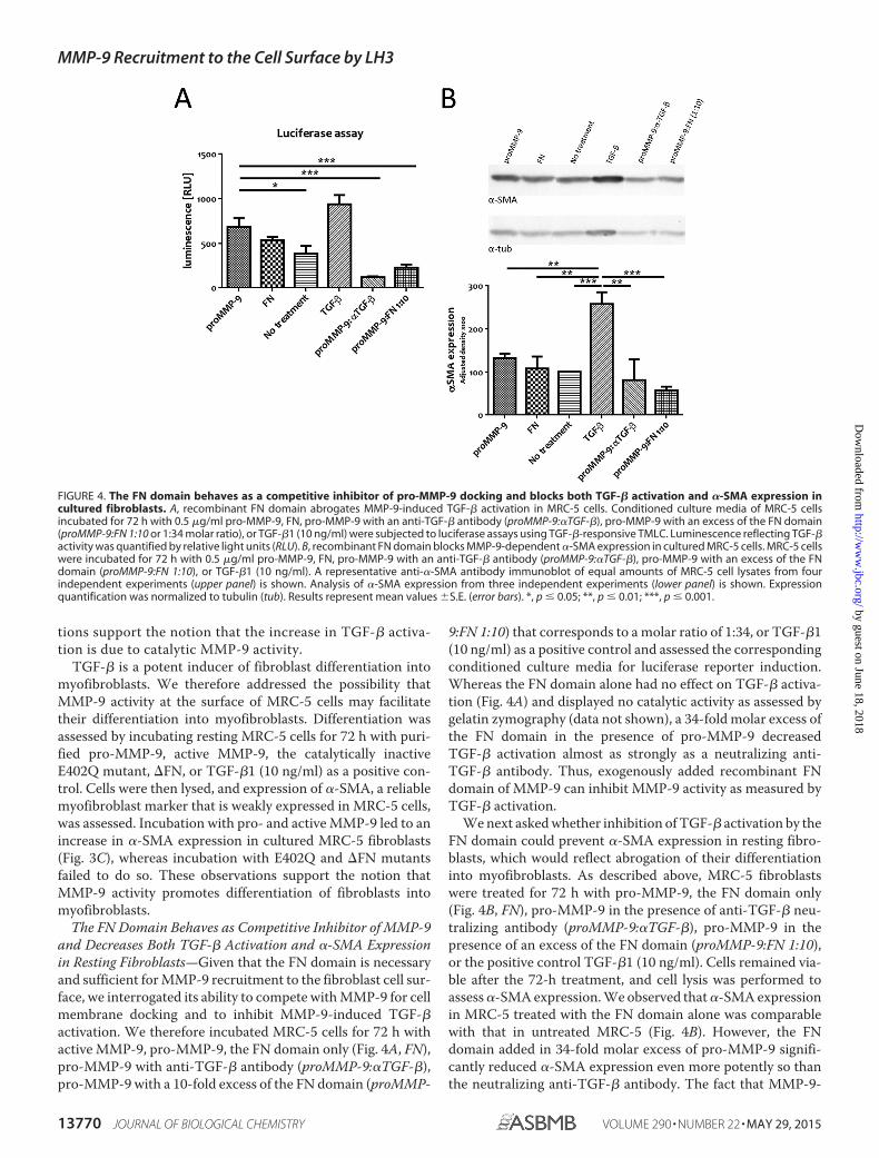
The FN Domain Behaves as Competitive Inhibitor of MMP-9and Decreases Both TGF-� Activation and �-SMA Expressionin Resting Fibroblasts—Given that the FN domain is necessaryand sufficient for MMP-9 recruitment to the fibroblast cell sur-face, we interrogated its ability to compete with MMP-9 for cellmembrane docking and to inhibit MMP-9-induced TGF-�activation. We therefore incubated MRC-5 cells for 72 h withactive MMP-9, pro-MMP-9, the FN domain only (Fig. 4A, FN),pro-MMP-9 with anti-TGF-� antibody (proMMP-9:�TGF-�),pro-MMP-9 with a 10-fold excess of the FN domain (proMMP-
9:FN 1:10) that corresponds to a molar ratio of 1:34, or TGF-�1(10 ng/ml) as a positive control and assessed the correspondingconditioned culture media for luciferase reporter induction.Whereas the FN domain alone had no effect on TGF-� activa-tion (Fig. 4A) and displayed no catalytic activity as assessed bygelatin zymography (data not shown), a 34-fold molar excess ofthe FN domain in the presence of pro-MMP-9 decreasedTGF-� activation almost as strongly as a neutralizing anti-TGF-� antibody. Thus, exogenously added recombinant FNdomain of MMP-9 can inhibit MMP-9 activity as measured byTGF-� activation.
We next asked whether inhibition of TGF-� activation by theFN domain could prevent �-SMA expression in resting fibro-blasts, which would reflect abrogation of their differentiationinto myofibroblasts. As described above, MRC-5 fibroblastswere treated for 72 h with pro-MMP-9, the FN domain only(Fig. 4B, FN), pro-MMP-9 in the presence of anti-TGF-� neu-tralizing antibody (proMMP-9:�TGF-�), pro-MMP-9 in thepresence of an excess of the FN domain (proMMP-9:FN 1:10),or the positive control TGF-�1 (10 ng/ml). Cells remained via-ble after the 72-h treatment, and cell lysis was performed toassess �-SMA expression. We observed that �-SMA expressionin MRC-5 treated with the FN domain alone was comparablewith that in untreated MRC-5 (Fig. 4B). However, the FNdomain added in 34-fold molar excess of pro-MMP-9 signifi-cantly reduced �-SMA expression even more potently so thanthe neutralizing anti-TGF-� antibody. The fact that MMP-9-
FIGURE 4. The FN domain behaves as a competitive inhibitor of pro-MMP-9 docking and blocks both TGF-� activation and �-SMA expression incultured fibroblasts. A, recombinant FN domain abrogates MMP-9-induced TGF-� activation in MRC-5 cells. Conditioned culture media of MRC-5 cellsincubated for 72 h with 0.5 �g/ml pro-MMP-9, FN, pro-MMP-9 with an anti-TGF-� antibody (proMMP-9:�TGF-�), pro-MMP-9 with an excess of the FN domain(proMMP-9:FN 1:10 or 1:34 molar ratio), or TGF-�1 (10 ng/ml) were subjected to luciferase assays using TGF-�-responsive TMLC. Luminescence reflecting TGF-�activity was quantified by relative light units (RLU). B, recombinant FN domain blocks MMP-9-dependent �-SMA expression in cultured MRC-5 cells. MRC-5 cellswere incubated for 72 h with 0.5 �g/ml pro-MMP-9, FN, pro-MMP-9 with an anti-TGF-� antibody (proMMP-9:�TGF-�), pro-MMP-9 with an excess of the FNdomain (proMMP-9:FN 1:10), or TGF-�1 (10 ng/ml). A representative anti-�-SMA antibody immunoblot of equal amounts of MRC-5 cell lysates from fourindependent experiments (upper panel) is shown. Analysis of �-SMA expression from three independent experiments (lower panel) is shown. Expressionquantification was normalized to tubulin (tub). Results represent mean values �S.E. (error bars). *, p � 0.05; **, p � 0.01; ***, p � 0.001.
MMP-9 Recruitment to the Cell Surface by LH3
13770 JOURNAL OF BIOLOGICAL CHEMISTRY VOLUME 290 • NUMBER 22 • MAY 29, 2015
by guest on June 18, 2018http://w
ww
.jbc.org/D
ownloaded from
MMP-9 Recruitment to the Cell Surface by LH3
MAY 29, 2015 • VOLUME 290 • NUMBER 22 JOURNAL OF BIOLOGICAL CHEMISTRY 13771
by guest on June 18, 2018http://w
ww
.jbc.org/D
ownloaded from
induced �-SMA expression can be inhibited by neutralizinganti-TGF-� antibody indicates that MMP-9-mediated differ-entiation of MRC-5 into �-SMA-expressing myofibroblastsunder our culture conditions occurs in large part through theTGF-� pathway. Moreover, abrogation by excess recombinantFN domain of the ability of MMP-9 to induce �-SMA expres-sion in cultured fibroblasts suggests that the FN domain caninhibit MMP-9 activity at the fibroblast cell surface.
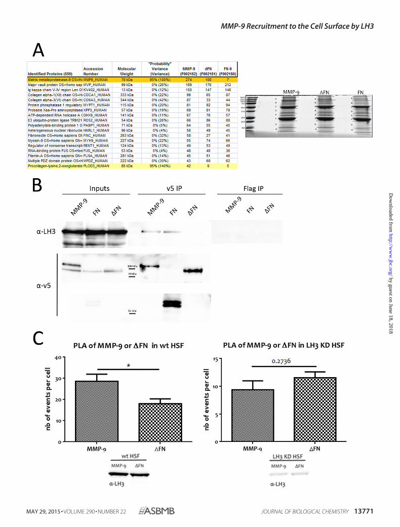
LH3 Provides the Docking Mechanism for MMP-9 Cell Sur-face Association via the FN Domain—CD44 has been shown tobe an MMP-9 docking molecule at the surface of TA3 mousemammary carcinoma, melanoma cells, and normal keratino-cytes (17). However, CD44 does not appear to be necessary forMMP-9 recruitment to the fibroblast membrane (data notshown), and recruitment therefore occurs by a CD44-indepen-dent mechanism. To identify candidate MMP-9 docking mole-cules on the fibroblast cell surface, we performed mass spec-trometry analysis of an anti-v5 antibody pulldown of MMP-9,FN, and �FN cross-linked to MRC-5 cells. MRC-5 cells wereincubated with MMP-9, FN, and �FN proteins and labeled withSulfo-SBED Biotin Label Transfer Reagent after which theputative interactions were cross-linked by UV light at 365 nmfor 8 min. Anti-v5 antibody was then used for immunoprecipi-tation from the corresponding cell lysates (Fig. 5 A), and theimmunoprecipitates were subjected to shotgun mass spec-trometry. Analysis of the pulldown revealed PLOD3_HUMAN,also known as LH3, to be a specific candidate binding partner ofMMP-9 and the FN domain (Fig. 5A).
To verify the interaction between MMP-9 and LH3, we incu-bated MRC-5 cells with recombinant v5-tagged and Sulfo-SBED-labeled MMP-9, FN, or �FN for 4 h at 37 °C, cross-linkedthe interaction, and collected cell lysates to perform anti-v5 andanti-FLAG control antibody immunoprecipitation. We usedanti-endogenous LH3 antibody to reveal the interaction. Byimmunoblot analysis, we could clearly demonstrate that bothv5-tagged MMP-9 and the FN domain can immunoprecipitateendogenous LH3, whereas �FN cannot (Fig. 5B). Thus, MMP-9forms a complex with LH3 via its FN domain.
To further explore this interaction in vivo without resortingto cross-linking, we performed PLAs. HSF were treated withv5-tagged MMP-9 or �FN overnight at 37 °C and the followingday incubated with mouse anti-v5 and rabbit anti-LH3 anti-body prior to paraformaldehyde fixation and subjection toproximity ligation (see “Experimental Procedures”). We com-pared v5-tagged MMP-9 and �FN interaction with endogenousLH3 by quantifying the number of events per cell (reflected byfluorescence signals) in WT HSF or in HSF depleted of LH3.We observed a significantly higher number of events in HSFincubated with MMP-9 than in HSF treated with �FN (Fig. 5C,left panel), confirming the requirement of the FN domain for
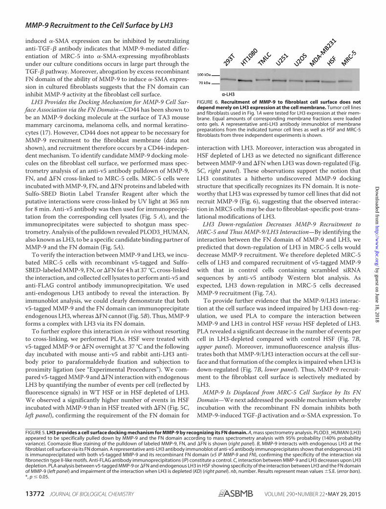
interaction with LH3. Moreover, interaction was abrogated inHSF depleted of LH3 as we detected no significant differencebetween MMP-9 and �FN when LH3 was down-regulated (Fig.5C, right panel). These observations support the notion thatLH3 constitutes a hitherto undiscovered MMP-9 dockingstructure that specifically recognizes its FN domain. It is note-worthy that LH3 was expressed by tumor cell lines that did notrecruit MMP-9 (Fig. 6), suggesting that the observed interac-tion in MRC5 cells may be due to fibroblast-specific post-trans-lational modifications of LH3.
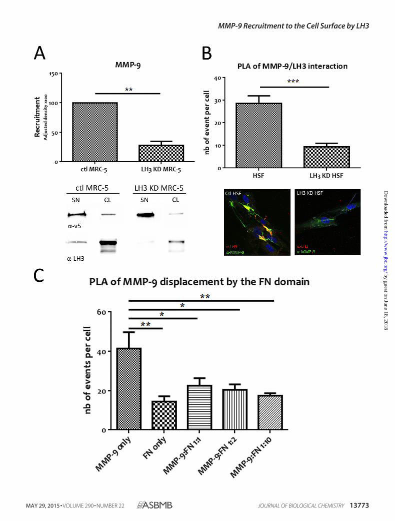
LH3 Down-regulation Decreases MMP-9 Recruitment toMRC-5 and Thus MMP-9/LH3 Interaction—By identifying theinteraction between the FN domain of MMP-9 and LH3, wepredicted that down-regulation of LH3 in MRC-5 cells woulddecrease MMP-9 recruitment. We therefore depleted MRC-5cells of LH3 and compared recruitment of v5-tagged MMP-9with that in control cells containing scrambled siRNAsequences by anti-v5 antibody Western blot analysis. Asexpected, LH3 down-regulation in MRC-5 cells decreasedMMP-9 recruitment (Fig. 7A).
To provide further evidence that the MMP-9/LH3 interac-tion at the cell surface was indeed impaired by LH3 down-reg-ulation, we used PLA to compare the interaction betweenMMP-9 and LH3 in control HSF versus HSF depleted of LH3.PLA revealed a significant decrease in the number of events percell in LH3-depleted compared with control HSF (Fig. 7B,upper panel). Moreover, immunofluorescence analysis illus-trates both that MMP-9/LH3 interaction occurs at the cell sur-face and that formation of the complex is impaired when LH3 isdown-regulated (Fig. 7B, lower panel). Thus, MMP-9 recruit-ment to the fibroblast cell surface is selectively mediated byLH3.
MMP-9 Is Displaced from MRC-5 Cell Surface by Its FNDomain—We next addressed the possible mechanism wherebyincubation with the recombinant FN domain inhibits bothMMP-9-induced TGF-� activation and �-SMA expression. To
FIGURE 5. LH3 provides a cell surface docking mechanism for MMP-9 by recognizing its FN domain. A, mass spectrometry analysis. PLOD3_HUMAN (LH3)appeared to be specifically pulled down by MMP-9 and the FN domain according to mass spectrometry analysis with 95% probability (140% probabilityvariance). Coomassie Blue staining of the pulldown of labeled MMP-9, FN, and �FN is shown (right panel). B, MMP-9 interacts with endogenous LH3 at thefibroblast cell surface via its FN domain. A representative anti-LH3 antibody immunoblot of anti-v5 antibody immunoprecipitates shows that endogenous LH3is immunoprecipitated with both v5-tagged MMP-9 and its recombinant FN domain (v5 IP MMP-9 and FN), confirming the specificity of the interaction viafibronectin type II-like motifs. Anti-FLAG antibody immunoprecipitations (IP) constitute a control. C, interaction between MMP-9 and LH3 decreases upon LH3depletion. PLA analysis between v5-tagged MMP-9 or �FN and endogenous LH3 in HSF showing specificity of the interaction between LH3 and the FN domainof MMP-9 (left panel) and impairment of the interaction when LH3 is depleted (KD) (right panel). nb, number. Results represent mean values �S.E. (error bars).*, p � 0.05.
FIGURE 6. Recruitment of MMP-9 to fibroblast cell surface does notdepend merely on LH3 expression at the cell membrane. Tumor cell linesand fibroblasts used in Fig. 1A were tested for LH3 expression at their mem-brane. Equal amounts of corresponding membrane fractions were loadedonto gels. A representative anti-LH3 antibody immunoblot of membranepreparations from the indicated tumor cell lines as well as HSF and MRC-5fibroblasts from three independent experiments is shown.
MMP-9 Recruitment to the Cell Surface by LH3
13772 JOURNAL OF BIOLOGICAL CHEMISTRY VOLUME 290 • NUMBER 22 • MAY 29, 2015
by guest on June 18, 2018http://w
ww
.jbc.org/D
ownloaded from
MMP-9 Recruitment to the Cell Surface by LH3
MAY 29, 2015 • VOLUME 290 • NUMBER 22 JOURNAL OF BIOLOGICAL CHEMISTRY 13773
by guest on June 18, 2018http://w
ww
.jbc.org/D
ownloaded from
do so, we asked whether the FN domain alone might competewith MMP-9 for docking to the fibroblast cell surface andimpair MMP-9�LH3 complex formation by displacing MMP-9from the cell membrane. MRC-5 fibroblasts were incubatedwith v5-tagged MMP-9 in the presence of increasing concen-trations of the FN domain (MMP-9:FN ratios of 1:1, 1:2, and1:10 or molar ratios of 1:3.4, 1:6.8, and 1:34) after which MMP-9/LH3 interaction was assessed by PLA using mouse anti-MMP-9 antibody, which does not recognize the FN domain,and rabbit anti-LH3 antibody. We observed an increase in thenumber of fluorescence signals in the cells treated with MMP-9only compared with cells treated with the FN domain only,confirming that the MMP-9 antibody does not recognize theFN domain (Fig. 7C). Moreover, we noted a strong decrease ofthe MMP-9/LH3 interaction in the presence of a 3.4 molarexcess of FN. Thus, recombinant FN domain prevents LH3-de-pendent MMP-9 anchoring to the MRC-5 cell surface and pro-vides a mechanism of inhibition of MMP-9-mediated TGF-�activation and fibroblast differentiation.
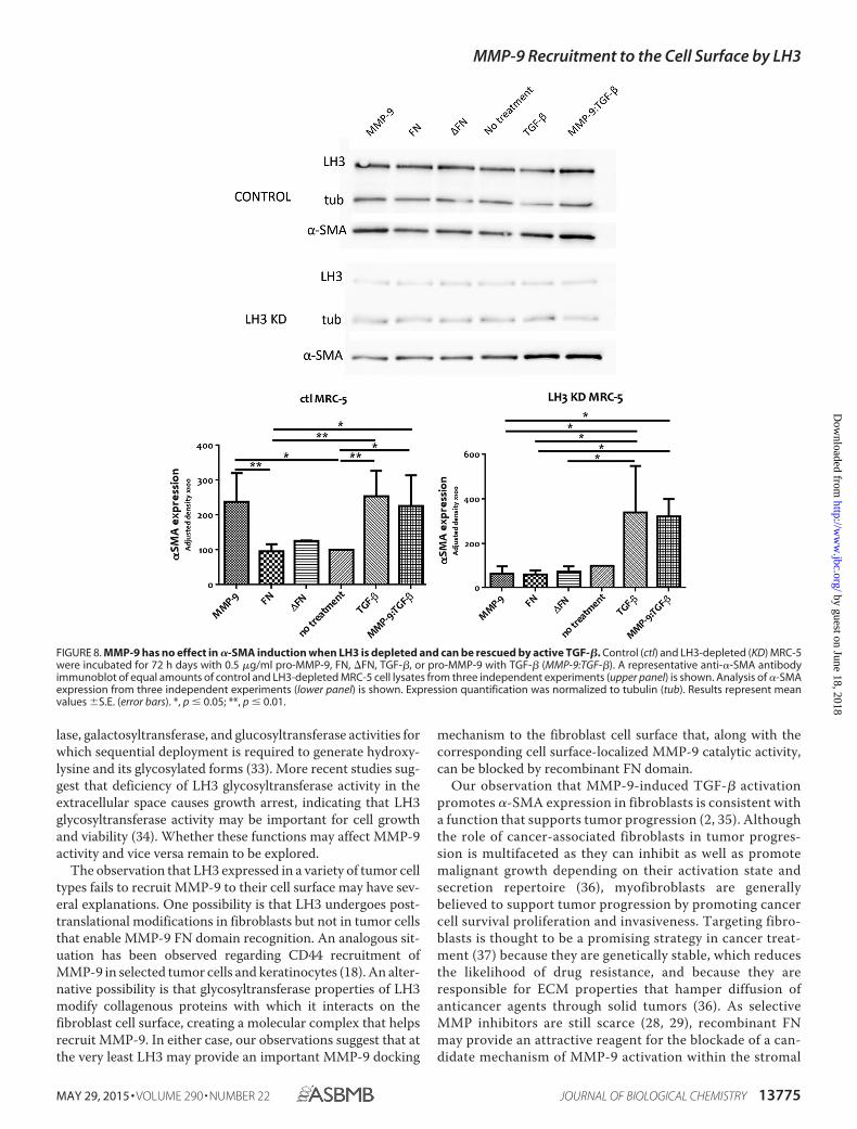
Finally, we assessed �-SMA induction in MRC5 cellsdepleted of LH3 in response to recombinant MMP-9. Expres-sion of �-SMA in these cells was not enhanced by addition ofrecombinant MMP-9 to the cell culture medium (Fig. 8). How-ever, induction of �-SMA was rescued by addition of activeTGF-� in the presence or absence of MMP-9. These observa-tions support the notion that recruitment by LH3 provides amechanism for deployment of MMP-9 catalytic activity at thefibroblast cell surface that promotes TGF-� activation and thecorresponding enhancement of �-SMA expression.
Discussion
MMP-9 can be recruited to the surface of diverse cell typeswhere it may play an important role in regulating growth factoractivation, receptor processing, and pericellular matrix turn-over, all of which are highly relevant to tissue remodeling inboth physiological and tumor-associated contexts. Thus far,the hyaluronan receptor CD44 has been shown to provide amajor docking site for MMP-9 on the surface of a variety oftumor cells and primary keratinocytes (17), although associa-tion with other cell surface receptors, including LRP-1, LRP-2,and membrane-anchored glycoprotein RECK, has beenreported as well (23–25). The MMP-9 structure responsible forinteraction with CD44 resides within the HEX domain (26),leaving open the possibility that other MMP-9 structures maybe implicated in cell surface docking to different receptors. Wehave shown here that MMP-9 can be recruited to the fibroblastcell surface via its fibronectin type II-like motifs by LH3 whereit activates latent TGF-� and induces myofibroblast differenti-ation as reflected by induction of �-SMA expression. LH3-re-
cruited pro-MMP-9 may become activated and remain at thecell surface, cleaving latent TGF-� in the pericellular matrix (asobserved in certain tumor cells (17)). Alternatively, pro-MMP-9 may be recruited to the cell surface for proteolytic acti-vation and then released into the ECM to liberate active TGF-�from its latency complex.
Among mammalian MMPs, only MMP-2 and MMP-9 sharefibronectin type II-like repeats that form a collagen-bindingdomain located within the catalytic region in the vicinity of theactive site. Fibronectin type II-like motifs are widespreadamong extracellular proteins and engage in interactions withcollagens and gelatin (4, 27). MMP FN type II repeats may haveanalogous functions, mediating interactions with diverse dock-ing structures and regulating MMP activation and correspond-ing proteolytic activity. They may thereby provide a means forselective MMP-9 (and possibly MMP-2) targeting in a variety ofpathological conditions.
Selective targeting of individual MMPs has been a major hur-dle toward therapeutic strategies aimed at blocking MMP-de-pendent tumor progression as most compounds with potentinhibitory properties are non-selective and tend to block all ornearly all MMP activity with adverse consequences (28, 29).Nevertheless, the continued search for selective means to blocksingle MMPs or subsets thereof has identified potentiallypromising avenues as illustrated by chemical compounds thattarget the HEX domain of MMP-9 and that inhibit tumorcell migration and proliferation by abrogating MMP-9homodimerization (30, 31). An alternative approach may be totarget structures that are unique to defined MMPs providedthey are shown to play a functionally relevant role in determin-ing MMP localization and activity. The FN domain appearsparticularly attractive in light of our present observations as, inaddition to constituting part of only two MMPs, its delivery inrecombinant form may provide selective inhibition of the effectof only this subset of MMPs on fibroblast functions that arehighly relevant to tumor progression. Enhanced selectivity ofMMP inhibitors has already been achieved by taking advantageof differences in secondary substrate binding sites or exositeswithin the MMP family (32). Thus, a triple helical peptide thatincorporates an FN type II-like motif-binding sequence selec-tively inhibits MMP-9 type V collagen-specific activity. Simi-larly, FN type II motif-mediated MMP-9 interaction withLH3 provides a targetable event with potentially beneficialconsequences.
Lysyl hydroxylase 3 is a multifunctional protein that localizesto the endoplasmic reticulum but is also secreted into the extra-cellular space and is associated with collagenous proteins on thecell surface (21). Its principal function resides in lysyl hydroxy-
FIGURE 7. Depletion of LH3 decreases MMP-9 cell surface recruitment as does the recombinant FN domain, which displaces MMP-9 from the fibroblastmembrane. A, LH3 down-regulation in MRC-5 decreases MMP-9 cell surface recruitment. Equal amounts of cell lysates (CL) from control (ctl) and LH3-depleted(KD) MRC-5 incubated with v5-tagged MMP-9 (SN) were loaded onto gels. Recruitment was quantified by densitometry. A representative anti-v5 antibodyimmunoblot of equal amounts of MRC-5 cell lysates from three independent experiments (lower panel) and the corresponding anti-LH3 antibody immunoblotare shown. B, PLA analysis of WT or LH3-depleted HSF treated with v5-tagged MMP-9. The histogram shows interaction between v5-tagged MMP-9 andendogenous LH3 that is decreased when LH3 is depleted. Lower panels show immunofluorescence images of MMP-9 recruitment (green) to membranes of HSFwith an overlap between MMP-9 and LH3 (yellow) that is decreased by LH3 depletion. C, the FN domain prevents MMP-9/LH3 interaction in a dose-dependentmanner. PLA analysis of MRC-5 cells treated with MMP-9 only, FN only, or MMP-9 with increased concentrations of the FN domain (1:1, 1:2, and 1:10 corre-sponding to 1:3.4, 1:6.8, and 1:34 molar ratios). The histogram shows interaction between MMP-9 and endogenous LH3 that decreases as the concentration ofFN increases. nb, number. Results represent mean values �S.E. (error bars). *, p � 0.05; **, p � 0.01; ***, p � 0.001.
MMP-9 Recruitment to the Cell Surface by LH3
13774 JOURNAL OF BIOLOGICAL CHEMISTRY VOLUME 290 • NUMBER 22 • MAY 29, 2015
by guest on June 18, 2018http://w
ww
.jbc.org/D
ownloaded from
lase, galactosyltransferase, and glucosyltransferase activities forwhich sequential deployment is required to generate hydroxy-lysine and its glycosylated forms (33). More recent studies sug-gest that deficiency of LH3 glycosyltransferase activity in theextracellular space causes growth arrest, indicating that LH3glycosyltransferase activity may be important for cell growthand viability (34). Whether these functions may affect MMP-9activity and vice versa remain to be explored.
The observation that LH3 expressed in a variety of tumor celltypes fails to recruit MMP-9 to their cell surface may have sev-eral explanations. One possibility is that LH3 undergoes post-translational modifications in fibroblasts but not in tumor cellsthat enable MMP-9 FN domain recognition. An analogous sit-uation has been observed regarding CD44 recruitment ofMMP-9 in selected tumor cells and keratinocytes (18). An alter-native possibility is that glycosyltransferase properties of LH3modify collagenous proteins with which it interacts on thefibroblast cell surface, creating a molecular complex that helpsrecruit MMP-9. In either case, our observations suggest that atthe very least LH3 may provide an important MMP-9 docking
mechanism to the fibroblast cell surface that, along with thecorresponding cell surface-localized MMP-9 catalytic activity,can be blocked by recombinant FN domain.
Our observation that MMP-9-induced TGF-� activationpromotes �-SMA expression in fibroblasts is consistent witha function that supports tumor progression (2, 35). Althoughthe role of cancer-associated fibroblasts in tumor progres-sion is multifaceted as they can inhibit as well as promotemalignant growth depending on their activation state andsecretion repertoire (36), myofibroblasts are generallybelieved to support tumor progression by promoting cancercell survival proliferation and invasiveness. Targeting fibro-blasts is thought to be a promising strategy in cancer treat-ment (37) because they are genetically stable, which reducesthe likelihood of drug resistance, and because they areresponsible for ECM properties that hamper diffusion ofanticancer agents through solid tumors (36). As selectiveMMP inhibitors are still scarce (28, 29), recombinant FNmay provide an attractive reagent for the blockade of a can-didate mechanism of MMP-9 activation within the stromal
FIGURE 8. MMP-9 has no effect in �-SMA induction when LH3 is depleted and can be rescued by active TGF-�. Control (ctl) and LH3-depleted (KD) MRC-5were incubated for 72 h days with 0.5 �g/ml pro-MMP-9, FN, �FN, TGF-�, or pro-MMP-9 with TGF-� (MMP-9:TGF-�). A representative anti-�-SMA antibodyimmunoblot of equal amounts of control and LH3-depleted MRC-5 cell lysates from three independent experiments (upper panel) is shown. Analysis of �-SMAexpression from three independent experiments (lower panel) is shown. Expression quantification was normalized to tubulin (tub). Results represent meanvalues �S.E. (error bars). *, p � 0.05; **, p � 0.01.
MMP-9 Recruitment to the Cell Surface by LH3
MAY 29, 2015 • VOLUME 290 • NUMBER 22 JOURNAL OF BIOLOGICAL CHEMISTRY 13775
by guest on June 18, 2018http://w
ww
.jbc.org/D
ownloaded from
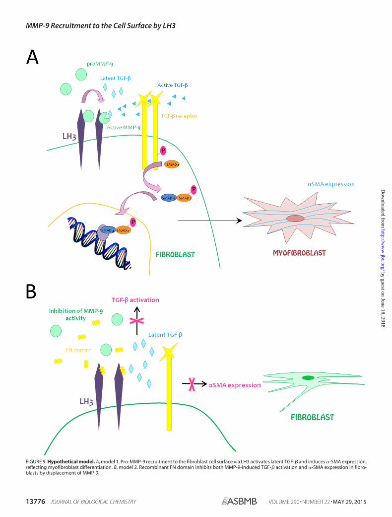
FIGURE 9. Hypothetical model. A, model 1. Pro-MMP-9 recruitment to the fibroblast cell surface via LH3 activates latent TGF-� and induces �-SMA expression,reflecting myofibroblast differentiation. B, model 2. Recombinant FN domain inhibits both MMP-9-induced TGF-� activation and �-SMA expression in fibro-blasts by displacement of MMP-9.
MMP-9 Recruitment to the Cell Surface by LH3
13776 JOURNAL OF BIOLOGICAL CHEMISTRY VOLUME 290 • NUMBER 22 • MAY 29, 2015
by guest on June 18, 2018http://w
ww
.jbc.org/D
ownloaded from
compartment as well as a structural basis for the design ofsmaller effective MMP-9 inhibitors.
We report a hitherto undiscovered mechanism of MMP-9recruitment to the surface of fibroblasts. Cell surface activity ofMMP-9 has been shown to be important for TGF-� activationwhether on the fibroblast surface or in the immediate pericel-lular fibroblast microenvironment and may play a critical rolein fibroblast differentiation into myofibroblasts, providing amechanism that underlies the constitution of at least a subsetof cancer-associated fibroblasts (Fig. 9A). Recombinant FNdomain blocks MMP-9-dependent, TGF-�-mediated myofi-broblast differentiation and thereby abrogates a potentiallyimportant fueling mechanism of tumor progression (Fig. 9B).
Acknowledgments—We thank Manfredo Quadroni from the ProteinAnalysis Facility of University of Lausanne for Mass SpectrometryAnalysis and all the members of the laboratory for support and help-ful discussion. We particularly thank Nils Degrauwe, Giulia Fregni,Carlo Fusco, Marie-Aude Le Bitoux, Patricia Martin, and AnnePlanche Roduit for precious help.
References1. Pietras, K., and Ostman, A. (2010) Hallmarks of cancer: interactions with
the tumor stroma. Exp. Cell Res. 316, 1324 –13312. Mueller, M. M., and Fusenig, N. E. (2004) Friends or foes— bipolar effects
of the tumour stroma in cancer. Nat. Rev. Cancer 4, 839 – 8493. Bhowmick, N. A., Neilson, E. G., and Moses, H. L. (2004) Stromal fibro-
blasts in cancer initiation and progression. Nature 432, 332–3374. Stamenkovic, I. (2000) Matrix metalloproteinases in tumor invasion and
metastasis. Sem. Cancer Biol. 10, 415– 4335. Stamenkovic, I. (2003) Extracellular matrix remodelling: the role of matrix
metalloproteinases. J. Pathol. 200, 448 – 4646. Coussens, L. M., Tinkle, C. L., Hanahan, D., and Werb, Z. (2000) MMP-9
supplied by bone marrow-derived cells contributes to skin carcinogenesis.Cell 103, 481– 490
7. Bergers, G., Brekken, R., McMahon, G., Vu, T. H., Itoh, T., Tamaki, K.,Tanzawa, K., Thorpe, P., Itohara, S., Werb, Z., and Hanahan, D. (2000)Matrix metalloproteinase-9 triggers the angiogenic switch during carci-nogenesis. Nat. Cell Biol. 2, 737–744
8. Xu, X., Chen, Z., Wang, Y., Yamada, Y., and Steffensen, B. (2005) Func-tional basis for the overlap in ligand interactions and substrate specificitiesof matrix metalloproteinases-9 and -2. Biochem. J. 392, 127–134
9. Collier, I. E., Krasnov, P. A., Strongin, A. Y., Birkedal-Hansen, H., andGoldberg, G. I. (1992) Alanine scanning mutagenesis and functional anal-ysis of the fibronectin-like collagen-binding domain from human 92-kDatype IV collagenase. J. Biol. Chem. 267, 6776 – 6781
10. Steffensen, B., Wallon, U. M., and Overall, C. M. (1995) Extracellular ma-trix binding properties of recombinant fibronectin type II-like modules ofhuman 72-kDa gelatinase/type IV collagenase. High affinity binding tonative type I collagen but not native type IV collagen. J. Biol. Chem. 270,11555–11566
11. Overall, C. M. (2002) Molecular determinants of metalloproteinase sub-strate specificity: matrix metalloproteinase substrate binding domains,modules, and exosites. Mol. Biotechnol. 22, 51– 86
12. Roomi, M. W., Monterrey, J. C., Kalinovsky, T., Rath, M., and Niedzwiecki,A. (2009) Patterns of MMP-2 and MMP-9 expression in human cancercell lines. Oncol. Rep. 21, 1323–1333
13. Malla, N., Sjøli, S., Winberg, J. O., Hadler-Olsen, E., and Uhlin-Hansen, L.(2008) Biological and pathobiological functions of gelatinase dimers andcomplexes. Connect. Tissue Res. 49, 180 –184
14. Deryugina, E. I., and Quigley, J. P. (2006) Matrix metalloproteinases andtumor metastasis. Cancer Metastasis Rev. 25, 9 –34
15. Fridman, R., Toth, M., Chvyrkova, I., Meroueh, S. O., and Mobashery, S.(2003) Cell surface association of matrix metalloproteinase-9 (gelatinase
B). Cancer Metastasis Rev. 22, 153–16616. Toth, M., Chvyrkova, I., Bernardo, M. M., Hernandez-Barrantes, S., and
Fridman, R. (2003) Pro-MMP-9 activation by the MT1-MMP/MMP-2axis and MMP-3: role of TIMP-2 and plasma membranes. Biochem. Bio-phys. Res. Commun. 308, 386 –395
17. Yu, Q., and Stamenkovic, I. (1999) Localization of matrix metalloprotei-nase 9 to the cell surface provides a mechanism for CD44-mediated tumorinvasion. Genes Dev. 13, 35– 48
18. Yu, Q., and Stamenkovic, I. (2000) Cell surface-localized matrix metallo-proteinase-9 proteolytically activates TGF-� and promotes tumor inva-sion and angiogenesis. Genes Dev. 14, 163–176
19. Mira, E., Lacalle, R. A., Buesa, J. M., de Buitrago, G. G., Jiménez-Baranda,S., Gómez-Moutón, C., Martínez-A, C., and Mañes, S. (2004) SecretedMMP9 promotes angiogenesis more efficiently than constitutive activeMMP9 bound to the tumor cell surface. J. Cell Sci. 117, 1847–1857
20. Heikkinen, J., Risteli, M., Wang, C., Latvala, J., Rossi, M., Valtavaara, M.,and Myllylä, R. (2000) Lysyl hydroxylase 3 is a multifunctional proteinpossessing collagen glucosyltransferase activity. J. Biol. Chem. 275,36158 –36163
21. Salo, A. M., Wang, C., Sipilä, L., Sormunen, R., Vapola, M., Kervinen, P.,Ruotsalainen, H., Heikkinen, J., and Myllylä, R. (2006) Lysyl hydroxylase 3(LH3) modifies proteins in the extracellular space, a novel mechanism formatrix remodeling. J. Cell. Physiol. 207, 644 – 653
22. Abe, M., Harpel, J. G., Metz, C. N., Nunes, I., Loskutoff, D. J., and Rifkin,D. B. (1994) An assay for transforming growth factor-� using cells trans-fected with a plasminogen activator inhibitor-1 promoter-luciferase con-struct. Anal. Biochem. 216, 276 –284
23. Hahn-Dantona, E., Ruiz, J. F., Bornstein, P., and Strickland, D. K. (2001)The low density lipoprotein receptor-related protein modulates levels ofmatrix metalloproteinase 9 (MMP-9) by mediating its cellular catabolism.J. Biol. Chem. 276, 15498 –15503
24. Van den Steen, P. E., Van Aelst, I., Hvidberg, V., Piccard, H., Fiten, P.,Jacobsen, C., Moestrup, S. K., Fry, S., Royle, L., Wormald, M. R., Wallis, R.,Rudd, P. M., Dwek, R. A., and Opdenakker, G. (2006) The hemopexin andO-glycosylated domains tune gelatinase B/MMP-9 bioavailability via in-hibition and binding to cargo receptors. J. Biol. Chem. 281, 18626 –18637
25. Takahashi, C., Sheng, Z., Horan, T. P., Kitayama, H., Maki, M., Hitomi, K.,Kitaura, Y., Takai, S., Sasahara, R. M., Horimoto, A., Ikawa, Y., Ratzkin,B. J., Arakawa, T., and Noda, M. (1998) Regulation of matrix metallopro-teinase-9 and inhibition of tumor invasion by the membrane-anchoredglycoprotein RECK. Proc. Natl. Acad. Sci. U.S.A. 95, 13221–13226
26. Redondo-Muñoz, J., Ugarte-Berzal, E., García-Marco, J. A., del Cerro,M. H., Van den Steen, P. E., Opdenakker, G., Terol, M. J., and García-Pardo, A. (2008) �4�1 integrin and 190-kDa CD44v constitute a cell sur-face docking complex for gelatinase B/MMP-9 in chronic leukemic butnot in normal B cells. Blood 112, 169 –178
27. Behrendt, N. (2004) The urokinase receptor (uPAR) and the uPAR-asso-ciated protein (uPARAP/Endo180): membrane proteins engaged in ma-trix turnover during tissue remodeling. Biol. Chem. 385, 103–136
28. Brinckerhoff, C. E., and Matrisian, L. M. (2002) Matrix metalloprotei-nases: a tail of a frog that became a prince. Nat. Rev. Mol. Cell Biol. 3,207–214
29. Coussens, L. M., Fingleton, B., and Matrisian, L. M. (2002) Matrix metal-loproteinase inhibitors and cancer: trials and tribulations. Science 295,2387–2392
30. Dufour, A., Sampson, N. S., Li, J., Kuscu, C., Rizzo, R. C., Deleon, J. L., Zhi,J., Jaber, N., Liu, E., Zucker, S., and Cao, J. (2011) Small-molecule antican-cer compounds selectively target the hemopexin domain of matrix met-alloproteinase-9. Cancer Res. 71, 4977– 4988
31. Dufour, A., Zucker, S., Sampson, N. S., Kuscu, C., and Cao, J. (2010) Role ofmatrix metalloproteinase-9 dimers in cell migration: design of inhibitorypeptides. J. Biol. Chem. 285, 35944 –35956
32. Lauer-Fields, J. L., Whitehead, J. K., Li, S., Hammer, R. P., Brew, K., andFields, G. B. (2008) Selective modulation of matrix metalloproteinase 9(MMP-9) functions via exosite inhibition. J. Biol. Chem. 283,20087–20095
33. Myllylä, R., Wang, C., Heikkinen, J., Juffer, A., Lampela, O., Risteli, M.,Ruotsalainen, H., Salo, A., and Sipilä, L. (2007) Expanding the lysyl hydrox-
MMP-9 Recruitment to the Cell Surface by LH3
MAY 29, 2015 • VOLUME 290 • NUMBER 22 JOURNAL OF BIOLOGICAL CHEMISTRY 13777
by guest on June 18, 2018http://w
ww
.jbc.org/D
ownloaded from
ylase toolbox: new insights into the localization and activities of lysyl hy-droxylase 3 (LH3). J. Cell. Physiol. 212, 323–329
34. Wang, C., Kovanen, V., Raudasoja, P., Eskelinen, S., Pospiech, H., andMyllylä, R. (2009) The glycosyltransferase activities of lysyl hydroxylase 3(LH3) in the extracellular space are important for cell growth and viability.J. Cell. Mol. Med. 13, 508 –521
35. Kalluri, R., and Zeisberg, M. (2006) Fibroblasts in cancer. Nat. Rev. Cancer
6, 392– 40136. Cirri, P., and Chiarugi, P. (2012) Cancer-associated-fibroblasts and tu-
mour cells: a diabolic liaison driving cancer progression. Cancer Metasta-sis Rev. 31, 195–208
37. Loeffler, M., Krüger, J. A., Niethammer, A. G., and Reisfeld, R. A. (2006)Targeting tumor-associated fibroblasts improves cancer chemotherapyby increasing intratumoral drug uptake. J. Clin. Investig. 116, 1955–1962
MMP-9 Recruitment to the Cell Surface by LH3
13778 JOURNAL OF BIOLOGICAL CHEMISTRY VOLUME 290 • NUMBER 22 • MAY 29, 2015
by guest on June 18, 2018http://w
ww
.jbc.org/D
ownloaded from
Cynthia Dayer and Ivan Stamenkovic) Activation and Fibroblast Differentiationβ(TGF-
βSurface by Lysyl Hydroxylase 3 (LH3) Triggers Transforming Growth Factor-Recruitment of Matrix Metalloproteinase-9 (MMP-9) to the Fibroblast Cell
doi: 10.1074/jbc.M114.622274 originally published online March 30, 20152015, 290:13763-13778.J. Biol. Chem.
10.1074/jbc.M114.622274Access the most updated version of this article at doi:
Alerts:
When a correction for this article is posted•
When this article is cited•
to choose from all of JBC's e-mail alertsClick here
http://www.jbc.org/content/290/22/13763.full.html#ref-list-1
This article cites 37 references, 15 of which can be accessed free at
by guest on June 18, 2018http://w
ww
.jbc.org/D
ownloaded from