Embed Size (px)
Citation preview

Research ArticleClinical Relevance of NGAL/MMP-9 Pathway in Patients withEndometrial Cancer
Aneta Cymbaluk-Płoska,1 Anita Chudecka-Głaz,1 Ewa Pius-Sadowska,2
Agnieszka Sompolska-Rzechuła,3 Karolina Chudecka,1 Michał Bulsa,1
Bogusław Machaliński,2 and Janusz Menkiszak1
1Department of Gynecological Surgery and Gynecological Oncology of Adults and Adolescents, Pomeranian Medical University,Al. Powstańców Wielkopolskich 72, 70-111 Szczecin, Poland2General Pathology Department, Pomeranian Medical University, Al. Powstańców Wielkopolskich 72, 70-111 Szczecin, Poland3Department of Mathematics Applications in Economy, West Pomeranian University of Technology, al. Piastów 17,70-310 Szczecin, Poland
Correspondence should be addressed to Aneta Cymbaluk-Płoska; [email protected]
Received 9 July 2017; Revised 21 August 2017; Accepted 10 September 2017; Published 27 September 2017
Academic Editor: Stamatios E. Theocharis
Copyright © 2017 Aneta Cymbaluk-Płoska et al. This is an open access article distributed under the Creative Commons AttributionLicense, which permits unrestricted use, distribution, and reproduction in any medium, provided the original work isproperly cited.
The objectives of the study were to assess the relationship between the serum levels of MMP-9 and NGAL and the clinical stagingand histopathological grade of the tumor. Lipocalin-2/NGAL and MMP-9 concentrations were quantified in serum by multiplexfluorescent bead-based immunoassays (Luminex Corporation, Austin, TX, USA). The AUC values for NGAL and MMP-9 were0.9 and 0.78, respectively. The diagnostic potential of NGAL and MMP-9 in differentiating high-stage (FIGO III and IV) andlow-stage (FIGO I and II) cancer and predicting the cell differentiation grade (G1 versus G3) on the basis of the analyses ofAUC values was determined to be 0.91 and 0.79 for NGAL and 0.82 and 0.84 for MMP-9, respectively. Multifactorial logisticregression analysis in the final method revealed that NGAL and MMP-9 variables were independent of the endometrial cancerrisk. OR values for NGAL and MMP-9 were 1.23 (95% CI 1.421–3.27; p = 0 034) and 1.09 (95% CI: 1.38–4.12; p = 0 026),respectively. The NGAL/MMP-9 complex may be useful in the assessment of tumor stage before surgical treatment.
1. Introduction
Endometrial cancer is one of the most common types oftumors. According to Globocan data, about 320,000 newcases of endometrial cancer were recorded in 2012 [1]. Theincidence rates in developed countries cannot be reduced.This is associated largely to the prevalence or endometrialcancer risk factors such as obesity, hypertension, and diabe-tes [2]. The 5-year survival rate in treated patients withlow-stage endometrial cancer is about 80%. Patients withhigh risk of recurrence pose a significant challenge forresearchers. Due to the high prevalence of the disease, thesearch for clinically useful prognostic and predictive factorsis under way. To date, no single protein was found that could
be considered an independent prognostic factor in endome-trial cancer. Recent publications reported high expressionof metalloproteinases in endometrial tumors. Metallopro-teinases are a group of extracellular endopeptidases involvedin the degradation of extracellular matrix. The main role inthe metalloproteinase family is played by MMP-9 alsoreferred to as gelatinase B. MMP-9 is an extracellular enzymedegrading the extracellular matrix and the basal membrane[3]. MMP-9 is also a lipocalin-2-associated endopeptidase.Lipocalin-2 is a protein comprising a risk factor of insulinresistance and diabetes [4, 5]. Higher levels of lipocalin-2were observed in obese patients as well as in patients withelevated levels of estrogens and glucocorticoids in bloodserum [6]. The role of lipocalin-2/MMP-9 complex consists
HindawiDisease MarkersVolume 2017, Article ID 6589262, 8 pageshttps://doi.org/10.1155/2017/6589262

in prevention of MMP-9 degradation and thus in thestrengthening of the role of MMP-9 as a factor involved intumor progression [7, 8].
2. Objectives
The objectives of the study are the following: to examine thebehavior of MMP-9 and NGAL levels in patients with endo-metrial cancer and benign endometrial disorders and toassess the relationship between the serum levels of MMP-9and NGAL and the clinical staging and histopathologicalgrade of the tumor.
3. Material
The study included 143 patients who were admitted due tovaginal bleeding. All patients signed informed consent toparticipate in the study. The study protocol was approvedby the Pomeranian Medical University Ethical Committee.Following surgical treatment, the results of histopathologicalexamination patients were assorted into four groups:
(1) Patients with endometrial cancer (n = 80)(2) Patients with normal endometrium (n = 23)(3) Patients with endometrial polyps (n = 20)(4) Patients with endometrial leiomyomas (n = 20)
Among the group of patients with endometrial cancers,we identified 67 patients with endometrial cancer, 9 patientswith endometrial cancer with serous component, and 3patient was diagnosed with adenocarcinoma with partialsquamous differentiation and one with adenocarcinoma withpart of clear cell carcinoma.
Patients from the endometrial cancer group weredivided according to tumor grading into G1=23, G2=36,and G3=21 subgroups, as well as depending on clinicaltumor staging:
(1) FIGO I and II patients, n = 61(2) FIGO III and IV patients, n = 19
Variables analyzed in the study included the depth ofmyometrial infiltration, vascular and lymphatic systeminvasion, and the status of lymph node metastases. Table 1presents the detailed distribution of individual subgroups ofthe population.
Five millilitres of blood was collected from each patientfor the determination of lipocalin-2, MMP-9 levels on theoccasion of routine preoperative testing, and centrifugation.The serum was subsequently frozen and stored at −70°C.
4. Methods
4.1. Assay Analysis MMP-9. 25μL of each standard, control,and samples was added to the plate together with multiplexantibody capture bead solution, and the plate was incubatedwith agitation for 2 h at room temperature. Subsequently,the well was washed with 200μL wash buffer 2 times by usinga hand-held magnet. 25μL of detection antibody cocktail waspipette to each well, and the plate was sealed and incubated atroom temperature for 1 hour on a plate shaker. After thisstep, 25μL streptavidin-phycoerythrin mixture was addedto the plate and incubated with agitation for 30 minutes inthe dark. Finally, after washing, the microspheres in each wellwere resuspended in 100μL sheath fluid and shaken at roomtemperature for 5 minutes. The plate was then read and ana-lyzed on the Luminex analyzer, and analyte concentrations
Table 1: Patients with endometrial cancer divided into subgroups.
Subgroups Distribution Numbers
The histopathological typeType I cancer (endometrial endometrioid adenocarcinoma) n = 67
Type II cancer patients (serous endometrial carcinoma, squamous adenocarcinoma,and clear cell carcinoma)
n = 13
Histopathological grade of the tumor
G1 n = 23G2 n = 36G3 n = 21
Clinical stage of the tumorFIGO I and II n = 61
FIGO III and IV n = 19
Myometrial infiltration depthSuperficial myometrial infiltration (<1/2 of the thickness) n = 53
Deep myometrial infiltration (>1/2 of the thickness) n = 27
Vascular space involvementWith vascular invasion n = 36
Without vascular invasion n = 44
Lymph vessel involvementWith lymph vessel invasion n = 27
Without lymph vessel invasion n = 53
Lymph node metastasesWith lymph node metastases n = 22
Without lymph node metastases n = 58
2 Disease Markers

were determinated from five different standard curves show-ing MFI (median fluorescence intensity) versus proteinconcentration.
Lipocalin-2/NGAL concentrations were quantified inserum by multiplex fluorescent bead-based immunoassays(Luminex Corporation, Austin, TX, USA) using commercialhuman cardiovascular disease (CVD) Magnetic Bead Panel 2(Merck Millipore, Billerica, MA, USA). 25μL of each stan-dard, control, and samples was added to the plate togetherwith multiplex antibody capture bead solution, and the platewas incubated with agitation overnight at 40°C. Subse-quently, the well was washed with 200μL wash buffer 3 timesby using a hand-held magnet. 50μL of detection antibodycocktail was pipette to each well, and the plate was sealedand incubated at room temperature for 1 hour on a plateshaker. After this step, 50μL streptavidin-phycoerythrinmixture was added to the plate and incubated with agitationfor 30 minutes in the dark. Finally, after washing, the micro-spheres in each well were resuspended in 150μL sheath fluidand shaken at room temperature for 5 minutes. The plate wasthen read and analyzed on the Luminex analyzer, and analyteconcentrations were determinated from five different stan-dard curves showing MFI (median fluorescence intensity)versus protein concentration.
The statistical analysis was performed using STASTICA10.0 PL program. The descriptive characteristic of the exam-ined population of patients was prepared, determining min-imum, maximum mean, and median values. Because thedistributions of the analyzed features to compare meanvalues are not normal distributions, median positionalparameters and nonparametric tests were used (theKruskal-Wallis test and post hoc Dunn test in the compari-son of three groups and Mann–Whitney U test in thecomparison of two groups). For the selected groups, thereceiver operating characteristic (ROC) curves were obtainedand the area under curve (AUC) was calculated with 95%confidence intervals according to the nonparametric methodof DeLong. A p value of <0.05 was considered statistically
significant. The study variables were analyzed using the logis-tic regression model.
The model facilitates the examination of the impacts ofmultiple independent variables on a binary dependent vari-able Y. The values of variable Y are coded as follows: 1: pres-ence of a particular trait and 0: absence of a particular trait.The function used in the description of the logistic regressionfollows an extended S-shaped curve. Logistic regression coef-ficients may be determined using the maximum likelihoodmethod or the generalized least squares method. Due to thenonlinearity of the model in relation to the independent var-iables and parameters, the logistic model is transformed intothe linear regression model using logarithmic transforma-tion. To this end, the concept of odds ratio (OR) is intro-duced as the ratio between the likelihood of a particularevent and the likelihood of that event not happening. There-fore, the odds ratio is used to express the factor of the
Table 2: Comparative analysis of the study groups.
Variable n Mean range Median (95.000%–95.000%) n Mean range Median (95.000%–95.000%) p
Carcinoma endometrium Normal endometrium
MMP-9pg/mL
80 8089.6 (6481.1–8891.7) 8234.2 (6341.6–8732.1) 23 5698.2 (3987.1–6341.1) 5712.1 (4251.4–6652.3) 0.001
NGALng/ml
80 180 (120–240) 171 (130–231) 23 110 (90–134) 113 (106–127) 0.002
Carcinoma endometrium Polyp endometrium
MMP-9pg/ml
80 8089.6 (6481.1–8891.7) 8234.2 (6341.6–8732.1) 20 6213.3 (4235.1–7123.8) 6438.1 (4521.2–7234.8) 0.003
NGALng/ml
80 180 (120–240) 171 (130–231) 20 118 (107–150) 110 (99–142) 0.004
Carcinoma endometrium Myoma
MMP-9pg/ml
80 8089.6 (6481.1–8891.7) 8234.2 (6341.6–8732.1) 20 4923.1 (3615.2–6213.4) 5022.1 (3871.4–6431.1) 0.0002
NGALng/ml
80 180 (120–240) 171 (130–231) 20 132 (113–170) 128 (120–161) 0.005
Pearson’s correlation coe�cient r = 0.811.Pearson’s linear correlation coe�cient is statistically signi�cant at p = 0.001.
9
8
7
6
5
4
NG
AL
(ng/
mL)
3
2
1
018 20 22 24 26 28
BMI30 32 34 36
Figure 1: Correlation between mean concentrations of NGAL andBMI in the whole study patient group.
3Disease Markers
increase or the decrease in the likelihood of a particular eventupon a unit change in the independent variable (with fixedvalues of the remaining independent variables).
5. Results
5.1. Comparative Analysis of the Study Groups. Table 2 pre-sents the mean serum levels of individual proteins.
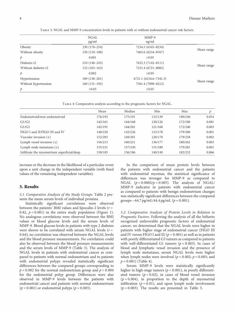
Statistically significant correlations were observedbetween the patients’ BMI values and lipocalin-2 levels (r =0 82, p = 0 001) in the entire study population (Figure 1).No analogous correlations were observed between the BMIvalues or blood glucose levels and the serum levels ofMMP-9. Blood glucose levels in patients with type 2 diabeteswere shown to be correlated with serum NGAL levels (r =0 64); no correlation was observed between the NGAL levelsand the blood pressure measurements. No correlation couldalso be observed between the blood pressure measurementsand the serum levels of MMP-9 (Table 3). The analysis ofNGAL levels in patients with endometrial cancer as com-pared to patients with normal endometrium and to patientswith endometrial polyps revealed statistically significantdifferences between the compared groups corresponding top = 0 002 for the normal endometrium group and p = 0 004for the endometrial polyp group. Differences were alsoobserved in MMP-9 levels between the patients withendometrial cancer and patients with normal endometrium(p = 0 001) or endometrial polyps (p = 0 003).
In the comparison of mean protein levels betweenthe patients with endometrial cancer and the patientswith endometrial myomas, the statistical significance ofdifferences was stronger for MMP-9 as compared toNGAL (p = 0 0002/p = 0 005). The analysis of NGAL/MMP-9 indicator in patients with endometrial canceras compared to patients with benign endometrium changeswas statistically significant differences between the comparedgroups—64.7 pg/mL/44.4 pg/mL (p = 0 001).
5.2. Comparative Analysis of Protein Levels in Relation toPrognostic Factors. Following the analysis of all the hithertorecognized unfavorable prognostic factors of endometrialcancer, we determined that the NGAL levels were higher inpatients with higher stage of endometrial cancer (FIGO IIIand IV versus FIGO I and II) (p = 0 001) as well as in patientswith poorly differentiated G3 tumors as compared to patientswith well-differentiated G1 tumors (p = 0 003). In cases ofblood and lymphatic vessel invasion and the presence oflymph node metastases, serum NGAL levels were higherwhen lymph nodes were involved (p = 0 002; p = 0 003; andp = 0 001) (Table 4).
Serum MMP-9 levels were statistically significantlyhigher in high-stage tumors (p = 0 001), in poorly differenti-ated tumors (p = 0 02), in cases of blood vessel invasion(p = 0 004), in proportion to the depth of myometrialinfiltration (p = 0 01), and upon lymph node involvement(p = 0 003). The results are presented in Table 5.
Table 3: NGAL and MMP-9 concentration levels in patients with or without endometrial cancer risk factors.
NGALpg/ml
MMP-9ng/ml
Obesity 230 (170–254) 7234.5 (6345–8234)Mean range
Without obesity 150 (110–188) 7465.6 (6234–8767)
p 0.001 >0.05Diabetes t2 210 (148–243) 7632.3 (7142–8111)
Mean rangeWithout diabetes t2 132 (103–163) 7231.4 (6721–8082)
p 0.002 >0.05Hypertension 189 (138–201) 6721.1 (6234.6–7341.3)
Mean rangeWithout hypertension 160 (131–192) 7561.4 (7098–8212)
p >0.05 >0.05
Table 4: Comparative analysis according to the prognostic factors for NGAL.
Mean Median Min Max p
Endometroid/non-endometroid 176/193 175/191 132/139 190/240 0.054
G1/G2 142/161 144/168 120/126 172/180 0.081
G1/G3 142/191 144/201 121/168 172/240 0.003
FIGO I and II/FIGO III and IV 140/220 142/226 123/178 179/280 0.001
Vascular invasion (±) 152/203 149/201 120/170 179/258 0.002
Lymph vessel invasion (±) 156/213 160/221 136/177 180/262 0.003
Lymph node metastasis (±) 155/231 157/230 131/180 179/281 0.001
Infiltrate the myometrium superficial/deep 158/185 156/186 140/149 182/232 0.066
4 Disease Markers

The statistically significant differences for the NGAL/MMP-9 indicator were found between the mean concentra-tions in patients according to staging and grading. Signifi-cantly higher indicator was in patients with high-stageand poor histopathological differentiation. It was 83.3 pg/mL/61.2 pg/mL (p = 0 004) and 77.5 pg/mL/62.2 pg/mL(p = 0 03), respectively.
5.3. Analysis of ROC Curves. In order to evaluate the diagnos-tic value of lipocalin-2 and MMP-9, the ROC curves wereplotted and the areas under the ROC curves (AUC) were
calculated. The analysis compared the patients with endome-trial cancer to patients with benign endometrial lesions. TheAUC values for NGAL and MMP-9 were 0.9 and 0.78,respectively. The AUC value for NGAL/MMP-9 indicatorwhich compared the patients with endometrial cancer topatients with benign endometrial changes was 0.92(Figure 2). The diagnostic potential of NGAL, MMP-9, andindicator NGAL/MMP-9 in differentiating high-stage (FIGOIII and IV) and low-stage (FIGO I and II) cancer and predict-ing the cell differentiation grade (G1 versus G3) on the basisof the analyses of AUC values was determined to be 0.91 and0.79 for NGAL, 0.82 and 0.84 for MMP-9, and 0.91, 0.86 forindicator NGAL/MMP-9, respectively. The curves are pre-sented in Figures 3, 4, 5, 6, 7, and 8. Four logistic regressionmodels were performed. Multifactorial logistic regressionanalysis in the final method revealed that NGAL andMMP-9 variables were independent of the endometrial can-cer risk. OR values for NGAL and MMP-9 were 1.23 (95%CI: 1.421–3.27; p = 0 034) and 1.09 (95% CI: 1.38–4.12; p =0 026), respectively. In addition, the same independent var-iables were analyzed with regard to the risk of lymph nodemetastases (yes/no), tumor stage FIGO III and IV versusFIGO I and II, and grading G3 versus G1. The respectiveOR values for NGAL and MMP-9 were as follows: 1.4 (95%CI: 0.9–2.98; p = 0 02)/2.02 (95% CI: 3.1–6.42; p = 0 004);3.66 (95% CI: 4.2–12.3; p = 0 005)/1.28 (95% CI: 1.58–7.2;
Table 5: Comparative analysis according to the prognostic factors for MMP-9.
Mean Median Min Max p
Endometroid/non-endometroid 7721.5/8909.8 7543.6/8823.3 7012.6/8002.3 8231/9567.6 0.04
G1/G2 5263/6789.2 5324.2/6881.2 4967.2/5122.5 5778.1/7657.7 0.061
G1/G3 5263/8782.1 5324.2/889.2 4967.2/6744.5 5778.1/989.0 0.02
FIGO I and II/FIGO III and IV 5433.1/9213.7 5367/9311.7 4977.2/7338.4 6877.2/10131.8 0.001
Vascular invasion (±) 5688.3/8672.9 5723.3/8721.5 5002.9/7112.4 5989.7/9881.7 0.004
Lymph vessels invasion (±) 6345.2/7187.2 6463.1/7132.2 4412.5/5323.9 7889.2/8999.3 0.07
Lymph nodes metastasis (±) 5998.1/9141.8 6021/9099.3 5121.9/7111 6781.9/10267 0.003
Infiltrate the myometrium superficial/deep 6891.3/9234.8 6631.4/9122.4 5899.6/7114.8 7346.8/10145.6 0.01
00.10.20.30.40.50.60.70.80.9
1
Sens
itivi
ty
1 – speci�city
NGAL/MMP‑9
AUC = 0.9211p = 0.0031
0 0.2 0.4 0.6 0.8 1
Figure 2: The ROC curves for indicator NGAL/MMP-9 in women.The analysis compared endometrial cancer patients to patients withbenign endometrial lesions.
00.10.20.30.40.50.60.70.80.9
1
0
Sens
itivi
ty
1 – speci�city0.1 0.20.3 0.40.5 0.6 0.7 0.8 0.9 1
Figure 3: The ROC curves for MMP-9 proteins in women. Theanalysis compared endometrial cancer patients to patients withbenign endometrial lesions.
00.10.20.30.40.50.60.70.80.9
1
0 0.1 0.2 0.3 0.4 0.5 0.6 0.7 0.8 0.9 1
Sens
itivi
ty
1 – speci�city
Figure 4: The ROC curves for NGAL proteins in women. Theanalysis compared endometrial cancer patients to patients withbenign endometrial lesions.
5Disease Markers

p = 0 03); and 1.09 (95% CI:1.8–5.2; p = 0 03)/1.83 (95% CI:2.4–8.9; p = 0 02) (Table 6).
6. Discussion
The main risk factors of type I endometrial cancer have beenknown for years and include obesity, diabetes, and arterialhypertension. The adipose tissue plays the role of a secretoryorgan releasing a number of adipokines that play an impor-tant role in the system. One of such released glycoproteinsis NGAL, that is, lipocalin-2. It acts as an insulin resistanceenhancer [9] and participates in the process of oncogenesis[10–12]. MMP-9 is a protein that plays an apparently crucialrole in the proliferation of cells in endometrial cancer. Itbelongs to the family of endoproteinases with iron-, zinc-,and calcium-dependent activity. Metalloproteinases degradethe components of extracellular matrix removing the barrierbetween tumor cells and normal tissue environment, thusinitiating the metastatic process. Numerous reports identifieda complex of MMP-9, that is, gelatinase B with NGAL (neu-trophil gelatinase-associated lipocalin), a low-molecular pro-tein. By strengthening the bonds in MMP-9 molecules,NGAL protects them from autodegradation. As demon-strated in in vitro studies, both MMP-9 and NGAL led tothe increase in tumor sizes [13, 14].
In our study, we were able to demonstrate that patientswith endometrial cancer presented with NGAL and MMP-9
1 MMP9
NGALAUC = 0.79
AUC = 0.81(1.0; 0.98)
(1.0; 0.98)
0.90.80.70.60.5
Sens
itivi
ty
0.40.30.20.1
0
10.90.80.70.60.5
Sens
itivi
ty
0.40.30.20.1
0
0 0.1 0.2 0.3 0.4 0.5 0.6 0.7 0.8 0.9 1
0 0.1 0.2 0.3 0.4 0.51 – speci�city
0.6 0.7 0.8 0.9 1
Figure 6: The ROC curves for MMP-9 and NGAL proteins in G1and G3 grading.
Table 6: Multifactorial logistic regression models for NGAL andMMP-9 for staging, grading, and lymph node metastasis.
Protein OR 95% CI p
Lymph node metastasis
NGAL 1.4 0.9–2.98 0.02
MMP-9 2.02 3.1–6.42 0.004
FIGO III and IV versus I and II
NGAL 3.66 4.2–12.3 0.005
MMP-9 1.28 1.58–7.2 0.03
GRADING III versus I
NGAL 1.09 1.8–5.2 0.03
MMP-9 1.83 2.4–8.9 0.02
00 0.2 0.4 0.6 0.8 1
0.10.20.30.40.50.60.70.80.9
1
Sens
itivi
ty
1 – speci�city
NGAL/MMP‑9 staging
AUC = 0.9152p = 0.0028
Figure 7: The ROC curves for indicator NGAL/MMP-9 dependingon staging.
00 0.2 0.4 0.6 0.8 1
0.10.20.30.40.50.60.70.80.9
1
Sens
itivi
ty
1 – speci�city
NGAL/MMP‑9 grading
AUC = 0.8631p = 0.0018
Figure 8: The ROC curves for indicator NGAL/MMP-9 dependingon grading.
00.10.20.30.40.50.60.70.80.9
1
Sens
itivi
ty
MMP9AUC = 0.82
(0.0; 0.06)
00.10.20.30.40.50.60.70.80.9
1
Sens
itivi
ty
1 – speci�city
(0.03; 0.7)
NGALAUC = 0.91
0 0.1 0.2 0.3 0.4 0.5 0.6 0.7 0.8 0.9 1
Figure 5: The ROC curves for MMP-9 and NGAL proteinsdepending on staging.
6 Disease Markers

levels higher than those with healthy endometrium or benignendometrial lesions. As observed by Mannelqvist et al.,NGAL expression was higher in endometrial cancer patientscompared to cancer-free patients [15]. Reports of higherexpression of MMP-9 in endometrial cancer patients werealso published [16].
In their recent study from 2016, Li et al. highlighted thatthe high expression of NGAL can be considered to be signif-icantly correlated with the expression of vimentin and themigration, invasion, and proliferation of tumor cells [11].
In our study, we were able to demonstrate statistically sig-nificant differences only with regard to the lipocalin-2 levelsin well- and poorly differentiated tumors with no differencesbeing observed between G1 and G2 tumors. On the otherhand, Li et al. demonstrated that NGAL levels in G1 tumorpatients are statistically significantly different from those inpatients with healthy endometrium. Thus, one may expectthat NGAL is a biomarker that may find its potential use inearly detection of endometrial cancer. This, however, mustbe confirmed in future studies. In addition, a study in endo-metrial cancer cell lines revealed that an increase in NGALexpression is observed during epithelial-mesenchymal trans-formation [11].
We found that the NGAL levels were significantly higherin patients with higher tumor stages and were correlated withinfiltration of lymphatic and blood vessels as well as lymphnode metastases. Similar reports were presented byMannelq-vist et al. who, in their multifactorial analysis, identifiedNGAL as an independent prognostic factor of tumor gradingand staging in endometrial cancer patients [15]. Lee et al.conducted a study in a transgenic mouse model to achieveinhibition of tumorigenesis following application of NGALinhibitor [17].
The effect of inflammation on carcinogenesis in genitalorgan tumors is another issue to be considered. Intheir meta-analysis of 832 cases of endometrial cancer,Delahanty et al. observed that inflammation played animportant role in the carcinogenesis of endometrial tumors[18]. Elevated expression of MMP-9 and IL-6 was alsoconfirmed. The authors take note of the importance ofNGAL as its levels are significantly increased in inflamma-tory processes [19, 20].
MMP-9 appears to play a crucial role in the formation ofmetastases of endometrial cancer. Increased MMP-9 expres-sion is associated with myometrial invasion as well as withinvasion into blood and lymphatic vessels [21]. Karahanet al. demonstrated that the increase in MMP-9 expressionmay be correlated to both tumor size and aggressiveness [16].
The increased expression of MMP-9 in endometrial can-cer is correlated with the overexpression of other proteins, forexample, with overexpression of Ki67 and tumor necrosisfactor alpha-induced protein-8 [22], epidermal growth factor[23], and fibroblast growth factor [24], as well as with theincreased activity of telomerase, and overexpression of cata-lytic protein hTERT [25, 26].
No correlation between MMP-9 expression and patientsurvival was presented by Fang Yu et al. who highlightedthe significant impact of myometrial infiltration depth, bloodand lymphatic vessel invasion, and lymph node metastases
but not of the MMP-9 levels on the predicted survival inendometrial cancer patients.
Our study was the first one to assess the total serum levelsof NGAL and MMP-9 using the logistic regression modelwhich facilitates attempted estimation of the preoperativerisk of endometrial cancer. We are looking for a new, easymethod to perform research of new cancer markers, whichwill allow us to evaluate and assess tumor progression. In thispaper, the analysis of individual proteins and then the indexwas calculated, but the NGAL/MMP-9 complex was not eval-uated. Determination of individual proteins is cheapermethod, and calculation of NGAL/MMP-9 indicator is notdifficult. Research may be carried out with a small quantityof test material. This method is relatively new and innova-tive, using a combination of two techniques, ELISA andcytometry. In the future, we are planning to compareNGAL/MMP-9 sensitivity and specificity indicator andNGAL/MMP-9 complex, both in serum and in urine.
7. Conclusion
Both NGAL and MMP-9 proteins may be useful in assessingthe stage of the cancer, before surgical treatment. Furtherstudies are needed to be conducted to confirm the prognosticsignificance of the GAL/MMP-9 indicator.
Conflicts of Interest
The authors declare that there is no conflict of interestregarding the publication of this article.
References
[1] J. Ferlay, I. Soerjomataram, M. Ervik et al., “GLOBOCAN 2012v1.0. Cancer incidence and mortality worldwide: IARCCancerBase No. 11,” 2013, http://globocan.iarc.fr/Pages/fact_sheets_cancer.aspx.
[2] A. G. Renehan and I. Soerjomataram, “Obesity as an avoidablecause of cancer (attributable risks),” Recent Results in CancerResearch, vol. 208, pp. 243–256, 2016.
[3] L. Yadav, N. Puri, V. Rastogi, P. Satpute, R. Ahmad, andG. Kaur, “Matrix metalloproteinases and cancer - roles inthreat and therapy,” Asian Pacific Journal of Cancer Preven-tion, vol. 15, no. 3, pp. 1085–1091, 2014.
[4] Y. Wang, K. S. Lam, E. W. Kraegen et al., “Lipocalin-2 is aninflammatory marker closely associated with obesity, insulinresistance, and hyperglycemia in humans,” Clinical Chemistry,vol. 53, no. 1, pp. 34–41, 2007.
[5] Q. W. Yan, Q. Yang, N. Mody et al., “The adipokine lipocalin2 is regulated by obesity and promotes insulin resistance,”Diabetes, vol. 56, no. 10, pp. 2533–2540, 2007.
[6] P. G. Kamble, M. J. Pereira, C. O. Sidibeh et al., “Lipocalin 2produces insulin resistance and can be upregulated by gluco-corticoids in human adipose tissue,” Molecular and CellularEndocrinology, vol. 427, pp. 124–132, 2016.
[7] H. H. Lin, C. J. Liao, Y. C. Lee, K. H. Hu, H. W. Meng, andS. T. Chu, “Lipocalin-2-induced cytokine productionenhances endometrial carcinoma cell survival and migra-tion,” International Journal of Biological Sciences, vol. 7,no. 1, pp. 74–86, 2011.
7Disease Markers

[8] S. Candido, M. Di Maso, D. Serraino et al., “Diagnosticvalue of neutrophil gelatinase-associated lipocalin/matrixmetalloproteinase-9 pathway in transitional cell carcinomaof the bladder,” Tumour Biology, vol. 37, no. 7, pp. 9855–9863, 2016.
[9] B. Cabia, S. Andrade, M. C. Carreira, F. F. Casanueva, andA. B. Crujeiras, “A role for novel adipose tissue-secretedfactors in obesity-related carcinogenesis,” Obesity Reviews,vol. 17, no. 4, pp. 361–376, 2016.
[10] C. J. Liao, Y. H. Huang, H. K. Au, L. M. Wang, and S. T. Chu,“The cancer marker neutrophil gelatinase-associated lipocalinis highly expressed in human endometrial hyperplasia,”Molec-ular Biology Reports, vol. 39, no. 2, pp. 1029–1036, 2012.
[11] T. Li, L. Yu, J. Wen, Q. Liao, and Z. Liu, “An early-screeningbiomarker of endometrial carcinoma: NGAL is associated withepithelio-mesenchymal transition,” Oncotarget, vol. 7, no. 52,pp. 86064–86074, 2016.
[12] Z. Marchewka, A. Tacik, and A. Piwowar, “KIM-1 and NGALas potential biomarkers for the diagnosis and cancer progres-sion,” Postȩpy Higieny i Medycyny Doświadczalnej (Online),vol. 70, pp. 329–336, 2016.
[13] E. Mira, R. A. Lacalle, J. M. Buesa et al., “Secreted MMP9promotes angiogenesis more efficiently than constitutive activeMMP9 bound to the tumor cell surface,” Journal of CellScience, vol. 117, Part 9, pp. 1847–1857, 2004.
[14] J. Yang and M. A. Moses, “Lipocalin 2: a multifaceted modula-tor of human cancer,” Cell Cycle, vol. 8, no. 15, pp. 2347–2352,2009.
[15] M. Mannelqvist, I. M. Stefansson, E. Wik et al., “Lipocalin 2expression is associated with aggressive features of endome-trial cancer,” BMC Cancer, vol. 12, p. 169, 2012.
[16] N. Karahan, M. Guney, S. Baspinar, B. Oral, N. Kapucuoglu,and T. Mungan, “Expression of gelatinase (MMP-2 andMMP-9) and cyclooxygenase-2 (COX-2) in endometrial carci-noma,” European Journal of Gynaecological Oncology, vol. 28,no. 3, pp. 184–188, 2007.
[17] Y. C. Lee, W. F. Tzeng, T. J. Chiou, and S. T. Chu, “MicroRNA-138 suppresses neutrophil gelatinase-associated lipocalinexpression and inhibits tumorigenicity,” PLoS One, vol. 7,no. 12, article e52979, 2012.
[18] R. J. Delahanty, Y. B. Xiang, A. Spurdle et al., “Polymorphismsin inflammation pathway genes and endometrial cancer risk,”Cancer Epidemiology, Biomarkers & Prevention, vol. 22, no. 2,pp. 216–223, 2013.
[19] J. Zhang, Y. Wu, Y. Zhang, D. Leroith, D. A. Bernlohr, andX. Chen, “The role of lipocalin 2 in the regulation of inflamma-tion in adipocytes and macrophages,” Molecular Endocrinol-ogy, vol. 22, no. 6, pp. 1416–1426, 2008.
[20] S. Kim, H. J. Kim, H. S. Ahn et al., “Is plasma neutrophilgelatinase-associated lipocalin a predictive biomarker for acutekidney injury in sepsis patients? A systematic review andmeta-analysis,” Journal of Critical Care, vol. 33, pp. 213–223, 2016.
[21] L. A. Di Nezza, A. Misajon, J. Zhang et al., “Presence of activegelatinases in endometrial carcinoma and correlation ofmatrix metalloproteinase expression with increasing tumorgrade and invasion,” Cancer, vol. 94, no. 5, pp. 1466–1475,2002.
[22] T. Liu, H. Gao, M. Yang, T. Zhao, Y. Liu, and G. Lou, “Corre-lation of TNFAIP8 overexpression with the proliferation,metastasis, and disease-free survival in endometrial cancer,”Tumour Biology, vol. 35, no. 6, pp. 5805–5814, 2014.
[23] M. Cho-Clark, D. O. Larco, B. R. Zahn, S. K. Mani, and T. J.Wu, “GnRH-(1-5) activates matrix metallopeptidase-9 torelease epidermal growth factor and promote cellular inva-sion,” Molecular and Cellular Endocrinology, vol. 415,pp. 114–125, 2015.
[24] S. N. Xue, J. Lei, C. Yang, D. Z. Lin, and L. Yan, “The biologicalbehaviors of rat dermal fibroblasts can be inhibited by highlevels of MMP9,” Experimental Diabetes Research, vol. 2012,Article ID 494579, 7 pages, 2012.
[25] D. Ding, P. Xi, J. Zhou, M. Wang, and Y. S. Cong, “Human tel-omerase reverse transcriptase regulates MMP expression inde-pendently of telomerase activity via NF-κB-dependenttranscription,” The FASEB Journal, vol. 27, no. 11, pp. 4375–4383, 2013.
[26] W. Kong, N. Lv, W. Z. Wysham et al., “Knockdown of hTERTand treatment with BIBR1532 inhibit cell proliferation andinvasion in endometrial cancer cells,” Journal of Cancer,vol. 6, no. 12, pp. 1337–1345, 2015.
8 Disease Markers

Submit your manuscripts athttps://www.hindawi.com
Stem CellsInternational
Hindawi Publishing Corporationhttp://www.hindawi.com Volume 2014
Hindawi Publishing Corporationhttp://www.hindawi.com Volume 2014
MEDIATORSINFLAMMATION
of
Hindawi Publishing Corporationhttp://www.hindawi.com Volume 2014
Behavioural Neurology
EndocrinologyInternational Journal of
Hindawi Publishing Corporationhttp://www.hindawi.com Volume 2014
Hindawi Publishing Corporationhttp://www.hindawi.com Volume 2014
Disease Markers
Hindawi Publishing Corporationhttp://www.hindawi.com Volume 2014
BioMed Research International
OncologyJournal of
Hindawi Publishing Corporationhttp://www.hindawi.com Volume 2014
Hindawi Publishing Corporationhttp://www.hindawi.com Volume 2014
Oxidative Medicine and Cellular Longevity
Hindawi Publishing Corporationhttp://www.hindawi.com Volume 2014
PPAR Research
The Scientific World JournalHindawi Publishing Corporation http://www.hindawi.com Volume 2014
Immunology ResearchHindawi Publishing Corporationhttp://www.hindawi.com Volume 2014
Journal of
ObesityJournal of
Hindawi Publishing Corporationhttp://www.hindawi.com Volume 2014
Hindawi Publishing Corporationhttp://www.hindawi.com Volume 2014
Computational and Mathematical Methods in Medicine
OphthalmologyJournal of
Hindawi Publishing Corporationhttp://www.hindawi.com Volume 2014
Diabetes ResearchJournal of
Hindawi Publishing Corporationhttp://www.hindawi.com Volume 2014
Hindawi Publishing Corporationhttp://www.hindawi.com Volume 2014
Research and TreatmentAIDS
Hindawi Publishing Corporationhttp://www.hindawi.com Volume 2014
Gastroenterology Research and Practice
Hindawi Publishing Corporationhttp://www.hindawi.com Volume 2014
Parkinson’s Disease
Evidence-Based Complementary and Alternative Medicine
Volume 2014Hindawi Publishing Corporationhttp://www.hindawi.com