Embed Size (px)
Citation preview
Rapid transcriptional autoregulation of a yeast metalloregulatory transcription factor is essential for liigh-level copper detoxification Pengbo Zhou and Dennis J. Thiele 1
Department of Biological Chemistry, University of Michigan Medical School, Ann Arbor, Michigan 48109-0606 USA
Copper detoxification in the yeast Candida glabrata is carried out in large part by a family of metallothionein (MT) genes: a unique MT-I gene, a tandemly amplified MT-IIa gene, and a single unlinked MT-IIb gene. In response to elevated environmental copper levels, members of this MT gene family are transcriptionally activated by a copper-dependent, sequence-specific DNA-binding transcription factor, AMT1. AMT1 shares several structural and functional features with the Saccharomyces cerevisiae copper metalloregulatory transcription factor ACE1, which is constitutively expressed and poised for rapid transcriptional responses to the toxic metal copper. In this paper, we demonstrate that AMT1 is subject to positive transcriptional autoregulation, which is exerted through binding of copper-activated AMT1 to a single copper responsive element in the AMT1 promoter. A nonautoregulatory amtl mutant displayed a marked decrease in both copper tolerance and expression of the MT-II genes, which are critical for high-level copper detoxification in Candida glabrata. Kinetic analysis demonstrated the remarkably rapid AMT1 mRNA accumulation in the presence of copper, which is followed by increased expression of the metallothionein gene products. These results demonstrate that AMTl-positive autoregulation plays a critical role in metal detoxification and suggest that the rapid autoactivation of the AMT1 metalloregulatory transcription factor biosynthesis is essential for C. glabrata to quickly build up a cellular defense line to protect cells upon exposure to high environmental copper levels.
[Key Words: AMT1; autoregulation; Candida glabrata; copper; metalloregulatory transcription factor; metallothionein]
Received April 20, 1993; revised version accepted June 22, 1993.
A common response of cells to fluctuations in the levels of nutrients, toxins, or developmental signals is to repro- gram the transcription of genes important for coping with the altered environment. In many cases, these genes encode both proteins that directly facilitate the adaptive or developmental response and regulatory genes encoding DNA-binding transcription regulation factors that control the expression of the structural genes (Angel et al. 1988). The metallothioneins (MTs) provide an im- portant system to study the homeostatic processes that regulate cell responses to both essential and toxic metal ions and the mechanisms by which these signals are in- terpreted and transduced.
MTs are low-molecular-weight, cysteine-rich proteins that bind copper and other metal ions and therefore pro- tect cells against metal toxicity (K/igi and Kojima 1987; K/igi and Schaffer 1988). Expression of MTs is activated, primarily at the level of gene transcription initiation, in
~Corresponding author.
response to elevated levels of copper, zinc, mercury, and many other metals {for review, see Thiele 1992). The baker's yeast Saccharomyces cerevisiae contains a single MT gene, designated CUP1 (Fogel and Welch 1982; Butt et al. 1984; Karin et al. 1984). In response to elevated copper concentrations, the CUP1 gene is transcription- ally induced by a copper-activated sequence-specific DNA-binding transcription factor denoted ACE1 (Thiele 1988, 1992). Upon the binding of copper through its amino-terminal cysteine-rich metal-binding domain, ACE1 is thought to undergo a conformational change, which acts as a "functional switch" from apo-ACE1 to an active protein competent for specific DNA-binding to four independent sites within the CUP1 upstream acti- vation sequence (UAScuP1) (Ffirst et al. 1988; Huibregste et al. 1989; Szczypka and Thiele 1989; Buchman et al. 1990; Evans et al. 1990; Hu et al. 1990; Thiele 1992). Previous experiments demonstrated that ACE1 exists as a single-copy gene in S. cerevisiae strains harboring ei- ther one copy (cupl s) or multiple tandem copies (CUP1 R) of CUP1 (Thiele 1988). Besides CUP1, ACE1 directly
1824 GENES & DEVELOPMENT 7:1824-1835 �9 1993 by Cold Spring Harbor Laboratory Press ISSN 0890-9369/93 $5.00
Cold Spring Harbor Laboratory Press on June 18, 2018 - Published by genesdev.cshlp.orgDownloaded from
Autoregulation of a metalloregulatory [actor
activates transcription of another gene, denoted SOD1, which encodes the copper-zinc superoxide dismutase; this protects cells from oxygen toxicity (Gralla et al. 1991). Furthermore, ACE1 biosynthesis is independent of copper, because constitutive levels of ACE1 are de- tected in S. cerevisiae cells growing either in the absence or presence of exogenous copper ions (Szczypka and Thiele 1989).
The opportunistic pathogenic yeast Candida glabrata provides an excellent system to study copper-dependent MT gene transcription. Like humans and other higher eukaryotes, this yeast harbors a family of MT genes that is divided into classes: a unique MT-I gene and two dis- tinct MT-II genes, composed of a tandemly amplified MT-IIa gene and a single unlinked MT-IIb gene (Mehra et al. 1989, 1992). Both the MT-I and MT-II genes are transcriptionally activated by copper and its electronic analog, silver (Mehra et al. 1989). Recently, we isolated a C. glabrata gene encoding a copper-dependent transcrip- tion factor, denoted AMT 1, which bears many structural and functional features of the ACE 1 protein in S. cerevi- siae (Zhou and Thiele 1991). AMT1 is composed of 265 amino acids organized to form two distinct domains; the amino-terminal domain (amino acid residues 1-115) is positively charged and contains the copper-activated DNA-binding function, which delivers monomeric AMT1 to binding sites in the MT target gene promoters. The carboxy-terminal region (residues 116-265) is nega- tively charged and, by analogy with ACE1, is likely to harbor the trans-activation function (Zhou and Thiele 1991; Zhou et al. 1992). Upon copper treatment, AMT1 binds to two and six binding sites in the MT-I and MT-IIa promoters, respectively, and activates target gene tran- scription in a copper concentration-dependent fashion (this paper; Zhou et al. 1992). Of the two MT classes in C. glabrata, the MT-II gene family was shown to play a major role in copper detoxification (Mehra et al. 1992}. The structural and functional characteristics of AMT1 demonstrate that this protein has dual functions, acting both as a copper sensory molecule and as a transcription activator: It senses the elevated copper levels and di- rectly initiates a genomic response to this signal by ac- tivating the transcription of copper detoxification genes (MTs).
To begin to investigate the mechanisms by which metalloregulatory transcription factors activate MT gene families, we analyzed the physiological factors that mod- ulate expression of the AMT1 gene. In contrast to its functional analog ACE1, which is constitutively ex- pressed both at the level of transcription and translation (Szczypka and Thiele 1989), AMT1 mRNA levels are dra- matically increased in response to copper or silver, the same metal ions that foster the AMTl-dependent MT-I and MT-H gene transcription. In this report we demon- strate that AMT1 is rapidly transcriptionally activated by its own gene product through a single binding site in the AMT1 promoter. Furthermore, through the genera- tion of yeast cells in which the sole functional AMT1 gene is incapable of positive transcriptional autoregula- tion, we have demonstrated that this autoregulatory
mechanism is critical for cell survival in the presence of toxic environmental copper concentrations.
R e s u l t s
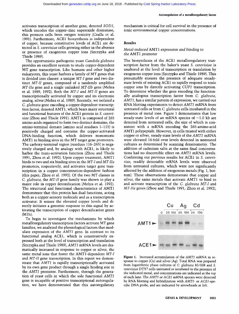
Metal-induced AMT1 expression and binding to the AMT1 promoter
The biosynthesis of the ACE1 metalloregulatory tran- scription factor from the baker's yeast S. cerevisiae is unaltered at the level of transcription or translation by exogenous copper ions (Szczypka and Thiele 1989). This presumably ensures the presence of adequate steady- state levels of existing ACE 1 to rapidly respond to toxic copper ions by directly activating CUP1 transcription. To determine whether the gene encoding the function- ally analogous transcription factor from C. glabrata, AMT1, has a similar pattern of expression, we carried out RNA blotting experiments to detect AMT1 mRNA from untreated cells or from C. glabrata cells incubated in the presence of metal ions. Figure 1 demonstrates that low steady-state levels of an mRNA species of -1 .0 kb are detected from untreated cells, the size of which is con- sistent with a mRNA encoding the 265-amino-acid AMT1 polypeptide. However, in cells treated with either copper or silver, steady-state levels of the AMT1 mRNA were elevated 16-fold over levels detected in untreated cultures as determined by scanning densitometry. The addition of cadmium salts at the same final concentra- tions had no discernible effect on AMT1 mRNA levels. Confirming our previous results for ACE1 in S. cerevi- siae, readily detectable mRNA levels were observed from untreated cultures, which were not significantly affected by the addition of exogenous metals (Fig. 1, bot- tom). These observations demonstrate that copper and silver, the same metals that activate AMT1 to bind to and activate transcription of the C. glabrata MT-I and MT-IIa genes (Zhou and Thiele 1991; Zhou et al. 1992),
Figure 1. Increased accumulation of the AMT1 mRNA in re- sponse to copper (Cu) and silver (Ag). Total RNA was prepared from logarithmic phase cultures of C. glabrata 85/038 and S. cerevisiae DTY7 cells untreated or incubated in the presence of the indicated metal, and concentrations are indicated at the top of each lane. The AMT1 or A CE1 mRNA species were detected by RNA blotting and hybridization with AMT1- or ACEl-spe- cific DNA probe, and are indicated by arrowheads at left.
GENES & DEVELOPMENT 1825
Cold Spring Harbor Laboratory Press on June 18, 2018 - Published by genesdev.cshlp.orgDownloaded from

Zhou and Thiele
also induce accumulation of the AMT1 mRNA steady- state levels in vivo.
Mapping of the AMTl-binding site in the AMT1 promoter
Recently, we demonstrated that AMT1 is essential for copper-responsive transcription of the MT gene family in C. glabrata through metal-activated DNA-binding to MT gene promoters (Zhou et al. 1992). This finding, in conjunction with the observation that AMT1 mRNA levels are dramatically induced in response to copper or silver, suggests that transcription of the AMT1 gene it- self is also subject, directly or indirectly, to regulation by copper. To test whether the AMT1 gene is a direct target for binding of copper-activated AMT1, we carried out DNA-binding studies by electrophoretic mobility shift assays. A 326-bp BglII-SspI DNA restriction fragment ( -457 to - 130 relative to the AMT1 transcription ini- tiation site) was radiolabeled and used as a probe in elec- trophoretic mobility shift assays using partially purified AMT1 protein produced in E. coli (Zhou et al. 1992). We detected the formation of a single copper- or silver-de- pendent DNA-protein complex and no further com- plexes, even at high extract concentrations (data not shown). Cadmium, however, failed to induce any D N A -
protein complex formation. This experiment demon- strated that copper- or silver-activated AMT1 directly interacts with a sequence in the AMT1 promoter, con- sistent with direct AMT1 metal-dependent positive au- toregulation. Furthermore, a copper (CuJ-AMT1 com- plex failed to form with a a~P-labeled 169-bp SspI-RsaI DNA restriction fragment (Zhou and Thiele 1991), which encompasses the downstream AMT1 promoter fragment ( -131 to +39), including the putative TATA box ( - 8 4 to -79) (data not shown). To specifically lo- cate the AMTl-binding site in the AMT1 promoter, we employed DNase I footprinting assays using partially pu- rified AMT1 protein produced in Escherichia coli (Zhou et al. 1992). As shown in Figure 2A, copper-activated AMT1 protein protected a major region, from - 1 9 5 to - 1 8 0 on the coding strand and from - 2 0 0 to - 1 8 0 on the noncoding strand of the AMT1 promoter against DNase I cleavage. This AMT1 recognition sequence is highly similar to a consensus AMTl-binding site derived from in vitro DNase I footprinting assays of the MT-I and MT-IIa promoters, which contains a conserved "GCTG" core sequence preceded by an A/T-rich region (Zhou et al. 1992; Fig. 3A). Furthermore, two nucleotides (G-205 and A-204) on the coding strand and one nucleotide (T- 207) on the noncoding strand were also protected by cop- per-activated AMT1 (Fig. 2A). Interestingly, 3 bp up-
Figure 2. Copper-activated AMT1 binds to a single site in the AMT1 promoter. (A) DNase I footprinting of the AMT1 pro- moter region. Footprinting reactions were carried out with either the a2P-labeled coding strand {CODING) or noncoding strand (NONCODING) of the AMT1 pro- moter fragments and the indicated amounts of the AMT1 extract {in micro- grams) at the top of each lane. Binding reactions were carried out in the absence (-) or presence (+) of 50 ~M C u S O 4 a t
room temperature for 10 min. G/A is the chemical sequencing reaction displaying adenine and guanine residues using the coding or noncoding probe fragments as templates. Solid vertical lines and num- bers indicate the positions of the Cu- AMTl-protected regions relative to the AMT1 transcription initiation site. (B) Methylation interference analysis of the Cu-AMT1 complex and the AMT1 pro- moter. The coding and noncoding AMT1 probe fragments, either free {F) or bound with Cu-AMT1 (C), were partially meth- ylated with dimethylsulfate, isolated on electrophoretic mobility shift assay gels, cleaved with piperidine, and analyzed on a 6% sequencing gel. The numbers and arrowheads indicate positions of critical G or A residues which, when methylated, interfere with Cu-AMT1 binding.
1826 GENES & DEVELOPMENT
Cold Spring Harbor Laboratory Press on June 18, 2018 - Published by genesdev.cshlp.orgDownloaded from

Figure 3. Copper-activated AMT1 fails to bind to the AMT1 (m) promoter. (A) Sequence of the AMTl-binding site in the AMT1 promoter. Base pairs are numbered relative to the AMTI tran- scription start site. Solid horizontal bars indicate the regions within the AMT1 promoter protected by Cu-AMT1 from DNase I cleavage. Boxed residues are guanines and adenines, which, when methylated, interfere with copper-activated AMTl-binding in vitro. The two brackets indicate the two con- served domains within the AMT1 recognition sequence that are derived from comparison of the AMTl-binding sites in the AMT1, MT-I, and MT-IIa promoters (Zhou et al. 1992). The two A's above the arrows represent the two point mutations intro- duced to the AMTl-binding site by site-directed mutagenesis to derive the AMTI(m) promoter fragment. (B)Electrophoretic mobility shift assays using 32P-labeled AMT1 promoter frag- ments containing either the wild-type (AMT1) or mutant [AMTI(m)] AMTl-binding sites. The amount of AMT1 extract, (indicated in lag) was used in binding reactions with ( + ) or with- out (-) 100 ~M CuSO4. (F) Free probe DNA; (C) copper-activated AMT1 complexed to the wild-type AMT1 promoter.
stream of the 5' extreme region of the AMTl-binding site lies a nucleotide sequence ( -225 to -210) com- posed exclusively of 16 adenine residues. This region is completely resistant to DNase I cleavage even in the absence of Cu-AMT1. Whether this poly(A) stretch plays any functional role with respect to AMT1 promoter activity is not currently known.
To determine the nucleotides in the single AMT1- binding site important for stable AMT1-AMT1 pro- moter-DNA interactions, we carried out methylation interference assays using the same 32p-labeled AMT1 promoter fragments used for the DNase I footprinting analysis (Fig. 2B). Methylation of the guanine residues in the GCTG core-binding sequence (G-191 and G-188 on the coding strand and G-190 on the noncoding strand) strongly interfered with Cu-AMT1 binding to its target sequence. Formation of this complex was also inhibited by methylation of A-195, A-194, and A-193 on the non- coding strand. Because the positions for methylation by dimethylsulfate are N-7 on guanine and N-3 on adenine,
Autoregulat ion of a meta l loregulatory [actor
which are located in the major and minor grooves of B-form double helical DNA, respectively, this methyl- ation interference assay also provided information on which regions of the binding site are in close proximity to bound copper-activated AMT1 protein {Thanos and Maniatis 1992). The observation that all of the critical G or A residues, as ascertained by methylation interfer- ence, are found within an 8-bp region suggests that Cu- AMT1 interacts with adjacent major and minor groove DNA, representing both core and A/T-rich domains of the AMTl-binding site, respectively (Fig. 3A).
AMTl-posi t ive autoregulation is mediated by the single AMTl-binding site
To examine the functional significance of the AMT1- binding site in the AMT1 promoter, we converted resi- dues G-191 and G-188 in the core-binding domain, the integrity of which is critical for binding as demonstrated by methylation interference analysis, to adenines by site-directed mutagenesis (Fig. 3A; Ausubel et al. 1987). The 326-bp AMT1 promoter fragments containing either the mutant or wild-type AMT 1-binding sites, designated AMTI(m) or AMT1, respectively, were radiolabeled and subjected to electrophoretic mobility shift assays using the partially purified AMT1 protein produced in E. coli. As shown in Figure 3B, mutation of the critical guanine residues in the core domain of the AMTl-binding site abolished Cu-AMT1 binding to the AMT1 (m) promoter fragment. The wild-type AMT1 promoter fragment gave rise to a single Cu-AMTl-dependent complex.
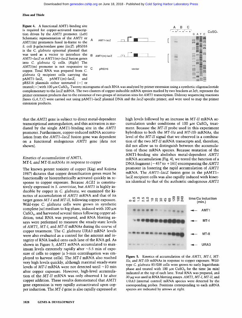
To ascertain the role of this binding site in AMT1 metal-responsive transcriptional autoregulation, we con- structed reporter gene fusions between a 613-bp BglII- StyI DNA restriction fragment containing the AMT1 wild-type or AMT1 (m) promoters and a portion of the AMTl-coding region, in-flame with the E. coli lacZ gene, encoding [3-galactosidase (Fig. 4). These two re- porter constructs were independently inserted into a S. cerevisiae plasmid, pRS316 (Sikorski and Hieter 1989), which is episomally maintained in multiple copies in C. glabrata (P. Zhou, M. Szczypka, R. Young and D.J. Thiele, in prep.). The resulting plasmids were then trans- formed into a C. glabrata strain harboring a ura3 gene mutation for plasmid selection and maintenance, but which has a wild-type AMT1 locus. Total RNA was pre- pared from the recipient cells either untreated or induced with 100 ~M CuSO4, and steady-state mRNA levels driven from the AMTI- IacZ and AMT1 (m)-lacZ fusions were detected by primer extension assays using an olig- onucleotide primer complementary to the lacZ gene- coding strand. Copper-induced A M T I - l a c Z mRNA ac- cumulation was observed in recipient cells harboring the wild-type A M T I - l a c Z fusion and yielded two clusters of AMTI- lacZ mRNA start sites, which correspond pre- cisely to authentic AMT1 mRNA start sites beginning at - 54 and - 79 relative to the ATG translation initiation codon (Fig. 4). However, no copper-dependent mRNA ac- cumulation was observed to be driven from the AMT1 (m)-lacZ fusion gene. These results demonstrate
GENES & DEVELOPMENT 1827
Cold Spring Harbor Laboratory Press on June 18, 2018 - Published by genesdev.cshlp.orgDownloaded from
Zhou and Thiele
Figure 4. A functional AMTl-binding site is required for copper-activated transcrip- tion driven by the AMT1 promoter. (Left) Schematic representation of the AMTI or AMT1 (m) promoters fused in-frame to the E. coli [3-galactosidase gene {lacZ). pRS316 is the C. glabrata episomal plasmid that was used as a vector to introduce the AMTI-lacZ or AMTI (m)-lacZ fusion genes into C. glabrata Q cells. (Right)The AMTI(m) promoter is not responsive to copper. Total RNA was prepared from C. glabrata Q recipient cells carrying the pAMT 1-lacZ, pAMT 1 (m)-lacZ, and pRS316 plasmids either untreated (-) or treated ( + } with 100 ~M CuSO4. Twenty micrograms of each RNA was analyzed by primer extension using a synthetic oligonucleotide complementary to the lacZ mRNA. The two clusters of copper-inducible mRNA species marked by two brackets at left, represent the primer extension products due to the existence of two groups of initiation sites for AMT1 transcription. Dideoxy sequencing reactions (lanes G,A,T,C} were carried out using pAMTI-lacZ plasmid DNA and the lacZ-specific primer, and were used to map the primer extension products.
that the AMT1 gene is subject to direct metal-dependent transcriptional autoregulation, and this activation is me- diated by the single AMTl-b ind ing site in the AMT1 promoter. Furthermore, copper-induced m R N A accumu- lation from the A M T I - l a c Z fusion gene was dependent on a functional endogenous AMT1 gene (data not shown).
Kinetics of accumulation of AMT1, MT-I, and MT-II m R N A s in response to copper
The known potent toxicity of copper (Kfigi and Kojima 1987) dictates that copper detoxification genes must be funct ional ly or biosynthet ical ly activated quickly in re- sponse to copper exposure. Because ACE1 is constitu- tively expressed in S. cerevisiae, but AMT1 is highly in- ducible by copper in C. glabrata, we examined the ki- netics of accumula t ion of AMT1 m R N A and that of its target genes MT-I and MT-II, following copper exposure. Wild-type C. glabrata cells were grown in synthetic complete (sc) m e d i u m to log phase, induced wi th 100 ~M CuSO4, and harvested several t imes following copper ad- dition; total RNA was prepared, and RNA blotting as- says were performed to measure the steady-state levels of AMT1, MT-I, and MT-II mRNAs during the course of copper treatment. The C. glabrata URA3 mRN~ levels were also evaluated as a control for the amount and in- tegrity of RNA loaded onto each lane of the RNA gel. As shown in Figure 5, AMT1 m R N A accumulated to max- i m u m levels extremely rapidly after -5 .5 min of expo- sure of cells to copper (a 5-min centrifugation was em- ployed to harvest cells). The MT-I m R N A also reached very high levels quickly, al though maximal steady-state levels of MT-I m R N A were not detected unt i l - 1 0 min after copper exposure. However, high-level accumula- tion of the MT-II m R N A was only observed 1 hr after copper addition. These results demonstrated that AMT1 gene expression is very rapidly autoactivated upon cop- per induction. The MT-I gene is also rapidly expressed at
high levels followed by an increase in MT-H m R N A ac- cumulat ion under conditions of 100 ~M CuSO4 treat- ment. Because the MT-II probe used in this experiment hybridizes to both the MT-IIa and MT-Hb mRNAs, the level of the MT-II signal that we observed is a combina- tion of the two MT-H m R N A transcripts and, therefore, did not allow us to dist inguish between the accumula- tion of these m R N A species. Because muta t ion of the AMTl-b inding site abolishes metal-dependent AMT1 m R N A accumulat ion (Fig. 4), we tested the funct ion of a DNA f r a g m e n t ( - 4 5 7 to + 161)encompassing the AMT1 promoter in fostering the rapid accumula t ion of AMT1 mRNA. The A M T I - l a c Z fusion gene in the pAMT1- lacZ recipient cells was also rapidly induced wi th kinet- ics identical to that of the authent ic endogenous AMT1
Figure 5. Kinetics of accumulation of the AMT1, MT-L MT- Ha, and MT-IIb mRNAs in response to copper exposure. Wild- type C. glabrata 85/038 cells were grown to early logarithmic phase and treated with 100 ~M CuSO4 for the time (in min) indicated at the top of each lane. Total RNA was prepared, and 30 ~g was used in RNA blotting assays. AMTI, MT-I, MT-1I, and URA3 (internal control) mRNA species were detected by the corresponding probes. Positions corresponding to each mRNA species are indicated by arrows at right.
1828 GENES & DEVELOPMENT
Cold Spring Harbor Laboratory Press on June 18, 2018 - Published by genesdev.cshlp.orgDownloaded from

gene as determined by primer extension assays (data not shown). These results strongly suggest that rapid AMT1 metal-dependent autoregulation is largely mediated through the AMT1 promoter-binding site and at the level of transcription initiation.
AMT1 metal-dependent autoregulation is critical for normal copper homeostasis
Due to experimental complexities, the physiological sig- nificance of transcription factor gene autoregulation in higher eukaryotes has not been directly tested in the complete absence of an endogenous functional allele. To determine whether AMT1 transcriptional autoregulation is critical for cellular copper detoxification in C. gla- brata, we uncoupled copper-dependent autoactivation from the AMT1 promoter in the context of an intact, single endogenous functional AMT1 gene. This was achieved by introducing the two point mutations in the AMTl-binding site, described in Figure 3A, in the com- plete AMT1 gene; therefore, this gene is identical to the wild-type AMT1 gene, with the exception of a nonfunc- tional AMTl-binding site in the AMT1 gene promoter. The wild-type or nonautoregulatory mutant AMT1 genes [AMT1 and AMT1 (m), respectively] were subcloned into a C. glabrata integrative plasmid, Ul(b), carrying the C. glabrata URA3 gene (P. Zhou, M. Szczypka, R. Young, and D. Thiele, in prep.) and inserted at the C. glabrata ura3 locus within a strain in which the endogenous AMT1 gene had been insertionally inactivated (Zhou et al. 1992). The targeted single-copy integration was ver- ified by Southem blotting (data not shown). The result- ant isogenic strains are designated AMTI::URA3 and AMTI (m): : URA3, respectively.
We then asked whether AMT1 gene transcriptional au- toregulation is critical for the protection of yeast cells in the presence of high environmental copper levels. The copper-resistant phenotype of the isogenic AMTI::URA3 and AMTI(m)::URA3 strains, as well as their parental strains C. glabrata 85/038 (wild-type) and amtl-1 (amtl gene insertionally inactivated), was tested for copper re- sistance. As shown in Figure 6, the parental wild-type and AMT1 :: URA3 cells are indistinguishable in their re- sistance to copper and grow on agar containing up to 1.5 ~M CuSO4. These observations demonstrate that inte- gration of the wild-type AMT1 gene at the ura3 chromo- somal locus does not affect its expression or function. The AMTI(m)::URA3 strain, however, gave rise to a maximal copper resistance level at 400 CuSO4, a three- to fourfold decrease compared with the wild-type or AMT1 :: URA3 strains. As demonstrated previously, the amtl-1 strain is hypersensitive to copper and fails to grow even in the presence of 25 ~M CuSO4 (Zhou et al. 1992). In these experiments, copper-sensitive cells ar- rested at the single-cell stage when challenged with toxic concentrations of CuSO4 (data not shown). These results clearly demonstrate that AMT1 gene-positive autoregu- lation is essential for C. glabrata resistance to high en- vironmental copper levels. The nonautoregulatory AMT1 allele [AMTI(m)::URA3], survives on media con-
Autoregulation of a metalloregulatory factor
0 25 ~M 200 IIM CuS04
300 pM 500 I~M 1 mM CuS04
Figure 6. AMT1 autoregulation is essential for C. glabrata cells to survive on high copper concentrations. The nonauto- regulatory AMT1 (m):: URA3 allele fails to confer wild-type cop- per resistance to C. glabrata. Isogenic C. glabrata strains 85/038, amtl-1, AMTI::URA3, and AMTI(m)::URA3 were streaked onto SC agar and SC agar containing the concentra- tions of CuSO 4 as indicated. Plates were incubated at 30~ for 2 days. The grid indicates the relevant genotypes of the four in- dividual strains streaked onto each sector of the plates.
taining - 1 0 times the concentration of exogenous cop- per as the amtl-1 strain. Although the mutation in the AMT1 promoter abolishes AMT1 binding and autoacti- vation, the AMTI(m):: URA3 allele drives the expression of the AMT1 mRNA and, presumably, protein, owing to AMTl-independent mechanisms of expression of this gene {see Fig. 7). Consistent with this possibility, we detected the formation of AMTl-independent DNA-pro- tein complexes using whole cell extracts from the amtl-1 strain and the 326-bp AMT1 promoter fragment described in Figure 2. Furthermore, the formation of these complexes was not affected by guanine mutations of the core AMTl-binding sequence [AMTllm)probe; data not shown].
Copper sensitivity of cells unable to autoactivate AMT1 correlates with a defect in MT mRNA accumulation
Previous studies suggested that because the C. glabrata MT-1 and MT-H genes are transcriptionally coinduced by copper in a concentration-dependent manner, the cop- per-responsive transcriptional activator AMT1 is not limiting in its abundance (Mehra et al. 1989, 1992). Our demonstrations that AMT1 is both transcriptionally pos- itively autoregulated and that this is essential for normal copper resistance suggest that positive autoregulation
GENES & DEVELOPMENT 1829
Cold Spring Harbor Laboratory Press on June 18, 2018 - Published by genesdev.cshlp.orgDownloaded from

Zhou and Thiele
Figure 7. Accumulation of the AMT1, MT-I, and MT-Ha, and MT-IIb mRNAs in response to increasing copper levels. AMTI:: URA3 and AMT1 (m):: URA3 cells were grown in SC medium lacking uracil (SC - ura) to logarithmic phase. Parallel cultures were untreated or treated for 10 min with the copper concentrations indicated at the top of each lane. Specific AMT1, AMTI::hIsG, MT-I, and MT-H mRNAs were detected by RNA blotting (left). As an internal control for the loading of RNA, C. glabrata actin mRNA levels were analyzed using a hybridization probe derived from the cloned S. cerevisiae actin gene, which hybridizes to the C. glabrata actin gene {Mason et al. 1987). The radioactivity {cpm) associated with each individual mRNA species was quantitated by counting the dried nitrocellulose membranes with a Beta-gen scanner. The amounts of each mRNA species were normalized to the corresponding actin mRNA level and plotted against copper concentration (in ~M). (Right) The three graphs indicate the steady-state mRNA levels from each gene in response to the range of copper levels. Each mRNA species is represented by the symbol shown at the right of each graph.
may ensure an adequate supply of the AMT1 transcrip- tion factor to activate the transcription of C. glabrata copper homeostasis genes. Because free intracellular cop- per could cause rapid macromolecular damage through redox chemistry (Halliwell and Gutteridge 1984), and AMT1 is expressed at very low levels in the absence of exogenous copper (Figs. 1 and 7), perhaps rapid AMT1 autoregulation is essential for cells to synthesize suffi- cient AMT1 protein to boost the synthesis of metal- lothioneins promptly before cells are irreversibly dam-
aged by sudden increases in intracellular copper concen- trations. To test this hypothesis, we carried out RNA blotting assays to determine the steady-state mRNA lev- els of the AMT1, MT-I, and MT-II genes after treating the AMTI:: URA3 and AMT1 (m):: URA3 cells with increas- ing doses of copper for a brief period of time (10-min induction plus 5-rain centrifugation) (Fig. 7, left). The S. cerevisiae actin gene, which hybridizes to a single mRNA species in C. glabrata, was used as a control for the amount and integrity of RNA loaded onto each lane
1830 GENES & DEVELOPMENT
Cold Spring Harbor Laboratory Press on June 18, 2018 - Published by genesdev.cshlp.orgDownloaded from

Autoregulation of a metalloregulatory factor
of the gel in this experiment. Figure 7 (right) shows a quantitation of the steady-state mRNA levels of AMT1, AMTI::hisG, MT-I, and MT-II in AMTI::URA3 and AMTI(m)::URA3 cells exposed to the range of copper concentrations. The wild-type AMT1 gene integrated at the C. glabrata ura3 locus was highly induced when cells were exposed to copper, reaching a maximal eight- fold induction after a 10-min exposure to 100 ~M CuSO4. The endogenous amtl-1 locus, which carries an inser- tion of the Salmonella hisG gene within the AMT1 open reading frame, synthesized a hybrid AMT1 ::hisG mRNA whose identity was confirmed by its hybridization to a 32p-labeled hisG probe by RNA blotting (data not shown). The expression of this hybrid gene was also in- duced by copper-activated AMT1 with a similar dose re- sponse and a 4.2-fold induction, presumably mediated through the intact AMTl-binding site within the pro- moter of the hisG insertionally inactivated amtl-1 lo- cus. In contrast, no copper-inducible mRNA accumula- tion was observed for the AMTI(m) gene in the AMTI(m)::URA3 strain. However, mRNA driven from the AMT1 ::hisG hybrid gene accumulated in response to exogenous copper, although there was an induction of only -1.6-fold. Low steady-state levels of the AMTI::hisG mRNA were also detected in the amtl-1 strain by RNA blotting upon prolonged exposure and were not altered upon copper treatment (data not shown). These findings demonstrate that copper-induced transcription driven by the AMT1 promoter is dependent on both the presence of a functional AMTl-binding site and a functional AMT1 protein. The AMT1 or AMTI::hisG mRNA levels in the AMTI::URA3 cells were highest when cells were treated with 100 ~M copper and slightly decreased at higher copper concentrations.
RNA blotting with the MT-1 probe showed that the MT-I gene was expressed at maximum levels even at low copper concentrations (100 ~M). The overall level of the MT-I mRNA in the AMTI(m):: URA3 strain is - 1.4-fold lower than that in the AMTI::URA3 strain. The MT-II mRNA levels (MT-1Ia and MT-IIb) in these two isogenic strains are similar upon addition of sublethal concentra- tions of copper, which are 100 and 300 ~M of CuSO4. A reproducible twofold difference of the MT-II mRNA level was observed when the strains were treated with lethal doses of copper for AMT1 (m):: URA3 cells (500 ~M and 1 raM). It should be noted that 10-min exposure of AMTI(m)::URA3 with 500 ~M or 1 mM copper did not immediately impair cell growth, as judged by similar in- creases of OD6oo as well as similar levels of actin mRNA observed between AMTI:: URA3 and AMT1 (m):: URA3 cells following copper treatment. Taken together, these results demonstrate that the MT-I and MT-H mRNA steady-state levels were significantly compromised in the nonautoregulatory AMT1 (m) : : URA3 allele.
MT-I and MT-H protein levels are correspondingly reduced in AMT1 nonautoregulatory cells
The steady-state levels of MT-I and MT-H protein under copper-induced conditions were analyzed by pulse-label-
ing proteins in AMT1 :: URA3 and AMT1 (m): : URA3 cells with [3SS]cysteine, followed by fluorographic analysis on a 25% native polyacrylamide gel (Fig. 8). This experi- ment demonstrates that the MT-I and MT-II proteins were undetectable in both AMTI::URA3 and AMTI(m)::URA3 cells grown in the absence of copper. Addition of increasing doses of copper resulted in the elevated synthesis of the MT-II protein with a signifi- cantly higher MT-II level observed in the AMT1 :: URA3 cells treated with all copper concentrations than that in the AMTI(m)::URA3 cells. The MT-I protein reached similar maximum levels upon treatment with low cop- per (100 ~M) in both strains, and this level was similar when the strains were induced with higher concentra- tions of copper. These results parallel the steady-state MT-I and MT-H mRNA levels that we obtained for AMTI::URA3 and AMTI(m)::URA3 cells under the same copper-induced conditions {Fig. 7).
D i s c u s s i o n
An interesting and important question in gene regula- tion is how transcription factor genes themselves are reg- ulated, and does this regulation play a key role in the function of the encoded protein? In prokaryotes, autog- enous regulation has been demonstrated for several tran- scriptional repressors and often results in down-regula- tion of genes encoding these DNA-binding proteins (for review, see Maloy and Stewart 1993). In eukaryotes, pos- itive transcriptional autoregulation has been observed for many transcription factors and is suggested to be an important regulatory mechanism in a large number of biological processes such as cell growth, differentiation, development, and others {for review, see Serfling 1989; Falvey and Schibler 1991). However, efforts to precisely delineate the physiological role of transcription factor autoregulation in these systems have been impeded by the fact that multiple regulatory mechanisms function through the upstream regulatory regions at the same dif-
Figure 8. The MT-I and MT-II protein levels in AMTI::URA3 and AMT1 (m):: URA3 cells in response to copper. [asS]Cysteine- labeled total soluble proteins were prepared from control or cop- per-treated AMTI::URA3 and AMTI(m)::URA3 cells at the concentrations indicated at the top of each lane. Each protein extract (1.0 ~g)was subjected to electrophoresis on a 25% non- denaturing polyacrylamide gel. The arrowheads at left indicate the MT-I and MT-II protein species.
GENES & DEVELOPMENT 1831
Cold Spring Harbor Laboratory Press on June 18, 2018 - Published by genesdev.cshlp.orgDownloaded from
Zhou and Thiele
ferentiation or developmental stage, and by the difficulty of genetic manipulations in higher eukaryotes, such as gene knockouts and homologous recombination.
As a unicellular eukaryotic microorganism, yeast pro- vides a powerful model system to study gene regulation and other biochemical processes, largely owing to the ease of genetic manipulations. In this work we demon- strated that the AMT1 metalloregulatory transcription factor of C. glabrata functions in a positive autoregula- tory loop, which is distinct from the constitutively ex- pressed ACE1 gene of S. cerevisiae (Szczypka and Thiele 1989). AMT1 is among the first reported yeast transcrip- tion factors that directly participates in its own tran- scription, and the physiological significance of this au- toregulation has been established with respect to its role in activating the transcription of target genes that play a critical role in cell defense mechanisms against metal toxicity. In this paper, we demonstrated that AMT1 au- toregulation is mediated by the single AMTl-binding site in the AMT1 promoter and requires functional AMT1 protein.
On the basis of previous studies {Mehra et al. 1989, 1992; Zhou and Thiele 1991; Zhou et al. 1992) and re- sults obtained in this work, we propose the following autoregulatory model for AMT1 expression and the cop- per-induced transcriptional response in C. glabrata cells. Under normal growth conditions and low environmental copper concentrations, C. glabrata cells synthesize low basal levels of the copper sensory molecule AMT1, as well as MTs and other putative copper homeostatic pro- teins. MTs may sequester the low concentration of bio- logically available cellular copper ions preferentially over AMT1, thereby preventing futile transcription of AMT1, MT-I, MT-II, and potentially other, as yet uniden- tified, AMTl-dependent copper-regulated genes. High environmental copper concentrations lead to an in- creased copper accumulation, and intracellular copper ions would be available to apo-AMT1 protein produced from basal level transcription of this gene. The coordi- nation of copper by the AMT1 amino-terminal DNA- binding domain activates sequence-specific DNA bind- ing and, once deposited on target gene promoters, first activates transcription of the AMT1 and MT-! genes, fol- lowed by the MT-H isoform genes. The specific proper- ties of the AMT1 promoter, either a high-affinity AMT1- binding site or interactions with other transcription fac- tors, may enhance the specific affinity of Cu-AMT1 or its transcriptional potency and contribute to the rapid autoactivation of the AMT1 gene. Although the relative in vivo affinity of Cu-AMT1 for the AMT1, MT-I, and MT-H promoters has not yet been determined, the re- sults that we observed for AMT1, MT-I, and MT-H mRNA levels suggest that even at low copper concen- trations (100 ~M), the AMT1 and MT-I genes are rapidly activated to very high levels while the MT-II genes are only partially activated. This observation suggests a pos- sible role of MT-I as a first line of defense to respond to low levels of copper in the environment. Increasing doses of copper treatment result in the elevated synthe- sis of the MT-II mRNA and protein presumably because
the MT-I protein alone is insufficient for sequestering intracellular copper ions. This high-level MT-II gene ex- pression is dependent on two factors: One is the AMT1 autoregulation that produces sufficient apo-AMT1 pro- tein; second is the increasing amount of copper that switches more apo-AMT1 into the transcriptionally competent Cu-AMT1 available to the C. glabrata MTs, especially the tandemly amplified MT-IIa gene. Al- though the MT-I and MT-II proteins bind cooper effi- ciently, we cannot exclude the possibility that MT-I, MT-II, or AMT1 proteins, or other proteins encoded by AMTl-dependent genes, have additional activities that protect cells from copper toxicity. Consistent with this possibility, Tamai et al. (1993) have recently demon- strated that S. cerevisiae Cu-MT protein has potent an- tioxidant activity. Furthermore, because Cu-ACE1 acti- vates SOD1 transcription (Gralla et al. 1991), it is possi- ble that Cu-AMT1 may also activate other target genes, and the decreased tolerance of the nonautoregulatory AMT1 mutant strain to copper may result from defective expression of several copper homeostatic genes regulated by AMT1. Further experiments will determine the rela- tive affinity of Cu-AMT1 for the AMT1, MT-I, and MT- H promoters and the precise mechanism by which AMT1 is rapidly transcriptionally autoactivated.
As a copper sensory molecule, AMT1 responds quickly to the increase in intracellular copper levels by an am- plification of its own gene product. Although the mech- anisms for copper transport and distribution in C. gla- brata and other organisms have not been elucidated in detail, copper uptake in S. cerevisiae has been demon- strated to be a rapid process, and the rate of intracellular copper accumulation is proportional to the medium cop- per concentrations (Lin and Kosman 1990}. The rapid copper-dependent AMT1 and MT-I transcription sug- gests that copper uptake in C. glabrata is not rate lim- iting; therefore, the cells may be immediately subjected to copper toxicity upon addition of copper to the growth media. The rapid AMT1 autoregulation ensures that AMT1 proteins are made promptly and at a concentra- tion sufficient for its function as a copper-dependent transcription factor to stimulate high-level expression of the multiple C. glabrata MT genes before extensive cop- per-induced cellular damage occurs. Recently, it has been observed that pretreatment of human HeLa H454 cells with a low dose of cadmium results in superinduc- tion of the human MT-II A gene in response to subse- quent cadmium administration (A. Leone, pers. comm.). This may reflect a similar autoregulatory expression of the gene, or genes, encoding putative metal response el- ement (MRE)-binding proteins in humans and other or- ganisms bearing an MT gene family.
In contrast to S. cerevisiae, in which the gene encoding the ACE1 copper metalloregulatory transcription factor is constitutively expressed (Szczypka and Thiele 1989), AMT1 autoregulation reflects another level of regulatory complexity for copper homeostatic genes in C. glabrata. Although the opportunistic pathogenic yeast C. glabrata and the baker's yeast S. cerevisiae are evolutionarily re- lated (Barns et al. 1991), the fact that C. glabrata cells are
1832 GENES & DEVELOPMENT
Cold Spring Harbor Laboratory Press on June 18, 2018 - Published by genesdev.cshlp.orgDownloaded from
Autoregulation of a metalloregulatory factor
found in a wide var ie ty of hab i t a t s inc lud ing soil, water , on an ima l s and h u m a n t i ssues or organs suggests tha t th is yeas t encoun te r s m a n y e n v i r o n m e n t a l e l emen t s w i t h qui te different copper con ten t s (Sinnot t et al. 1987). As a un ice l lu la r eukaryo t i c microorganism, a yeas t cell is h igh ly accessible to e n v i r o n m e n t a l changes in me ta l concent ra t ions . The deve lopmen t of a rapid posi t ive au- toregula tory m e c h a n i s m for a m e t a l sens ing switch, AMT1, could a l low C. glabrata to a c c u m u l a t e suff ic ient a m o u n t s of copper w h e n avai lable concen t ra t ions are low but respond rapidly w h e n cha l lenged w i t h toxic en- v i r o n m e n t a l copper concen t ra t ions .
M a t e r i a l s and m e t h o d s
Strains, plasmids, and growth conditions
The parental wild-type C. glabrata strain 85/038 was a gift of P. Magee (University of Minnesota, St. Paul). The ura3- {Q) strain and the AMT1 disruption strain amtl-I were constructed as described in detail (Zhou et al. 1992) and were used as recipient hosts for transformation by C. glabrata episomal or integrative plasmids described in the following sections. The amtl-I strain is the parental strain for the integration of wild-type (AMTI) or mutant [AMT1 (m)] genes at the ura3 locus to construct isogenic AMT1 ::URA3 and AMTI (m):: URA3 strains by transformation and homologous recombination (Aubusel et al. 1987; P. Zhou, M. Szczypka, R. Young, and D. Theile, in prep.). Yeast cells were grown in rich (YPD) or SC medium lacking uracil (Ausubel et al. 1987). Copper resistance tests, copper treatment, and the length of incubation are described in the respective figure legends. The S. cerevisiae strain DTY7 (Szczypka and Thiele 1989), which contains the wild-type ACE1 and three copies of the tandemly amplified CUP1 genes, was used to examine the mRNA levels from the ACE1 gene under control or metal-induced conditions. E. coli strain XL-1 blue (Stratagene) was employed for the con- struction and maintenance of plasmids using standard tech- niques {Ausubel et al. 1987).
To study the role that the single AMTl-binding site plays in AMT1 autoregulation, two guanine residues (G-192 and G-189) in the GCTG core-binding sequence were changed to adenines by oligonucleotide-directed mutagenesis using the mutagenic primer 5'- CGCCCACCACTACTTTTAAGTTAGTCAAATT- AGC-3', which hybridizes to AMT1 promoter nucleotide posi- tions -201 to - 168 relative to the + 1 transcription start site (this paper; Ausubel et al. 1987; Zhou and Thiele 1991). The in vivo autoactivation of the AMT1 gene was demonstrated using AMTI-lacZ or AMT1 (m)-lacZ fusion genes containing either the wild-type or mutant AM3al-binding site, respectively. The 616-bp BglII-StyI DNA restriction fragment, which contains the AMT1 or AMT1 (m) promoter and the first 36 AMT1 codons was first blunt-ended at the StyI site by the Klenow fragment of DNA polymerase I and then subcloned into the plasmid YEp356(R) to construct AMTI-lacZ or AMT1 (m)-lacZ in-flame fusion genes (Myers et al. 1986). The 4.3-kb HindIII-StuI DNA restriction fragments containing the AMTI-lacZ or AMT1 (m)- lacZ fusions were subcloned into the C. glabrata episomal plas- mid pRS316 to make plasmids pAMTI-lacZ and pAMTI(m)- lacZ (Sikorski and Hieter 1989). These two pRS316-based plas- raids can stably transform the strain Q (ura3-) and replicate in multiple copies in C. glabrata cells {P. Zhou, M. Szczypka, R. Young, and D. Theile, in prep.).
To characterize the biological role of AMT1 autoregulation, two integrative plasmids were constructed as follows: A 4.0 kb genomic fragment containing the C. glabrata URA3 gene was
inserted at the BamHI site of pRS425 to make plasmid Ul(b) (Sikorski and Hieter 1989; P. Zhou, M. Szczypka, R. Young, and D. Theile, in prep.). The 1.6-kb BglII-SpeI DNA restriction frag- ments containing either the wild-type IAMTll or mutant [AMTI(m)] AMT1 genes were then subcloned into Ul(b) to make integrative plasmids YIpAMT1 ::URA3 and YIpAMTI(m)::URA3. These two plasmids were digested to completion with StuI restriction enzyme, which has a unique site located in the URA3 open reading frame to facilitate se- quence-specific recombination. The linearized integrative plas- mids were integrated in single copy at the URA3 locus of the amtl-1 strain, in which the authentic AMT1 locus was dis- rupted by an insertion of the Salmonella hisG gene (Zhou and Thiele 1991).
Because our original AMT1 genomic clone lacks the 3' un- translated sequences of this gene, which contains the AMT1 transcription termination signal, we carried out inverse PCR (IPCR) and DNA cloning to isolate a 1.6-kb BglII-SpeI DNA restriction fragment that encompasses the entire AMT1 mRNA transcript (Ochman et al. 1990; GenBank accession number M69146). This experiment also allowed us to identify a short extragenic sequence that was ligated 3' to the AMTl-coding sequence during our initial cloning; however, this did not alter the AMT1 promoter or coding sequence.
RNA analysis
The steady-state levels of AMTI, MT-I, and MT-H mRNA before or after induction by copper or other metal ions were analyzed by either RNA blotting or primer extension assays as described (Ausubel et al. 1987). C. glabrata wild-type 85/038, Q (ura3-), or amtl-1 cells containing episomal or integrative plasmids were grown to logarithmic phase and induced with different copper concentrations and time periods as indicated in each figure legend. Specific AMT1, MT-I, MT-II, URA3, and actin mRNA species were detected using 32P-labeled DNA fragments, respectively, as follows: 1.6-kb BglII-SpeI (this paper; Zhou and Thiele 1991); 0.7-kb EcoRI-ApaI (Mehra et al. 1989); 0.7-kb EcoRI-SmaI (Mehra et al. 1990); 1.5-kb XhoI-PstI (P. Zhou, M. Szczypka, R. Young, and D. Theile, in prep.); and 1.6-kb HindIII (Ng and Abelson 1980) DNA restriction fragments. It is note- worthy that the MT-II probe used in RNA blot hybridization detected both the MT-Ha and MT-Hb mRNAs. Deoxyoligonu- cleotides were synthesized for use as primers in extension re- actions and were labeled at 5' termini with polynucleotide ki- nase and [~/-3zP]ATP; the AMTl-specific primer contained 26 nucleotides with the sequence 5'-GATTACTACCATGGTG- CAAATGTGTG-3', complementary to nucleotide positions + 65 to + 40 of the AMT1 gene (Zhou and Thiele 1991) and was used to determine the AMTI transcription initiation sites. The AMTl-specific primer was also employed in the primer exten- sion reactions using the AMTI-lacZ mRNA as a template. This experiment demonstrated that the transcription of the AMT1- lacZ fusion gene initiated at sites identical to those of the au- thentic AMT1 gene (P. Zhou, unpubl.).
Analysis of DNA-protein interactions
AMT1 DNA-binding studies were carried out with partially pu- rified AMT1 protein expressed in E. coli using the T7 RNA polymerase system {Studier et al. 1990; Zhou and Thiele 1991; Zhou et al. 19921. Electrophoretic mobility shift assays were used to detect binding of the copper-activated AMT1 protein to the 32P-labeled 326-bp BgllI-SspI DNA restriction fragments containing either the wild-type or mutant AMTl-binding site.
DNase I footprinting and methylation interference assays
GENES & DEVELOPMENT 1833
Cold Spring Harbor Laboratory Press on June 18, 2018 - Published by genesdev.cshlp.orgDownloaded from
Zhou and Thiele
were used to map the AMTl-binding site within the AMT1 promoter. Two plasmids were constructed for the isolation of the AMT1 promoter DNA restriction fragment probes. The 326- bp BglII-SspI fragment was inserted into the BamHI-EcoRV sites of pBluescript SK(+) to make plasmid pAMTI(U). The 369-bp MvaI-RsaI DNA restriction fragment was end-repaired by the Klenow fragment of DNA polymerase I and subcloned into the EcoRV site of pBluescript SK(+) to make plasmid pAMTI(L). Probes labeled on the coding or noncoding strand of the AMT1 promoter were prepared by digestion of pAMTI(U) and pAMTI{L) with appropriate restriction enzymes and then radiolabeled with [a-32P]dCTP and the Klenow fragment of DNA polymerase I (Ausubel et al. 1987). DNase I footprinting and methylation interference assays were carried out as de- scribed in detail (Aubusel et al. 1987; Zhou et al. 1992).
35S-Labeling of the MTs and gel electrophoresis
To analyze the MT-I and MT-II protein levels in C. glabrata cells induced with copper, a 5-ml culture of the isogenic AMTI::URA3 and AMTI(m)::URA3 cells were grown to log phase in SC media lacking cysteine and methionine (SC - Cys - Met). Cells were pelleted and resuspended in 0.4 ml of SC - Cys - Met and incubated at 30~ for 10 rain with shaking in the presence of 8.0 ~1 of [aSS]cysteine (ICN, 227 ~xCi/ ml) and CuSO4 at concentrations indicated in Figure 8. Cells were then harvested, suspended in 400 ~1 of lysis buffer (10.0 mM Tris-HC1 at pH 7.8, 10 mM PMSF, 1 mM dithiothreitol), and 400 ~1 of acid-washed glass beads, and lysed by vortexing at top speed four times for 1 rain each at 4~ The soluble proteins were isolated by centrifugation at 14,000 rpm for 15 rain, and protein concentrations were determined by the Bradford assay (Aubusel et al. 1987). Protein (1.0 ~g) was treated with 500 ~M CuSO4 at room temperature for 10 rain. This step saturated MTs with copper and converted all MT-I and MT-II protein isoforms into the uniformly copper-coordinated conformations that migrate as distinct bands on native polyacrylamide gel (Fig. 8). Equal volumes of buffer (0.1 M Tris-HC1 at pH 6.8, 25% glycerol, 0.35 M 2-mercaptoethanol, 0.1% bromphenol blue) were added to each protein extract. The samples were then loaded on 25% nondenaturing polyacrylamide gels and sub- jected to electrophoresis at 200 V until the bromphenol blue reached the bottom of the gel. The proteins were fixed in the gel with 10% acetic acid and 30% methanol for 1 hr and were fluorographed with EN3HANCE solution as described by the supplier (DuPont). The gel was then dried under vacuum and exposed to Kodak XAR-5 film with an intensifying screen at -80~ [3sS]Cysteine-labeled protein extracts from C. glabrata 2001-L5 and RM4 (MT-Ilazl, MT-Ilbzl) cells were coelectro- phoresed on a 25% native polyacrylamide gel and served as controls to identify the MT-I and MT-II polypeptides. It should be noted that the MT-IIa and MT-IIb genes encode identical protein species, although their mRNA transcripts are slightly different in the 3' untranslated regions {Mehra et al. 1992).
A c k n o w l e d g m e n t s
we thank D. Engelke, D. Friedman, A. Seasholtz, and K. Koch for critical reading of the manuscript; members of the Thiele laboratory for helpful discussions; D. Winge and J. Thorvaldsen for generously providing us with the C. glabrata strains 2001- L5, RM2, and RM4; L. Gedamu for advice on MT protein anal- ysis; and R. Young for technical assistance. We are grateful to Geraldine Butler for providing information on the AMT1 nude- otide sequence. P.Z. was supported in part by a Rackham pre-
doctoral fellowship from the Horace H. Rackham School of Graduate Studies at the University of Michigan and a Loeb pre- doctoral fellowship from the University of Michigan Cancer Center. This work was funded by grants GM41840 from the National Institutes of Health, and by grant M01-RR00042 to the General Clinical Research Center, University of Michigan Med- ical Center.
The publication costs of this article were defrayed in part by payment of page charges. This article must therefore be hereby marked "advertisement" in accordance with 18 USC section 1734 solely to indicate this fact.
References
Angel, P., K. Hattori, T. Smeal, and M. Karin. 1988. The jun proto-oncogene is positively autoregulated by its product, Jun/Ap-1. Cell 55: 875-885.
Ausubel, F.M., R. Brent, R.E. Kingston, D.D. Moore, J.G. Seid- man, J.A. Smith, and K. Struhl, eds. 1987. Current protocols in molecular biology. Greene/Wiley, New York.
Barns, S.M., D.J. Lane, M.L. Sogin, C. Bibeau, and W.G. Weis- burg. 1991. Evolutionary relationships among pathogenic Candida species and relatives. J. Bacteriol. 173: 2250-2255.
Buchman, C., P. Skroch, W. Dixon, T.D. Tullius, and M. Karin. 1990. A single amino acid change in CUP2 alters its mode of DNA-binding. Mol. Cell. Biol. 10: 4778-4787.
Butt, T.R., E. Steinberg, J. Herd, and S.T. Crooke. 1984. Cloning and expression of a yeast copper metallothionein gene. Gene 27: 23-33.
Evans, C.F., D.R. Engelke, and D.J. Thiele. 1990. ACE1 tran- scription factor produced in Escherichia coli binds multiple regions within yeast metallothionein upstream activation sequences. Mol. Cell. Biol. 10: 426-429.
Falvey, E. and U. Schibler. 1991. How are the regulators regu- lated? FASEB J. 5: 309-314.
Fogel, S. and J. W. Welch. 1982. Tandem gene amplification mediates copper resistance in yeast. Proc. Natl. Acad. Sci. 79: 5342-5346.
Fiirst, P., S. Hu, R. Hackett, and D.H. Hamer. 1988. Copper activates metallothionein gene transcription by altering the conformation of a specific DNA-binding protein. Cell 55: 705-717.
Gralla E.B., D.J. Thiele, P. Silar, and J.S. Valentine. 1991. ACE1, a copper-dependent transcription factor, activates expression of the yeast copper, zinc superoxide dismutase gene. Proc. Natl. Acad. Sci. 88: 8558-8562.
Halliwell, B. and J.M.C. Gutteridge. 1984. Oxygen toxicity, ox- ygen radicals, transition metals and disease. Biochem. 1. 219: 1-14.
Hu, S., P. Fiirst, and D.H. Hamer. 1990. The DNA and copper binding function of ACE1 are interdigitated within a single domain. New Biol. 2: 1-13.
Huibregste, J.M., D.R. Engelke, and D.J. Thiele. 1989. Copper- induced binding of cellular factors to yeast metallothionein upstream activation sequences. Proc. Natl. Acad. Sci. 86: 65-69.
K~igi, J.H.R. and Y. Kojima. 1987. Chemistry and biochemistry of metallothionein. In Metallothionein II (ed. J.H.R. K/igi and Y. Kojima), pp. 25-61. Birkh/iuser Verlag, Basel, Switzerland.
K~igi, J.H. and A. Schaffer. 1988. Biochemistry of metallothio- nein. Biochemistry 27:8509-8515.
Karin, M., R. Najarain, A. Haslinger, P. Valenzuela, J.W. Welch, and S. Fogel. 1984. Primary structure and transcription of an amplified genetic locus: The CUP1 locus of yeast. Proc. Natl. Acad. Sci. 81: 337-341.
1834 GENES & DEVELOPMENT
Cold Spring Harbor Laboratory Press on June 18, 2018 - Published by genesdev.cshlp.orgDownloaded from
Lin, C.M. and D.J. Kosman. 1990. Copper uptake in wild-type and copper metallothionein-deficient Saccharomyces cere- visiae. 1. Biol. Chem. 265: 9194-9200.
Maloy, S. and V. Stewart. 1993. Autogenous regulation of gene expression. J. Bacteriol. 175: 307-316.
Mason, M.M., B.A. Lasker, and W.S. Riggsby. 1987. Molecular probes for identification of medically important Candida species and Torulopsis glabrata. J. Clin. Microbiol. 25: 563- 566.
Mehra, R.K., J.R. Garey, T.R. Butt, W.R. Gray, and D.R. Winge. 1989. Candida glabrata metallothioneins. J. Biol. Chem. 264: 19747-19753.
Mehra, R.K., J.R. Garey, and D.R. Winge. 1990. Selective and tandem amplification of a member of the metallothionein gene family in Candida glabrata. J. Biol. Chem. 265: 6369- 6375.
Mehra, R.K., J.L. Thorvaldsen, I.G. Macreadie, and D.R. Winge. 1992. Disruption analysis of metallothionein-encoding genes in Candida glabrata. Gene 114: 75-80.
Myers, A.M., A. Tzagoloff, D.M. Kinney, and C.J. Lusty. 1986. Yeast shuttle and integrative vectors with multiple cloning sites suitable for construction of lacZ fusions. Gene 45: 299- 310.
Ng, R. and J. Abelson. 1980. Isolation and sequence of the gene for actin in Saccharomyces cerevisiae. Proc. Natl. Acad. Sci. 77: 3912-3916.
Ochman, H., M.M. Medhora, D. Garza, and D.L. Hartl. 1990. Amplification of flanking sequences by inverse PCR. In PCR protocols, a guide to methods and applications (ed. M.A. Innis, D.H. Gelfand, J.J. Sininsky, and T.J. White), pp. 219- 227. Academic Press, San Diego, CA.
Settling, E. 1989. Autoregulation--A common property of eu- karyotic transcription factors? Trends Genet. 5: 131-133.
Sikorski, R.S. and P. Hieter. 1989. A system of shuttle vectors and yeast host strains designed for efficient manipulation of DNA in Saccharomyces cerevisiae. Genetics 122: 19-27.
Sinnott, J.T., J.P. Cullison, and M.P. Sweeney. 1987. Candida (Torulopsis) glabrata. Infect. Control 8: 334-336.
Studier, F.W., A.H. Rosenberg, J.J. Dunn, and J.W. Dubendorff. 1990. Use of T7 RNA polymerase to direct expression of cloned genes. Methods Enzymol. 185: 60-89.
Szczypka, M.S. and D.J. Thiele. 1989. A cysteine-rich nuclear protein activates yeast metallothionein gene transcription�9 Mol�9 Cell. Biol. 9: 421-429.
Tamai, K.T., E.B. Gralla, L. Ellerby, J.S. Valentine and D.J. Thiele. 1993. Yeast and mammalian metallothioneins func- tionally substitute for yeast copper-zinc superoxide dismu- tase. Proc. Natl. Acad. Sci. (in press)�9
Thanos, D. and T. Maniatis. 1992. The high mobility group protein HMG I(Y) is required for NF-KB-dependent virus in- duction of the human IFN-B gene. Cell 71: 777-789.
Thiele, D.J. 1988. ACE1 regulates expression of the Saccharo- myces cerevisiae metallothionein gene. Mol. Cell. Biol. 8: 2745-2752.
�9 1992. Metal-regulated transcription in eukaryotes. Nu- cleic Acids Res. 20: 1183-1191.
Zhou, P. and D.J. Thiele. 1991. Isolation of a metal-activated transcription factor gene in Candida glabrata by comple- mentation in Saccharomyces cerevisiae. Proc. Natl. Acad. Sci. 88: 6112-6116.
Zhou, P., M.S. Szczypka, T. Sosinowski, and D.J. Thiele. 1992. Expression of a yeast metallothionein gene family is acti- vated by a single metalloregulatory transcription factor. Mol. Cell. Biol. 12: 3766--3775.
Autoregulation of a metalloregulatory factor
GENES & DEVELOPMENT 1835
Cold Spring Harbor Laboratory Press on June 18, 2018 - Published by genesdev.cshlp.orgDownloaded from
10.1101/gad.7.9.1824Access the most recent version at doi: 7:1993, Genes Dev.
P Zhou and D J Thiele transcription factor is essential for high-level copper detoxification.Rapid transcriptional autoregulation of a yeast metalloregulatory
References
http://genesdev.cshlp.org/content/7/9/1824.full.html#ref-list-1
This article cites 32 articles, 20 of which can be accessed free at:
License
ServiceEmail Alerting
click here.right corner of the article or
Receive free email alerts when new articles cite this article - sign up in the box at the top
Copyright © Cold Spring Harbor Laboratory Press
Cold Spring Harbor Laboratory Press on June 18, 2018 - Published by genesdev.cshlp.orgDownloaded from


![Edinburgh Research Explorer · from PNC expression to SOP fate determination and sub-sequent neural development is the initiation of positive autoregulation [10,11]. Autoregulation](https://img.pdfslide.us/doc/110x75/5e2f3ca4fec2bd1ace550d1c/edinburgh-research-from-pnc-expression-to-sop-fate-determination-and-sub-sequent.jpg)