Embed Size (px)
Citation preview
APPLIED AND ENVIRONMENTAL MICROBIOLOGY, Feb. 1990, p. 389-394 Vol. 56, No. 20099-2240/90/020389-06$02.00/0Copyright © 1990, American Society for Microbiology
Rapid Detection of Chlorine-Induced Bacterial Injury by theDirect Viable Count Method Using Image Analysis
AJAIB SINGH,t FEI-PENG YU, AND GORDON A. McFETERS*
Department of Microbiology, Montana State University, Bozeman, Montana 59717
Received 26 July 1989/Accepted 13 November 1989
A modified direct viable count method to detect living bacteria was used with image analysis for the rapidenumeration of chlorine-injured cells in an Escherichia coli culture. The method was also used for determiningchlorine-induced injury in coliform isolates and enteric pathogenic bacteria. Cultures were incubated inphosphate-buffered saline, containing 0.3% Casamino Acids (Difco Laboratories, Detroit, Mich.), 0.03% yeastextract, and optimal concentrations of nalidixic acid. Samples were withdrawn before and after incubationand stained with acridine orange, and cell lengths and breadths were measured by computerized imageanalysis. After incubation, cells which exceeded the mean preincubation length (viable cells) were enumeratedand the results were compared with those obtained by the plate count method. Injury in the chlorine-exposedcell population was determined from the difference in viable count obtained with a nonselective CasaminoAcids-yeast extract-nalidixic acid medium and a selective Casamino Acids-yeast extract-nalidixic acid mediumcontaining sodium deoxycholate or sodium lauryl sulfate. The levels of injury determined by the directviable count technique by using image analysis were comparable to those determined by the plate countmethod. The results showed that image analysis, under optimal conditions, enumerated significantly highernumbers of stressed E. coli than the plate count method did and detected injury in various cultures in4 to 6 h.
One of the major problems in studying indicator andpathogenic bacteria within aquatic environments, includingdrinking water, is their accurate enumeration. Enumerationof indicator bacteria is routinely done to assess the microbi-ological quality of water, and the presence of indicatorbacteria in excessive numbers signals the possible existenceof pathogens. Quantitative recovery of pathogens from thesource water may be important in understanding the natureof waterborne disease outbreaks. These microbiologicaltasks are complicated by drinking water environments thatare generally nutritionally limiting and that cannot supportthe growth of coliform and enteric pathogenic bacteria. Also,bacteria in treated drinking water are exposed to bothphysical and chemical stress factors (4-6). Chlorine andother stressors, at low levels, are lethal only for a smallnumber of exposed cells but induce injury in a large propor-tion of the remaining bacterial population (4, 26). As a result,injured bacteria become sensitive to selective agents that arecommonly incorporated in media used for their routineenumeration and are unable to form colonies (6, 15, 18).Thus, injury can lead to a significant underestimation ofindicator bacteria, since more than 90% of the population indrinking water may become injured (19). The media cur-rently used for quantitative recovery of coliforms fromdrinking water, such as m-Endo agar, have been associatedwith low detection rates of injured bacteria as well as poordifferentiation of coliforms from noncoliform bacteria (17,24). More recently, m-T7 medium was developed for theimproved detection and enumeration of injured coliformsfrom chlorinated drinking water (14). However, these pro-cedures require an incubation period of 24 h.
In 1979, Kogure et al. (13) described a direct viable count
* Corresponding author.t Present address: Delaware Department of Public Health, P.O.
Box 618, Dover, DE 19903.
(DVC) method for the detection of living bacteria that werepresent in natural waters by incubating samples with lowlevels of nutrients and nalidixic acid. Although high concen-trations of nalidixic acid (-50 ,ug/ml) interfere with severalmetabolic activities of gram-negative bacteria (8), a moder-ate concentration specifically inhibits bacterial DNA synthe-sis without affecting other metabolic activities of the cell (7,12). Thus, in the presence of suitable concentrations ofnalidixic acid and nutrients, viable gram-negative bacteriacontinue to grow without cell division, resulting in theformation of elongated cells. The enlarged bacteria countedby epifluorescence microscopy, after acridine orange stain-ing, represent the viable proportion of the total bacterialpopulation. This method has been used by several investi-gators for the enumeration of actively growing cells as wellas physiologically dormant bacteria in the environment (22,23, 27). The major disadvantages of this method is thetedious and subjective process of visually differentiatingnormal cells from marginally elongated cells by conventionalmicroscopy. Recent applications of image analysis permitfast and accurate determination of morphological measure-ments (1) and biochemical changes associated with bacterialgrowth (10). Using image analysis and a modified yeastextract-nalidixic acid method, Singh et al. (25a) have de-scribed an efficient and quantitative method of countingviable bacteria in a relatively short time.Here we report on a modification of the yeast extract-
nalidixic acid method in which conditions for enumeratingchlorine-injured bacteria were optimized and made compat-ible with image analysis. Chlorine-induced injury was as-sessed by determining the difference in DVCs that wasobtained after the cells were incubated in both nonselectiveand selective media. This is a rapid and efficient procedurefor enumerating injured bacteria and assessing the injurylevels in bacterial cultures. The results compared favorablywith the traditional plate count method.
389
on June 7, 2018 by guesthttp://aem
.asm.org/
Dow
nloaded from
on June 7, 2018 by guesthttp://aem
.asm.org/
Dow
nloaded from
on June 7, 2018 by guesthttp://aem
.asm.org/
Dow
nloaded from
APPL. ENVIRON. MICROBIOL.
MATERIALS AND METHODS
Bacterial cultures. The following cultures used in thisstudy either were obtained from the culture collection of theDepartment of Microbiology, Montana State University, or
were obtained earlier from different sources. Escherichiacoli Cl and C2 were previously isolated from the GallatinRiver in the Bozeman, Mont., area, and Enterobacter cloa-cae and Klebsiella pneumoniae were isolated from urbandistribution water samples. Salmonella typhimurium SL3201was obtained from B. A. D. Stocker (Department of MedicalMicrobiology, Stanford University, Stanford, Calif.), Yer-sinia enterocolitica 0:8 was supplied by D. A. Schiemann(Department of Microbiology, Montana State University),and Vibrio cholerae strains were obtained from M. M.Levine (Center of Vaccine Development, University ofMaryland, Baltimore). Stock cultures were stored at -700Cin 1% peptone water containing 20% glycerol.
Preparation of injured cells. Cultures of E. coli and Y.enterocolitica were grown in tryptic soy broth withoutglucose supplemented with 1% lactose and 0.3% yeastextract (TLY); V. cholerae was grown in Casamino Acids(Difco Laboratories, Detroit, Mich.) containing 0.3% yeastextract, 0.6% Na2HPO4, 0.12% NaH2PO4 - H20, and 0.85%NaCl (CAA); E. cloacae and K. pneumoniae were grown intryptic soy broth without glucose; and S. typhimurium was
grown in minimal medium containing 0.2% glucose; 0.01%MgSO4 7H20; 0.1% (NH4)2SO4; 0.01% sodium citrate (di-hydrate); 0.12% NaH2PO4- H20; 0.6% Na2HPO4; and 5 mgeach of leucine, cysteine, and histidine per 100 ml (MM).After 24 h of incubation at 35°C, cells from each culture were
harvested by centrifugation, washed twice with sterile, re-
agent-grade water (Milli Q water system; Millipore Corp.,Bedford, Mass.), and suspended in Milli Q water; the V.cholerae cells, however, were washed and suspended insterile saline solution (0.85% NaCl). Bacterial concentra-tions in the washed suspensions were adjusted to obtainapproximately 109 viable cells per ml.To induce chlorine injury, cells were exposed to different
chlorine concentrations (0 to 0.8 mg/liter) for 10 min at 4°C as
described earlier (26), and then the chlorine was neutralizedwith sodium thiosulfate solution (final concentration,0.01%).
Assessment of viability and injury by the plate countmethod. Injury in chlorine-exposed cultures was assessed bydetermining the difference between the number of CFU per
milliliter on nonselective and selective media. The nonselec-tive and selective media used for E. coli and Y. enteroco-
litica were TLY agar (TLY containing 1.5% agar) and TLYDagar (TLY agar containing 0.1% sodium deoxycholate),respectively; for V. cholerae they were CAA agar andCAAD agar (CAA agar containing 0.01 to 0.05% deoxycho-late), respectively; for E. cloacae and K. pneumoniae theywere tryptic soy agar and tryptic soy agar containing 0.05%sodium deoxycholate, respectively; and for S. typhimuriumthey were MM agar and MML agar (MM agar containing0.1% sodium lauryl sulfate). All plates were counted after 24to 48 h of incubation, and results were expressed as thepercentage of the total population enumerated on nonselec-tive media, as described previously (17).
Assessment of viability and injury by image analysis. Via-bility of the cultures was determined by the modified methodof Kogure et al. (13) as described earlier (25a). Briefly, thewashed bacterial cultures were incubated at 35°C in phos-phate-buffered saline containing 0.3% Casamino Acids,0.03% yeast extract, and appropriate concentrations of nal-
idixic acid (CA-YE-NA medium). Samples were withdrawnat timed intervals and processed by the method described byHobbie et al. (9), with minor modifications. The withdrawnsamples were fixed immediately with Formalin (final concen-tration, 4%), heated in a boiling water bath for 5 min, andcooled rapidly by immersing them in ice cold water. Sampleswere filtered through polycarbonate (pore size, 0.2 ,um;diameter, 25 mm; black membranes; Nuclepore Corp.,Pleasanton, Calif.). The membranes were moistened withfilter-sterilized water, and a low filtration pressure wasapplied to ensure uniform spreading of bacteria on the filter.The retained bacteria were washed with 5 ml of water andstained by slowly filtering through the filters 3 to 5 ml of0.02% acridine orange solution to allow 2 to 3 min of contactand then by filtering through the filters 3 to 5 ml of water toremove unbound stain. The wash water, Formalin solution,staining solution, and incubation medium were all filteredthrough 0.2-,um-pore-size membranes before use. Eachstained membrane was air dried and placed on a drop ofimmersion oil on a glass slide. Another drop of immersion oilwas then placed on the filter, and a cover slip was firmlypressed over it. The slides were examined under oil immer-sion by using an epifluorescence microscope connected to animage analyzer.Image analysis was done with an image analyzer (Q10;
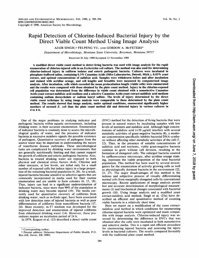
Cambridge Instruments, Buffalo, N.Y.) equipped with aprocessor image analyzer unit (68,000; Cambridge Instru-ments), four independent binary image planes, and a CP-Mcontrolling unit (64,000 RAM). The system was integratedwith an epifluorescence microscope (model BH-2; Olym-pus). The analyzer had the capability of digitizing images in0 (black) to 64 (white) grey levels of variable slices into abinary image with 512 by 480 pixels. Cellular measurementsof individual features such as length and breadth were madein 10 microscopic fields. Field measurements, including thetotal number of features detected, average feature size,standard deviation, and frame area, were also made by thecomputer. The system allowed editing of each image framedisplayed on the screen by the addition of a feature that wasnot detected by the system but that was visible through themicroscope or by separation of the images that were pro-duced by closely placed and entangled cells before finalmeasurements were made. In addition, outputs of measure-ments of different distribution histograms at 0.5-,um intervalswere calculated and printed. Some of the field measurementsand typical distribution histograms of an E. coli culture attime zero and after 4 h of incubation are shown in Fig. IAand B, respectively. DVCs were calculated from the num-bers of elongated cells after incubation in CA-YE-NA me-dium, and calculations were made as follows: DVC/ml =(average elongated cells/field) x (filter area [pum2]/frame area[,.m2]) x dilution.
Injury in chlorine-exposed cultures was assessed by de-termining the difference between the number of viable(enlarged) cells in a nonselective CA-YE-NA medium and aselective CA-YE-NA medium containing sodium deoxycho-late or sodium lauryl sulfate. The nonselective mediumsupported the growth of both uninjured and injured bacteria,whereas the selective medium allowed only uninjured cellsto grow. The results were expressed as percent injury in thetest population.MIC of nalidixic acid. Two methods for estimating the
MIC of nalidixic acid were used. (i) In the microdilutionbroth procedure (11), washed cell suspensions were used toinoculate Mueller-Hinton broth tubes (106 to 107 cells perml). The tubes were incubated at 35°C for 18 h and examined
390 SINGH ET AL.
on June 7, 2018 by guesthttp://aem
.asm.org/
Dow
nloaded from
DETECTION OF INJURY BY IMAGE ANALYSIS 391
TABLE 1. Effect of nalidixic acid on the length and breadth ofE. coli C2 after incubation at 35°C
Incubation Nalidixic acidtime (h) concn (,ug/ml) Length (,m) Breadth (,um)
0 10 1.97 ± 0.82 1.38 + 0.54
4 10 6.05 ± 4.20 1.88 ± 0.844 20 5.76 ± 3.63 1.60 ± 0.514 40 4.49 ± 2.30 1.69 ± 0.53
8 10 6.77 ± 4.60 1.86 ± 0.818 20 6.05 ± 4.06 1.79 ± 0.748 40 4.12 ± 2.31 1.62 ± 0.53
12 10 6.78 ± 4.91 1.83 ± 0.9012 20 6.25 ± 4.20 1.82 ± 0.9512 40 4.51 ± 2.78 1.65 ± 0.74
visually to determine the MIC. (ii) For the disk agar diffusiontest, antibiotic-impregnated disks were prepared by soakingsterile blank disks (diameter; 1/4 in. [7 mm]; Difco) insolutions (20 ,ul per disk) of the desired concentrations ofnalidixic acid. The disks were completely air dried beforeuse. The tests were performed by the procedure of theNational Committee for Clinical Laboratory Standards (21)by using Mueller-Hinton agar.
Statistical analysis. Regression analysis was performed byusing the MSUSTAT statistical analysis package, microcom-puter version 4.10, developed by Richard E. Lund (MontanaState University).
RESULTS
Determination of optimal conditions for DVC. The optimalrecovery and the assessment of injured bacteria depend onthe efficiency of determination of a differential count ofviable bacteria on nonselective and selective media. Thus,conditions for the DVC method were optimized and madecompatible with image analysis. Previous results (25a) indi-cated that the concentration of nalidixic acid (20 ,ug/ml)suggested by Kogure et al. (13) causes an underestimation ofviable cells in uninjured cultures. To determine the effect ofincubation time and nalidixic acid concentrations on cellenlargement, unexposed cultures of E. coli C2 were used.Cells (107/ml) were incubated in CA-YE-NA medium con-taining different concentrations of nalidixic acid. The results(Table 1) show that 10 ,ug of nalidixic acid per ml causedapproximately 1.3- and 3-fold increases in breadths andlengths of cells, respectively, after 4 h of incubation. Further
incubation for up to 12 h did not cause a substantial increasein the mean cell enlargement. Nalidixic acid concentrationslower than 10 jig/ml allowed cell division of E. coli C2cultures and were not suitable for determining viability.However, higher concentrations (>10 ,ug/ml) yielded lowermean cell lengths (Table 1) and may result in underestima-tion of viable cells, as reported elsewhere (25a).
Determination of MICs and optimal levels of nalidixic acidfor different cultures. The MICs of nalidixic acid weredetermined for both unexposed and chlorine-exposed cul-tures in an attempt to find the optimal levels for use with theDVC method. The results are shown in Table 2.
Preliminary trials indicated that the MICs of nalidixic acidobtained by the microdilution or the disk agar diffusionmethod did not cause optimal enlargement of incubatedcells. Thus, different concentrations of nalidixic acid close tothe MICs were used to determine the optimal levels thatwere required for maximal cell enlargement in unexposedand chlorine-exposed cultures (data not shown) by using aprocedure similar to that used for E. coli C2 cultures. TheMIC data provided range-finding values which facilitateddetermination of optimal concentrations of nalidixic acid(Table 2) for the image analysis method. We also measuredthe length and breadth distribution of bacteria in each sample(data not shown) before they were incubated in CA-YE-NAmedium. After incubation, cells longer than the mean lengthat 0 h were considered viable, as shown in Fig. 1A and B forE. coli C2 cultures. Other useful modifications of this pro-cedure have been described elsewhere (25a).Comparison of viable cell enumeration methods in chlorine-
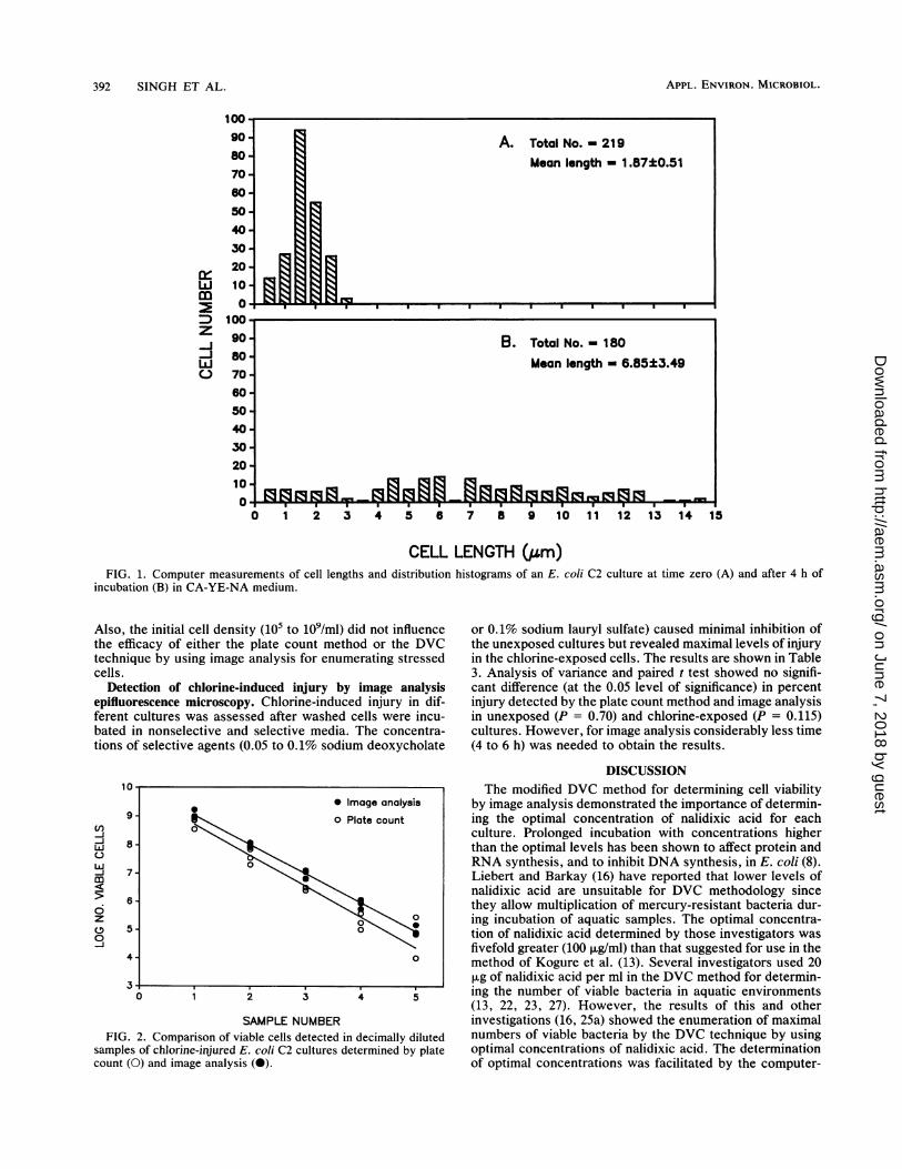
injured cultures of E. coli C2. Comparisons of viable cellenumerations done by plating and image analysis methodswere made in serially diluted strain C2 cultures that werechlorine injured (82 to 87% injury). The viability of cells wasdetermined by both the plate count method by using nonse-lective TLY agar and the DVC method by using CA-YEmedium containing 10 jig of nalidixic acid per ml. The resultsobtained by these two methods are given in Fig. 2. Linearregression analysis showed that both image analysis and theplate count method can be used for the determination ofrelative numbers of viable cells in serially diluted chlorine-injured cell cultures. Further analysis with a reduced modelshowed that a single regression line with one slope and oneintercept was insufficient to fit all the datum points generatedby the two methods (P = 0.00165). Thus, these regressionlines represent different levels of viable cell detection. Fig-ure 2 and statistical analysis indicated that higher numbers ofchlorine-injured E. coli cells could be determined by theimage analysis technique than by the spread plate method.
TABLE 2. MICs of nalidixic acid for uninjured and chlorine-injured cultures and optimal levels used in the DVC method
MIC by: Optimal levels (p.g/ml) used forDVC of the following cells:
Organism Tube dilution (pLg/ml) Disk diffusion (p.g/disk)Uninjured Chlorine-injured
Uninjured Chlorine-injured Uninjured Chlorine-injured
Salmonella typhimurium 16 8 16 16 10 10Yersinia enterocolitica 16 8 8 4 20 10Escherichia coli Cl 8 8 32 16 10 10Escherichia coli C2 16 8 32 16 10 10Vibrio cholerae CVD101 8 8 4 2 2.5 2.5Vibrio cholerae 569B 8 4 2 1 5 2.5Enterobacter cloacae 16 4 32 16 10 2.0Klebsiella pneumoniae 4 4 32 16 10 2.0
VOL. 56, 1990
on June 7, 2018 by guesthttp://aem
.asm.org/
Dow
nloaded from
APPL. ENVIRON. MICROBIOL.
1 2 3 4 5 6 7 8 9 10 11 12 13 14 15
CELL LENGTH (gm)FIG. 1. Computer measurements of cell lengths and distribution histograms of an E. coli C2 culture at time zero (A) and after 4 h of
incubation (B) in CA-YE-NA medium.
Also, the initial cell density (105 to 109/ml) did not influencethe efficacy of either the plate count method or the DVCtechnique by using image analysis for enumerating stressedcells.
Detection of chlorine-induced injury by image analysisepifluorescence microscopy. Chlorine-induced injury in dif-ferent cultures was assessed after washed cells were incu-bated in nonselective and selective media. The concentra-tions of selective agents (0.05 to 0.1% sodium deoxycholate
1U0 Image analysis
count(O)and0 Plate count-J
-I 7
zo 50
4- 0
3I0 1 2 3 4 5
SAMPLE NUMBERFIG. 2. Comparison of viable cells detected in decimally diluted
samples of chlorine-injured E. coli C2 cultures determined by platecount (0) and image analysis (0).
or 0.1% sodium lauryl sulfate) caused minimal inhibition ofthe unexposed cultures but revealed maximal levels of injuryin the chlorine-exposed cells. The results are shown in Table3. Analysis of variance and paired t test showed no signifi-cant difference (at the 0.05 level of significance) in percentinjury detected by the plate count method and image analysisin unexposed (P = 0.70) and chlorine-exposed (P = 0.115)cultures. However, for image analysis considerably less time(4 to 6 h) was needed to obtain the results.
DISCUSSIONThe modified DVC method for determining cell viability
by image analysis demonstrated the importance of determin-ing the optimal concentration of nalidixic acid for eachculture. Prolonged incubation with concentrations higherthan the optimal levels has been shown to affect protein andRNA synthesis, and to inhibit DNA synthesis, in E. coli (8).Liebert and Barkay (16) have reported that lower levels ofnalidixic acid are unsuitable for DVC methodology sincethey allow multiplication of mercury-resistant bacteria dur-ing incubation of aquatic samples. The optimal concentra-tion of nalidixic acid determined by those investigators wasfivefold greater (100 ,ug/ml) than that suggested for use in themethod of Kogure et al. (13). Several investigators used 20pLg of nalidixic acid per ml in the DVC method for determin-ing the number of viable bacteria in aquatic environments(13, 22, 23, 27). However, the results of this and otherinvestigations (16, 25a) showed the enumeration of maximalnumbers of viable bacteria by the DVC technique by usingoptimal concentrations of nalidixic acid. The determinationof optimal concentrations was facilitated by the computer-
LLJm
z-C-J
C.
9o080-70-60-50-40-30-20-10.0
B. Total No. m180Mean length - 6.85±3.49
94 -
0L-.m b;ll MI -
. .r%.] Do
392 SINGH ET AL.
2R0 SI zg rgOn. vq 1:3 rq-IF-
-opon
on June 7, 2018 by guesthttp://aem
.asm.org/
Dow
nloaded from
DETECTION OF INJURY BY IMAGE ANALYSIS 393
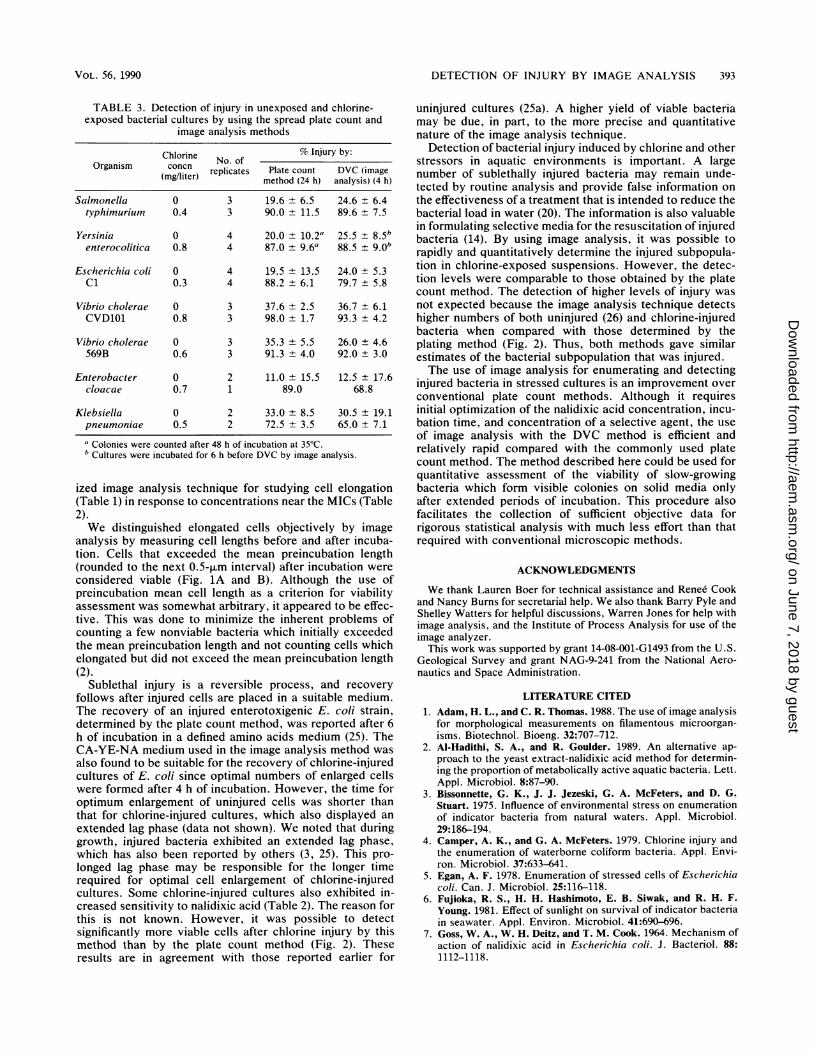
TABLE 3. Detection of injury in unexposed and chlorine-exposed bacterial cultures by using the spread plate count and
image analysis methods
Chlorine No. of % Injury by:Organism concn replicates Plate count DVC (image
(mg/liter) method (24 h) analysis) (4 h)
Salmonella 0 3 19.6 ± 6.5 24.6 ± 6.4typhimurium 0.4 3 90.0 ± 11.5 89.6 ± 7.5
Yersinia 0 4 20.0 ± 10.2a 25.5 ± 8.5benterocolitica 0.8 4 87.0 ± 9.6a 88.5 ± 9.0*
Escherichia coli 0 4 19.5 ± 13.5 24.0 + 5.3C1 0.3 4 88.2 ± 6.1 79.7 ± 5.8
Vibrio cholerae 0 3 37.6 ± 2.5 36.7 ± 6.1CVD101 0.8 3 98.0 ± 1.7 93.3 ± 4.2
Vibrio cholerae 0 3 35.3 ± 5.5 26.0 ± 4.6569B 0.6 3 91.3 ± 4.0 92.0 ± 3.0
Enterobacter 0 2 11.0 ± 15.5 12.5 ± 17.6cloacae 0.7 1 89.0 68.8
Klebsiella 0 2 33.0 ± 8.5 30.5 ± 19.1pneumoniae 0.5 2 72.5 + 3.5 65.0 ± 7.1
a Colonies were counted after 48 h of incubation at 35'C.b Cultures were incubated for 6 h before DVC by image analysis.
ized image analysis technique for studying cell elongation(Table 1) in response to concentrations near the MICs (Table2).We distinguished elongated cells objectively by image
analysis by measuring cell lengths before and after incuba-tion. Cells that exceeded the mean preincubation length(rounded to the next 0.5-pLm interval) after incubation were
considered viable (Fig. 1A and B). Although the use ofpreincubation mean cell length as a criterion for viabilityassessment was somewhat arbitrary, it appeared to be effec-tive. This was done to minimize the inherent problems ofcounting a few nonviable bacteria which initially exceededthe mean preincubation length and not counting cells whichelongated but did not exceed the mean preincubation length(2).
Sublethal injury is a reversible process, and recoveryfollows after injured cells are placed in a suitable medium.The recovery of an injured enterotoxigenic E. coli strain,determined by the plate count method, was reported after 6h of incubation in a defined amino acids medium (25). TheCA-YE-NA medium used in the image analysis method was
also found to be suitable for the recovery of chlorine-injuredcultures of E. coli since optimal numbers of enlarged cellswere formed after 4 h of incubation. However, the time foroptimum enlargement of uninjured cells was shorter thanthat for chlorine-injured cultures, which also displayed an
extended lag phase (data not shown). We noted that duringgrowth, injured bacteria exhibited an extended lag phase,which has also been reported by others (3, 25). This pro-longed lag phase may be responsible for the longer timerequired for optimal cell enlargement of chlorine-injuredcultures. Some chlorine-injured cultures also exhibited in-creased sensitivity to nalidixic acid (Table 2). The reason forthis is not known. However, it was possible to detectsignificantly more viable cells after chlorine injury by thismethod than by the plate count method (Fig. 2). Theseresults are in agreement with those reported earlier for
uninjured cultures (25a). A higher yield of viable bacteriamay be due, in part, to the more precise and quantitativenature of the image analysis technique.
Detection of bacterial injury induced by chlorine and otherstressors in aquatic environments is important. A largenumber of sublethally injured bacteria may remain unde-tected by routine analysis and provide false information onthe effectiveness of a treatment that is intended to reduce thebacterial load in water (20). The information is also valuablein formulating selective media for the resuscitation of injuredbacteria (14). By using image analysis, it was possible torapidly and quantitatively determine the injured subpopula-tion in chlorine-exposed suspensions. However, the detec-tion levels were comparable to those obtained by the platecount method. The detection of higher levels of injury wasnot expected because the image analysis technique detectshigher numbers of both uninjured (26) and chlorine-injuredbacteria when compared with those determined by theplating method (Fig. 2). Thus, both methods gave similarestimates of the bacterial subpopulation that was injured.The use of image analysis for enumerating and detecting
injured bacteria in stressed cultures is an improvement overconventional plate count methods. Although it requiresinitial optimization of the nalidixic acid concentration, incu-bation time, and concentration of a selective agent, the useof image analysis with the DVC method is efficient andrelatively rapid compared with the commonly used platecount method. The method described here could be used forquantitative assessment of the viability of slow-growingbacteria which form visible colonies on solid media onlyafter extended periods of incubation. This procedure alsofacilitates the collection of sufficient objective data forrigorous statistical analysis with much less effort than thatrequired with conventional microscopic methods.
ACKNOWLEDGMENTS
We thank Lauren Boer for technical assistance and Renee Cookand Nancy Burns for secretarial help. We also thank Barry Pyle andShelley Watters for helpful discussions, Warren Jones for help withimage analysis, and the Institute of Process Analysis for use of theimage analyzer.
This work was supported by grant 14-08-001-G1493 from the U.S.Geological Survey and grant NAG-9-241 from the National Aero-nautics and Space Administration.
LITERATURE CITED1. Adam, H. L., and C. R. Thomas. 1988. The use of image analysis
for morphological measurements on filamentous microorgan-isms. Biotechnol. Bioeng. 32:707-712.
2. Al-Hadithi, S. A., and R. Goulder. 1989. An alternative ap-proach to the yeast extract-nalidixic acid method for determin-ing the proportion of metabolically active aquatic bacteria. Lett.Appl. Microbiol. 8:87-90.
3. Bissonnette, G. K., J. J. Jezeski, G. A. McFeters, and D. G.Stuart. 1975. Influence of environmental stress on enumerationof indicator bacteria from natural waters. Appl. Microbiol.29:186-194.
4. Camper, A. K., and G. A. McFeters. 1979. Chlorine injury andthe enumeration of waterborne coliform bacteria. Appl. Envi-ron. Microbiol. 37:633-641.
5. Egan, A. F. 1978. Enumeration of stressed cells of Escherichiacoli. Can. J. Microbiol. 25:116-118.
6. Fujioka, R. S., H. H. Hashimoto, E. B. Siwak, and R. H. F.Young. 1981. Effect of sunlight on survival of indicator bacteriain seawater. Appl. Environ. Microbiol. 41:690-696.
7. Goss, W. A., W. H. Deitz, and T. M. Cook. 1964. Mechanism ofaction of nalidixic acid in Escherichia coli. J. Bacteriol. 88:1112-1118.
VOL. 56, 1990
on June 7, 2018 by guesthttp://aem
.asm.org/
Dow
nloaded from
APPL. ENVIRON. MIcRoBIoL.
8. Goss, W. A., W. H. Deitz, and T. M. Cook. 1965. Mechanism ofaction of nalidixic acid on Escherichia coli. lI. Inhibition ofdeoxyribonucleic acid synthesis. J. Bacteriol. 89:1068-1074.
9. Hobbie, J. E., R. J. Daley, and S. Jasper. 1977. Use ofNuclepore filters for counting bacteria by fluorescence micros-copy. Appl. Environ. Microbiol. 33:1225-1228.
10. Horwitz, M. A., and F. R. Maxfield. 1984. Legionella pneumo-
phila inhibits acidification of its phagosomes in human mono-
cytes. J. Cell Biol. 99:1936-1943.11. Jones, R. N., A. L. Barry, T. L. Gavan, and J. A. Washington II.
1985. Susceptibility tests: microdilution and macrodilutionbroth procedures, p. 972-987. In E. H. Lennette, A. Balows,W. J. Hausler, Jr., and H. J. Shadomy (ed.), Manual of clinicalmicrobiology, 4th ed. American Society for Microbiology,Washington, D.C.
12. Kantor, G. J., and R. A. Deering. 1968. Effect of nalidixic acidand hydroxyurea on division ability of Escherichia coli Fil+ andLon- strains. J. Bacteriol. 95:520-530.
13. Kogure, K., U. Simidu, and N. Taga. 1979. A tentative directmethod for counting living marine bacteria. Can. J. Microbiol.25:415-420.
14. LeChevallier, M. W., S. C. Cameron, and G. A. McFeters. 1983.New medium for improved recovery of coliform bacteria fromdrinking water. Appl. Environ. Microbiol. 45:484-492.
15. LeChevallier, M. W., and G. A. McFeters. 1985. Enumeration ofinjured coliforms in drinking water. J. Am. Water Works Assoc.77:81-87.
16. Liebert, C., and T. Barkay. 1988. A direct viable countingmethod for measuring tolerance of aquatic microbial communi-ties to Hg2+. Can. J. Microbiol. 34:1090-1095.
17. McFeters, G. A., S. C. Cameron, and M. W. LeChevallier. 1982.Influence of diluents, media, and membrane filters on thedetection of injured waterborne coliform bacteria. Appl. Envi-ron. Microbiol. 43:97-103.
18. McFeters, G. A., and A. K. Camper. 1983. Enumeration of
indicator bacteria exposed to chlorine. Adv. Appi. Microbiol.29:171-193.
19. McFeters, G. A., J. S. Kippin, and M. W. LeChevallier. 1986.Injured coliforms in drinking water. Appl. Environ. Microbiol.51:1-5.
20. McFeters, G. A., M. W. LeChevallier, A. Singh, and J. S.Kippin. 1986. Health significance and occurrence of injuredbacteria in drinking water. Water Sci. Technol. 18:227-231.
21. National Committee for Clinical Laboratory Standards. 1984.Performance standards for antimicrobial disk susceptibilitytests. Approved standard M2-A3. National Committee for Clin-ical Laboratory Standards, Villanova, Pa.
22. Peele, E. R., and R. R. Colwell. 1981. Application of a directmicroscopic method of enumeration of substrate-responsivemarine bacteria. Can. J. Microbiol. 27:1071-1075.
23. Roszak, D. B., D. J. Grimes, and R. R. Colwell. 1984. Viable butnon-recoverable stage of Salmonella enteritidis in aquatic sys-tems. Can. J. Microbiol. 30:334-338.
24. Schiff, L. J., S. M. Morrison, and J. V. Mayevx. 1970. Syner-gistic false-positive coliform reaction on m-Endo MF medium.Appl. Environ. Microbiol. 20:778-781.
25. Singh, A., and G. A. McFeters. 1986. Recovery, growth, andproduction of heat-stable enterotoxin by Escherichia coli aftercopper-induced injury. Appl. Environ. Microbiol. 51:738-742.
25a.Singh, A., B. H. Pyle, and G. A. McFeters. 1989. Rapid enumer-ation of viable bacteria by image analysis. J. Microbiol. Meth-ods 10:91-101.
26. Singh, A., R. Yeager, and G. A. McFeters. 1986. Assessment ofin vivo revival, growth, and pathogenicity of Escherichia colistrains after copper- and chlorine-induced injury. Appl. Envi-ron. Microbiol. 52:832-837.
27. Xu, H.-S., N. Roberts, F. L. Singleton, R. W. Attwell, D. J.Grimes, and R. R. Colwell. 1982. Survival and viability ofnon-culturable Escherichia coli and Vibrio cholerae in theestuarine and marine environment. Microb. Ecol. 8:313-323.
394 SINGH ET AL.
on June 7, 2018 by guesthttp://aem
.asm.org/
Dow
nloaded from
ERRATA
Rapid Detection of Chlorine-Induced Bacterial Injury by the Direct ViableCount Method Using Image AnalysisAJAIB SINGH, FEI-PENG YU, AND GORDON A. McFETERS
Department of Microbiology, Montana State University, Bozeman, Montana 59717
Volume 56, no. 2, p. 590, column 2, lines 3 to 16: These sentences should read as follows. "The withdrawn samples wereheated in a boiling water bath for 5 min and cooled rapidly by immersing them in ice-cold water. Samples were filtered throughpolycarbonate (pore size, 0.2 ,um; diameter, 25 mm; black membranes; Nuclepore Corp., Pleasanton, Calif.) by applying a lowfiltration pressure to ensure uniform spreading of bacteria on the filter. The retained bacteria were washed with 5 ml of waterand fixed by passing 3 to 5 ml of4% Formalin through the filter, allowing 1 to 2 min of contact. The fixed bacteria were washedwith water as described above and stained by slowly passing through the filter 2 to 5 ml of 0.02% acridine orange solution,allowing 2 to 3 min of contact. Finally, 3 to 5 ml of wash water was passed through the filter to remove unbound stain."
Nucleotide Sequence and Distribution of the pTR2030 Resistance Determinant(hsp) Which Aborts Bacteriophage Infection in Lactococci
COLIN HILL, LYNETTE A. MILLER, AND TODD R. KLAENHAMMER
Departments ofFood Science and Microbiology, Southeast Dairy Foods Research Center,North Carolina State University, Raleigh, North Carolina 27695-7624
Volume 56, no. 7, p. 2257, Fig. 2: nucleotide 1735, "C," should read "G"; the corresponding amino acid residue, "L,"should read "V."
2949