Embed Size (px)
Citation preview
Neurobiology of Disease
Rapamycin Delays Disease Onset and Prevents PrP PlaqueDeposition in a Mouse Model of Gerstmann–Straussler–Scheinker Disease
Constanza J. Cortes, Kefeng Qin, Julie Cook, Ani Solanki, and James A. MastrianniDepartment of Neurology, The University of Chicago Pritzker School of Medicine, Chicago, Illinois 60637
Autophagy is a cell survival response to nutrient deprivation that delivers cellular components to lysosomes for digestion. In recent years,autophagy has also been shown to assist in the degradation of misfolded proteins linked to neurodegenerative disease (Ross and Poirier,2004). In support of this, rapamycin, an autophagy inducer, improves the phenotype of several animal models of neurodegenerativedisease. Our Tg(PrP-A116V) mice model Gerstmann–Straussler–Scheinker disease (GSS), a genetic prion disease characterized by prom-inent ataxia and extracellular PrP amyloid plaque deposits in brain (Yang et al., 2009). To determine whether autophagy induction canmitigate the development of GSS, Tg(PrP-A116V) mice were chronically treated with 10 or 20 mg/kg rapamycin intraperitoneally thriceweekly, beginning at 6 weeks of age. We observed a dose-related delay in disease onset, a reduction in symptom severity, and an extensionof survival in rapamycin-treated Tg(PrP-A116V) mice. Coincident with this response was an increase in the autophagy-specific markerLC3II, a reduction in insoluble PrP-A116V, and a near-complete absence of PrP amyloid plaques in the brain. An increase in glial cellapoptosis of unclear significance was also detected. These findings suggest autophagy induction enhances elimination of misfolded PrPbefore its accumulation in plaques. Because ataxia persisted in these mice despite the absence of plaque deposits, our findings alsosuggest that PrP plaque pathology, a histopathological marker for the diagnosis of GSS, is not essential for the GSS phenotype.
IntroductionAutophagy is a cell survival mechanism whereby cellular compo-nents are phagocytized by double membrane autophagosomesthat transport cargo to lysosomes for degradation. Initially rec-ognized as a cellular response to nutrient deprivation, autophagyhas since been shown to participate in the clearance of aggregate-prone cytosolic proteins linked to a variety of neurodegenerativedisorders (Ross and Poirier, 2004). Autophagy is negativelyregulated by the mammalian target of rapamycin (mTOR)(Schmelzle and Hall, 2000). Rapamycin is a lipophilic macrolideantibiotic (Noda and Ohsumi, 1998) that inhibits mTOR andpotently induces autophagy. Numerous reports describe protec-tive effects of rapamycin in cell, fly, and mouse models of severalneurodegenerative diseases that result from accumulation ofmisfolded aggregate-prone cytosolic proteins, including Parkin-son’s disease (PD) (Webb et al., 2003), amyotrophic lateral scle-rosis (Fornai et al., 2008), Huntington’s disease (HD) (Berger et
al., 2006), spinocerebellar ataxia (Ravikumar et al., 2002, 2004),and frontotemporal dementia (Williams et al., 2006). Despite thelack of cytosolic aggregates in Alzheimer’s disease (AD), au-tophagy appears to play a complex role in A� production andclearance, and recent work suggests a beneficial effect of rapamy-cin in some AD mouse models (Nixon, 2007; Spilman et al., 2010;Yang et al., 2011).
Prion diseases are transmissible neurodegenerative disor-ders linked to accumulation of misfolded pathogenic isoforms(PrP Sc) of normal prion protein (PrP C) (Prusiner, 1998). Phar-macologic induction of autophagy appears to promote the clear-ance of PrP Sc in vitro (Heiseke et al., 2009a,b), and a limitednumber of in vivo studies have found either no effect (Sarkar etal., 2007) or a modest effect (Heiseke et al., 2009b) to prolongsurvival following scrapie infection. However, whether au-tophagy induction can delay the development of genetic priondisease has not been addressed.
Gerstmann–Straussler–Scheinker disease (GSS) is a geneticprion disease, typified by the onset of progressive ataxia and thepresence of PrP amyloid plaque deposits in brain, a feature rem-iniscent of AD. We previously constructed the Tg(PrP-A116V)transgenic mouse line that recapitulates the major clinicopatho-logic features of GSS, notably the onset of progressive ataxia andaccumulation of thioflavin S-positive PrP amyloid plaques, espe-cially within the cerebellum (Yang et al., 2009).
To determine whether autophagy functions in genetic priondisease and amyloid plaque pathology, we chronically treatedTg(PrP-A116V) mice with rapamycin, beginning at 6 weeks ofage. We report a dose-dependent delay in disease onset, symptom
Received Dec. 13, 2011; revised July 11, 2012; accepted July 20, 2012.Author contributions: C.J.C. and J.A.M. designed research; C.J.C., K.Q., J.C., and A.S. performed research; A.S.
contributed unpublished reagents/analytic tools; C.J.C., K.Q., and J.A.M. analyzed data; C.J.C., K.Q., and J.A.M. wrotethe paper.
This work was supported by NIH Grant R01NS051480 and The Brain Research Foundation (J.A.M.).The authors declare no competing financial interests.Correspondence should be addressed to Dr. James A. Mastrianni, Prion Laboratory, Department of Neurology,
MC2030, University of Chicago Pritzker School of Medicine, 5841 South Maryland Avenue, Chicago, IL 60637. E-mail:[email protected].
C. J. Cortes’s present address: Department of Pediatrics, University of California, San Diego, San Diego, CA 92037.J. Cook’s present address: Indiana University School of Medicine Northwest, Gary, IN 46408.DOI:10.1523/JNEUROSCI.6189-11.2012
Copyright © 2012 the authors 0270-6474/12/3212396-10$15.00/0
12396 • The Journal of Neuroscience, September 5, 2012 • 32(36):12396 –12405

severity, and improved survival of rapamycin-treated Tg(PrP-A116V) mice. These improvements were accompanied by anincrease of the specific autophagy marker LC3II in brain, areduction in insoluble PrP-A116V, a near-total absence of PrPamyloid plaques, and interestingly, but of unclear signifi-cance, an increase in glial cell apoptosis.
Our results suggest a role for autophagy in genetic prion dis-ease and extracellular plaque generation, and they support au-tophagy induction as a potential therapeutic approach. Theconspicuous absence of plaque pathology yet persistence ofataxia, albeit with reduced severity, supports the concept that PrPamyloid plaque deposits, while pathognomonic for GSS, are notrequired for the GSS phenotype.
Materials and MethodsTransgenic miceThe Tg(PrP-A116V) mice have been described previously (Yang et al.,2009). In short, the A116V mutation and 128V polymorphism were in-troduced into the mouse Prnp coding segment by site-directed mutagen-esis, ligated into the XhoI-cut MoPrP.Xho vector (ATCC JHU-2;American Type Culture Collection), and injected into pronuclei of fer-tilized eggs from PrP knock-out [Tg(Prnp o/o)] parental mice at the Uni-versity of Chicago Transgenic Mouse Facility. A founder that expressedapproximately four to six times normal mouse PrP were bred to Tg-(Prnp o/o) mice, so they express only the mutated mouse PrP-A116V/128V protein. Male and female mice were used randomly throughout thestudy.
Rapamycin injectionsRapamycin (Sirolimus; LC Laboratories) was dissolved in pure ethanol asa 20 mg/ml stock solution and diluted in injection buffer (4% ethanol,5% Tween 80, 5% PEG400 in dH2O) on the day of injection to a dose of10 or 20 mg/kg in a final volume of �0.1 ml. Mice were weighed weeklythroughout the experiment, and dosages were adjusted accordingly.Control mice received a similar volume of injection buffer without activedrug. Beginning at 6 weeks of age, injections were administered intra-peritoneally Monday, Wednesday, and Friday, until killed for analysis.
Clinical assessmentAt the start of the treatment, mice were monitored daily for typical signsof prion disease, including gait ataxia, roughened fur, hunched posture,and poor righting reflex. We developed a detailed clinical assessmentscale to monitor the progression of disease. Since ataxia and mobilityproblems are key features of this mouse model, these features are heavilyweighted in the assessment. The following scoring from A0 to A6 is used:A0, no ataxia; A0/1, no ataxia, but may show other signs such as rough furor walking lower to the ground; A1, more consistent, but still a subtlechange in gait (wider, lower to ground), but not definitive ataxia; A1/A2,subtle but definite wobble that is intermittent; A2, persistent and obviouswobble—this stage defines the clear onset of ataxia; A3, stumble/loss offooting occasionally (once or twice during a 2 min observation); A4,stumble/loss of footing every few steps; mouse may occasionally start tosway (rock back and forth) when in a stationary position; A4/A5, early-stage A5, with brief spells of falling to one side and more swaying; A5,falling nearly constantly, weight loss generally obvious, clear change incoat roughness; A6, lethargic, very hunched, emaciated. Mice are gener-ally killed during stage A5 or early in A6 (A5/A6), as they can no longerfeed and death is imminent. Ataxia typically develops at �130 –140 d ofage.
PrP analysisWestern blots. Fresh frozen brain was used to prepare 10% (w/v) brainhomogenates in lysis buffer (20 mM Tris-HCl, pH 7.4, 150 mM NaCl, 1mM EDTA, 0.5% Triton X-100, 0.5% Na-deoxycholate). Brain homoge-nate (5 or 2%) was separated by SDS-PAGE on a 14% gel. Followingelectrophoresis, samples were transferred to polyvinylidene difluoridemembranes (Bio-Rad) that were washed in TBST (20 mM Tris-HCl, 0.9%NaCl, 0.1% Tween 20, pH 7.6), blocked for 1 h with 5% milk, washed,
and incubated in TBST with 1% milk containing anti-mouse PrP anti-bodies D13 (InPro) at 1:3000 or SAF32 (SPI Bio) at 1:200, or antibodiesfor LC3B (Cell Signaling) at 1:1000, or �-tubulin (Santa Cruz) at1:10,000, all incubated overnight at 4°C. After washing, secondary anti-bodies (anti-human-HRP for D13, anti-mouse-HRP for SAF32, and�-tubulin) were added at concentrations from 1:5000 to 1:2000. Blotswere imaged with West Pico ECL (Thermo Fisher Scientific) and cap-tured with a Bio-Rad Alpha Document Imager. Densitometry quantifi-cations were performed using NIH ImageJ or TotalLab TL100 software(TotalLab).
Solubility assay. Brain homogenates were prepared in lysis buffer. Afraction was removed for analysis and centrifuged at 4°C for 1 h at100,000 � g in a Sorvall RC M120EX ultramicrocentrifuge, using aRP100-AT4 rotor. The pellet was washed once with lysis buffer, centri-fuged, and resuspended in the starting volume of lysis buffer containing1% SDS. Equal supernatant and pellet fractions were subjected to West-ern blotting and densitometry analysis, as above.
Histological studiesCerebellum sections. Mice were asphyxiated with CO2 and slowly perfusedvia cardiac puncture with 20 ml of PBS followed by 20 ml of 4% parafor-maldehyde (Sigma-Aldrich). Brains were stored in 4% paraformalde-hyde for 48 h and then transferred to PBS containing 0.1% sodium azide(Sigma-Aldrich) until paraffin embedding. The blocks of mouse cere-bella were cut at similar landmarks of the cerebellum into 5 �m coronalsections. Sections were depariffinized with xylene (Thermo Fisher Scien-tific) immediately before staining.
Thioflavin S staining. Sections were stained with 0.05% thioflavin S(Sigma-Aldrich) for detection of PrP amyloid deposits and with 10�g/ml 4�,6-diamidino-2-phenyl-indole, dihydrochloride (DAPI) (Invit-rogen) to visualize nuclei, and then observed with a Zeiss Axioplan flu-orescence microscope at excitation wavelength of 520 nm for thioflavin Sand 450 nm for DAPI. Plaque burden was calculated as the area of thio-flavin S staining per area of entire cerebellar section, using NIH ImageJ.Several sections were analyzed from each group to obtain the mean areaof thioflavin S staining per cerebellar area for each treatment group. Theaverage fraction of plaque area per cerebellar area of the 160-d-oldvehicle-treated mice was normalized to a value of 100, with which allother groups were compared. The data are presented as the relativeplaque burden. The absolute plaque counts were zero in all sectionsexamined in mice receiving rapamycin at 20 mg/kg.
Terminal deoxynucleotidyl transferase-mediated biotinylated UTP nickend labeling. DNA fragmentation was assessed with the terminal deoxy-nucleotidyl transferase-mediated biotinylated UTP nick end labeling(TUNEL) apoptosis detection kit (Millipore), following the manufactur-er’s instruction. Briefly, sections were incubated with proteinase K for 30min. After incubation with a reaction mix containing biotin-dUTP andthe terminal deoxynucleotidyl transferase (TdT) for 60 min, sectionswere incubated with the avidin–FITC solution for 30 min in the dark. Aspositive control, the fixed sections were incubated with 5 �g/ml DNase Iin PBS for 60 min at 37°C before labeling following the manufacturer’sinstruction. Sections treated with the TdT reaction mix containing noTdT were used as negative controls. For nuclear staining, sections wereincubated with 10 �g/ml DAPI (Sigma-Aldrich) at room temperature for1 min. Sections were washed with PBS, air dried, mounted, and observedwith a Zeiss Axioplan fluorescence microscope at excitation wavelengthof 520 nm for FITC and 450 nm for DAPI. For quantitative analysis ofapoptosis, the percentage area of TUNEL-positive signal relative to thearea of DAPI stain in each cerebellar section was calculated, using NIHImageJ, and the mean determined and plotted.
Immunofluorescence of NeuN and GFAP. Brain sections were firstTUNEL stained, with the proteinase K step eliminated, blocked with 2%BSA in PBS for 1 h, and then incubated with rabbit anti-glial fibrillaryacidic protein (GFAP) antibody (1:100) (Dako) or mouse anti-NeuNantibody (1:50) (Millipore Bioscience Research Reagents) overnight. Dy-Light 649-conjugated AffiniPure goat anti-rabbit or mouse IgG, F(ab�)was used as secondary antibody. Results were visualized by a fluorescencemicroscopy and verified under a confocal microscope (BX61; Olympus).
Cortes et al. • Rapamycin Improves the Phenotype of GSS Mice J. Neurosci., September 5, 2012 • 32(36):12396 –12405 • 12397

Semiquantitative RT-PCRBrains of Tg(PrP-A116V) mice treated with rapamycin (0, 10, or 20mg/kg) were harvested and quick frozen at �80°C. Total RNA was ex-tracted using the RNAqueous-4PCR kit (Ambion). A total of 500 ng ofRNA from each sample was used for RT-PCR using SuperScript One-Step RT-PCR with Platinum Tag (Invitrogen) to synthesize cDNA ofmouse PrP and LC3. �-Actin was used as control. The PCR primers weredesigned as follows: mouse PrP C, forward, 5�-TGG GGA CAA CCT CATGGT GGT, and reverse, 5�-GAT ATT GAC GCA GTC GTG CAC-3�(expected PCR product, 329 bp), mouse LC3, forward, 5�-TTC AAGCAG CGG CGC ACG TTC-3�, and reverse, 5�-CTC CTC TTG ACT CAGAAG CCG AAG GTT-3� (expected PCR product, 360 bp), mouse�-actin, forward, 5�-TGG AAT CCT GTG GCA TCC ATG AAA-3�, andreverse, 5�-TAA AAC GCA GCT CAG TAA CAG TCC G (expected PCRproduct, 348 bp). The PCR products were separated on a 1% 1� Tris-acetate EDTA (Tris-acetate EDTA)-agarose gel and analyzed on a docu-ment imaging system (Bio-Rad). Densitometric analysis was performedusing NIH ImageJ. The mean signal of PrP and LC3 cDNA from threeexperiments each were normalized to �-actin cDNA, and the relativepercentage change in PrP or LC3 mRNA among the three treatmentgroups was reported.
ResultsRapamycin impairs weight gain of Tg(PrP-A116V) micePrior work suggests that rapamycin impairs weight gain in ro-dents (Walpoth et al., 2001; Ravikumar et al., 2004). To assess forthis possibility in Tg(PrP-A116V) mice, weight measurementswere recorded weekly from a presymptomatic period of 100 d.When assessed as a group, mice treated with rapamycin at 20mg/kg had similar weights as vehicle-treated mice over the entiremonitoring period (Fig. 1A); however, when separated by gen-
der, there were differences. From the beginning of the weightassessment period and at each time point thereafter, males receiv-ing either dose of rapamycin weighed less than vehicle-treatedmales (Fig. 1B). This suggests an initial reduction in weight gainfrom the start of injections at 6 weeks until the start of the record-ing period at 100 d, but no differential effect on weight thereafter.The 10 mg/kg dose of rapamycin resulted in a statistically similareffect as the 20 mg/kg dose (data not shown). In contrast,rapamycin-treated female Tg(PrP-A116V) mice weighed slightlymore at the beginning of the measurement period, and slightlyless at the end of the measurement period, but at none of the timepoints did this difference reach statistical significance (Fig. 1B).At necropsy, the mean brain weight of the entire group ofrapamycin-treated Tg(PrP-A116V) mice was lower than vehicle-treated mice (p � 0.001) (Fig. 1C), but when corrected for bodyweight and separated by gender, this difference barely reachedsignificance for male mice (p � 0.0496) and was not significantfor female mice (Fig. 1D). All doses of rapamycin were thereforecalculated for each mouse individually, based on its weight at thetime of dosing.
Rapamycin delays disease onset and improves clinicalphenotype of Tg(PrP-A116V) miceTg(PrP-A116V) mice typically exhibit normal behavior and ac-tivity until �5 months of age, when they begin to display earlysigns of gait ataxia, as the primary manifestation of GSS (Yang etal., 2009). The ataxia progresses steadily, culminating in severedisability and death, typically by �30 d. To assess the effect ofrapamycin on the onset, progression, and duration of disease inthese mice, we developed a scaled scoring system that ranges fromA0 (no disease) to A6 (terminal). Based on this scale, the onset ofdisease is defined as A2, when ataxia is clearly present (see Mate-rials and Methods for details of the ataxia assessment), and miceare killed at the end of stage A5 or the beginning of stage 6, whenthey are unable to appropriately feed.
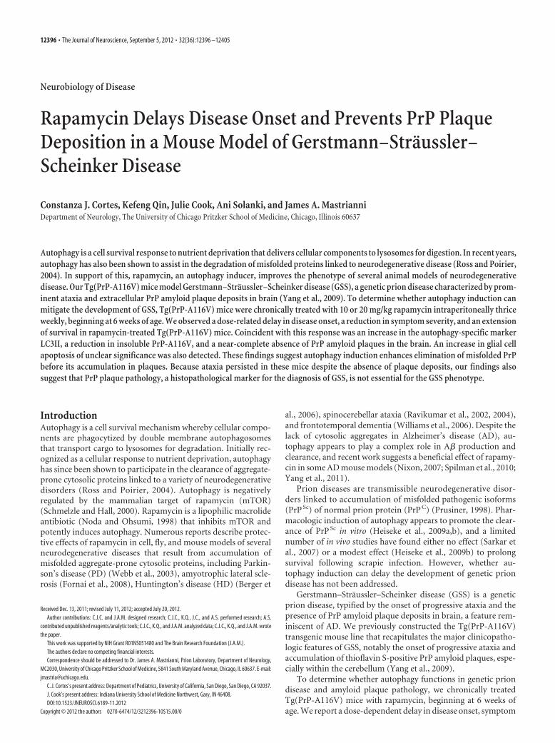
The mean age at disease onset of vehicle-injected mice was134 � 2.6 d, which agrees well with that of untreated Tg(PrP-A116V) mice (Yang et al., 2009), suggesting the stress of injec-tions and handling did not influence disease onset (Fig. 2A). Themean age of onset was significantly delayed in mice treated withrapamycin, in a dose-related manner; mice receiving 10 mg/kgrapamycin had an onset of 149 � 3.7 d (p � 0.05, compared withvehicle) and those treated with 20 mg/kg had an onset of 159 �4.2 d (p � 0.0001, compared with vehicle). While these numbersreflect a 10 and 18% delay in disease onset, respectively, and reachdifferent levels of significance, using a Bonferroni post-test anal-ysis, they were not statistically significantly different from eachother.
To assess the effect of rapamycin on the progression of diseasein Tg(PrP-A116V) mice, we calculated the average disabilityscore of mice within each treatment group at several time points,beginning at 120 d, just before the typical onset of disease, until190 d, when the majority of vehicle-treated mice were dead. Atmost time points, a significant dose-related improvement in dis-ability scores was evident, suggesting that rapamycin shifts thedisability curve to the right (Fig. 2B). These data are also visuallypresented as a bar graph that displays the percentage of micewithin each clinical disability stage at each time point (Fig. 2C–E).Rapamycin-treated mice show a predominance of lighter sec-tions (lower disability scores), compared with vehicle-treatedmice throughout the observation period. (Fig. 2C–E).
Figure 1. Rapamycin reduces weight gain in male Tg(PrP-A116V) mice. A, Averageweight � SE, in grams, of all mice treated with rapamycin at 20 mg/kg or vehicle from 6 weeksof age, measured between 100 (presymptomatic) and 180 (symptomatic) days of age (n � 12per group). No significant differences at any time point. B, Data from A, but separated bygender. Males (squares) and females (triangles) treated with rapamycin, compared withvehicle-treated, mice within the same gender group. Rapamycin-treated males weighed sig-nificantly less than vehicle-treated males at the start of the weight recordings, suggestingreduced weight gain with the initiation of rapamycin. *p � 0.05, Student’s t test, measured ateach time point. Data from mice receiving 10 mg/kg rapamycin are not shown, to simplify thefigure, but no statistical difference compared with 20 mg/kg was detected. C, Brain weight (ingrams) from �160-d-old male and female Tg(PrP-A116V) mice treated with vehicle or rapa-mycin at 20 mg/kg (n � 16 each, 10 males, 6 females). **p � 0.001, Student’s t test. D, Ratioof brain to body weight in 160-d-old Tg(PrP-A116V) male and female mice treated with vehicleor rapamycin at 20 mg/kg (n � 6 each). *p � 0.0496, Student’s t test.
12398 • J. Neurosci., September 5, 2012 • 32(36):12396 –12405 Cortes et al. • Rapamycin Improves the Phenotype of GSS Mice
Rapamycin improves survival of Tg(PrP-A116V) miceTg(PrP-A116V) mice are killed at the terminal clinical stage ofdisease (end of A5, beginning of A6), when the mice are severelydisabled and no longer able to feed themselves. We comparedsurvival times among the three treatment groups to determinewhether rapamycin delays death in Tg(PrP-A116V) mice. Sur-vival curves for each group are plotted in Figure 3A. A significant(p � 0.018) rightward shift in the survival curve was present in
mice that received 20 mg/kg rapamycin,but not in those receiving 10 mg/kg (p �0.7). A similar relationship was found bycomparing the mean age at death of thethree treatment groups. Whereas 20mg/kg rapamycin significantly delayedthe mean age at death to 189 � 3.8 d, com-pared with 173 � 3.7 d for the vehicle-treated group, the mean age at death forthe 10 mg/kg dose was 175 � 3.8 d,which did not differ from the controlgroup (Fig. 3B).
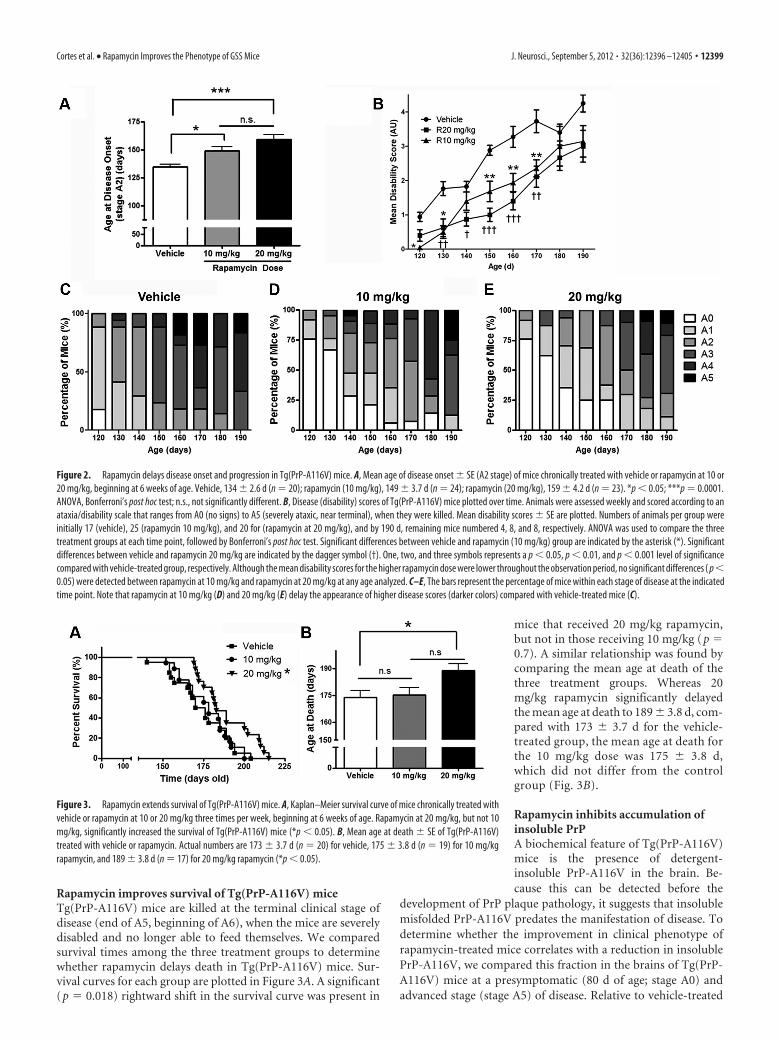
Rapamycin inhibits accumulation ofinsoluble PrPA biochemical feature of Tg(PrP-A116V)mice is the presence of detergent-insoluble PrP-A116V in the brain. Be-cause this can be detected before the
development of PrP plaque pathology, it suggests that insolublemisfolded PrP-A116V predates the manifestation of disease. Todetermine whether the improvement in clinical phenotype ofrapamycin-treated mice correlates with a reduction in insolublePrP-A116V, we compared this fraction in the brains of Tg(PrP-A116V) mice at a presymptomatic (80 d of age; stage A0) andadvanced stage (stage A5) of disease. Relative to vehicle-treated
Figure 3. Rapamycin extends survival of Tg(PrP-A116V) mice. A, Kaplan–Meier survival curve of mice chronically treated withvehicle or rapamycin at 10 or 20 mg/kg three times per week, beginning at 6 weeks of age. Rapamycin at 20 mg/kg, but not 10mg/kg, significantly increased the survival of Tg(PrP-A116V) mice (*p � 0.05). B, Mean age at death � SE of Tg(PrP-A116V)treated with vehicle or rapamycin. Actual numbers are 173 � 3.7 d (n � 20) for vehicle, 175 � 3.8 d (n � 19) for 10 mg/kgrapamycin, and 189 � 3.8 d (n � 17) for 20 mg/kg rapamycin (*p � 0.05).
Figure 2. Rapamycin delays disease onset and progression in Tg(PrP-A116V) mice. A, Mean age of disease onset � SE (A2 stage) of mice chronically treated with vehicle or rapamycin at 10 or20 mg/kg, beginning at 6 weeks of age. Vehicle, 134 � 2.6 d (n � 20); rapamycin (10 mg/kg), 149 � 3.7 d (n � 24); rapamycin (20 mg/kg), 159 � 4.2 d (n � 23). *p � 0.05; ***p � 0.0001.ANOVA, Bonferroni’s post hoc test; n.s., not significantly different. B, Disease (disability) scores of Tg(PrP-A116V) mice plotted over time. Animals were assessed weekly and scored according to anataxia/disability scale that ranges from A0 (no signs) to A5 (severely ataxic, near terminal), when they were killed. Mean disability scores � SE are plotted. Numbers of animals per group wereinitially 17 (vehicle), 25 (rapamycin 10 mg/kg), and 20 for (rapamycin at 20 mg/kg), and by 190 d, remaining mice numbered 4, 8, and 8, respectively. ANOVA was used to compare the threetreatment groups at each time point, followed by Bonferroni’s post hoc test. Significant differences between vehicle and rapamycin (10 mg/kg) group are indicated by the asterisk (*). Significantdifferences between vehicle and rapamycin 20 mg/kg are indicated by the dagger symbol (†). One, two, and three symbols represents a p � 0.05, p � 0.01, and p � 0.001 level of significancecompared with vehicle-treated group, respectively. Although the mean disability scores for the higher rapamycin dose were lower throughout the observation period, no significant differences ( p�0.05) were detected between rapamycin at 10 mg/kg and rapamycin at 20 mg/kg at any age analyzed. C–E, The bars represent the percentage of mice within each stage of disease at the indicatedtime point. Note that rapamycin at 10 mg/kg (D) and 20 mg/kg (E) delay the appearance of higher disease scores (darker colors) compared with vehicle-treated mice (C).
Cortes et al. • Rapamycin Improves the Phenotype of GSS Mice J. Neurosci., September 5, 2012 • 32(36):12396 –12405 • 12399
Tg(PrP-A116V) mice, rapamycin treat-ment was associated with a dose-relatedreduction in the fraction of brain-derivedinsoluble PrP-A116V in both presymp-tomatic (Fig. 4A) and advanced stagesymptomatic (Fig. 4B) mice. This findingnot only suggests that rapamycin acts toreduce the overall load of insoluble PrPthroughout the course of disease, but thatthe symptoms of advanced disease develop,albeit with a notable delay, despite the sig-nificantly lower levels of insoluble PrP.
Rapamycin inhibits PrP amyloidplaque generationThe pathognomonic feature of GSS in hu-mans, and the most prominent histo-pathologic feature in Tg(PrP-A116V)mice, is the deposition of PrP amyloidplaques, especially within the granularlayer of the cerebellum (Yang et al., 2009).In Tg(PrP-A116V) mice, plaques are de-tectable by �100 d of age and the numberof plaques increase with disease progres-sion. Because they are true amyloid, theycan be detected by thioflavin S. To deter-mine whether the reduction in insolublePrP-A116V and mitigation of the clinicalphenotype are tied to a reduction in PrPplaque deposition, we compared theplaque burden within the cerebella ofmice from each treatment group at �160 dof age, when �50% of rapamycin-treated mice were clearly symp-tomatic (i.e., reached stage A2). The average area of thioflavin Sstaining per total area of cerebellar tissue section was measured usingImageJ. Vehicle-treated Tg(PrP-A116V) mice displayed well devel-oped and numerous plaques distributed throughout the cerebellargranule cell layer (Fig. 5A, top row). In stark contrast, mice treatedwith rapamycin were nearly or completely devoid of plaques. In micereceiving 10 mg/kg, the measured plaque area was 28.7 � 9.6% (n �4; p�0.01) of that measured in vehicle-treated mice (Fig. 5A, secondrow and graph), whereas in mice receiving 20 mg/kg, no plaqueswere detected in any sections examined (n�6) (Fig. 5A, bottom rowand graph).
We questioned whether rapamycin delays the production ofplaques in Tg(PrP-A116V) mice or it persistently suppresses thegeneration of plaques throughout the course of disease, as sug-gested by the insolubility data in Figure 4. To determine this, weexamined the plaque burden within the cerebella of mice thatreceived 20 mg/kg rapamycin and were allowed to survive toterminal stages of disease. As expected, the plaque burden ofvehicle-treated mice was increased, relative to that in 160-d-oldvehicle-treated mice (125.3�14.1%). However, surprisingly, termi-nal mice treated with rapamycin were devoid of plaque deposits (Fig.5B and graph). These findings not only suggests that the generationof plaques is prevented by rapamycin, but they also support theconcept that plaque pathology is not required for the GSS pheno-type, since rapamycin-treated mice developed ataxia and eventuallyreached end-stage levels of disability.
PrP expression is not affected by rapamycinIn addition to its role as an autophagy supressor, mTOR is also amaster regulator of protein translation, via its downstream tar-
gets p70s6k and 4E-BP1 (Bove et al., 2011). To investigatewhether the decrease in insoluble PrP-A116V and the markedreduction in plaque deposition resulted from reduced PrP ex-pression, we analyzed the effect of each treatment on the steady-state level of PrP-A116V protein and mRNA. Western blot ofdetergent-extracted lysates of brain homogenate from vehicleand rapamycin-treated mice revealed comparable levels of PrP(Fig. 6A). Similarly, semiquantitative RT-PCR did not show asignificant difference in the level of PrP-A116V transcriptsamong the treatment groups (Fig. 6B). Although a slight reduc-tion in PrP-A116V mRNA relative to actin mRNA was present inmice treated with 10 mg/kg (92.8 � 15.7% of baseline) and 20mg/kg (91.4 � 5.0% of baseline) rapamycin, these were not sig-nificantly different from vehicle-injected mice (p � 0.05 forboth), and not likely to account for the striking reduction inplaque deposits observed in rapamycin-treated mice.
LC3 is increased in the brain of rapamycin-treatedTg(PrP-A116V) miceTo confirm that intraperitoneal administration of rapamycin wassufficient to activate autophagy within the brain of Tg(PrP-A116V) mice, we probed whole-brain lysates prepared from�160-d-old mice for typical markers of autophagy. As shown inFigure 7A, LC3II, a component of the autophagosome mem-brane and a specific marker for autophagy activation, was signif-icantly increased in the brains of rapamycin-treated mice relativeto vehicle-treated brains. Increased levels of LC3II might reflectincreased autophagosome formation or decreased autophagicflux. Because the total signal of LC3 was much greater in micetreated with rapamycin, we questioned whether rapamycin al-tered the expression of LC3. RT-PCR of LC3 revealed a dramatic
Figure 4. Rapamycin reduces the fraction of insoluble PrP-A116V. A, Western blot of supernatant (S) and pellet (P) fractions ofPrP-A116V prepared from brain lysates of asymptomatic (stage A0) 80-d-old Tg(PrP-A116V) mice chronically treated with vehicleor 10 or 20 mg/kg rapamycin. Sample preparation is described in Materials and Methods. The markers on left are in kilodaltons. PrPdetected with SAF-32 anti-mouse PrP antibody. Densitometric signal of each fraction was semiquantified using TotalLab, and theinsoluble fraction is plotted as the percentage of total (S P) signal. Actual values for each are as follows: vehicle, 10.3 � 1.2%;rapamycin at 10 mg/kg, 6.5 � 1.2%; rapamycin at 20 mg/kg, 0.5 � 0.2%. B, Western blot and bar graph displays the insolublefraction of PrP-A116V in the brains of symptomatic Tg(PrP-A116V) mice during late-stage (stage A5) disease. Actual values for eachare as follows: vehicle, 10.1 � 0.60%; rapamycin at 10 mg/kg, 5.7 � 1.16%; rapamycin at 20 mg/kg, 1.4 � 0.4%. The markers onleft are in kilodaltons. *p � 0.05, **p � 0.01, n � 3 for each treatment per time point, Student’s t test, from vehicle control. Errorbars indicate SEM.
12400 • J. Neurosci., September 5, 2012 • 32(36):12396 –12405 Cortes et al. • Rapamycin Improves the Phenotype of GSS Mice

dose-dependent increase in LC3 transcript in the brains ofrapamycin-treated mice, compared with vehicle-treated controls(Fig. 7B). The 10 mg/kg rapamycin dose produced a �7-foldincrease in LC3 mRNA, while the 20 mg/kg dose produced a�14-fold increase. These findings support activation of au-tophagy within the brain of these mice.
Rapamycin induces apoptosis in Tg(PrP-A116V) miceDuring the histological assessment of these mice, we noted thepresence of pyknotic nuclei in scattered cells within the cerebellargranule layer. Since rapamycin is known to induce apoptosis incell culture, xenograft tumor models and, more recently, inmouse models of amyotrophic lateral sclerosis (ALS) (Huang etal., 2001; Tirado et al., 2005; Zhang et al., 2011), we questionedwhether rapamycin treatment induced apoptosis in Tg(PrP-A116V) mice, despite its overall positive effect on disease. Apo-ptosis was assessed by TUNEL staining within cerebellar tissuesections adjacent to those used for assessment of plaque burdenin Figures 5 and 8. The fraction of apoptotic cells was estimated asthe TUNEL-positive signal relative to the signal of DAPI-stainednuclei within each tissue section, using ImageJ. In vehicle-treatedmice, the fraction of TUNEL-positive cells was 1.9 � 1.0% (n �
7) compared with 4.8 � 0.6% (n � 4; p � 0.01) in mice treatedwith 10 mg/kg rapamycin, and 6.6 � 3.6% (n � 5; p � 0.01compared with vehicle, but p � 0.42 compared with 10 mg/kg) inmice treated with 20 mg/kg rapamycin. These data suggest theapoptosis was not dose related. To determine whether the sever-ity of apoptosis corresponded with progression of disease, weexamined a group of mice treated with 20 mg/kg rapamycin andallowed to survive to end stage. The level of apoptosis in thesemice was not significantly different from that in 160-d-old mice(p � 0.34), and in fact, it appeared to trend downward to 4.4 �0.6% (n � 3). Thus, these findings suggest that rapamycin doesinduce apoptosis in our mouse model of GSS, although it doesnot correlate directly with dose or disease progression, at leastbeyond 160 d.
Rapamycin induces apoptosis in glial cells but not in neuronsWe questioned the paradoxical increase in apoptosis of disease-mitigated rapamycin-treated Tg(PrP-A116V) mice. Prior worksuggested that high doses or prolonged administration of rapa-mycin have the potential to cross-inhibit the mTOR2 complex,resulting in a reduction in Akt/PKB activity in specific cell lines(Sarbassov et al., 2006). We considered that rapamycin was in-
Figure 5. Rapamycin reduces PrP plaque burden in Tg(PrP-A116V) mice. A, Representative cerebellar sections from �160-d-old Tg(PrP-A116V) mice treated with vehicle or rapamycin (10 or 20mg/kg). Sections were stained with thioflavin S to reveal PrP amyloid plaques. Nuclei were stained with DAPI. Sections were visualized by fluorescence microscopy on a Zeiss Axioplan microscope.Images are at 10� magnification. The area of thioflavin S staining, as a fraction of the total area of the cerebellar section, was determined using ImageJ and plotted as the relative plaque burden,by normalizing each group to the 160-d-old vehicle-treated group. The actual values are as follows: vehicle, 100.0 � 36.7; rapamycin at 10 mg/kg, 28.7 � 9.6; rapamycin at 20 mg/kg, nonedetected. B, Representative cerebellar sections from Tg(PrP-A116V) mice in the terminal stage of disease (�A5) following treatment with vehicle or rapamycin at 20 mg/kg and stained withthioflavin S and DAPI, as above (magnification, 10�). The relative plaque burden was determined as in A, using 160-d-old vehicle-treated mice as control. The actual values are 125.3 � 14.1 forvehicle-treated mice and none detected for rapamycin-treated mice. The bars represent the mean � SD for each group (n � 6 –9 samples per group). **p � 0.01 from vehicle treatment group,Student’s t test.
Cortes et al. • Rapamycin Improves the Phenotype of GSS Mice J. Neurosci., September 5, 2012 • 32(36):12396 –12405 • 12401

ducing apoptosis in a non-neuronal subpopulation of cells thatare not integral to disease. To test this, we combined TUNELstaining with immunofluorescence staining with either GFAP an-tibody to detect glial cells, or NeuN antibody to detect neurons.Representative images from cerebellar sections of �160-d-oldmice treated with 20 mg/kg rapamycin are presented in Figure 9.TUNEL-positive nuclei were nearly completely associated withthe cytoplasmic glial marker GFAP (Fig. 9A) and nearly com-pletely separate from NeuN-positive nuclei (Fig. 9B). These re-sults strongly suggest the enhanced apoptosis observed withrapamycin in the Tg(PrP-A116V) mice is selective for non-neuronal cells, primarily astrocytic glia.
DiscussionWe show that in vivo inhibition of mTOR mitigates the pheno-typic expression of genetic prion disease in a mouse model ofGSS. Chronic treatment of Tg(PrP-A116V) mice with rapamycinsignificantly delayed disease onset, reduced disability, and im-proved survival. These clinical improvements were coincidentwith a reduction in the fraction of insoluble PrP-A116V and astriking reduction in PrP amyloid plaque deposits in brain.
These findings compare with those reported for other modelsof neurodegenerative disease (Bove et al., 2011). Rapamycin re-duced the load of huntingtin aggregates and improved motorperformance in a mouse model of HD (Ravikumar et al., 2004)and it reduced �-synuclein accumulation in PD transgenic mice(Crews et al., 2010). In addition, it was recently shown to improvecognitive deficits, decrease soluble A�,and reduce A� plaque burden in some(Spilman et al., 2010; Yang et al., 2011),but not all (Zhang et al., 2010), transgenicmouse models of AD.
Ours is not the first report to suggest apotential link between autophagy andprion disease. Ultrastructural evidence forautophagy was initially described in ani-mals and then humans with prion disease(for review, see Liberski et al., 2004). In-duction of autophagy in experimentalmodels of prion disease has also been at-tempted, although the results have beenmixed. Imatinib, a cancer drug that acti-vates autophagy, enhanced the lysosomaldegradation of PrP Sc, the proteinase K(PK)-resistant pathogenic isoform of PrP,in vitro (Ertmer et al., 2007; Heiseke et al.,2009b) and it delayed the onset of symp-toms and appearance of PrP Sc in the CNSof mice peripherally inoculated withscrapie (Yun et al., 2007), while rapamy-cin modestly extended survival in scrapie-infected mice (Heiseke et al., 2009a).Trehalose, a novel autophagy inducer,also reduced the load of PK-resistantPrP Sc in cells chronically infected withscrapie prions; however, it did not affectthe course of disease in scrapie-infectedmice (Aguib et al., 2009). While thesestudies focused on transmissible prion disease, our report pro-vides the first evidence that rapamycin can mitigate the develop-ment of genetically induced prion disease.
Based on work in Drosophila that links the cytoprotective ef-fects of rapamycin to the expression of autophagy-related genes
(Berger et al., 2006; Pandey et al., 2007), autophagy induction islikely to mediate its protective effects. This is supported by therobust upregulation of LC3 and the increased conversion of LC3Ito LC3II in the brains of rapamycin-treated Tg(PrP-A116V)mice. However, rapamycin does possess other properties to be
Figure 6. Rapamycin does not alter protein or transcript levels of PrP. A, Western blot com-paring PrP levels in total brain lysates prepared from �160-d-old Tg(PrP-A116V) mice chron-ically treated with vehicle or rapamycin at 10 or 20 mg/kg. Tubulin (TBLN) represents a loadingcontrol. The markers on the left are in kilodaltons. Densitometry quantification � SD of PrPsignal with each dose of rapamycin relative to vehicle was determined using ImageJ softwareand is plotted in the adjacent graph. No significant difference was detected among the threegroups (ANOVA). B, RT-PCR of PrP-A116V and actin (control) transcripts from mice chronicallyadministered vehicle or rapamycin at 10 or 20 mg/kg. The adjacent graph displays the ratio ofPrP transcript signal � SD relative to actin signal for each treatment group (n � 3 mice perexperiment). No significant differences were detected by ANOVA.
Figure 7. LC3II levels and LC3 transcripts are increased in the CNS of rapamycin-treated Tg(PrP-A116V) mice. A, RepresentativeWestern blot prepared from brain lysates of �160-d-old mice chronically treated with vehicle or rapamycin (Rapa) at 20 mg/kg,and probed for LC3. Tubulin (TBLN) is a loading control. Each sample represents 30 �g of total protein. The markers on the left arein kilodaltons. The adjacent bar graph displays the ratio of LC3II/LC3I densitometric signal, as determined by ImageJ, relative tothat of vehicle-treated mice (n � 3 mice for each treatment). B, RT-PCR of LC3 mRNA from the brains of �160-d-old micechronically treated with vehicle or rapamycin at 10 or 20 mg/kg. RT-PCR of actin mRNA is presented as a reference control. Adjacentbar graph presents the ratio of LC3/actin RT-PCR signal for each treatment (n � 3 for each). *p � 0.05; **p � 0.001. Error barsindicate SEM.
12402 • J. Neurosci., September 5, 2012 • 32(36):12396 –12405 Cortes et al. • Rapamycin Improves the Phenotype of GSS Mice

Figure 8. Rapamycin induces apoptosis in the CNS of Tg(PrP-A116V) mice. TUNEL (green) and DAPI staining of representative cerebella of 160-d-old and end-stage (A5) Tg(PrP-A116V) micechronically treated with vehicle or rapamycin at 10 or 20 mg/kg. Cerebellar sections adjacent to those used for assessment of plaque burden in Figure 5 were used to assess apoptosis. Adjacent bargraph displays the ratio of TUNEL-positive area relative to the total neuron load, as estimated by DAPI signal area, using ImageJ software. The bars represent the mean � SE (n � 6 mice per group).**p � 0.01, significantly different from vehicle, Student’s t test. No statistical differences were detected between any rapamycin-treated groups at 160 d compared with end-stage time points,suggesting this feature did not progress with disease.
Figure 9. Apoptosis induced by rapamycin is selective for astrocytes. A, B, Cerebellar sections from mice treated with 20 mg/kg rapamycin (Rapa) were costained for TUNEL (green) and eitherrabbit anti-GFAP antibody (A), to detect astrocytes, or mouse anti-NeuN monoclonal antibody (B), to detect nuclei of neuronal cells. TUNEL-positive nuclei (green) were associated primarily withinGFAP-positive cells, although the difference in subcellular localization of the two markers made it difficult to rule out an effect on neuronal cells. NeuN staining (B) of neuronal nuclei confirmed anearly complete absence of colocalization with TUNEL staining. Of 200 NeuN-positive neurons, only 11 were found to be TUNEL positive. These data suggest the enhanced apoptosis of rapamycin wasselective for astrocytes and not neurons.
Cortes et al. • Rapamycin Improves the Phenotype of GSS Mice J. Neurosci., September 5, 2012 • 32(36):12396 –12405 • 12403

considered. Its potent immunosuppressive action (Huang et al.,2003) could theoretically affect prion disease. The immune sys-tem, especially follicular dendritic cells, participates in the deliv-ery of prions from the periphery to the CNS (Prinz et al., 2002;Aguzzi et al., 2003). However, spontaneous and genetic priondiseases, which originate in the CNS, lack a typical immune re-sponse and the lymphoreticular system does not appear to play arole. Thus, while the immunosuppressive feature of rapamycinmight play a role in models of peripheral scrapie infection, it isnot likely to contribute to the beneficial effect observed inTg(PrP-A116V) mice. Another important effect of rapamycin issuppression of protein translation through inhibition of mTOR(Sarbassov et al., 2005). King et al. (2008) determined that thisfeature, rather than autophagy-induced clearance, accounted forthe reduction of insoluble huntingtin in a cell-based model ofHD. We found that neither PrP-A116V mRNA transcripts norsteady-state protein levels were significantly affected by rapamy-cin treatment, suggesting this was not the mechanism for its ben-eficial effect in our mice.
Although total levels of PrP-A116V were not significantly af-fected, rapamycin selectively reduced the insoluble fraction ofPrP-A116V. Because this was evident during the late symptom-atic and presymptomatic phases of disease, we speculate thatrapamycin limits production, facilitates elimination, or seques-ters misfolded PrP away from the pathway that would otherwisecontribute to the generation of extracellular PrP plaques. Thisargument is strengthened by the dramatic reduction in plaqueburden observed in rapamycin-treated mice. At �160 d of age,when PrP plaques are normally plentiful, they were barely detect-able in mice that received the 10 mg/kg dose, while virtually nonewere detectable in mice that received the 20 mg/kg dose. Remark-ably, plaque pathology was also absent in terminal stage micetreated with rapamycin, suggesting it acts to eliminate plaqueproduction rather than simply delay their development. This hasimportant implications with respect to the underlying pathogen-esis of GSS. PrP amyloid plaques are an invariant feature of GSSthat distinguishes it from other prion disease subtypes, and theirpresence is required for the histopathological diagnosis of GSS(for review, see Mastrianni, 2010). Whether plaque pathologycontributes to the phenotypic expression of GSS has long beenquestioned. Our results show that, despite the absence of plaquesin rapamycin-treated mice, the course of disease, although de-layed, was phenotypically similar to that of untreated mice. Theseresults complement those in transgenic mice that express PrPlacking the glycosylphosphatidylinositol (GPI) anchor and de-velop abundant extracellular PrP plaques, but no obvious pheno-type (Chesebro et al., 2005). Our findings clearly demonstratethat plaque pathology is not essential for the expression of GSS,and the pathogenesis of GSS must be linked to a feature thatprecedes or parallels plaque deposition.
Unlike most neurodegenerative proteins that produce cytoso-lic aggregates susceptible to clearance by rapamycin-inducedmacroautophagy, PrP is a GPI-anchored protein that follows thesecretory pathway to the plasma membrane (Campana et al.,2005). Plaques in Tg(PrP-A116V) mice appear to be comprisedof full-length PrP-A116V molecules (Yang et al., 2009), suggest-ing it is cleaved from the plasma membrane at the GPI anchor, orsecreted, as in PrP(GPI-) mice (Chesebro et al., 2005). Whereautophagy might specifically function in this pathway is, as yet,unclear. However, because PrP carrying familial mutations isknown to misfold within the ER, and our findings support areduction in misfolded PrP during the presymptomatic phase, wespeculate that autophagy might act to eliminate misfolded PrP
that accumulates within this compartment, as an early qualitycontrol mechanism, as has been reported with �1-antitrypsin zmutant (Teckman and Perlmutter, 2000; Kamimoto et al., 2006),vasopressin (Castino et al., 2005), and dysferlin (Fujita et al.,2007). In addition, we acknowledge that an additional nonauto-phagic mechanism contributing to the effect of rapamycin can-not be completely ruled out.
Despite the overall beneficial effect of rapamycin, we detectedan increase in apoptosis within the cerebellum, as assessed byTUNEL staining. Interestingly, this effect was not related to thedose of rapamycin, nor did it correlate with disease progression,since it did not significantly increase at end stage, compared withthe 160 d time point. This paradox was initially difficult to ex-plain. Although a reduction in PrP C is predicted to have an apo-ptotic effect, based on its known antiapoptotic effect againstserum deprivation (Kuwahara et al., 1999) and Bax-mediated celldeath (Bounhar et al., 2001; Roucou et al., 2003), Tg(PrP-A116V)mice express only mutated PrP, the levels of which were notsignificantly affected by rapamycin. Prior reports suggested rapa-mycin can induce apoptosis in several cell lines and xenografttumor models (Huang et al., 2001; Tirado et al., 2005), and in atransgenic mouse model of ALS, it induced apoptosis within thespinal cord with associated acceleration of motor neuron disease,manifested as enhanced motor dysfunction (Zhang et al., 2011).However, our mice exhibited improvement in all disease measures,which led us to consider that non-neuronal cells might be selectivelyaffected and, therefore, not contribute to disease manifestation. Infact, costaining cerebellar sections with TUNEL and markers forneurons or glia revealed astrocytes to be the predominant cell typeundergoing apoptosis. Although this finding helps to resolve theparadoxical increase in CNS apoptosis with an improvement in clin-ical and histopathological features, the etiology of glial cell apoptosisis, as yet, unclear. Whether this is a direct result of long-durationrapamycin treatment, or it is directly linked to the expression ofPrP-A116V, is currently under study.
In summary, we provide the first evidence for a therapeuticbenefit of rapamycin in a genetic model of prion disease. To ourknowledge, all prion disease therapies have been tested usingmodels of scrapie infection. An array of therapies has been tested,with mixed results, including aggregate-inhibiting compounds, an-timalarial dugs, �-sheet breaker peptides, and immunotherapy (forreview, see Trevitt and Collinge, 2006; Aguzzi and O’Connor, 2010),in addition to the limited studies of autophagy-inducing drugs notedabove. A consistent finding among these studies is that initiation oftreatment before, or simultaneously with, prion inoculation, pre-dicts a better response. Genetic screening in advance of disease onsetcan identify individuals at risk for inherited prion diseases, whichmakes this group ideally suited for preventative therapies that delaythe onset of disease. Based on our results, the benefit for humancarriers of the PrP-A117V mutation could be substantial; the 18%delay in onset observed in GSS mice could translate to at least a 7 yeardelay in a carrier of a GSS mutation who would normally develop thedisease by age 40. Because rapamycin and its analogs are alreadyapproved for use in humans, their application to prion disease couldbe quickly adapted.
ReferencesAguib Y, Heiseke A, Gilch S, Riemer C, Baier M, Schatzl HM, Ertmer A
(2009) Autophagy induction by trehalose counteracts cellular prion in-fection. Autophagy 5:361–369.
Aguzzi A, O’Connor T (2010) Protein aggregation diseases: pathogenicityand therapeutic perspectives. Nat Rev Drug Discov 9:237–248.
Aguzzi A, Heppner FL, Heikenwalder M, Prinz M, Mertz K, Seeger H, Glatzel
12404 • J. Neurosci., September 5, 2012 • 32(36):12396 –12405 Cortes et al. • Rapamycin Improves the Phenotype of GSS Mice
M (2003) Immune system and peripheral nerves in propagation of pri-ons to CNS. Br Med Bull 66:141–159.
Berger Z, Ravikumar B, Menzies FM, Oroz LG, Underwood BR, PangalosMN, Schmitt I, Wullner U, Evert BO, O’Kane CJ, Rubinsztein DC (2006)Rapamycin alleviates toxicity of different aggregate-prone proteins. HumMol Genet 15:433– 442.
Bounhar Y, Zhang Y, Goodyer CG, LeBlanc A (2001) Prion protein protectshuman neurons against Bax-mediated apoptosis. J Biol Chem 276:39145–39149.
Bove J, Martínez-Vicente M, Vila M (2011) Fighting neurodegenerationwith rapamycin: mechanistic insights. Nat Rev Neurosci 12:437– 452.
Campana V, Sarnataro D, Zurzolo C (2005) The highways and byways ofprion protein trafficking. Trends Cell Biol 15:102–111.
Castino R, Davies J, Beaucourt S, Isidoro C, Murphy D (2005) Autophagy isa prosurvival mechanism in cells expressing an autosomal dominant fa-milial neurohypophyseal diabetes insipidus mutant vasopressin trans-gene. FASEB J 19:1021–1023.
Chesebro B, Trifilo M, Race R, Meade-White K, Teng C, LaCasse R, RaymondL, Favara C, Baron G, Priola S, Caughey B, Masliah E, Oldstone M (2005)Anchorless prion protein results in infectious amyloid disease withoutclinical scrapie. Science 308:1435–1439.
Crews L, Spencer B, Desplats P, Patrick C, Paulino A, Rockenstein E, HansenL, Adame A, Galasko D, Masliah E (2010) Selective molecular altera-tions in the autophagy pathway in patients with Lewy body disease and inmodels of alpha-synucleinopathy. PLoS One 5:e9313.
Ertmer A, Huber V, Gilch S, Yoshimori T, Erfle V, Duyster J, Elsasser HP,Schatzl HM (2007) The anticancer drug imatinib induces cellular au-tophagy. Leukemia 21:936 –942.
Fornai F, Longone P, Cafaro L, Kastsiuchenka O, Ferrucci M, Manca ML,Lazzeri G, Spalloni A, Bellio N, Lenzi P, Modugno N, Siciliano G, IsidoroC, Murri L, Ruggieri S, Paparelli A (2008) Lithium delays progression ofamyotrophic lateral sclerosis. Proc Natl Acad Sci U S A 105:2052–2057.
Fujita E, Kouroku Y, Isoai A, Kumagai H, Misutani A, Matsuda C, HayashiYK, Momoi T (2007) Two endoplasmic reticulum-associated degrada-tion (ERAD) systems for the novel variant of the mutant dysferlin: ubiq-uitin/proteasome ERAD(I) and autophagy/lysosome ERAD(II). HumMol Genet 16:618 – 629.
Heiseke A, Aguib Y, Riemer C, Baier M, Schatzl HM (2009a) Lithium in-duces clearance of protease resistant prion protein in prion-infected cellsby induction of autophagy. J Neurochem 109:25–34.
Heiseke A, Aguib Y, Schatzl HM (2009b) Autophagy, prion infection andtheir mutual interactions. Curr Issues Mol Biol 12:87–97.
Huang S, Liu LN, Hosoi H, Dilling MB, Shikata T, Houghton PJ (2001)p53/p21(CIP1) cooperate in enforcing rapamycin-induced G1 arrest anddetermine the cellular response to rapamycin. Cancer Res 61:3373–3381.
Huang S, Bjornsti MA, Houghton PJ (2003) Rapamycins: mechanism ofaction and cellular resistance. Cancer Biol Ther 2:222–232.
Kamimoto T, Shoji S, Hidvegi T, Mizushima N, Umebayashi K, PerlmutterDH, Yoshimori T (2006) Intracellular inclusions containing mutantalpha1-antitrypsin Z are propagated in the absence of autophagic activity.J Biol Chem 281:4467– 4476.
King MA, Hands S, Hafiz F, Mizushima N, Tolkovsky AM, Wyttenbach A(2008) Rapamycin inhibits polyglutamine aggregation independently ofautophagy by reducing protein synthesis. Mol Pharmacol 73:1052–1063.
Kuwahara C, Takeuchi AM, Nishimura T, Haraguchi K, Kubosaki A, Matsu-moto Y, Saeki K, Matsumoto Y, Yokoyama T, Itohara S, Onodera T(1999) Prions prevent neuronal cell-line death. Nature 400:225–226.
Liberski PP, Sikorska B, Bratosiewicz-Wasik J, Gajdusek DC, Brown P (2004)Neuronal cell death in transmissible spongiform encephalopathies (priondiseases) revisited: from apoptosis to autophagy. Int J Biochem Cell Biol36:2473–2490.
Mastrianni JA (2010) The genetics of prion diseases. Genet Med12:187–195.
Nixon RA (2007) Autophagy, amyloidogenesis and Alzheimer disease. J CellSci 120:4081– 4091.
Noda T, Ohsumi Y (1998) Tor, a phosphatidylinositol kinase homologue,controls autophagy in yeast. J Biol Chem 273:3963–3966.
Pandey UB, Nie Z, Batlevi Y, McCray BA, Ritson GP, Nedelsky NB, SchwartzSL, DiProspero NA, Knight MA, Schuldiner O, Padmanabhan R, Hild M,Berry DL, Garza D, Hubbert CC, Yao TP, Baehrecke EH, Taylor JP(2007) HDAC6 rescues neurodegeneration and provides an essential linkbetween autophagy and the UPS. Nature 447:859 – 863.
Prinz M, Montrasio F, Klein MA, Schwarz P, Priller J, Odermatt B, Pfeffer K,Aguzzi A (2002) Lymph nodal prion replication and neuroinvasion inmice devoid of follicular dendritic cells. Proc Natl Acad Sci U S A99:919 –924.
Prusiner SB (1998) Prions. Proc Natl Acad Sci U S A 95:13363–13383.Ravikumar B, Duden R, Rubinsztein DC (2002) Aggregate-prone proteins
with polyglutamine and polyalanine expansions are degraded by au-tophagy. Hum Mol Genet 11:1107–1117.
Ravikumar B, Vacher C, Berger Z, Davies JE, Luo S, Oroz LG, Scaravilli F,Easton DF, Duden R, O’Kane CJ, Rubinsztein DC (2004) Inhibition ofmTOR induces autophagy and reduces toxicity of polyglutamine ex-pansions in fly and mouse models of Huntington disease. Nat Genet36:585–595.
Ross CA, Poirier MA (2004) Protein aggregation and neurodegenerativedisease. Nat Med 10 [Suppl]:S10 –S17.
Roucou X, Guo Q, Zhang Y, Goodyer CG, LeBlanc AC (2003) Cytosolicprion protein is not toxic and protects against Bax-mediated cell death inhuman primary neurons. J Biol Chem 278:40877– 40881.
Sarbassov DD, Ali SM, Sabatini DM (2005) Growing roles for the mTORpathway. Curr Opin Cell Biol 17:596 – 603.
Sarbassov DD, Ali SM, Sengupta S, Sheen JH, Hsu PP, Bagley AF, MarkhardAL, Sabatini DM (2006) Prolonged rapamycin treatment inhibitsmTORC2 assembly and Akt/PKB. Mol Cell 22:159 –168.
Sarkar S, Davies JE, Huang Z, Tunnacliffe A, Rubinsztein DC (2007) Treh-alose, a novel mTOR-independent autophagy enhancer, accelerates theclearance of mutant huntingtin and alpha-synuclein. J Biol Chem282:5641–5652.
Schmelzle T, Hall MN (2000) TOR, a central controller of cell growth. Cell103:253–262.
Spilman P, Podlutskaya N, Hart MJ, Debnath J, Gorostiza O, Bredesen D,Richardson A, Strong R, Galvan V (2010) Inhibition of mTOR by rapa-mycin abolishes cognitive deficits and reduces amyloid-beta levels in amouse model of Alzheimer’s disease. PLoS One 5:e9979.
Teckman JH, Perlmutter DH (2000) Retention of mutant alpha(1)-antitrypsin Z in endoplasmic reticulum is associated with an autophagicresponse. Am J Physiol Gastrointest Liver Physiol 279:G961–G974.
Tirado OM, Mateo-Lozano S, Notario V (2005) Rapamycin induces apo-ptosis of JN-DSRCT-1 cells by increasing the Bax:Bcl-xL ratio throughconcurrent mechanisms dependent and independent of its mTOR inhib-itory activity. Oncogene 24:3348 –3357.
Trevitt CR, Collinge J (2006) A systematic review of prion therapeutics inexperimental models. Brain 129:2241–2265.
Walpoth BH, Pavlicek M, Celik B, Nicolaus B, Schaffner T, Althaus U, HessOM, Carrel T, Morris RE (2001) Prevention of neointimal proliferationby immunosuppression in synthetic vascular grafts. Eur J CardiothoracSurg 19:487– 492.
Webb JL, Ravikumar B, Atkins J, Skepper JN, Rubinsztein DC (2003)Alpha-Synuclein is degraded by both autophagy and the proteasome.J Biol Chem 278:25009 –25013.
Williams A, Jahreiss L, Sarkar S, Saiki S, Menzies FM, Ravikumar B, Rubin-sztein DC (2006) Aggregate-prone proteins are cleared from the cytosolby autophagy: therapeutic implications. Curr Top Dev Biol 76:89 –101.
Yang DS, Stavrides P, Mohan PS, Kaushik S, Kumar A, Ohno M, Schmidt SD,Wesson D, Bandyopadhyay U, Jiang Y, Pawlik M, Peterhoff CM, Yang AJ,Wilson DA, St George-Hyslop P, Westaway D, Mathews PM, Levy E,Cuervo AM, Nixon RA (2011) Reversal of autophagy dysfunction in theTgCRND8 mouse model of Alzheimer’s disease ameliorates amyloid pa-thologies and memory deficits. Brain 134:258 –277.
Yang W, Cook J, Rassbach B, Lemus A, DeArmond SJ, Mastrianni JA (2009) Anew transgenic mouse model of Gerstmann-Straussler-Scheinker syndromecaused by the A117V mutation of PRNP. J Neurosci 29:10072–10080.
Yun SW, Ertmer A, Flechsig E, Gilch S, Riederer P, Gerlach M, Schatzl HM,Klein MA (2007) The tyrosine kinase inhibitor imatinib mesylate delaysprion neuroinvasion by inhibiting prion propagation in the periphery.J Neurovirol 13:328 –337.
Zhang S, Salemi J, Hou H, Zhu Y, Mori T, Giunta B, Obregon D, Tan J (2010)Rapamycin promotes beta-amyloid production via ADAM-10 inhibition.Biochem Biophys Res Commun 398:337–341.
Zhang X, Li L, Chen S, Yang D, Wang Y, Zhang X, Wang Z, Le W (2011)Rapamycin treatment augments motor neuron degeneration inSOD1(G93A) mouse model of amyotrophic lateral sclerosis. Au-tophagy 7:412– 425.
Cortes et al. • Rapamycin Improves the Phenotype of GSS Mice J. Neurosci., September 5, 2012 • 32(36):12396 –12405 • 12405