Embed Size (px)
Citation preview

Radiologic Manifestations of Lead Poisoning
Jonathan Kenyon, Harvard Medical School, Year IIIJonathan Kenyon, Harvard Medical School, Year IIIGillian Lieberman, MDGillian Lieberman, MD
January 2001
Jonathan KenyonGillian Lieberman, MD

2
Lead Poisoning or Plumbism
Remains a significant problem in the United Remains a significant problem in the United States, despite an effort to reduce lead States, despite an effort to reduce lead contaminants.contaminants.The department of health and human The department of health and human services called lead poisoning “the most services called lead poisoning “the most important environmental problem for young important environmental problem for young children.”children.”
Jonathan KenyonGillian Lieberman, MD

3
Epidemiology
Most common sources for lead in a home Most common sources for lead in a home are: leadare: lead--based paint, drinking water, and based paint, drinking water, and pottery.pottery.Lead is either ingested or inhaled.Lead is either ingested or inhaled.98% of lead poisoning cases in children 98% of lead poisoning cases in children between 2between 2--6 years old.6 years old.
Jonathan KenyonGillian Lieberman, MD

4
Toxicity of Lead Exposure
Mild ToxicityMild Toxicity Moderate ToxicityModerate Toxicity Severe ToxicitySevere Toxicity
MyalgiasMyalgiasParesthesiasParesthesiasFatigueFatigueIrritabilityIrritability
Abdominal PainAbdominal PainVomitingVomitingArthralgiasArthralgiasHeadacheHeadacheTremorTremor
ParesisParesisEncephalopathyEncephalopathyDeathDeath
Jonathan KenyonGillian Lieberman, MD

5
Chronic Lead Exposure
Shown to have neurological, behavioral, Shown to have neurological, behavioral, and cognitive effects.and cognitive effects.Has been correlated with a direct decrease Has been correlated with a direct decrease in exposed children’s IQ scores.in exposed children’s IQ scores.
Jonathan KenyonGillian Lieberman, MD

6
Radiology and Lead Poisoning
Between 70%Between 70%--95% of total body lead is 95% of total body lead is found in osseous tissues.found in osseous tissues.The halfThe half--life of lead in bone is up to 10 yrs.life of lead in bone is up to 10 yrs.Nearly 70 years ago, Nearly 70 years ago, CaffeyCaffey and Park and Park described radiologic manifestations of described radiologic manifestations of chronic exposure to lead in children.chronic exposure to lead in children.
Jonathan KenyonGillian Lieberman, MD

7
Patient A: 2.5 Year Old Female
Image courtesy of Children’s Hospital, Boston, MA
Film findings:Dense Line at the metaphyses- a “lead line” or “lead band.” This appears at a blood lead level of between 70-80 micrograms/dl.
Jonathan KenyonGillian Lieberman, MD

8
The Lead Line, A Disruption of Balance
Normal growth at the Normal growth at the metaphysismetaphysis is the result of a is the result of a balance between balance between osteoblasticosteoblastic bone deposition and bone deposition and osteoclasticosteoclastic bone bone resorptionresorption at the Zone of at the Zone of Provisional Calcification.Provisional Calcification.Lead ions are preferentially deposited at the ZPC, Lead ions are preferentially deposited at the ZPC, and disrupt this balance by inhibiting and disrupt this balance by inhibiting osteoclasticosteoclasticactivity.activity.Thus a “lead line” does NOT represent the Thus a “lead line” does NOT represent the radiopacityradiopacity of the lead itself, but rather increased of the lead itself, but rather increased calcium deposition.calcium deposition.
Jonathan KenyonGillian Lieberman, MD

9
Differential Diagnosis of Dense Metaphyseal Lines
1)1) Normal variantNormal variant2)2) Lead PoisoningLead Poisoning3)3) Treated LeukemiaTreated Leukemia4)4) Healing RicketsHealing Rickets5)5) Other Heavy Metal PoisoningOther Heavy Metal Poisoning6)6) Recovery from ScurvyRecovery from Scurvy7)7) Vitamin D Vitamin D hypervitamintosishypervitamintosis8)8) HypothyroidismHypothyroidism9)9) HypoparathyroidismHypoparathyroidism10)10) TransplacentalTransplacental InfectionsInfections
Jonathan KenyonGillian Lieberman, MD

10Image courtesy of Children’s Hospital, Boston, MA
Patient B: 20 Month Old Female
Note FibularDensity
Jonathan KenyonGillian Lieberman, MD
11
Plumbism or Normal Variant?
Identification of a Identification of a metaphysealmetaphyseal density in density in the proximal fibula is a strong indicator of the proximal fibula is a strong indicator of lead poisoning.lead poisoning.This sign is the most reliable marker in This sign is the most reliable marker in differentiating between a normal and differentiating between a normal and pathologic state.pathologic state.
Jonathan KenyonGillian Lieberman, MD

12
Diagnosis
Lead bands are NOT an early manifestation of Lead bands are NOT an early manifestation of lead toxicity and thus should NOT be used to lead toxicity and thus should NOT be used to assess acute toxicity.assess acute toxicity.A clinical history of lead ingestion, symptoms, A clinical history of lead ingestion, symptoms, and blood lead levels are more reliable indicators.and blood lead levels are more reliable indicators.However, if blood lead levels are not readily However, if blood lead levels are not readily available, radiography of knees should be available, radiography of knees should be considered in a symptomatic patient. Though not considered in a symptomatic patient. Though not helpful in acute poisoning, they demonstrate helpful in acute poisoning, they demonstrate findings with chronic exposure.findings with chronic exposure.
Jonathan KenyonGillian Lieberman, MD

13
Patient C; The evolution of a lead line
4.5 year old female admitted with coma. 4.5 year old female admitted with coma. History notable for fever, vomiting, and History notable for fever, vomiting, and progressive lethargy. progressive lethargy. Parents also noted that she had a “history of Parents also noted that she had a “history of eating paint from the veranda of her home.”eating paint from the veranda of her home.”
Jonathan KenyonGillian Lieberman, MD

14
Patient C 8/20/66
Image courtesy of Children’s Hospital, Boston, MA
Jonathan KenyonGillian Lieberman, MD

15
Patient C 1/18/67
Image courtesy of Children’s Hospital, Boston, MA
Jonathan KenyonGillian Lieberman, MD

16
Migration of Lead LinesLead lines undergo a constant migration Lead lines undergo a constant migration from the ZPC into the from the ZPC into the diaphysisdiaphysis. . Different bones have different migration Different bones have different migration ratesratesDistal femur Distal femur -- ~22mm/year~22mm/yearProximal tibia Proximal tibia -- ~15mm/year~15mm/yearThis growth related migration occurs for This growth related migration occurs for about 4 years, after which the lines about 4 years, after which the lines disappear.disappear.
Jonathan KenyonGillian Lieberman, MD

17Image courtesy of Children’s Hospital, Boston, MA
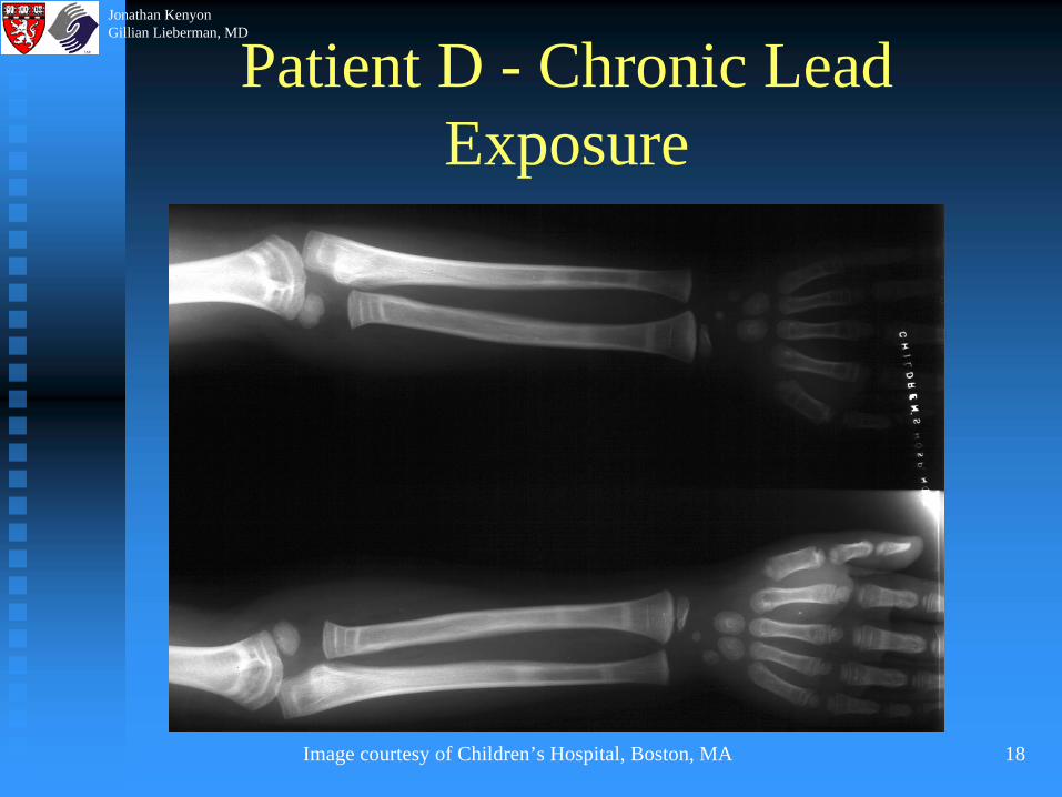
Patient D;Unknown History,Evidence of Chronic Lead Exposure
Note the alternatingNarrow-Broad-Narrow patternconsistent among different bones
Jonathan KenyonGillian Lieberman, MD
18
Patient D - Chronic Lead Exposure
Image courtesy of Children’s Hospital, Boston, MA
Jonathan KenyonGillian Lieberman, MD

19
KUB as a means of Diagnosis
Evidence of lead ingestion Evidence of lead ingestion -- Multiple Multiple radiopaqueradiopaque flakes representing paint chips flakes representing paint chips may be seen on plain abdominal film.may be seen on plain abdominal film.
Jonathan KenyonGillian Lieberman, MD

20
Patient E
Image courtesy of Children’s Hospital, Boston, MA
Film findingsRadioopaque lead ingested in paint chips
Jonathan KenyonGillian Lieberman, MD

21
Another Radiographic Manifestation
Widened cranial sutures may also be Widened cranial sutures may also be present in chronic lead poisoning, present in chronic lead poisoning, secondary to increased intracranial pressuresecondary to increased intracranial pressure
Jonathan KenyonGillian Lieberman, MD

22
SummaryLead poisoning is a serious and potentially Lead poisoning is a serious and potentially lifelife--threatening condition affecting threatening condition affecting primarily children.primarily children.Chronically, lead poisoning results in the Chronically, lead poisoning results in the inhibition of inhibition of osteoclasticosteoclastic activity. This is activity. This is visualized on plain films as dense visualized on plain films as dense metaphysealmetaphyseal thickening in growing bones.thickening in growing bones.Acutely lead toxicity should be diagnosed Acutely lead toxicity should be diagnosed by H+P, and measurement of blood lead by H+P, and measurement of blood lead levels, though sometimes evidence of lead levels, though sometimes evidence of lead ingestion is visible on KUB. ingestion is visible on KUB.
Jonathan KenyonGillian Lieberman, MD
23
SourcesSachs HK. The evolution of the radiologic lead Sachs HK. The evolution of the radiologic lead line. Radiology 1981; 139: 81line. Radiology 1981; 139: 81--85.85.BlickmanBlickman JG, Wilkinson RH, JG, Wilkinson RH, GraefGraef JW. The JW. The radiologic “lead band” revisited. American radiologic “lead band” revisited. American Journal of Journal of RoentgenologyRoentgenology 1986; 146: 2451986; 146: 245--247.247.RaberRaber S. The dense S. The dense metaphysealmetaphyseal band sign. band sign. Radiology 1999; 211: 773Radiology 1999; 211: 773--774774CaffeyCaffey J. Pediatric XJ. Pediatric X--ray Diagnosis. 7th edition. ray Diagnosis. 7th edition. 19781978EllenhorEllenhor M. M. Ellenhorn’sEllenhorn’s Medical Toxicology. Medical Toxicology. 19971997Children’s Hospital Radiology teaching files. Children’s Hospital Radiology teaching files. Boston, MABoston, MA
Jonathan KenyonGillian Lieberman, MD

24
AcknowlegementsBeverleeBeverlee Turner for her support and PowerPoint expertiseTurner for her support and PowerPoint expertiseLarry Barbaras and Ben Crandall our Larry Barbaras and Ben Crandall our WebMastersWebMasters