Embed Size (px)
Citation preview
Neuroimmune DisordersAKA
Neurologic manifestations of autoimmune diseases
Bridget T Walsh, DOCatalina Pointe Arthritis & Rheumatology Specialists, PC
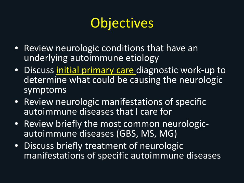
Objectives
• Review neurologic conditions that have an underlying autoimmune etiology
• Discuss initial primary care diagnostic work-up to determine what could be causing the neurologic symptoms
• Review neurologic manifestations of specific autoimmune diseases that I care for
• Review briefly the most common neurologic-autoimmune diseases (GBS, MS, MG)
• Discuss briefly treatment of neurologic manifestations of specific autoimmune diseases

Disclaimer
I am a rheumatologist not a neurologist or a magician so…..I will do my best and try to focus on a more primary care approach to these neurologic problems.

Immune-mediated neuropathies:Acute
• Guillain-Barré syndrome (GBS)-is an acute monophasic illness resulting from an immune response to a preceding infection that cross-reacts with peripheral nerve components because of molecular mimicry. The immune response can be directed towards the myelin or the axon of peripheral nerve, resulting in demyelinating and axonal forms of GBS.
Chronic1. Multiple Sclerosis2. Myasthenia gravis3. Chronic inflammatory demyelinating
polyneuropathy (CIDP) (prototype)4. Multifocal motor neuropathy
(MMN)5. POEMS syndrome (osteosclerotic
myeloma: polyneuropathy, organomegaly, endocrinopathy, monoclonal protein, skin changes)
6. Anti-myelin associated glycoprotein (MAG)-related neuropathies
Note: # 4,5 &6 are distinct from classic CIDP on the basis of clinical, electrodiagnostic, and therapeutic differences.

What did I get myself into?
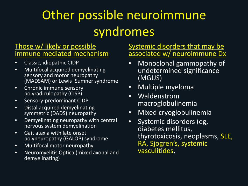
Other possible neuroimmunesyndromes
Those w/ likely or possible immune mediated mechanism• Classic, idiopathic CIDP• Multifocal acquired demyelinating
sensory and motor neuropathy (MADSAM) or Lewis–Sumner syndrome
• Chronic immune sensory polyradiculopathy (CISP)
• Sensory-predominant CIDP• Distal acquired demyelinating
symmetric (DADS) neuropathy• Demyelinating neuropathy with central
nervous system demyelination• Gait ataxia with late onset
polyneuropathy (GALOP) syndrome• Multifocal motor neuropathy• Neuromyelitis Optica (mixed axonal and
demyelinating)
Systemic disorders that may be associated w/ neuroimmune Dx• Monoclonal gammopathy of
undetermined significance (MGUS)
• Multiple myeloma• Waldenstrom
macroglobulinemia• Mixed cryoglobulinemia• Systemic disorders (eg,
diabetes mellitus, thyrotoxicosis, neoplasms, SLE, RA, Sjogren’s, systemic vasculitides,

So where to begin?
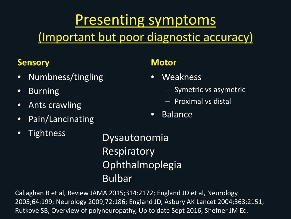
Presenting symptoms (Important but poor diagnostic accuracy)
Sensory• Numbness/tingling• Burning• Ants crawling• Pain/Lancinating• Tightness
Motor• Weakness
– Symetric vs asymetric– Proximal vs distal
• Balance
DysautonomiaRespiratoryOphthalmoplegiaBulbar
Callaghan B et al, Review JAMA 2015;314:2172; England JD et al, Neurology 2005;64:199; Neurology 2009;72:186; England JD, Asbury AK Lancet 2004;363:2151; Rutkove SB, Overview of polyneuropathy, Up to date Sept 2016, Shefner JM Ed.
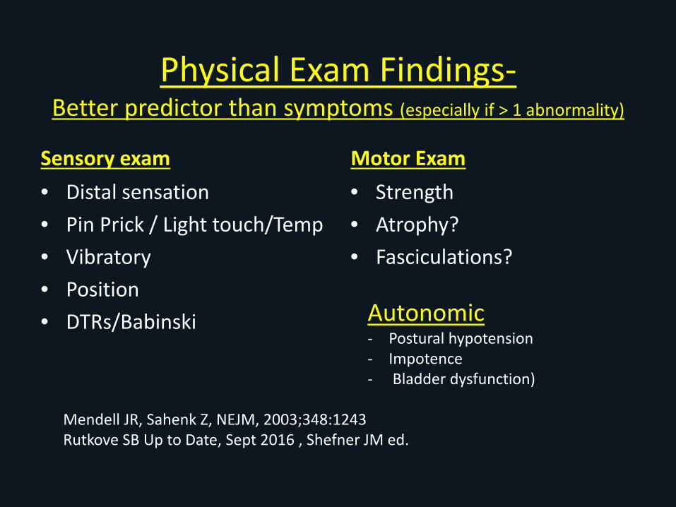
Physical Exam Findings-Better predictor than symptoms (especially if > 1 abnormality)
Sensory exam• Distal sensation• Pin Prick / Light touch/Temp• Vibratory• Position • DTRs/Babinski
Motor Exam• Strength • Atrophy?• Fasciculations?
Autonomic - Postural hypotension- Impotence- Bladder dysfunction)
Mendell JR, Sahenk Z, NEJM, 2003;348:1243Rutkove SB Up to Date, Sept 2016 , Shefner JM ed.
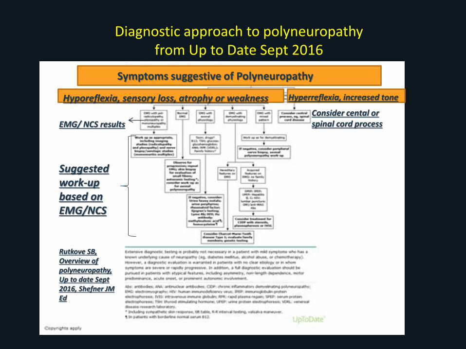
Diagnostic approach to polyneuropathy from Up to Date Sept 2016
EMG/ NCS resultsConsider cental or spinal cord process
Suggested work-up based on EMG/NCS
Rutkove SB, Overview of polyneuropathy, Up to date Sept 2016, Shefner JM Ed
Symptoms suggestive of Polyneuropathy
Hyporeflexia, sensory loss, atrophy or weakness Hyperreflexia, increased tone
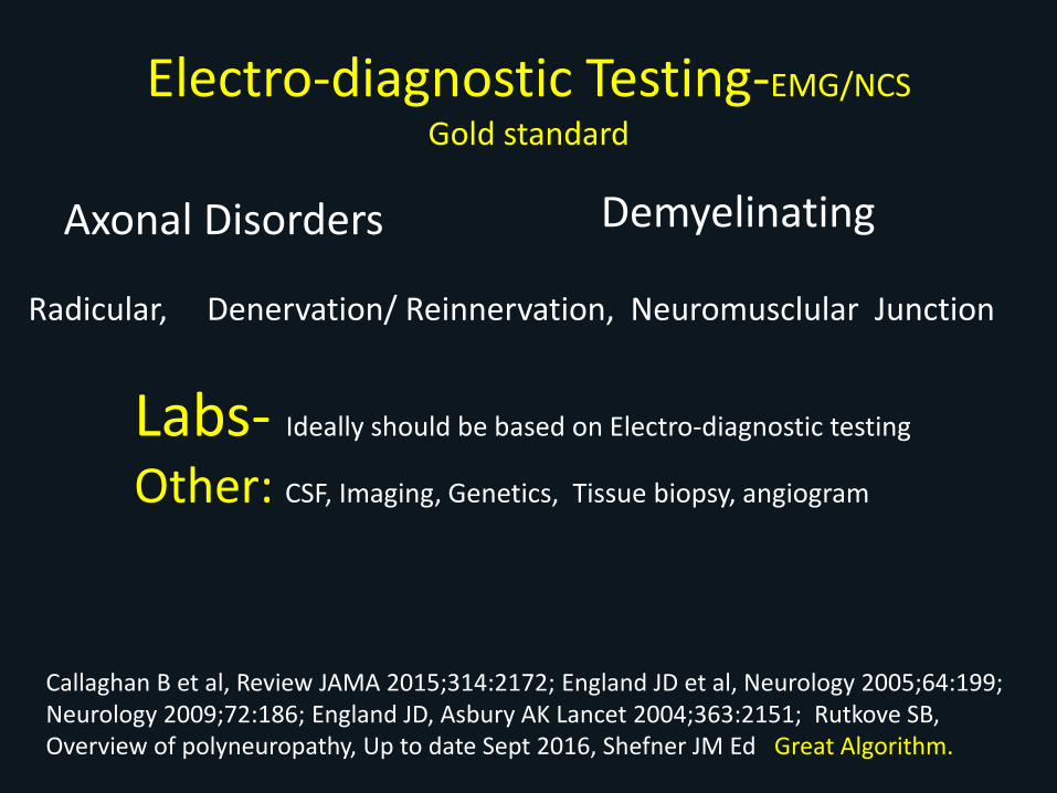
Electro-diagnostic Testing-EMG/NCSGold standard
Axonal Disorders Demyelinating
Radicular, Denervation/ Reinnervation, Neuromusclular Junction
Labs- Ideally should be based on Electro-diagnostic testing
Other: CSF, Imaging, Genetics, Tissue biopsy, angiogram
Callaghan B et al, Review JAMA 2015;314:2172; England JD et al, Neurology 2005;64:199; Neurology 2009;72:186; England JD, Asbury AK Lancet 2004;363:2151; Rutkove SB, Overview of polyneuropathy, Up to date Sept 2016, Shefner JM Ed Great Algorithm.
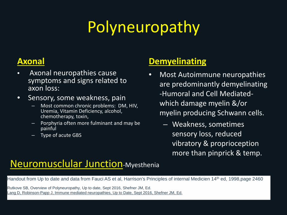
Polyneuropathy
Axonal• Axonal neuropathies cause
symptoms and signs related to axon loss:
• Sensory, some weakness, pain– Most common chronic problems: DM, HIV,
Uremia, Vitamin Deficiency, alcohol, chemotherapy, toxin,
– Porphyria often more fulminant and may be painful
– Type of acute GBS
Demyelinating • Most Autoimmune neuropathies
are predominantly demyelinating -Humoral and Cell Mediated-which damage myelin &/or myelin producing Schwann cells.– Weakness, sometimes
sensory loss, reduced vibratory & proprioception more than pinprick & temp.
Handout from Up to date and data from Fauci AS et al, Harrison’s Principles of internal Medicien 14th ed, 1998,page 2460
Rutkove SB, Overview of Polyneuropathy, Up to date, Sept 2016, Shefner JM, Ed.Lang D, Robinson-Papp J, Immune mediated neuropathies, Up to Date, Sept 2016, Shefner JM, Ed.
Neuromusclular Junction-Myesthenia
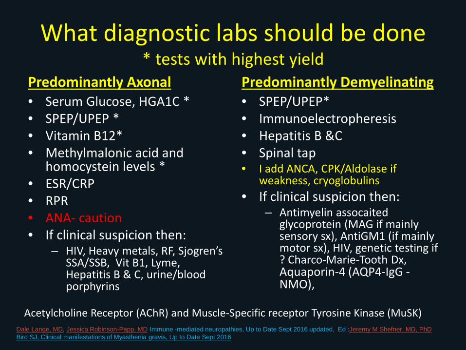
What diagnostic labs should be done* tests with highest yield
Predominantly Axonal• Serum Glucose, HGA1C *• SPEP/UPEP *• Vitamin B12*• Methylmalonic acid and
homocystein levels *• ESR/CRP• RPR• ANA- caution • If clinical suspicion then:
– HIV, Heavy metals, RF, Sjogren’sSSA/SSB, Vit B1, Lyme, Hepatitis B & C, urine/blood porphyrins
Predominantly Demyelinating• SPEP/UPEP*• Immunoelectropheresis• Hepatitis B &C• Spinal tap• I add ANCA, CPK/Aldolase if
weakness, cryoglobulins• If clinical suspicion then:
– Antimyelin assocaitedglycoprotein (MAG if mainly sensory sx), AntiGM1 (if mainly motor sx), HIV, genetic testing if ? Charco-Marie-Tooth Dx, Aquaporin-4 (AQP4-IgG -NMO),
Dale Lange, MD, Jessica Robinson-Papp, MD Immune -mediated neuropathies, Up to Date Sept 2016 updated, Ed :Jeremy M Shefner, MD, PhDBird SJ, Clinical manifestations of Myasthenia gravis, Up to Date Sept 2016
Acetylcholine Receptor (AChR) and Muscle-Specific receptor Tyrosine Kinase (MuSK)

Anti-Nuclear Antibody -ANA
• Screening ANA- high rate of false + (5-30% or more depending on age). If one assumes that the prevalence of ANA-associated diseases in the population is 1 percent, the majority of these patients will have a false positive result.
• Order if pretest probability high• MUST ORDER A TITER- not just a screen. If
screen is + must f/u with a titer and should be > 1:40 to be considered significant
Solomon DH, Kavanaugh AJ, Schur PH ACR Ad Hoc Committee on Immunologic Testing Guidelines A&R, 2002;47,434

Conditions where ANA can be +
Systemic Auto-immune• SLE 98-100%• Scleroderma 95%• MCTD 100%• Drug Induced 80-95%• Sjogren’s 60-90%• Rheumatoid Arth. 45%• Raynauds 40%• Polymyositis/DM 35%• Juvenile Idiopathic arthritis
(JIA) 15-40%
Organ Specific Autoimmune• Hashimotos Thyroiditis 50%
• Graves’ Disease 50%
• Autoimmune Hepatitis 70%
• Primary Biliary Cirrhosis 50-70%
Other diseases:Infections- Viral: EBV,HIV, Parvovirus 19
Bacterial SBE, SyphilisMalignancies: Lymphoproliferative, paraneoplastic syndromesInflammatory bowel DsInterstitial Pulmonary FibrosisBlock DB, Up to date, Sept 2016, Ed:
Schmerling RH
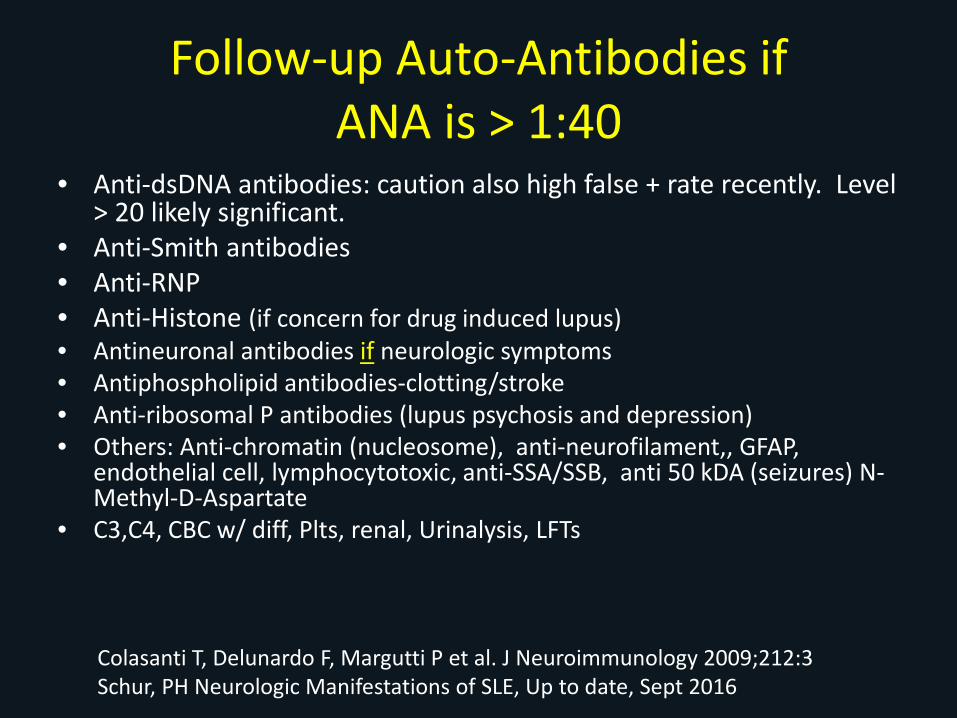
Follow-up Auto-Antibodies if ANA is > 1:40
• Anti-dsDNA antibodies: caution also high false + rate recently. Level > 20 likely significant.
• Anti-Smith antibodies• Anti-RNP• Anti-Histone (if concern for drug induced lupus)• Antineuronal antibodies if neurologic symptoms• Antiphospholipid antibodies-clotting/stroke• Anti-ribosomal P antibodies (lupus psychosis and depression)• Others: Anti-chromatin (nucleosome), anti-neurofilament,, GFAP,
endothelial cell, lymphocytotoxic, anti-SSA/SSB, anti 50 kDA (seizures) N-Methyl-D-Aspartate
• C3,C4, CBC w/ diff, Plts, renal, Urinalysis, LFTs
Colasanti T, Delunardo F, Margutti P et al. J Neuroimmunology 2009;212:3Schur, PH Neurologic Manifestations of SLE, Up to date, Sept 2016
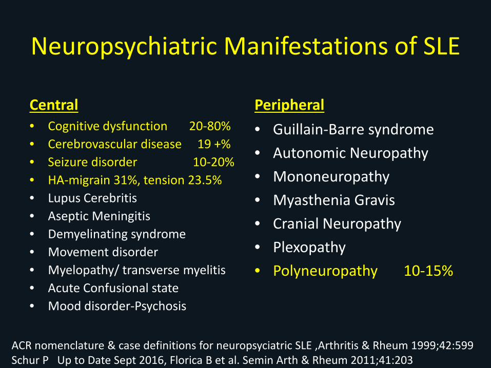
Neuropsychiatric Manifestations of SLE
Central• Cognitive dysfunction 20-80%• Cerebrovascular disease 19 +%• Seizure disorder 10-20%• HA-migrain 31%, tension 23.5%• Lupus Cerebritis• Aseptic Meningitis• Demyelinating syndrome• Movement disorder• Myelopathy/ transverse myelitis• Acute Confusional state• Mood disorder-Psychosis
Peripheral• Guillain-Barre syndrome• Autonomic Neuropathy• Mononeuropathy• Myasthenia Gravis• Cranial Neuropathy• Plexopathy• Polyneuropathy 10-15%
ACR nomenclature & case definitions for neuropsyciatric SLE ,Arthritis & Rheum 1999;42:599Schur P Up to Date Sept 2016, Florica B et al. Semin Arth & Rheum 2011;41:203
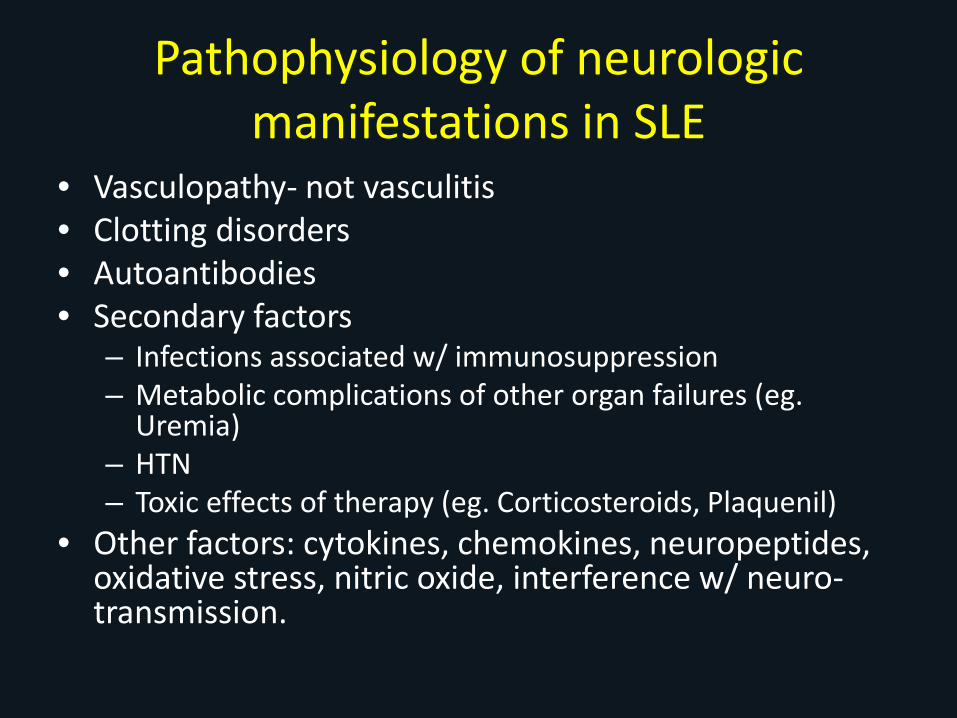
Pathophysiology of neurologic manifestations in SLE
• Vasculopathy- not vasculitis• Clotting disorders• Autoantibodies• Secondary factors
– Infections associated w/ immunosuppression– Metabolic complications of other organ failures (eg.
Uremia)– HTN– Toxic effects of therapy (eg. Corticosteroids, Plaquenil)
• Other factors: cytokines, chemokines, neuropeptides, oxidative stress, nitric oxide, interference w/ neuro-transmission.
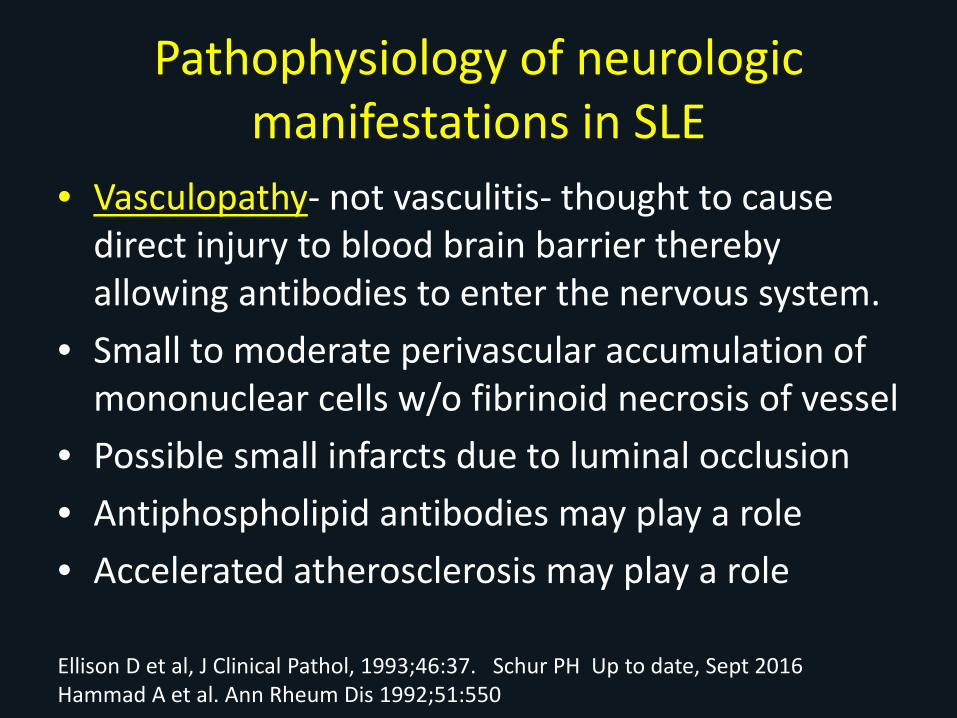
Pathophysiology of neurologic manifestations in SLE
• Vasculopathy- not vasculitis- thought to cause direct injury to blood brain barrier thereby allowing antibodies to enter the nervous system.
• Small to moderate perivascular accumulation of mononuclear cells w/o fibrinoid necrosis of vessel
• Possible small infarcts due to luminal occlusion• Antiphospholipid antibodies may play a role• Accelerated atherosclerosis may play a role
Ellison D et al, J Clinical Pathol, 1993;46:37. Schur PH Up to date, Sept 2016Hammad A et al. Ann Rheum Dis 1992;51:550
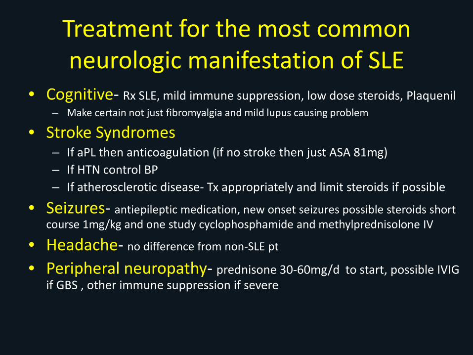
Treatment for the most common neurologic manifestation of SLE
• Cognitive- Rx SLE, mild immune suppression, low dose steroids, Plaquenil– Make certain not just fibromyalgia and mild lupus causing problem
• Stroke Syndromes– If aPL then anticoagulation (if no stroke then just ASA 81mg)– If HTN control BP– If atherosclerotic disease- Tx appropriately and limit steroids if possible
• Seizures- antiepileptic medication, new onset seizures possible steroids short course 1mg/kg and one study cyclophosphamide and methylprednisolone IV
• Headache- no difference from non-SLE pt
• Peripheral neuropathy- prednisone 30-60mg/d to start, possible IVIG if GBS , other immune suppression if severe
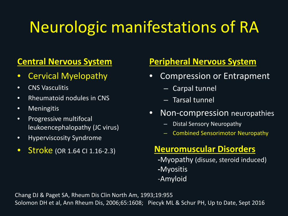
Neurologic manifestations of RA
Central Nervous System• Cervical Myelopathy• CNS Vasculitis• Rheumatoid nodules in CNS• Meningitis • Progressive multifocal
leukoencephalopathy (JC virus)• Hyperviscosity Syndrome
• Stroke (OR 1.64 CI 1.16-2.3)
Peripheral Nervous System• Compression or Entrapment
– Carpal tunnel– Tarsal tunnel
• Non-compression neuropathies– Distal Sensory Neuropathy– Combined Sensorimotor Neuropathy
Neuromuscular Disorders-Myopathy (disuse, steroid induced)-Myositis-Amyloid
Chang DJ & Paget SA, Rheum Dis Clin North Am, 1993;19:955Solomon DH et al, Ann Rheum Dis, 2006;65:1608; Piecyk ML & Schur PH, Up to Date, Sept 2016
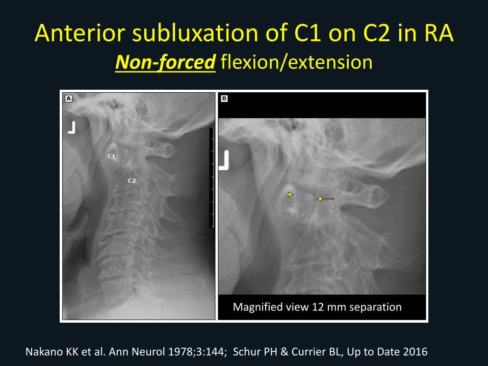
Anterior subluxation of C1 on C2 in RANon-forced flexion/extension
Magnified view 12 mm separation
Nakano KK et al. Ann Neurol 1978;3:144; Schur PH & Currier BL, Up to Date 2016

Anterior subluxation C1 on C2 w/ pannus causing impingement of cord
5 mm Seperation
Pannusformation
Schur PH & Currier BL, Up to Date 2016
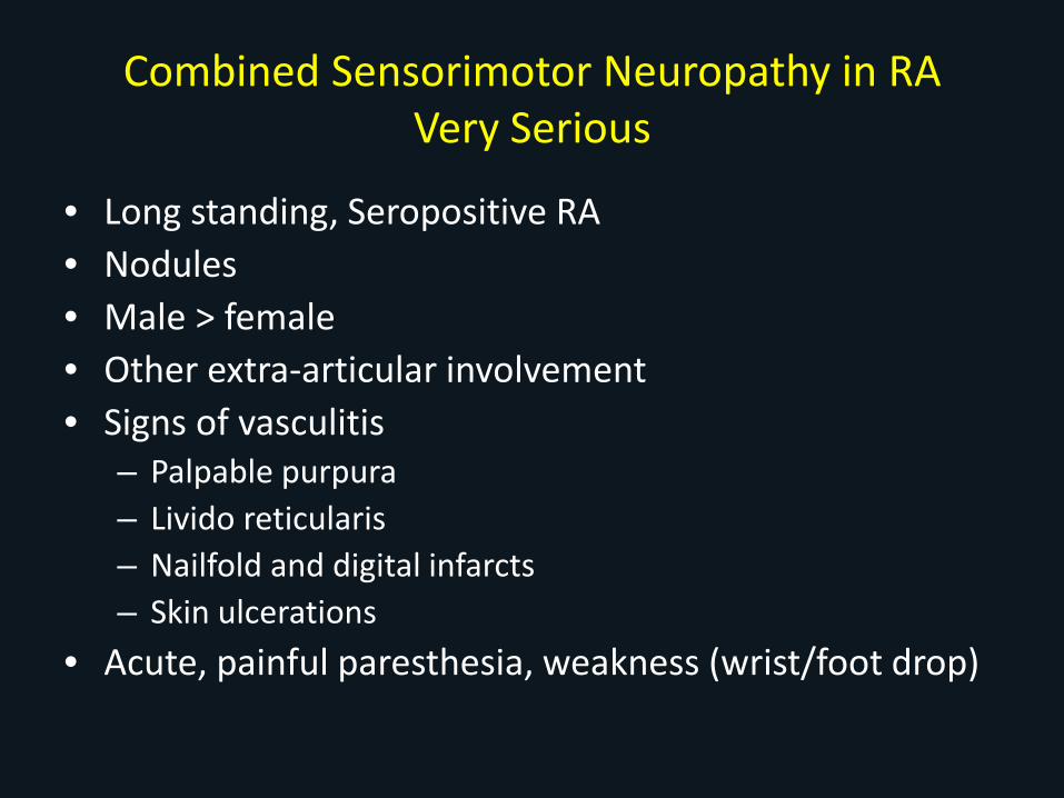
Combined Sensorimotor Neuropathy in RAVery Serious
• Long standing, Seropositive RA• Nodules• Male > female• Other extra-articular involvement• Signs of vasculitis
– Palpable purpura– Livido reticularis– Nailfold and digital infarcts– Skin ulcerations
• Acute, painful paresthesia, weakness (wrist/foot drop)
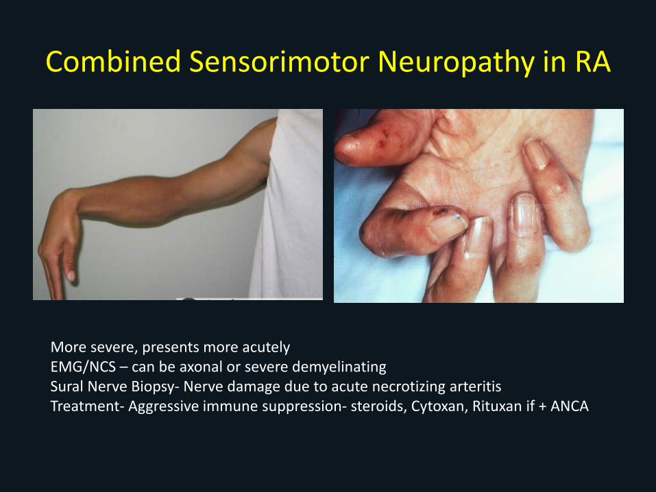
Combined Sensorimotor Neuropathy in RA
More severe, presents more acutelyEMG/NCS – can be axonal or severe demyelinatingSural Nerve Biopsy- Nerve damage due to acute necrotizing arteritisTreatment- Aggressive immune suppression- steroids, Cytoxan, Rituxan if + ANCA
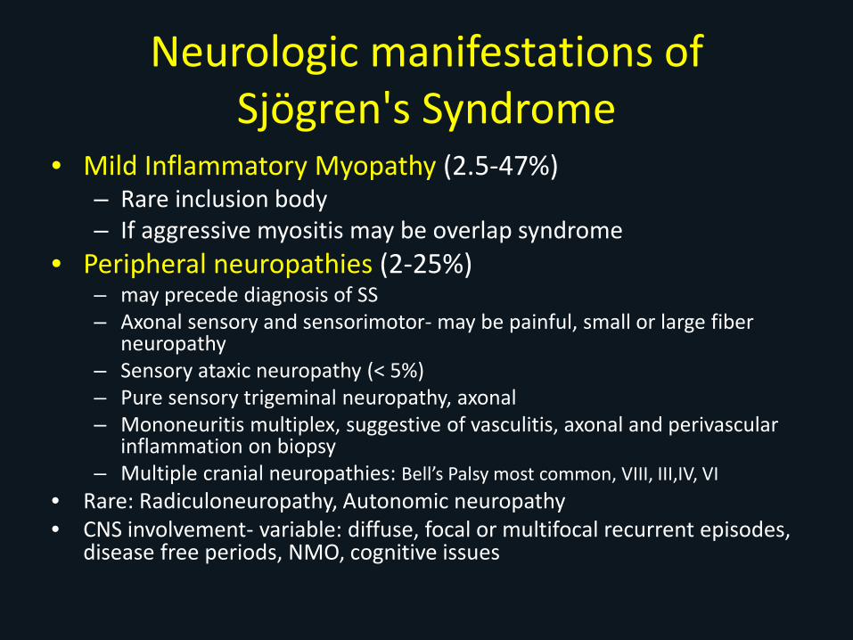
Neurologic manifestations of Sjögren's Syndrome
• Mild Inflammatory Myopathy (2.5-47%)– Rare inclusion body– If aggressive myositis may be overlap syndrome
• Peripheral neuropathies (2-25%) – may precede diagnosis of SS– Axonal sensory and sensorimotor- may be painful, small or large fiber
neuropathy– Sensory ataxic neuropathy (< 5%)– Pure sensory trigeminal neuropathy, axonal– Mononeuritis multiplex, suggestive of vasculitis, axonal and perivascular
inflammation on biopsy– Multiple cranial neuropathies: Bell’s Palsy most common, VIII, III,IV, VI
• Rare: Radiculoneuropathy, Autonomic neuropathy• CNS involvement- variable: diffuse, focal or multifocal recurrent episodes,
disease free periods, NMO, cognitive issues
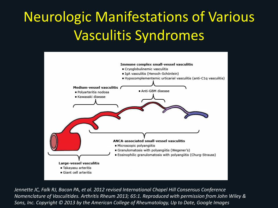
Neurologic Manifestations of Various Vasculitis Syndromes
Jennette JC, Falk RJ, Bacon PA, et al. 2012 revised International Chapel Hill Consensus Conference Nomenclature of Vasculitides. Arthritis Rheum 2013; 65:1. Reproduced with permission from John Wiley & Sons, Inc. Copyright © 2013 by the American College of Rheumatology, Up to Date, Google Images
Neurologic Manifestations of Various Vasculitis Syndromes
• Large Vessel– Headache, Stroke, amaurosis fugax- GCA, PMR– Claudication-Takayasu/s
• Medium vessel– Mononeuritis multiplex– Sensory neuropathy– Cranial neuropathy– CNS– External ophthalmoplegias and sensory neural hearing loss
• Small Vessel– Clinically significant neuropathy uncommon– Mainly sensory neuropathy
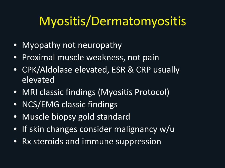
Myositis/Dermatomyositis
• Myopathy not neuropathy• Proximal muscle weakness, not pain• CPK/Aldolase elevated, ESR & CRP usually
elevated• MRI classic findings (Myositis Protocol)• NCS/EMG classic findings• Muscle biopsy gold standard• If skin changes consider malignancy w/u• Rx steroids and immune suppression
No talk on autoimmune neurologic conditions would be complete without:
• Guillain Barre Syndrome• Multiple Sclerosis• Myasthenia Gravis
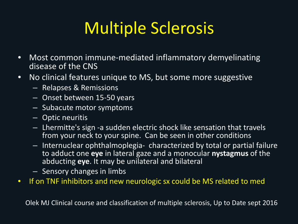
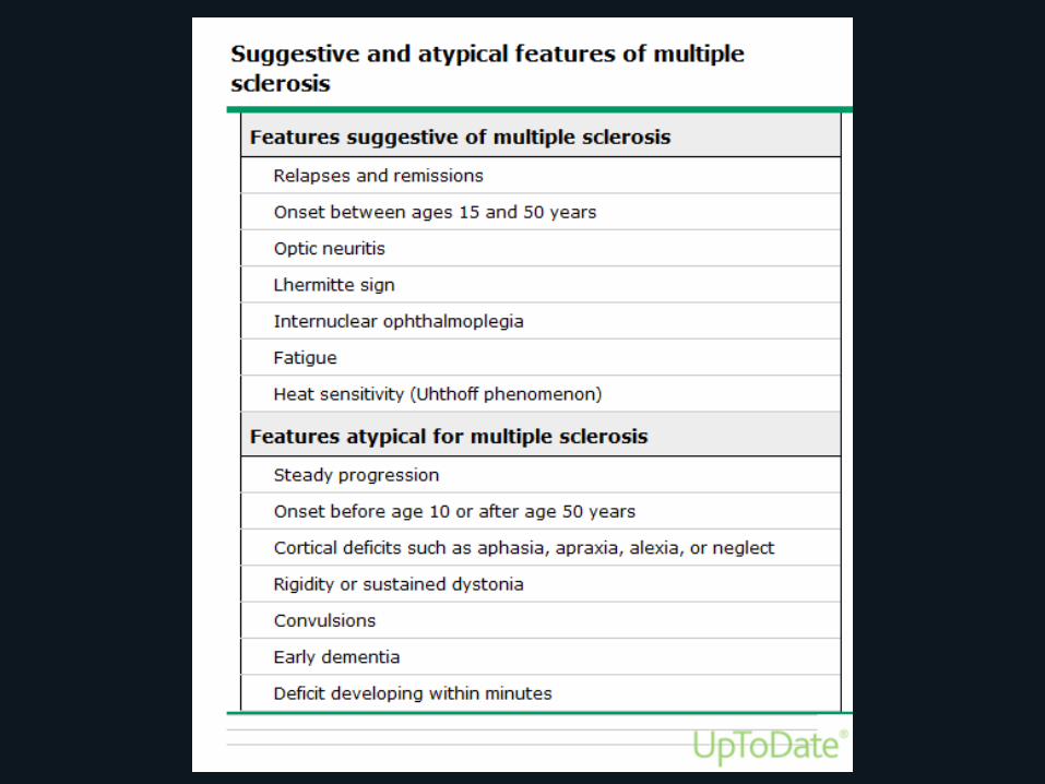
Multiple Sclerosis• Most common immune-mediated inflammatory demyelinating
disease of the CNS• No clinical features unique to MS, but some more suggestive
– Relapses & Remissions– Onset between 15-50 years– Subacute motor symptoms– Optic neuritis– Lhermitte's sign -a sudden electric shock like sensation that travels
from your neck to your spine. Can be seen in other conditions– Internuclear ophthalmoplegia- characterized by total or partial failure
to adduct one eye in lateral gaze and a monocular nystagmus of the abducting eye. It may be unilateral and bilateral
– Sensory changes in limbs• If on TNF inhibitors and new neurologic sx could be MS related to med
Olek MJ Clinical course and classification of multiple sclerosis, Up to Date sept 2016
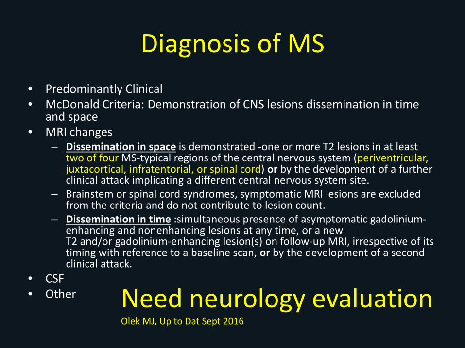
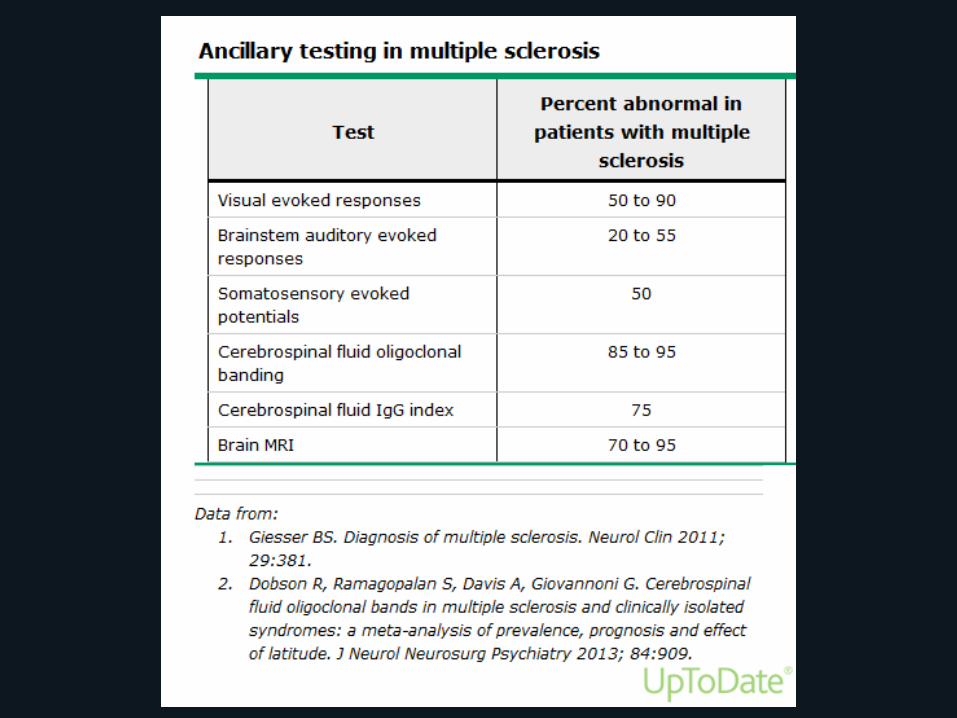
Diagnosis of MS• Predominantly Clinical• McDonald Criteria: Demonstration of CNS lesions dissemination in time
and space• MRI changes
– Dissemination in space is demonstrated -one or more T2 lesions in at least two of four MS-typical regions of the central nervous system (periventricular, juxtacortical, infratentorial, or spinal cord) or by the development of a further clinical attack implicating a different central nervous system site.
– Brainstem or spinal cord syndromes, symptomatic MRI lesions are excluded from the criteria and do not contribute to lesion count.
– Dissemination in time :simultaneous presence of asymptomatic gadolinium-enhancing and nonenhancing lesions at any time, or a new T2 and/or gadolinium-enhancing lesion(s) on follow-up MRI, irrespective of its timing with reference to a baseline scan, or by the development of a second clinical attack.
• CSF• Other Need neurology evaluation
Olek MJ, Up to Dat Sept 2016
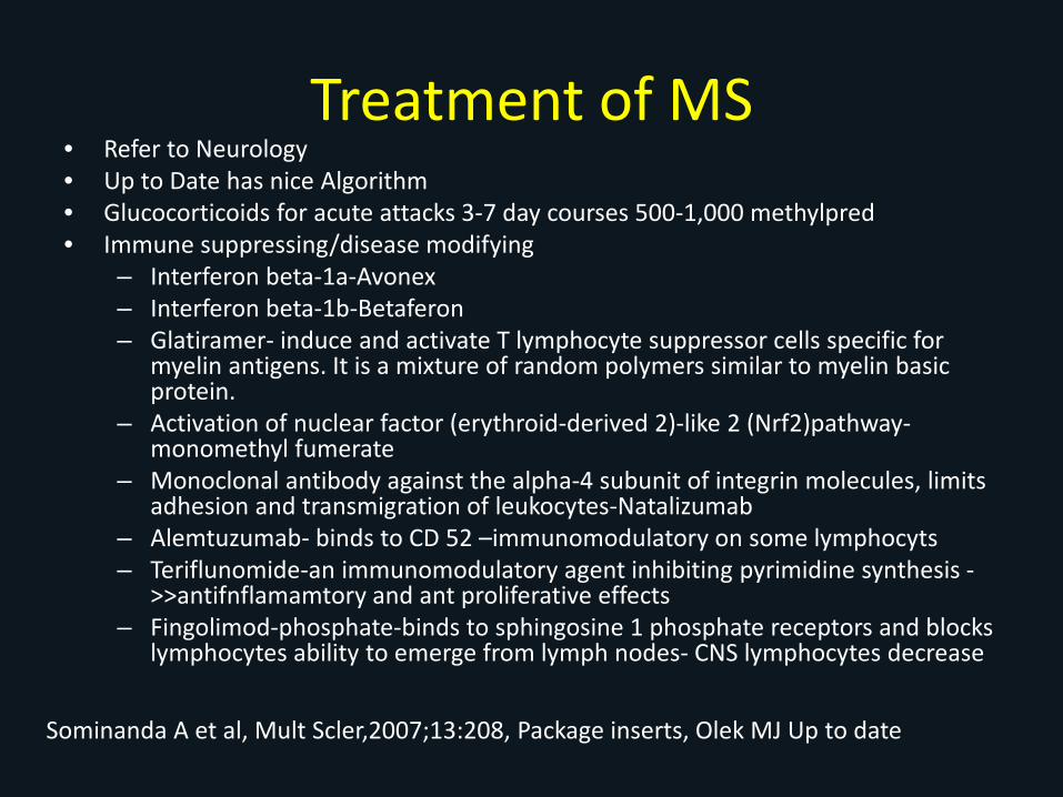
Treatment of MS• Refer to Neurology• Up to Date has nice Algorithm• Glucocorticoids for acute attacks 3-7 day courses 500-1,000 methylpred• Immune suppressing/disease modifying
– Interferon beta-1a-Avonex– Interferon beta-1b-Betaferon– Glatiramer- induce and activate T lymphocyte suppressor cells specific for
myelin antigens. It is a mixture of random polymers similar to myelin basic protein.
– Activation of nuclear factor (erythroid-derived 2)-like 2 (Nrf2)pathway-monomethyl fumerate
– Monoclonal antibody against the alpha-4 subunit of integrin molecules, limits adhesion and transmigration of leukocytes-Natalizumab
– Alemtuzumab- binds to CD 52 –immunomodulatory on some lymphocyts– Teriflunomide-an immunomodulatory agent inhibiting pyrimidine synthesis -
>>antifnflamamtory and ant proliferative effects– Fingolimod-phosphate-binds to sphingosine 1 phosphate receptors and blocks
lymphocytes ability to emerge from lymph nodes- CNS lymphocytes decrease
Sominanda A et al, Mult Scler,2007;13:208, Package inserts, Olek MJ Up to date
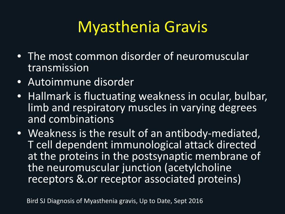
Myasthenia Gravis
• The most common disorder of neuromuscular transmission
• Autoimmune disorder• Hallmark is fluctuating weakness in ocular, bulbar,
limb and respiratory muscles in varying degrees and combinations
• Weakness is the result of an antibody-mediated, T cell dependent immunological attack directed at the proteins in the postsynaptic membrane of the neuromuscular junction (acetylcholine receptors &.or receptor associated proteins)
Bird SJ Diagnosis of Myasthenia gravis, Up to Date, Sept 2016

Myasthenia Gravis Clinical Forms
• Ocular Myasthenia- weakness limited to eye lids and extra-ocular muscles (ptosis, diplopia >50%)
• Generalized- weakness ocular muscles but also combination of bulbar (15%), limb (<5%)and respiratory
• Specific muscle weakness and not generalized fatigue
Bird SJ Diagnosis of Myasthenia gravis, Up to Date, Sept 2016
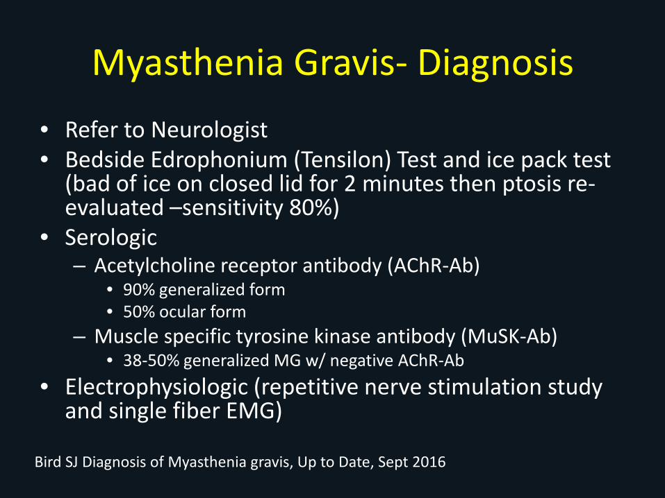
Myasthenia Gravis- Diagnosis• Refer to Neurologist• Bedside Edrophonium (Tensilon) Test and ice pack test
(bad of ice on closed lid for 2 minutes then ptosis re-evaluated –sensitivity 80%)
• Serologic– Acetylcholine receptor antibody (AChR-Ab)
• 90% generalized form• 50% ocular form
– Muscle specific tyrosine kinase antibody (MuSK-Ab)• 38-50% generalized MG w/ negative AChR-Ab
• Electrophysiologic (repetitive nerve stimulation study and single fiber EMG)
Bird SJ Diagnosis of Myasthenia gravis, Up to Date, Sept 2016
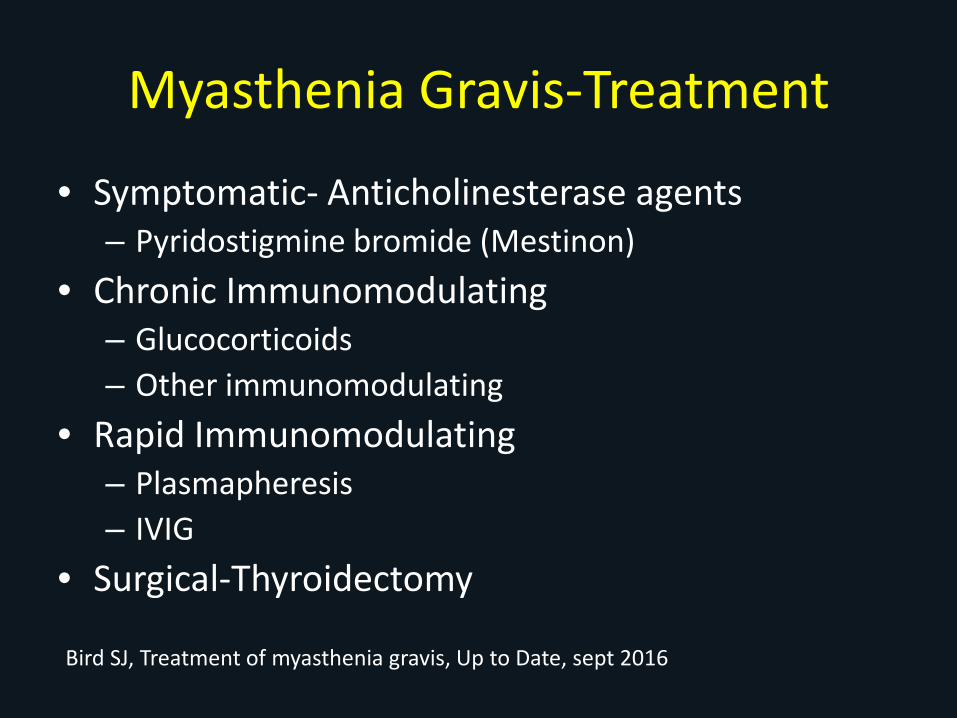
Myasthenia Gravis-Treatment
• Symptomatic- Anticholinesterase agents– Pyridostigmine bromide (Mestinon)
• Chronic Immunomodulating– Glucocorticoids– Other immunomodulating
• Rapid Immunomodulating– Plasmapheresis– IVIG
• Surgical-Thyroidectomy
Bird SJ, Treatment of myasthenia gravis, Up to Date, sept 2016
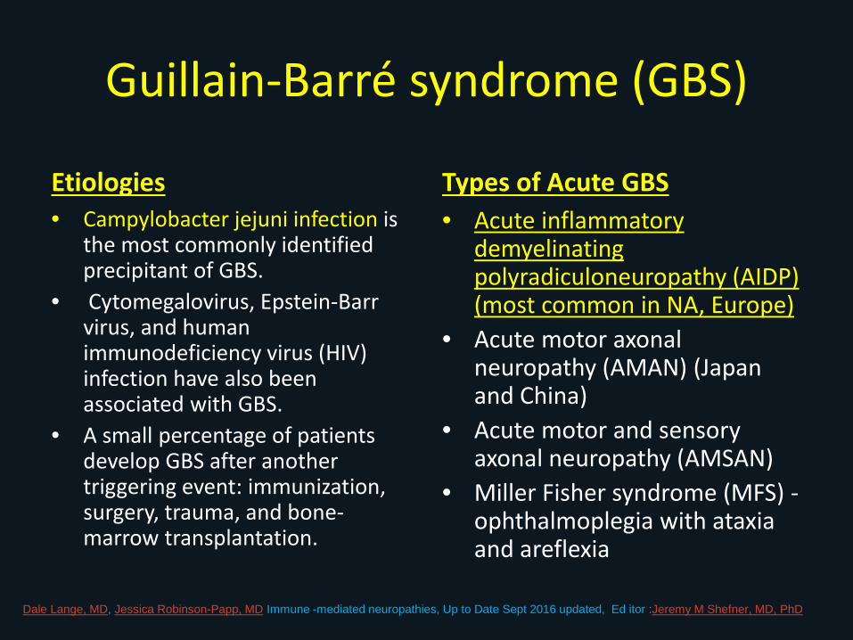
Guillain-Barré syndrome (GBS)
Etiologies• Campylobacter jejuni infection is
the most commonly identified precipitant of GBS.
• Cytomegalovirus, Epstein-Barr virus, and human immunodeficiency virus (HIV) infection have also been associated with GBS.
• A small percentage of patients develop GBS after another triggering event: immunization, surgery, trauma, and bone-marrow transplantation.
Types of Acute GBS• Acute inflammatory
demyelinating polyradiculoneuropathy (AIDP) (most common in NA, Europe)
• Acute motor axonal neuropathy (AMAN) (Japan and China)
• Acute motor and sensory axonal neuropathy (AMSAN)
• Miller Fisher syndrome (MFS) -ophthalmoplegia with ataxia and areflexia
Dale Lange, MD, Jessica Robinson-Papp, MD Immune -mediated neuropathies, Up to Date Sept 2016 updated, Ed itor :Jeremy M Shefner, MD, PhD
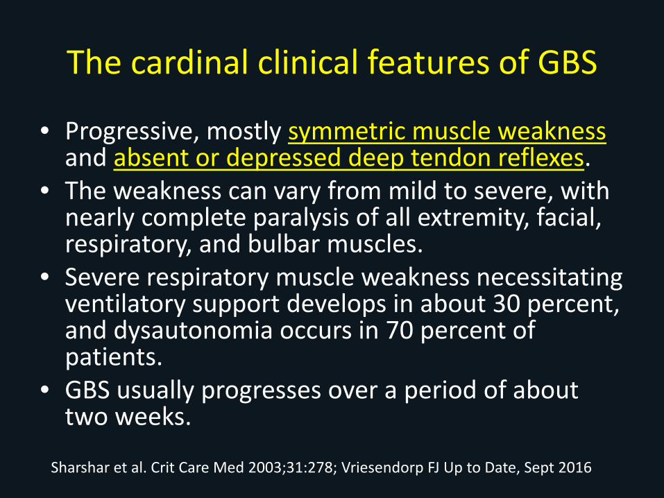
The cardinal clinical features of GBS
• Progressive, mostly symmetric muscle weakness and absent or depressed deep tendon reflexes.
• The weakness can vary from mild to severe, with nearly complete paralysis of all extremity, facial, respiratory, and bulbar muscles.
• Severe respiratory muscle weakness necessitating ventilatory support develops in about 30 percent, and dysautonomia occurs in 70 percent of patients.
• GBS usually progresses over a period of about two weeks.
Sharshar et al. Crit Care Med 2003;31:278; Vriesendorp FJ Up to Date, Sept 2016
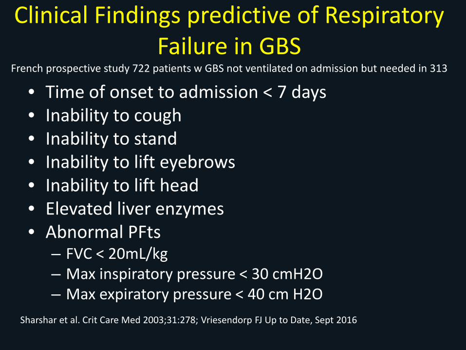
Clinical Findings predictive of Respiratory Failure in GBS
French prospective study 722 patients w GBS not ventilated on admission but needed in 313
• Time of onset to admission < 7 days• Inability to cough• Inability to stand• Inability to lift eyebrows• Inability to lift head• Elevated liver enzymes• Abnormal PFts
– FVC < 20mL/kg– Max inspiratory pressure < 30 cmH2O– Max expiratory pressure < 40 cm H2O
Sharshar et al. Crit Care Med 2003;31:278; Vriesendorp FJ Up to Date, Sept 2016

Diagnosis of GBS• Clinical presentation –index of suspicion • Dx confirmed by cerebrospinal fluid (CSF) analysis –
– An increased cerebrospinal fluid (CSF) protein with a normal CSF white blood cell count, called albuminocytologic dissociation
– Found in 50 to 66 percent of patients with GBS in the first week after the onset of symptoms and in ≥75 percent of patients in the third week.
• Neurophysiology studies / Electrodiagnostic studies help determine type– Acute polyneuropathy with either predominantly demyelinating
(ie, AIDP) – Axonal (ie, AMAN or AMSAN) features. – Testing for serum IgG antibodies to GQ1b is useful for the
diagnosis of Miller Fisher syndrome.
Sharshar et al. Crit Care Med 2003;31:278; Vriesendorp FJ Up to Date, Sept 2016

Guillain-Barré syndrome-Treatment
• Plasma Exchange or IVIG hastens recovery– Equivalent effects– Combination not beneficial– Plasma exchange for non-ambulatory adults who start
treatment in 4 weeks or ambulatory starting in 2 weeks
– IVIG recommended for non-ambulatory adults within 2-4 weeks
• Glucocorticoids not recommended• Supportive Care (30% develop respiratory failure)
Patwa HS et al. Guidelines from American Academy of Neurology Neurology 2012;78:1009Hughes RA et al, Neurology 2003;61:736; Vriesendorp FJ Up to Date, Sept 2016, Shefner JM Ed.

Summary• Neuroimmune disorders or Neurologic manifestations of
Autoimmune diseases is not for the faint of heart• Basic good clinical practice will guide most evaluation and
treatment/referral decisions ie- a good Hx and Px• Consider EMG/NCS before labs testing (although I am not certain
how cost effective that is. I usually do basics labs)– At least glucose, HGA1C, Vitamin B12, Homocysteine and
methylmeonic acid, SPEP/UPEP, basic chem and CBC panel and would get UA to check for protein
– ANA w/ caution, ANCA, Hep B& C, cryoglobulins, C3,C4 and CRP/ESR if suspect vasculitis (If obese ESR and CRP less reliable)
– Other:based on clinical suspicion (HIV, RPR, Heavy metals. Ro/La)• Neurology referral – pick up to phone or admit if concern

TGIO
Almost……. Questions?