Embed Size (px)
Citation preview
Amin et. al. / Alexandria Journal of Veterinary Sciences 2014, 41: 68-79
68
Radiographic and Computed Tomographic Anatomy of the Fetlock, Pastern and Coffin
joints of the Manus of the Donkey (Equus asinus)
Mohamed E. Amin, Raafat M.A. Elbakary, Mohamed A.M. Alsafy, Naglaa Fathi Anatomy and Embryology Department, Faculty of Veterinary Medicine, Alexandria University
Key words Abstract:
Donkey, CT,
Radiology,
Cast, Gross
anatomy
The digit of the donkey as a draught animal is commonly susceptible to much affection. The
purpose of the present study was to provide a detailed anatomic reference of radiographic and
computed tomographic images in conjunction with cross and sagittal sections of the normal
fetlock, pastern and coffin joints of the donkey for anatomists, surgeons and veterinary students.
Eight adult donkeys of both sexes free from any joints affection were used in our study. The digit
of two donkeys had undergone radiographic and computed tomographic scanning; the other
donkey’s specimens were used to anatomical dissection and sectional anatomy. In the computed
tomography (CT) of the fetlock joint all bone structures of the joint appeared also the soft tissue
structures that could be identified and evaluated on the different soft tissue window planes
included the common digital extensor tendon, lateral digital extensor tendon, superficial digital
flexor tendon (SDFT), deep digital flexor tendon (DDFT), straight, oblique, and cruciate distal
sesamoidean and intersesamoidean ligaments. For the pastern joint the structures that can be
identified including the proximal phalanx, DDFT and digital cushion. In the coffin joint the
collateral sesamoidean ligament (CSL) is difficult to identify on CT images.
Corresponding Author: Mohamed Alsafy, [email protected]
1. Introduction
The donkey is an important farming animal used
heavily by the Egyptian farmers because of their
great tolerance as a draught animal. Computed
tomography examinations have become more
common in veterinary medicine due to the
availability of the computed tomography machine
in teaching institutions and private veterinary
hospitals (Pollard and Puchalski, 2011). The using
of the new anatomical techniques in studying
anatomy as the radiology, computed tomography
and others techniques being useful for both
student and anatomist as well as surgeon. Imaging
techniques play a major meanwhile a few CT
studies on horse's foot have been role in the
modern biomedical research (Olsen et al., 2007)
and (Olsenan andWinterdahl, 2009). CT, through
its high spatial resolution and moderate
differentiation of tissue contrast is a fastened
exceptionally useful technique for visualizing
general anatomy (Dixon and Dacre, 2005).
Accurate interpretation of ultrasonography or CT
of the foot requires a thorough knowledge of the
cross sectional anatomy of the region and accurate
interpretation of the plan metric CT is necessary
for the study and evaluation of the pathological
condition or damaged tissues (Raji et al., 2008)
and (Weissengruber et al., 2006). CT is
particularly useful for looking at complex bony
structures such as the skull, spine or joints. The
involvement of the new techniques (Digital X-ray
images, computed tomography, magnetic
resonance imaging, ultrasonography, endoscopy,
fluoroscopy, 3D Image, animation, CDs) in the
anatomy tutorial in Egypt, as well as in the other
Arabic countries becomes essential to follow the
international steps and to prepare the students to
the clinical stage of veterinary study (Saber,
2008). CT was efficient imaging modality that
provides a cross-sectional image with superior
soft tissue differentiation and no superimposition
of the overlying structures, which can be used for
better diagnosis of foot and foot pad abnormalities
(Badawy, 2011) .Radiography remains the main
stay of equine musculoskeletal imaging due to its
low cost, ready accessibility and global evaluation
of bony structures (Kinns and Nelson, 2010). The
using of the normal radiographic anatomy,
Alexandria Journal of Veterinary Sciences 2014, 41: 68-79
ISSN 1110-2047, www.alexjvs.com
DOI: 10.5455/ajvs.156620
Amin et. al. / Alexandria Journal of Veterinary Sciences 2014, 41: 68-79
69
computed tomographic scans, cross and sagittal
sections and the gross anatomical dissection of the
digit is very important for anatomists, surgeons
and veterinary students to the accurate
investigations of the fetlock, pastern and coffin
joints of the donkey. So the current study is
focused on the anatomical details of these joints.
2. Material and methods
Eight adult donkeys of both sexes free from any
joints affection were used in our study.
Gross anatomy
Eight fore limbs of donkey used for studying the
gross anatomy of the fetlock, pastern and coffin
joints. The donkeys are being bled after being
anesthetized then undergone routine preservative
technique (10% formalin mixed with 4% glycerin
and 1% phenol for 10-15 days until complete
fixation and then the fore limbs were separated
and the three joints dissected for studying the
gross anatomy. Three of these donkeys fore limb
were injected using gum milk latex with paint in
the joint cavity of fetlock, pastern and coffin
joints then preserved in freezer for three days then
dissected for studying the joint capsule of each
joint and its pouches if present.
Computed tomography
Two fore limbs of donkey were used for studying
CT of the three joints. The donkey was being
anesthetized then being bled and the three joints
were severed and transferred to the CT center
within 24 hours and imaged using a Toshiba
asteion super 4 multi slice 4 CT apparatus. The
distance between the slices taken was 0.5 cm.
Radiology
Two fore limbs of donkey were obtained after the
donkey being anesthetized and being bled and
transferred to the x-ray center within two hours
and imaged using Toshiba 500ml apparatus for x-
rays in dorsopalmar view.
Cross and sagittal sections
Four donkeys’ fore limbs were obtained after the
donkey being anesthetized and being bled well
then the digits was cut using electric band saw
with 0.5 cm slices interval. The slices were
preserved in freezer for 24 hrs and being imaged
using a digital camera, matched with the
correlated CT images.
3. Results
Fetlock joint
Gross anatomy
The Articular surfaces of the fetlock joint are the
head of the metacarpus, proximal extremity of the
1st phalanx (Fig.1). The head of the large
metacarpal bone consists of two condyles
separated by a sagittal ridge; the medial condyle is
the largest (Fig.1/B). The proximal extremity of
the proximal phalanx is consists of two articular
cavities for articulation with the two condyles of
the large metacarpal bone (Fig.1/A). The medial
cavity is slightly larger than the lateral one. The
two proximal sesamoid bones have the shape of
three sided pyramids, their dorsal surface
articulates with the condyles of the large
metacarpal bone (Fig.1/C). Each of the proximal
sesamoid bones carries a sagittaly elongated
articular facet for articulation with the palmar part
of the crossponding condyle on the distal
extremity of the large metacarpal bone.
The joint capsule is capacious and attached
around the articular surfaces. It consists of fibrous
and synovial layer. The fibrous layer is distinct on
the dorsal aspect of the joint. It is somewhat thick
and on the side it is closely adhered to the
collateral ligaments from which it cannot be
identified as an independent structure. On the
palmar surface the fibrous capsule is difficult to
distinguish and it is represented by the synovial
layer only. There is a bursa which interposed
between the tendons of common digital extensor
muscle dorsally and the fibrous capsule of the
fetlock joint palmarly. The synovial layer forms
two pouches, dorsal and palmar pouch. The dorsal
pouch is cone shape with the base directed distally
(Fig.2/B). It extends for about 2 cm on the 3rd
metacarpal bone. The palmar pouch is thin walled
lies between the distal extremity of 3rd
metacarpal bone and the suspensory ligament to
the point of its bifurcation (Fig.2/A). It is covered
by the suspensory ligament, superficial and deep
digital flexor tendons. This pouch forms a
proximal palmar pouch under suspensory
ligament and a distal palmar pouch.
Ligaments
The ligaments can be classified into collateral and
sesamoidean ligaments. The collateral ligaments
are medial and lateral collateral ligaments. They
are divided into two layers, superficial layer
which arises from the eminence on the side of the
distal end of the 3rd metacarpal bone and passes to
the rough area distal to the margin of the articular
surface of the proximal phalanx; the deep layer is
Amin et. al. / Alexandria Journal of Veterinary Sciences 2014, 41: 68-79
70
shorter and stronger. It arises from the depression
on the side of the distal end of the 3rd metacarpal
bone and passes obliquely distally and palmarly
and inserted on the abaxial surface of the
proximal sesamoid bone and the proximal end of
the 1st phalanx. The suspensory ligament; the
fibers of the suspensory ligament or M.
Interosseous is attached proximally to the palmar
aspect of distal row of carpal bones, the palmar
carpal ligament and the proximal part of the
palmar surface of the large metacarpal bone. They
coalesce to form a strong musculotendinous band
that runs down in the metacarpal groove between
two splint bones on the palmar surface of the
large metacarpal bone. At the distal fourth of the
metacarpus it divides into two diverging branches.
Each branch passes to the abaxial surface of the
crossponding sesamoid bone. Then it passes
obliquely distally and dorsally to the dorsal
surface of the proximal phalanx to attach with the
common digital extensor tendon (extensor
branch). There is a bursa between extensor branch
and the proximal end of the 1st phalanx. The
lateral and medial collateral sesamoidean
ligaments connect the proximal sesamoid bones
to the metacarpus proximally and to the 1st
phalanx distally. The straight sesamoidean
ligament is a strong band of fibers originates
proximal to the sesamoid bones and inserts on the
palmar border of the base of the 1st phalanx. The
oblique sesamoidean ligament was represented
by strong bands. It is narrow proximally and wide
distally passes from the base of the proximal
sesamoid bones and the adjacent part of the
intersesamoidean ligament to the proximal 4th of
the palmar surface of the proximal phalanx. The
cruciate sesamoidean ligament includes two
bands crucially disposed across the distopalmar
aspect of the fetlock joint. The fibers of each band
are attached proximally to the base of the
respective sesamoid bone and adjacent part of the
intersesamoidean ligament. They extend obliquely
towards the opposite palmar tubercle on the base
of the 1st phalanx. The intersesamoidean
ligament is represented by a narrow dense
fibrocartilaginous mass filling the interval
between the adjacent proximal sesamoid bones.
Proximally, this mass extends beyond the
sesamoid bones for about 0.5 cm and thus
provides an additional surface for articulation
with the ridge on the head of the large metacarpal
bone. The short sesamoidean ligament is strong
but short. They extends from the base of the
sesamoid bone close to its articular surface and
passes distally between the joint capsule of the
fetlock joint dorsally and the distal sesamoidean
ligament palmarly to be attached on the palmar
border of the base of the 1st phalanx close to the
articulation with the crossponding sesamoid bone.
Computed tomography
Three precontrast CT images were selected
(Fig.3) and matched with their corresponding
anatomic sections: in a transverse plane and 2 in
a sagittal plane (Fig.4). All bone structures,
including the diaphysis of MCIII, the condyles
and the sagittal ridge of MCIII, the proximal
sesamoid bones and the proximal phalanx were
seen on transverse and sagittal images. All images
had excellent delineation between the cortex and
medulla of the bones. The soft tissue structures
that could be identified and evaluated on the
different soft tissue window planes included the
common digital extensor tendon, lateral digital
extensor tendon, SDFT, DDFT; straight, oblique,
and cruciate distal sesamoidean ligaments and
intersesamoidean ligament. The collateral
sesamoidean ligaments and the short distal
sesamoidean ligaments could be seen but not
always clearly identified. The
metacarpointersesamoidean ligament could not be
identified. The common and lateral digital
extensor tendons were oval shaped on the
transverse images and clearly seen. The flattened
SDFT (on transverse images) was smoothly
marginated and its margins were clearly
demarcated on the transverse and sagittal
reconstructions. The straight distal sesamoidean
ligament originates from the base of the proximal
sesamoid bones and the intersesamoidean
ligament and inserts distally on the second
phalanx where it forms with the SDFT the scutum
medium. Proximally, the straight distal
sesamoidean ligament had a trapezoidal shape; in
the middle, a rectangular to square shapes; and
distally, became oval. The oblique distal
sesamoidean ligaments had a heterogeneous
appearance. In the middle of the proximal phalanx
the obliquedistal sesamoidean ligaments appeared
as small triangular structures deep to the straight
distal sesamoidean ligament adjacent to the bony
surface of the proximal phalanx. The cruciate
distal sesamoidean ligaments were best evaluated
Amin et. al. / Alexandria Journal of Veterinary Sciences 2014, 41: 68-79
71
on the transverse plane. On the sagittal
reconstructions, the differentiation between the
cruciate, oblique, and straight distal sesamoidean
ligaments could not be made at the origin site on
the base of the proximal sesamoid bones. The
short distal sesamoidean ligaments extend from
the dorsal aspect of the base of the proximal
sesamoid bones to the palmar margin of the
articular surface of the proximal phalanx. The
short distal sesamoidean ligaments were quite
difficult to identify, resulting from the difficulty
to differentiate them from the oblique
sesamoidean ligaments. The separation between
the short and oblique distal sesamoidean
ligaments was best seen on the transverse images,
although it was difficult to discern. The short
distal sesamoidean ligaments were not
recognizable as separate from the oblique
sesamoidean ligaments in the sagittal planes. The
metacarpointersesamoidean ligament originates
on the palmar distal aspect of MCIII and fuses
with the intersesamoidean ligament. This ligament
could not be identified.
Pastern joint
Gross anatomy
The articular surfaces of the pastern joint consist
of the distal extremity of the proximal phalanx
and the proximal extremity of the middle phalanx
(Fig.5). The articular surface on the distal
extremity of the proximal phalanx is composed of
a shallow sagittal groove separating two condyles.
The medial condyle is slightly larger, and the two
are separated by a notch (Fig.5/B). The proximal
extremity of the middle phalanx (Fig.5/A)
consists of two articular cavities separated by a
low ridge for articulation with the distal extremity
of the proximal phalanx. The two articular
cavities are oval in outlines and are equal in
width. On the dorsal border there is an elevation
which forms extensor process.
The joint capsule blended with the common
digital extensor tendon dorsally, the collateral
ligaments medially and laterally and the straight
sesamoidean ligament palmarly. On the dorsal
surface of the joint the fibrous layer of the joint
capsule is thick and is intimately blended with the
fascia of the region. On the sides the fibrous layer
is adherent to the collateral ligaments from which
it cannot be separated as an independent structure.
On the palmar surface the fibrous layer cannot be
identified. The synovial layer forms two pouches,
dorsal and proximal palmar pouch. The dorsal
pouch is extends for about 1.5cm on the dorsal
aspect of the shaft of the proximal phalanx. It is
covered somewhat by the common digital
extensor tendon. The proximal palmar pouch
extends along the distal 4th of the palmar aspect of
the proximal phalanx. Its free convex borders
have a foston like appearance (Fig.6).
Ligaments
The ligaments of the pastern joint are collateral
and palmar ligaments. The Medial and lateral
collateral ligamentarise from a depression
surrounded by a tubercle onthe corresponding
aspect of the proximal phalanx and gain
attachment to an eminence on the corresponding
aspect on the base of the middle phalanx. The
Palmar ligaments consist of four ligaments which
are central pair and medial and lateral ligaments.
The medial and lateral ligaments attached
proximally to the middle of the border of the 1st
phalanx while the central pair attached more
distally and on the margin of the rough triangular
area of the 1st phalanx.
Computed tomography
In CT images of the pastern joint of donkey the
structures that can be identified including the
proximal phalanx, DDFT and digital cushion
(Fig.7).
Coffin joint
Gross anatomy
The articular surfaces of the coffin joint include
the distal extremity of the middle phalanx, the
proximal extremity of the distal phalanx and distal
sesamoid bone (Fig.8). The distal extremity of the
middle phalanx consists of two articular condyles
separated by a sagittal groove. The two articular
condyles encroach on the dorsal surface and more
on the palmar surface (Fig.8/A). The articular
surface of the distal phalanx consists of two
articular cavities separated by a sagittal low ridge.
The two cavities are oval in shape and are
concave dorsally and convex palmarly (Fig.8/B).
The articular surface tapers dorsally to form
extensor process. The distal sesamoid bone
consists of two articular facets separated by a
vertical ridge (Fig.8/C). These facets are facing
proximally and dorsally. Its dorsal articular
surface contacts the distal end of PII; a narrow
distal facet touches PIII.
The joint capsule is attached around the margin of
the articular surfaces of the bones entering in the
Amin et. al. / Alexandria Journal of Veterinary Sciences 2014, 41: 68-79
72
formation of the coffin joint. It is reinforced on
either side by the collateral ligaments of the joint.
Dorsally; the capsule is supported by the tendon
of the common digital extensor muscle while
palmarly the tendon of the deep digital flexor
muscle provides the necessary support. On
distension, the joint capsule forms two pouches,
dorsal and palmar. The dorsal pouch is located
undercover of the terminal part of the tendon of
insertion of the common digital extensor muscle.
On the other hand, the palmar pouch is larger than
the dorsal one and extends along the distal 3rd of
the palmar surface of the middle phalanx.
Palmarly, the pouch is related to the collateral
sesamoidean ligament distally and the tendon of
the deep digital flexor muscle proximally.
Ligaments
The ligaments of the coffin joint include the
collateral ligaments and the ligaments of the distal
sesamoid bone. The medial and lateral collateral
ligamentare short and strong bands attached
proximally in a depression on either side of the
distal part of the 2nd phalanx and distally end on
the depression on either side of the extensor
process. The ligaments of the distal sesamoid
bone; the distal sesamoidean impar ligament
extends from the distal border of the distal
sesamoid bone to the palmar border of the 3rd
phalanx. The Collateral sesamoidean ligaments
were attached proximal to the depressions on each
side of the distal end of the 1st phalanx and ends
on the proximal border of the distal sesamoid
bone but give a branch to the axial surface of each
cartilage and angle of the 3rd phalanx.
Computed tomography
Two precontrast CT images were selected and
matched with their corresponding anatomic
sections (Fig.9). All bone structures including the
2nd phalanx, 3rd phalanx, condyles of 2nd phalanx,
articular cavities of 3rd phalanx and distal
sesamoid bone were seen on transverse and
sagittal images. All images have excellent
delineation between the cortex and medulla of the
bones. The DDFT and digital cushion were
identified. The sagittal images show the complete
distal extremity of 2nd phalanx and proximal
extremity of 3rd phalanx.The distal
interphalangeal collateral ligaments is difficult to
be identified especially near their distal
attachment to the distal phalanx.the CSL is
difficult to identify on CT images.
Radiology of the fetlock, pastern and coffin
joints
On dorsopalmar radiographs (Fig.10), the fetlock
joint is approximately symmetrical about the
prominent sagittal ridge of the distalmetacarpus,
although the medial condyle is slightly wider than
the lateral. The sagittal ridge articulates with a
groove in the proximal phalanx. The joint space is
approximately at right angles to the long axis of
the third metacarpal bone. The middle phalanx is
approximately half the length of the proximal
phalanx. There are two prominent bony ridges on
either side of the distal aspect of the bone, where
the collateral ligaments of the coffin joint
originate. The articular surface of the distal end of
the middle phalanx normally has a smooth curved
outline, which extends dorsally into a point. The
central third of the articular surface may be
relatively flatter than the more dorsal and palmar
aspects. The articular surfaces of the proximal and
middle phalanges in the pastern joint are
otherwise reasonably congruous. The navicular
bone is largely obscured by the extensor process
of the distal phalanx.
4. Discussion
The present investigation was carried out to
characterize the anatomic features of the fetlock,
pastern and coffin joints of the donkey by use of
gross anatomy, CT and radiology.
The general morphological features of the
articular surfaces entering in the formation of the
fetlock joint of the donkey show great
resemblance to these of the horse (Getty, 1975;
Dyce et al., 2010). Concerning the joint capsule of
fetlock joint, it is observed that the joint capsule
forming a dorsal pouch and palmar pouch, a
similar result was also reported by Getty (1975) and
Skerritt and Mclelland (1984) in horse . In the donkey
the palmar pouch have two parts a proximal and distal
palmar pouch. The ligaments of the fetlock joint of the
donkey are similar to that observed in horse (Getty,
1975; Dyce et al., 2010).
Results of the study indicated that not only the
bony structures but also the clinically important
soft tissue structures could be well identified by
use of CT. Computed tomography has proven its
usefulness in the diagnosis of subchondral
bonecysts (Rijkenhuizen et al. 2005),
osteomyelitis of the axial border of the proximal
sesamoid bones (Barbee et al. 1987, Hanson et al.
Amin et. al. / Alexandria Journal of Veterinary Sciences 2014, 41: 68-79
73
1996, Rijkenhuizen et al. 2005) and condylar
fractures (Morgan et al.2006).
Computed tomography is an excellent imaging
modality for evaluation of bony structures. In the
present study, CT provided excellent
discrimination between the cortex and medulla of
MCIII, the proximal sesamoid bones, and the
proximal phalanx. Differentiation between the
superficial and deep parts of the collateral
ligaments of the fetlock joint could not be made.
Because of their attachment sites, separation at
those sites was a possibility. This is in agreement
with results of CT of the carpus (Kaser-Hotz,
1994). Differentiation between the cruciate,
oblique, and straight distal sesamoidean ligaments
and between the short and oblique sesamoidean
ligaments at their origin site on the proximal
sesamoid bones could not be made on the sagittal
reconstructions as well as differentiation between
the sagittal part of the oblique distal sesamoidean
ligament and straight distal sesamoidean ligament
on the transverse planes and between the
collateral sesamoidean ligaments and superficial
part of the collateral ligaments. Differentiation
between the short and oblique distal sesamoidean
ligaments on the transverse images was difficult.
Separation of the structures with the same density,
such as the short, cruciate, oblique, and straight
sesamoidean ligaments, the superficial and deep
part of the collateral ligaments and the collateral
sesamoidean ligaments with the superficial part of
the collateral ligaments, remains difficult with CT
(Tucker et al., 2001). Computed tomography
allowed a full assessment of the fetlock joint
because of the good soft tissue and bone images
that were obtained at the same time.Therefore,
knowledge of the normal anatomy is essential,
and results of the present study could be used as a
basis for evaluation of CT images of the limbs of
donkeys with fetlock joint injuries.
The results obtained on the articular surfaces and
the capsule of the pastern joint of the donkey are
essentially similar to those recorded on the
corresponding articulation of the horse (Getty,
1975), (Skerritt and Mclclland, 1984) and (Dyce
et al., 2010).
Dissection has revealed that in donkey, as in horse
(Getty, 1975) and (Dyce et al., 2010) the pastern
joint is provided with collateral and palmar
ligaments. The collateral ligaments of the joint in
the donkey have a similar character to those
reported by (Getty, 1975) in the horse.
Concerning the palmar ligaments, the present
findings have revealed that they represented by
two branches (axial and abaxial) which are
attached proximally to a rough area about the
middle of the corresponding border of the 1st
phalanx to the complementary fibrocartilage of
the 2nd phalanx. These findings agreed with (Dyce
et al., 2010) in horse.
Concerning the CT of the pastern joint of the
donkey, computed tomography scan is excellent
imaging modality. Its usage in veterinary
medicine is, however, limited as it is expensive
and the animal should be anaesthetized (Raji et
al., 2008 and Garland et al., 2002).Nevertheless, it
has some potential advantages over the routine
radiography; it provides across-sectional image
with superior soft tissue differentiation and no
superimposition of the overlying structures, which
can be used for better diagnosis of abnormalities
and for evaluating the extent and severity of the
lesion (Walker et al., 1993).
On CT images the joints and the digital blood
vessels were represented the exact radiolucent
(hypodense) structures having the least density,
these might be attributed to their contents are
fluids (Raji et al., 2008 and Jain and Gupta 2004).
The articular surfaces and the capsule of the
coffin joint in donkey show a close similarity to
that of the horse (Getty, 1975 and (Dyce et al.,
2010).
The synovial membrane forms dorsal and palmar
pouches in the present work as well as in the
horse. Getty (1975) observed that the coffin joint
of ox has three pouches a considerable pouch
behind and on each side small pouches projecting
against the cartilage of the 3rd phalanx. In camel
the only pouch which was observed by Morcos
(1955) is the dorsal one. Concerning the number
and attachments of the ligaments of the coffin
joint in donkey are similar in general to that
mentioned by Getty (1975) and Dyce et al (2010)
in horse.
Concerning CT of the coffin joint advances in
diagnostic techniques are continuously sought to
assist clinical practitioners of veterinary medicine
with making a definitive diagnosis, providing an
accurate prognosis and determining the most
appropriate treatment strategy. In the present
study the CT images of the donkey coffin joint
Amin et. al. / Alexandria Journal of Veterinary Sciences 2014, 41: 68-79
74
provides acceptable details of the anatomical
structures and were correlated well with its
corresponding gross anatomical specimens. In
accordance with some authors (Barbee et al.,
1987, Peterson & Bowman, 1988, Dick, 1995) in
horse, in bovine (Raji et al., 2008.) and in small
ruminant (Bahgat, 2007), in dog (Fike et al., 1981,
1984). The CT provides good discrimination
between bone and soft tissue architectures.
Thus the computed tomography (CT) has become
an important diagnostic imaging modality in the
diagnosis of the musculoskeletal disorders
(Bienert & Stadler, 2006). The coffin joint
comprised of the distal phalanges, the navicular
bone, the impar ligament and the collateral
sesamoidean ligament (CSL). Normally, the peri-
articular and subchondral bone surfaces are
smooth and well delineated. The cartilage of the
joint is relatively thick but not identified on CT
unless a CT arthrogram is performed (Puchalski et
al., 2005) in horse. In the present study the coffin
joint of donkey in CT sections and cross
anatomical sections appeared bounded palmarly
by digital cushion. This is similar to that observed
by (El-shafy and Sayed-Ahmed, 2012) in one-
humped camel and Egyptian water buffalo.
The condyles of the MCIII or MTIII bone and
sesamoid bones should be superimposed on each
other and the MCP/MTP joint space should be
identifiable (Park, 2000).
5. Conclusion
The knowledge of the advanced techniques as CT
and radiograph of the fetlock, pastern and coffin
joints of the donkey beside the normal anatomy
help in treatment of several injuries occurring in
these joints. Also these techniques help students
in learning and studying the anatomy of these
joints with more details and easily.
6. References Badawy, M. 2011. Computed Tomographic Anatomy
of the Fore Foot in One-Humped Camel. Global
Veterinaria 6 (4): 417-423.
Bahgat, H. 2007. Computed Tomography and Cross
Sectional Anatomy of the Metacarpus and Digits of the
Small Ruminants. BenhaVet. Med. J., 18:63-84.
Bienert, A., Stadler, P. 2006. Computed tomographic
examination of the locomotor apparatus of horses a
review. Pferdeheilk 22:218-26.
Barbee, D.D., Allen, J.R., Grant, B.D. 1987. Detection
by computed tomography of occult osteochondral
defects in the fetlock of a horse. Equine Vet J.19:556-
558.
Dyce, K.M., Sack, W.O., Wensing, C.J.G. 2010: Text
book Of Veterinary Anatomy, 4th Ed. Saunders.
Dixon, P.M., Dacre, I. 2005. A review of equine dental
disorders. Veterinary J., 169(2): 165-187.
Dick, K. J. 1995. Computed tomography of the head of
horses. Magyar Allatorvosok Lapjy, 50:309-11.
EL-shafy, A., Sayed-Ahmed, A. 2012.Computed
tomography and cross sectional anatomy of the
metacarpus and digits of the one-humped camel and
Egyptian water buffalo. Int. J. Morphol. 30(2):473-
482.
Fike, J. R., LeCouteur, R. A., Cann, C. E. 1984.
Anatomy of the canine orbital region. Vet.
Radiol.Ultrasound, 25:32-6.
Fike, J. R., LeCouteur, R. A., Cann, C. E.
1981.Anatomy of the canine brain using high
resolution computed tomography. Vet. Radiol.
Ultrasound, 22:236-43.
Garland, M.R., Lawler, L.P., Whitaker, B.R., Walker,
I.D.F., Corl, F.M. and Fishman, E.K. 2002. Modern
CT applications in veterinary medicine. Radiographics,
22(1): 399-415.
Getty, R., 1975: Sission and Grossmans the Anatomy
of Domestic Animals, 5th Ed, vol.1. Philadelphia, PA:
W.B. Saunders.
Hanson, J.A., Seeherman, H.J., Kirker-Head, C.A.
1996. The role ofcomputed tomography in evaluation
of subchondral osseouslesions in seven horses with
chronic synovitis. Equine Vet J. 28:480–488.
Jain, R.K., Gupta, A.N. 2004. Arterial supply of the
fetlock, pastern and coffin joints of fore limb in camel
(Camelus Dromedarius). Haryana Veterinarian, 43: 15-
18.
Kinns, J., Nelson, N. 2010: Imaging tarsal trauma.
Equine Vet. Edu. 22, 296-298.
Kaser-Hotz, B., Sartoretti-Schefe, r S., Weiss, R. 1994.
Computed tomography and magnetic resonance
imaging of the normal equinecarpus. Vet
RadiolUltrasound. 35:457-461.
Morgan, J.W., Santschi, E.M., Zekas, L.J. 2006.
Comparison of radiography and computed tomography
to evaluate metacarpo/metatarsophalangeal joint
pathology of paired limbs of thoroughbred
racehorseswith severe condylar fracture. Vet Surg.
35:611-617.
Morcos, W. M. 1955. Bovine Anatomy. 1st, Ed.
Burgesa, Minneapolis.
Olsen, A.K. and Winterdahl, M. 2009. Imaging
techniques in large animals. Scandinavian J.
Laboratory Animal Sci., 36(1): 55-66.
Olsen, A.K., Zeidler, D., Pedersen, K., Sorensen, M.,
Jensen, S.B., Munk, O.L. 2007. Imaging techniques:
CT, MRI and Pet scanning, pp 387-395. In Swindle
MM: Swine in laboratory. Surgery, anesthesia,
imaging and experimental techniques. CRC press.
Amin et. al. / Alexandria Journal of Veterinary Sciences 2014, 41: 68-79
75
Pollard, R, Puchalski, S. 2011. CT contrast media and
applications. In: Schwarz T and Saunders J, (Eds.):
Veterinary Computed Tomography. Wiley-Blackwell,
2011:57-65.
Puchalski, S. M., Snyder, J.R., Hornof, W.J.,
Macdonald, M.H., Galuppo, L.D. 2005. Contrast
enhanced computed tomography of the equine distal
extremity . In: 51 Annual Convention of the AAEP,
ivis.org, Seattle, WA, USA.
Park, R.D. 2000. Optimal radiographic views for
evaluating thoroughbred yearlings - quality control of
the radiographic image. In: 46th Annual Convention of
the American Association of Equine Practitioners. San
Antonio, Texas, USA. Pp: 357-358.
Peterson, P. R., Bowman, K .F. 1988. Computed
tomographic anatomy of the distal extremity of the
horse. Vet. Radiol. Ultrasound, 29:147-56.
Raji, A.R., Sardari, K., Mohammadi, H.R. 2008.
Normal cross-sectional anatomy of the bovine digit:
comparison of computed tomography and limb
anatomy. AnatomiaHistologiaEmberyologia, Journal
of Veterinary Medicine. C., 37: 188-191.
Rijkenhuizen, A.B.M., van den Top, G.B., van den
Belt, A.J. 2005. The role of computed tomography in
the surgical management of cystic lesions. Pferdeheilk.
21:317-321.
Saber, A.S. 2008. Implementing Imaging Facilities and
Multimedia in Teaching Veterinary Anatomy. J. vet.
anat.Vol 1 No1: 48-53.
Skerritt, G.C., Mclelland, J. 1984. Functional Anatomy
of the Domestic Animals. John Wright and Sons Ltd.
Tucker, R.L., Sande, R.D. 2001. Computed
tomography and magnetic resonance imaging of equine
musculoskeletal conditions. VetClin North Am
EquinePract. 17:145-157.
Walker, M., S. Hartsfield, N. Matthews, G. White, M.
Slater and J. Thoos, 1993. Computed tomography and
blood gas analysis of anesthetized bloodhounds with
induced pneumothorax. Veterinary Radiology and
Ultrasound, 34: 93-98.
Weissengruber, G.E., Egger, G.F., Hutchinson, J.R.,
Groewold, H.B., Elsasser, L., Famini, D. and
Forstenpointner, G. 2006. The structure of the cushions
in the feet of African elephants (Loxodonta Africana),
J. Anatomy. 209: 781-792.
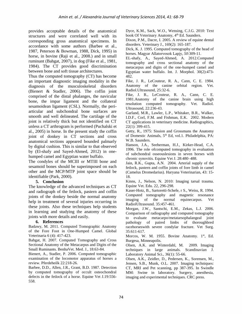
Figure (1) bones of fetlock joint of donkey. Image A: proximal extremity of 1st phalanx.1= medial condyle. 2=
lateral condyle. 3= sagittal ridge. B: distal extremity of MCIII. 1=lateral articular surface. 2= medial articular
surface. 3= sagittal groove. C: 1, 2= proximal sesamoid bones.
Figure (2) fetlock joint injected with colored latex showing the joint capsule of the joint and its pouches. A= image
of palmar pouch. B= image of dorsal pouch. 1= MCIII. 2= 1st phalanx. 3= dorsal pouch. 4= palmar pouch. 5=
proximal palmar pouch. 6= distal palmar pouch.
Amin et. al. / Alexandria Journal of Veterinary Sciences 2014, 41: 68-79
76
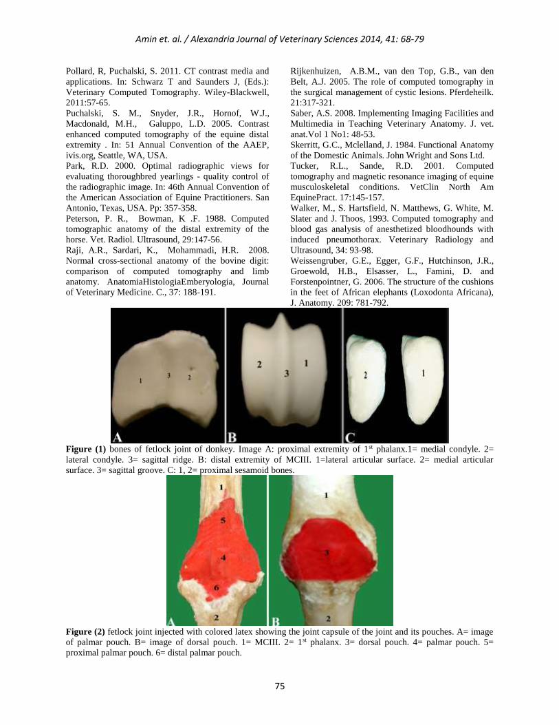
Figure (3) photograph of transverse anatomic section (right) and transverse CT views (left) of normal fetlock joint
in donkey sequentially displayed from proximal to distal. 1=MCIII. 1a= sagittal ridge of MCIII. 2= proximal
sesamoid bone. 3=DDFT. 4= SDFT. 5= intersesamoidean ligament. 6= suspensory ligament.
Amin et. al. / Alexandria Journal of Veterinary Sciences 2014, 41: 68-79
77
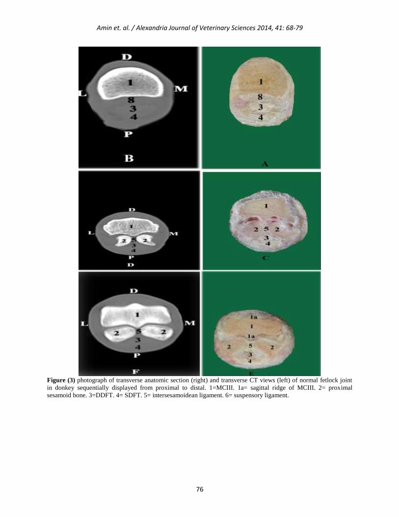
Figure (4) photographs of the sagittal anatomic section (B) and sagittal reconstructed CT image of the normal
fetlock joint (A). 1= MCIII. 1a= distal extremity of MCIII. 2= 1st phalanx. 3= 2nd phalanx. 4=3rd phalanx.
5=proximal sesamoid bone. 6= intersesamoidean ligament. 7= suspensory ligament. 8= DDFT. 9= palmar annular
ligament. 10=oblique sesamoidean ligament. 11= straight sesamoidean ligament of distal sesamoid bone. 12=middle
scutum. 13= SDFT. 14= distal sesamoid bone. 15= collateral sesamoidean ligament of the distal sesamoid bone. 16=
digital cushion. 17= proximal palmar pouch of MCP joint. 18= dorsal digital extensor tendon.
Figure (5) photograph of the articular surfaces of the pastern joint of the donkey. B= distal extremity of the 1st
phalanx. 1=medial condyle. 2=lateral condyle. 3= sagittal groove. A= proximal extremity of 2nd phalanx. 1= lateral
articular cavity. 2= medial articular cavity. 3=sagittal ridge.
Amin et. al. / Alexandria Journal of Veterinary Sciences 2014, 41: 68-79
78
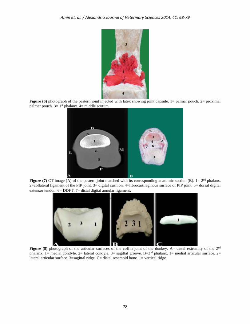
Figure (6) photograph of the pastern joint injected with latex showing joint capsule. 1= palmar pouch. 2= proximal
palmar pouch. 3= 1st phalanx. 4= middle scutum.
Figure (7) CT image (A) of the pastern joint matched with its corresponding anatomic section (B). 1= 2nd phalanx.
2=collateral ligament of the PIP joint. 3= digital cushion. 4=fibrocartilaginous surface of PIP joint. 5= dorsal digital
extensor tendon. 6= DDFT. 7= distal digital annular ligament.
Figure (8) photograph of the articular surfaces of the coffin joint of the donkey. A= distal extremity of the 2nd
phalanx. 1= medial condyle. 2= lateral condyle. 3= sagittal groove. B=3rd phalanx. 1= medial articular surface. 2=
lateral articular surface. 3=sagittal ridge. C= distal sesamoid bone. 1= vertical ridge.
Amin et. al. / Alexandria Journal of Veterinary Sciences 2014, 41: 68-79
79
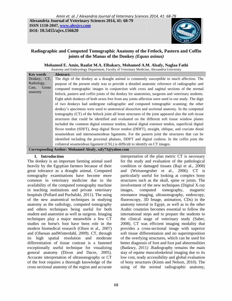
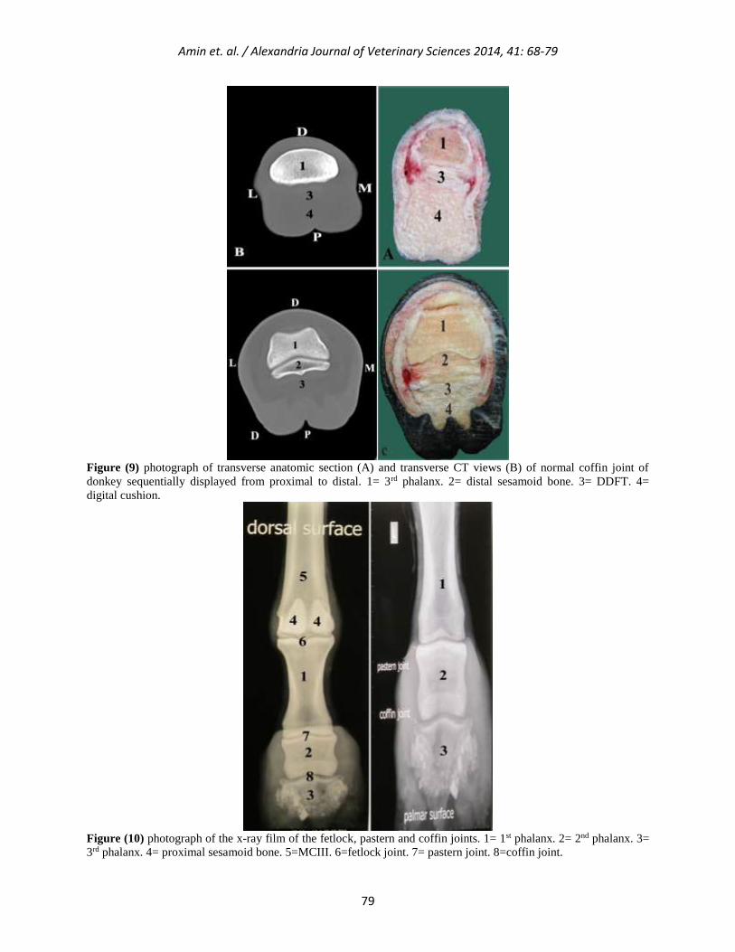
Figure (9) photograph of transverse anatomic section (A) and transverse CT views (B) of normal coffin joint of
donkey sequentially displayed from proximal to distal. 1= 3rd phalanx. 2= distal sesamoid bone. 3= DDFT. 4=
digital cushion.
Figure (10) photograph of the x-ray film of the fetlock, pastern and coffin joints. 1= 1st phalanx. 2= 2nd phalanx. 3=
3rd phalanx. 4= proximal sesamoid bone. 5=MCIII. 6=fetlock joint. 7= pastern joint. 8=coffin joint.


![Welcome [applications.emro.who.int]applications.emro.who.int/docs/RC64_2017_bulletin_1_20097_en.pdf · their talents.This year, almost 50 drawings from across the Region were awarded](https://img.pdfslide.us/doc/110x75/5f31da622bb02e749c290170/welcome-their-talentsthis-year-almost-50-drawings-from-across-the-region-were.jpg)