Embed Size (px)
Citation preview
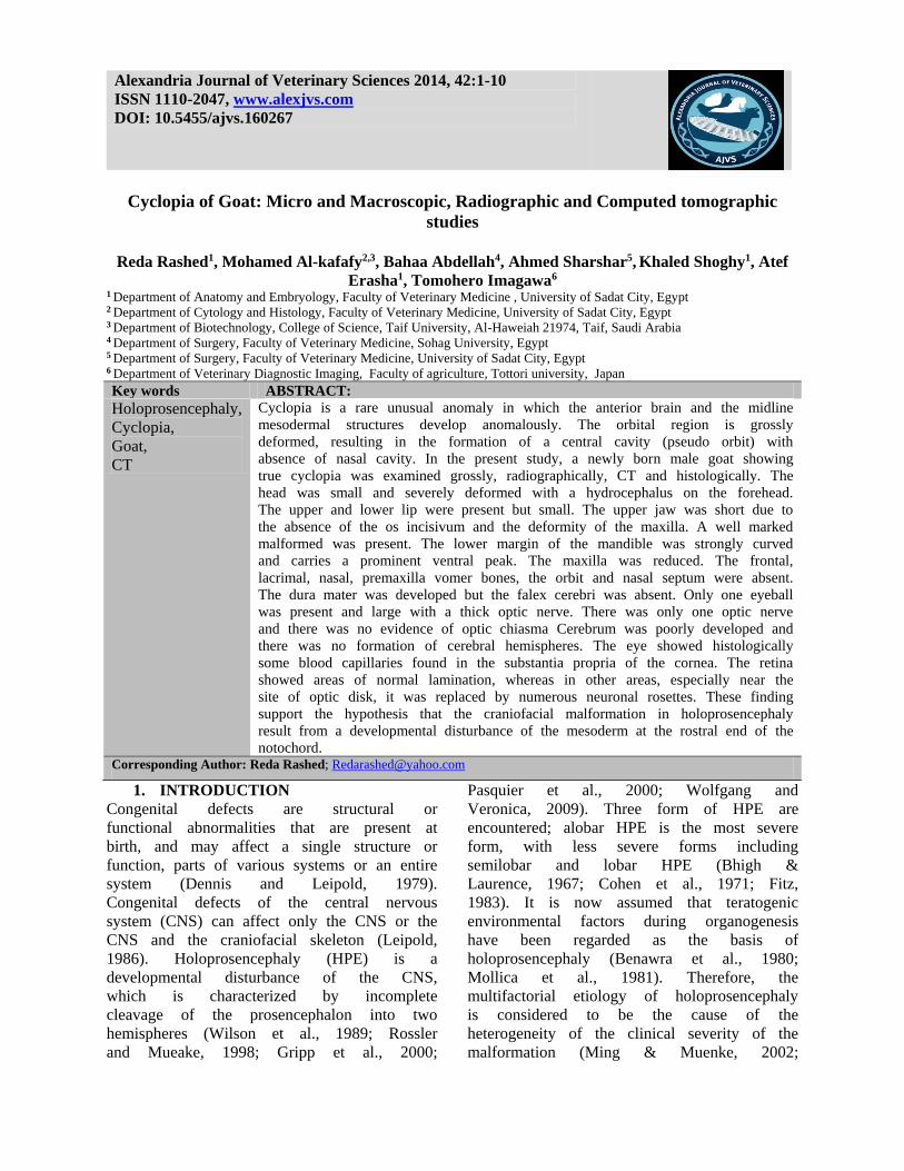
Cyclopia of Goat: Micro and Macroscopic, Radiographic and Computed tomographic
studies
Reda Rashed1, Mohamed Al-kafafy2,3, Bahaa Abdellah4, Ahmed Sharshar5, Khaled Shoghy1, Atef
Erasha1, Tomohero Imagawa6 1 Department of Anatomy and Embryology, Faculty of Veterinary Medicine , University of Sadat City, Egypt 2 Department of Cytology and Histology, Faculty of Veterinary Medicine, University of Sadat City, Egypt 3 Department of Biotechnology, College of Science, Taif University, Al-Haweiah 21974, Taif, Saudi Arabia 4 Department of Surgery, Faculty of Veterinary Medicine, Sohag University, Egypt 5 Department of Surgery, Faculty of Veterinary Medicine, University of Sadat City, Egypt 6 Department of Veterinary Diagnostic Imaging, Faculty of agriculture, Tottori university, Japan
Key words ABSTRACT:
Holoprosencephaly,
Cyclopia,
Goat,
CT
Cyclopia is a rare unusual anomaly in which the anterior brain and the midline
mesodermal structures develop anomalously. The orbital region is grossly
deformed, resulting in the formation of a central cavity (pseudo orbit) with
absence of nasal cavity. In the present study, a newly born male goat showing
true cyclopia was examined grossly, radiographically, CT and histologically. The
head was small and severely deformed with a hydrocephalus on the forehead.
The upper and lower lip were present but small. The upper jaw was short due to
the absence of the os incisivum and the deformity of the maxilla. A well marked
malformed was present. The lower margin of the mandible was strongly curved
and carries a prominent ventral peak. The maxilla was reduced. The frontal,
lacrimal, nasal, premaxilla vomer bones, the orbit and nasal septum were absent.
The dura mater was developed but the falex cerebri was absent. Only one eyeball
was present and large with a thick optic nerve. There was only one optic nerve
and there was no evidence of optic chiasma Cerebrum was poorly developed and
there was no formation of cerebral hemispheres. The eye showed histologically
some blood capillaries found in the substantia propria of the cornea. The retina
showed areas of normal lamination, whereas in other areas, especially near the
site of optic disk, it was replaced by numerous neuronal rosettes. These finding
support the hypothesis that the craniofacial malformation in holoprosencephaly
result from a developmental disturbance of the mesoderm at the rostral end of the
notochord. Corresponding Author: Reda Rashed; [email protected]
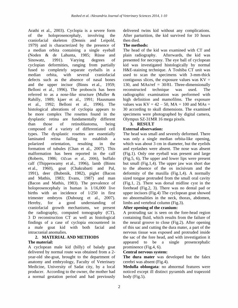
1. INTRODUCTION
Congenital defects are structural or
functional abnormalities that are present at
birth, and may affect a single structure or
function, parts of various systems or an entire
system (Dennis and Leipold, 1979).
Congenital defects of the central nervous
system (CNS) can affect only the CNS or the
CNS and the craniofacial skeleton (Leipold,
1986). Holoprosencephaly (HPE) is a
developmental disturbance of the CNS,
which is characterized by incomplete
cleavage of the prosencephalon into two
hemispheres (Wilson et al., 1989; Rossler
and Mueake, 1998; Gripp et al., 2000;
Pasquier et al., 2000; Wolfgang and
Veronica, 2009). Three form of HPE are
encountered; alobar HPE is the most severe
form, with less severe forms including
semilobar and lobar HPE (Bhigh &
Laurence, 1967; Cohen et al., 1971; Fitz,
1983). It is now assumed that teratogenic
environmental factors during organogenesis
have been regarded as the basis of
holoprosencephaly (Benawra et al., 1980;
Mollica et al., 1981). Therefore, the
multifactorial etiology of holoprosencephaly
is considered to be the cause of the
heterogeneity of the clinical severity of the
malformation (Ming & Muenke, 2002;
Alexandria Journal of Veterinary Sciences 2014, 42:1-10
ISSN 1110-2047, www.alexjvs.com
DOI: 10.5455/ajvs.160267
Rashed et al. /Alexandria Journal of Veterinary Sciences 2014, 1-10
2
Arathi et al., 2003). Cyclopia is a severe form
of the holoprosencephaly, involving the
craniofacial skeleton (Dennis and Leipold,
1979) and is characterized by the presence of
a median orbita containing a single eyeball
(Noden & de Lahunta, 1985; Rüsse and
Sinowatz, 1991). Varying degrees of
cyclopian deformities, ranging from partially
fused to completely separate eyeballs in a
median orbita, with several craniofacial
defects such as the absence of nasal bones
and the upper incisor (Binns et al., 1959;
Belloni et al., 1996). The proboscis has been
referred to as a nose-like structure (Muller &
Rahilly, 1989; kjaer et al., 1991; Hausmann
et al., 1992; Belloni et al., 1996). The
histological alterations of cyclopia appears to
be more complex The rosettes found in the
dysplastic retina are fundamentally different
than those of retinoblastoma, being
composed of a variety of differentiated cell
types. The dysplastic rosettes are essentially
laminated retina failing to establish a
polarized orientation, resulting in the
formation of tubules (Chan et al., 2007). This
malformation has been reported in the calf
(Roberts, 1986; OZcan et al., 2006), buffalo
calf (Thippeswamy et al., 1996), lamb (Binns
et al., 1960), goat (Chakrabarti and Pal,
1991), deer (Bubenik, 1982), piglet (Bacon
and Mathis, 1983; Evans, 1987) and man
(Bacon and Mathis, 1983). The prevalence of
holoprosencephaly in human is 1/16,000 live
births with an incidence of 1/250 in first
trimester embryos (Dubourg et al., 2007).
Hereby, for a good understanding of
craniofacial growth mechanisms, we present
the radiography, computed tomography (CT),
3 D reconstruction CT as well as histological
findings of a case of cyclopia encountered in
a male goat kid with both facial and
intracranial anomalies.
2. MATERIAL AND METHODS
The material:
A cyclopean male kid (billy) of balady goat
delivered by normal route was obtained from a 2-
year-old she-goat, brought to the department of
anatomy and embryology, Faculty of Veterinary
Medicine, University of Sadat city, by a local
producer. According to the owner, the mother had
a normal gestation period and had previously
delivered twins kid without any complications.
After parturition, the kid survived for 10 hours
then died.
The methods:
The head of the kid was examined with CT and
plain radiography. Afterwards, the kid was
presented for necropsy. The eye ball of cyclopean
kid was investigated histologically by normal
H&E-staining technique. A Toshiba CT unit was
used to scan the specimens with 3-mm-thick
contiguous slices, the exposure values was KV =
130, and MAs/ref = 30/81. Three-dimensionally
reconstructed technique was used. The
radiographic examination was performed with
high definition and mamofilms. The exposure
values was KV = 42 – 50, MA = 100 and MAs =
30 according to skull dimensions. The examined
specimens were photographed by digital camera,
Olympus SZ-31MR 16 mega pixels.
3. RESULT
External observation:
The head was small and severely deformed. There
was only a single median orbita-like opening,
which was about 3 cm in diameter, but the eyelids
and eyelashes were absent. The nose was absent
(Fig.1). Only one eyeball was present and large
(Fig.5, 6), The upper and lower lips were present
but small (Fig.1,4). The upper jaw was short due
to the absence of the os incisivum and the
deformity of the maxilla (Fig.1,4). A normally
sized tongue protruded from the small oral cavity
(Fig.1, 2). There was dorsal midline cyst in the
forehead (Fig.2, 3). There was no dental pad or
upper incisors (Fig.4) The cyclopean goat showed
no abnormalities in the neck, thorax, abdomen,
limbs and vertebral column (Fig.3).
After opening of the cranium:
A protruding sac is seen on the fore-head region
containing fluid, which results from the failure of
the neural groove to close (Fig.2). After opening
of this sac and cutting the dura mater, a part of the
nervous tissue was exposed and protruded inside
the sac of the fore head, and with investigation it
appeared to be a single prosencephalic
prominence (Fig.4, 6).
Central nervous system:
The dura mater was developed but the falex
cerebri was absent (Fig.4).
Medulla oblongata: no abnormal features were
noticed except ill distinct pyramids and trapezoid
body (Fig.5).
Rashed et al. /Alexandria Journal of Veterinary Sciences 2014, 1-10
3
Pons: it was enlarged but no abnormal feature are
detected (Fig.5).
Cerebellum: It was poorly developed. No
abnormal feature was noticed in the cerebellum
and it differentiated into the most primitive part;
the flocculo-nodular lobe, and small median part;
the vermis; and two small lateral cerebellar
hemispheres (Fig.6, 7).
Midbrain: Macroscopically no abnormality was
detected. The rostral and caudal colliculi were
very large (Fig.6, 7). The cerebral crura were
enlarged and the intercrurl sulcus was replaced by
faint intercrural fissure (Fig.5).
Diencephalon: thalami were fused rostrally but
caudally a portion of third ventricle was seen in
between the malformed thalami (Fig.7). Tuber
cenerium and infundibulum of pituitary gland are
absent (Fig.5).
Optic nerves: There was only one optic nerve and
there was no evidence of optic chiasma (Fig.5).
The entrance of the optic nerve to the eyeball was
normal grossly (Fig.6).
Cerebrum: it was poorly developed and there was
no formation of cerebral hemispheres (Fig.4).
Both the lateral ventricles and the third ventricle
were absent. Only, in the floor of the sac, the
opening of the mesencephalic aqueduct was
observed between the two thalami in front and the
colliculi behind (Fig.7). The lower part of the third
ventricle in between two thalami was recognizable
and it opened widely above in the dilated sac
(Fig.7).
Hard palate:
The palate was structurally abnormal, the rostral
part showed an acute angle at the premaxilla
region (Fig.8). The palatine ridge showed different
manner of curvature and absent in the caudal third
of the palate (Fig.8). The palatine raphae was faint
rostrally, grooved in the middle part and elevated
caudally (Fig.8).
Cranial bones:
A well marked malformed mandible was present.
The lower margin was strongly curved and carries
a prominent ventral peak (Fig.9). Resulted from
this curvature, the incisive teeth were positioned
near to the coronoid process (Fig. 9). The
mandible was massive and it's right and left halves
diverge at relatively large angle as illustrated by X
ray and C.T (Figs. 11, 13). There were four
incisive, three premolars teeth and one molar tooth
in each mandible (Fig. 10).
The maxilla was reduced and appeared as
irregular bony mass and was malformed and 4
teeth analog were found in each mass (Fig. 10,
12). The frontal, lacrimal, nasal, premaxilla vomer
bones, the orbit and nasal septum were absent
(Fig.10, 11, 12, 13, 14, 15, 16). The base of skull
formed by body of occipital and sphenoid bones
appeared normal (Fig.13). The two ethmoidal
fossa and etmoidal crest were observed (Fig.15,
18). As a result of absence of the above mentioned
bones, the cranial cavity was small and malformed
(Fig.16, 17).
CT and X-ray findings;
It showed a malformed maxilla with four teeth
analog. There are four incisive teeth, three
premolar teeth and one molar tooth on each
mandible, the mandible was curved, with wide
deep temporal fossa. The right and left halves of
the mandible diverge at relatively large angle. The
orbit was absent. The frontal, nasal, vomer and
incisive bones were absent. Volume 3D dimension
CT showed the large opening of the cranium due
to the absence of orbit and the frontal bone.
Sagittal and coronal CT scan showed one lens in
on eyeball.
Histological findings: Ocular findings
The cornea
Except for some blood capillaries found in
the substantia propria (Fig.19a), the cornea
appeared to have the normal histological
organization. Cornea consisted of anterior
epithelium (stratified squamous epithelium),
substantia propria and corneal endothelium
(simple squamous epithelium).
The retina
It showed areas of normally laminated
neural retina (Fig.19b), whereas in other
areas, especially near the site of optic disk, it
was replaced by numerous neuronal rosettes
(Figs.20a, b; 21b).
The optic nerve
It was thick and spaced by the central artery of the
retina (Fig. 21a)
4. Discussion
Many reports on cyclopia describe the presence of
a single median orbita that contains either a single
eyeball (true cyclopia) (Binns et al., 1960; Binns
et al., 1963; Bacon and Mathis, 1983; Camon
et al., 1990; Chakrabarti and Pal, 1991) or
incompletely fused eyeballs (synophthalmia)
(Evans, 1987; Jie and Shi, 1991). Apart from the

Rashed et al. /Alexandria Journal of Veterinary Sciences 2014, 1-10
4
classical description of cyclopia, several authors
(Roberts, 1986; Van Allen et al., 1993; Cannistra
et al., 2001) describe that anophthalmia may also
be encountered in cases of cyclopia. In general,
cyclopia is considered to result from defects at the
neural plate stage of development and involves
more specifically the rostral portion of the
notochord and the mesoderm surrounding it (Jubb
and Huxtable, 1993). Depending on the severity of
the inhibiting agent that causes this congenital
defect, various forms of craniofacial deformations
ranging from a true cyclopia to synophthalmia can
occur. However, if the inhibiting agent is severe
enough, anophthalmia may be formed. In this
respect, our study matches the classical
description of cyclopia. In addition, in many cases
of cyclopia, the presence of a proboscis dorsal to
the median orbita has been described (Binns et al.,
1963; Bacon and Mathis, 1983; Evans, 1987;
Camon et al., 1990; Cannistra et al., 2001).
However, such a structure was not present in this
cyclop. In this male goat, deformations and
deficiencies in the craniofacial bones such as
arrhinia and brachygnathia superior and defects of
the CNS such as prosencephalic aplasia were
similar to those described in the literature (Binns
et al., 1960; Bubenik, 1982; Evans, 1987;
Thippeswamy et al., 1996). In the light of these
similarities and differences, we assumed that the
current case is a typical cyclopia.
In the present case we the neural retina was
laminated, except near the site of optic disk, it was
replaced by numerous neuronal rosettes. The
neural rosettes are fundamentally laminated retina
failing to set up a polarized orientation, resulting
in the formation of tubules (Chan et al., 2007).
Cyclopian malformation was reported in newborn
lambs from ewes fed with Veratrum californicum
on the 14th day of gestation (Binns et al., 1965).
Three steroidal alkaloids, i.e. jervine, cyclopamine
and cycloposine, were suggested to be capable of
inhibiting the neural development in lambs
(Keeler, 1984). A detailed botanical investigation
might be necessary for further information about
the aetiology of this malformation. Several other
teratogens, such as radiation, viral infections and
hypovitaminosis are in the scope of possible other
factors that may cause this congenital defect.
Since there was no recorded history about the
mother of the goat kid and due to the inability to
detect a causative agent it wasn't possible to
ascertain the cause of this malformation.
FIG. 1: Rostral view of the cyclopean goat head shows a single median orbita-like opening (black arrow) with one
eyeball. The tongue (black star) protrudes from the small oral orifice. The eyelids, eyelashes and nose are absent.
FIG. 2: Lateral view of the cyclopean goat head shows dorsal midline cyst (black star) in the forehead, small upper
jaw (white arrow), and large protruded lower jaw (black arrow).
2 1

Rashed et al. /Alexandria Journal of Veterinary Sciences 2014, 1-10
5
FIG. 3: Lateral view of the cyclopean goat shows no abnormalities in the neck, thorax, abdomen, limbs and
vertebral column.
FIG. 4: Dorsal view of the cyclopean goat head after opening of the cyst of the forehead showing protrusion of the
brain tissue (black star) into the cyst of the forehead. The dura mater (white star) is developed with absence of falex
cerebri. The forceps elevate the small upper lip to illustrate the absence of dental pad and upper incisors.
FIG. 5: Ventral view of the brain of the cyclopean goat shows absence of the telencephalon, large single eyeball
(red star), very short single optic nerve which ends within the diencephalon by two divergent optic tract (black star),
enlarged cerebral crura (c), narrow intercrural fissure (if), enlarged pons (p), ill distinct trapezoid body (tb) and ill
distinct pyramid (py). The optic chiasm, tuber cenerium and infundibulum of pituitary gland are absent (white
arrow). The optic nerve is single.
FIG. 6: Lateral view of the brain of the cyclopean goat shows absence of cerebral hemisphere, malformed thalami
appear as a prosencephalic prominence (th), enlarged rostral colliculus (rc) and caudal colliculus (cc), large single
eyeball (red star). The cerebellum is poorly developed with no abnormality.
3 4
c
if tb
p
th rc cerebellum
cc
py
Rashed et al. /Alexandria Journal of Veterinary Sciences 2014, 1-10
6
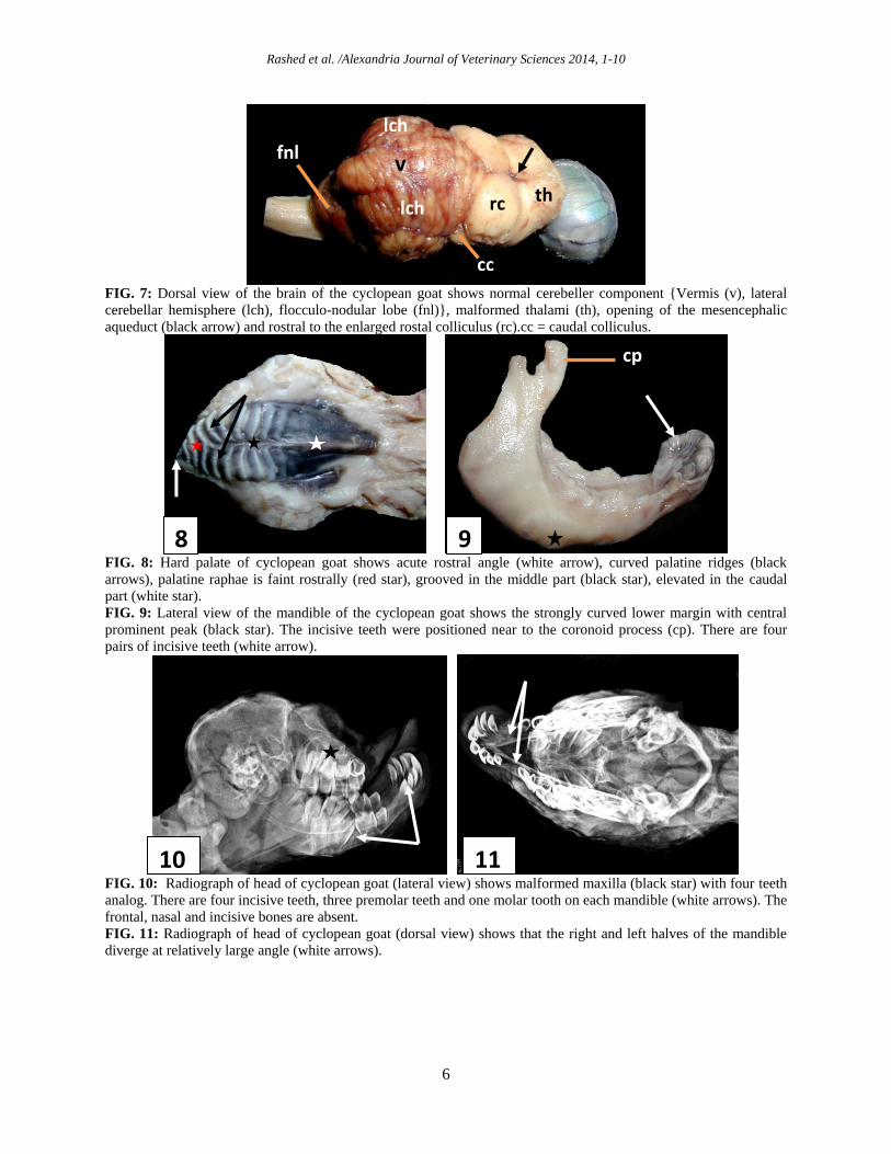
FIG. 7: Dorsal view of the brain of the cyclopean goat shows normal cerebeller component {Vermis (v), lateral
cerebellar hemisphere (lch), flocculo-nodular lobe (fnl)}, malformed thalami (th), opening of the mesencephalic
aqueduct (black arrow) and rostral to the enlarged rostal colliculus (rc).cc = caudal colliculus.
FIG. 8: Hard palate of cyclopean goat shows acute rostral angle (white arrow), curved palatine ridges (black
arrows), palatine raphae is faint rostrally (red star), grooved in the middle part (black star), elevated in the caudal
part (white star).
FIG. 9: Lateral view of the mandible of the cyclopean goat shows the strongly curved lower margin with central
prominent peak (black star). The incisive teeth were positioned near to the coronoid process (cp). There are four
pairs of incisive teeth (white arrow).
FIG. 10: Radiograph of head of cyclopean goat (lateral view) shows malformed maxilla (black star) with four teeth
analog. There are four incisive teeth, three premolar teeth and one molar tooth on each mandible (white arrows). The
frontal, nasal and incisive bones are absent.
FIG. 11: Radiograph of head of cyclopean goat (dorsal view) shows that the right and left halves of the mandible
diverge at relatively large angle (white arrows).
10
8 9
11
rc
v
lch
fnl
lch
th
cc
cp

Rashed et al. /Alexandria Journal of Veterinary Sciences 2014, 1-10
7
FIG. 12: Volume 3D dimension CT scan of a fetal skull (lateral view) shows malformed maxilla (mx), curved
malformed mandible (m), wide, deep temporal fossa (black star). The orbit is absent. The frontal, nasal and incisive
bones are absent.
FIG. 13: Volume 3D dimension CT scan of a fetal skull (ventral view) shows that the right and left halves of the
mandible diverge at relatively large angle (white arrows).The base of the skull appears normal {occipital bone (oc),
sphenoid bone (s)}. The vomer bone is absent.
FIG. 14: Volume 3D dimension CT scan of a fetal skull (rostro-dorsal view) shows the large opening of the cranium
due to the absence of orbit and the frontal bone (white fork). The nasal and vomer bones and nasal septum were
absent.
FIG. 15: Volume 3D dimension CT scan of a fetal skull (rostral view) shows etmoidal fossa (white arrows) and
ethmoidal crest (black arrow). The vomer bone is absent.
12 13
15 14
16 17
mx
m
oc s
mx m
Rashed et al. /Alexandria Journal of Veterinary Sciences 2014, 1-10
8
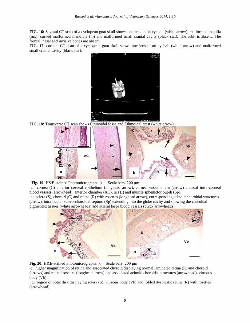
FIG. 16: Sagittal CT scan of a cyclopean goat skull shows one lens in on eyeball (white arrow), malformed maxilla
(mx), curved malformed mandible (m) and malformed small cranial cavity (black stat). The orbit is absent. The
frontal, nasal and incisive bones are absent.
FIG. 17: coronal CT scan of a cyclopean goat skull shows one lens in on eyeball (white arrow) and malformed
small cranial cavity (black star).
FIG. 18: Transverse CT scan shows Ethmoidal fossa and Ethmoidal crest (white arrow).
Fig. 19: H&E-stained Photomicrographs. ). Scale bars: 200 μm
a; cornea (C) anterior corneal epithelium (longhead arrow), corneal endothelium (arrow) unusual intra-corneal
blood vessels (arrowhead), anterior chamber (AC), iris (I) and muscle sphenictor pupili (Sp).
b; sclera (S), choroid (C) and retina (R) with rosettes (longhead arrow), corresponding acinoid choroidal structures
(arrow), intra-ocular sclero-choroidal septum (Sp) extending into the globe cavity and showing the choroidal
pigmented tissues (white arrowheads) and scleral large blood vessels (black arrowheads).
Fig. 20: H&E-stained Photomicrographs. ). Scale bars: 200 μm
c; higher magnification of retina and associated choroid displaying normal laminated retina (R) and choroid
(arrows) and retinal rosettes (longhead arrow) and associated acinoid choroidal structures (arrowhead), vitreous
body (Vb).
d; region of optic disk displaying sclera (S), vitreous body (Vb) and folded dysplastic retina (R) with rosettes
(arrowhead).

Rashed et al. /Alexandria Journal of Veterinary Sciences 2014, 1-10
9
Fig. 21: H&E-stained Photomicrographs. ). Scale bars: 100 μm
e; higher magnification of optic disk region showing optic nerve (ON) spaced by a large cavity at its sagital plane
(asterisk) and folded retina (R) with rosettes (arrowhead), sclera (S) and vitreous body (Vb).
f; higher magnification of dysplastic retina (DR) with rosettes (arrowhead) sandwiched between choroid (C) at one
side and normal retina (R) with ganglionic cells (arrow) next to vitreous body (Vb)
5. REFERENCES
Arathi, N., Mahadevan, A., Santosh, V., Yasha, T.C.,
Shankar, S.K. 2003. Holoprosencephaly with cyclopia:
report of a pathological study. Neurol India 51: 279-
282.
Bacon, W., Mathis, R. 1983. Craniofacial characteristics
of cyclopia in man and swine. Angle Orthod. 53: 290-
310.
Belloni, E., Muenke, M., Roessler, E., Traverso, G.,
Siegel-Bartelt, J., Frumkin, A., Mitchell, H.F., Donis-
Keller, H., Helms, C., Hing, A.V. 1996. Identification
of Sonic hedgehog as a candidate gene responsible for
holoprosencephaly. Nat Genet. 14: 353-356.
Benawra, R., Mangurten, H.H., Duttell, D.R. 1980.
Cyclopia and other anomalies following maternal
ingestion of salicylates. J Pediatrn: 1069-1071.
Binns, W., Anderson, W.A., Sullivan, D. J. 1960. Further
observations on a congenital cyclopian-type
malformation in lambs. J. Am. Vet. Med. Assoc.
137:515-521.
Binns, W., James, L.F., Shupe, J. L., Everett, G. 1963. A
congenital cyclopian-type malformation in lambs
induced by maternal ingestion of a range plant,
Veratrum californicum. Am. J. Vet. Res. 24: 1164-
1175.
Binns, W., Shupe, J.L., Keeler, R.F., James, L.F. 1965.
Chronologic evaluation of teratogenicity in sheep fed
Veratrum californicum. J. Am. Vet. Med. Assoc. 147:
839-842.
Binns, W., Thacker, E. J., James, L. F., Huffman, W. T.
1959. A congenital cyclopian-type malformation in
lambs. J. Am. Vet. Med. Assoc. 134: 180–183.
Bligh, A.S., Laurence, K.M. 1967. The radiological
appearances in arhinencephaly. Clin Radiol. 18:383-91.
Bligh, A.S., Laurence, K.M. 1967. The radiological
appearances in arhinencephaly. Clin Radiol. 18:383-91.
Bubenik, G. 1982. Cyclopia combined with anencephalia
in white-tailed deer, Odocoileus virgianus
(Zimmerman, 1780). Säugetierkd. Mitt. 30:158-160.
Camon, J., Sabate, D., Franch, J., Lopez-Bejar, M.A.,
Pastor, J., Rutllant, J., Ordeig, J., Degollada, E., Verdu,
J. 1990. Associated multiple congenital malformations
in domestic animals. Contribution of four cases. J. Vet.
Med. A 37:659-668.
Cannistra, C., Barbet, P., Parisi, P., Iannetti, G. 2001.
Cyclopia: a radiological and anatomical craniofacial
post mortem study. J. CranioMaxillofac. Surg. 29:150-
155.
Chakrabarti, A., Pal, A. 1991. Cyclopia prostomus
arrhynchus in a black bengal goat. Indian Vet. J. 68:
985-986.
Chan, A., Lakshminrusimha, S., Heffner, R., Gonzalez-
Fernandez, F. 2007. Histogenesis of retinal dysplasia in
trisomy 13. Diagnostic Pathol. 2, 48:1-8.
Cohen, M.M., Jirasek, J.E., Guzman, R.T., Gorlin, R.J.,
Peterson, M.Q. 1971. Holoprosencephaly and facial
dysmorphia: nosology, etiology and pathogenesis. Birth
Defects 7:125-135.
Dennis, S.M., Leipold, H.W. 1979. Ovine congenital
defects. Bulletin 49 (4): 233-239.
Dubourg, C., Bendavid, C., Pasquier, L., Henry, C.,
Odent, S., David, V. 2007. Holoprosencephaly.
Orphanet J Rare Dis. 2, 8:1-14.
Evans, H. E. 1987. Cyclopia, situs inversus and widely
patent ductus arteriosus in a new-born pig, Sus scrofa.
Anat. Histol. Embryol. 16:221-226.
Fitz, C.R. 1983. Holoprosencephaly and related entities.
Neuroradiology 25:225-238.
Gripp, K.W., Wotton, D., Edwards, M.C., Roessler, E.,
Ades, L., Meinecke, P., Richieri-Costa, A., Zackai,
E.H., Massague, J., Muenke, M., Elledge, S.J. 2000.
Mutations in TGIF cause holoprosencephaly and link
NODAL signalling to human neural axis determination.
Nat Genet. 25: 205-208.
Rashed et al. /Alexandria Journal of Veterinary Sciences 2014, 1-10
10
Hausmann, N., Stefani, F.H., Lund, O.E. 1992.
Diplophthalmia versus cyclopia and synophthalmia.
Mechanisms of doubling of the eye. Doc Ophthalmol
79:201-219.
Jie, Z., Shi, Z. 1991. Case of atypical cyclopia. Oral Surg.
Oral Med. Oral Pathol. 72:332-333.
Jubb, K.V.F., Huxtable, C. R. 1993. The nervous system.
In: Pathology of Domestic Animals, Vol. 1 (K. V.
F.Jubb, P. C.Kennedy, and N.Palmer, eds). California:
Academic Press, pp. 441–529.
Keeler, R.F., 1984. Mammalian teratogenicity of steroidal
alkaloids. In: Biochemistry and Function of
Isopentenoids in Plants (W. D.Ness, G.Fuller, and L.
S.Tsai, eds). New York: Dekker Inc., pp. 531–562.
Kjaer, I., Keeling, J.W., Graem, N. 1991. The midline
craniofacial skeleton in holoprosencephalic fetuses. J
Med Genet. 28: 846-855.
Krauss, R.S. 2007. Holoprosencephaly: new models, new
insights. Expert. Rev. Mol. Med. 9:1-17.
Leipold, H.W. 1986. Neonatal disease and disease
management, congenital defects in cattle. In: Current
Veterinary Therapy 2: Food Animal Practice (J.
L.Howard, ed.). Philadelphia: W.B. Saunders
Company, pp. 89-98.
Ming, J.E., Muenke, M. 2002 Multiple hits during early
embryonic development: digenic diseases and
holoprosencephaly. Am. J. Hum. Genet. 71:1017-
1032.
Mollica, F., Pavone, L., Sorge, G. 1981. Maternal drug
ingestion and cyclopia. J. Pediatr. 98: 680.
Muller, F., O'Rahilly, R. 1989. Mediobasal
prosencephalic defects, including holoprosencephaly
and cyclopia, in relation to the development of the
human forebrain. Am. J. Anat. 185: 391-414.
Noden, D. M., De Lahunta, A. 1985. The embryology of
domestic animals, developmental mechanisms and
malformations (G.Stamathis, ed.). Baltimore: Williams
& Wilkins, pp. 1-8.
ÖZcan, K., Gürbulak, K., Takçi̇, İ., ÖZen, H., Kaçar, C.
and Pancarci̇, M. Ş. (2006), Atypical Cyclopia in a
Brown Swiss Cross Calf: A Case Report. Anatomia,
Histologia, Embryol. 35: 152–154.
Pasquier, L., Dubourg, C., Blayau, M., Lazaro, L., Le
Marec, B., David, V., Odent, S. 2000. A new mutation
in the six-domain of SIX3 gene causes
holoprosencephaly. Eur J Hum Genet. 8:797-800.
Roberts, S. J. 1986. Veterinary Obstetric and Genital
Disease (Theriogenology). Vermont, pp. 51-91.
Roessler, E., Muenke, M. 1998. Holoprosencephaly: a
paradigm for the complex genetics of brain
development. J. Inherited Metabolic Dis. 21:481-497.
Roessler, E., Muenke, M. 1998. Holoprosencephaly: a
paradigm for the complex genetics of brain
development. J. Inherit. Metab. Dis. 21: 481-497.
Rüsse, I., Sinowatz, F. 1991. In: Lehrbuch der
Embryologie der Haustiere (A.Von Den Driesch, ed.).
Berlin and Hamburg: Verlag Paul Parey, pp. 419-460.
Thippeswamy, T., Prasad, R. V., Kakade, K. 1996. A
clinical case of cyclopia prostomus arrhynchus in a
buffalo calf (Bubalus bubalis). Indian Vet. J. 73: 674-
676.
Van Allen, M.I., Ritchie, S., Toi, A., Fong, K., Winsor, E.
1993. Trisomy 4 in a fetus with cyclopia and other
anomalies. Am. J. Med. Genet. 46:193-197.
Wilson, W.G., Shanks, D.E., Sudduth, K.W., Couper,
K.A., McIlhenny, J. 1989. Holoprosencephaly and
interstitial deletion of 2 (p2101 p2109). Am. J. Med.
Genet. 34:252-254.
Wolfgang, H., Veronika, M.A. 2009. 3-D reconstruction
of a human fetus with combined holoprosencephaly
and cyclopia. Head and Face Medicine 5,14:1-11.