Embed Size (px)
Citation preview
Quantitative Analysis of Histone Modifications:Formaldehyde Is a Source of Pathological N6-Formyllysine That Is Refractory to Histone DeacetylasesBahar Edrissi1, Koli Taghizadeh2, Peter C. Dedon1,2*
1 Department of Biological Engineering, Massachusetts Institute of Technology, Cambridge, Massachusetts, United States of America, 2 Center for Environmental Health
Sciences, Massachusetts Institute of Technology, Cambridge, Massachusetts, United States of America
Abstract
Aberrant protein modifications play an important role in the pathophysiology of many human diseases, in terms of bothdysfunction of physiological modifications and the formation of pathological modifications by reaction of proteins withendogenous electrophiles. Recent studies have identified a chemical homolog of lysine acetylation, N6-formyllysine, as anabundant modification of histone and chromatin proteins, one possible source of which is the reaction of lysine with 39-formylphosphate residues from DNA oxidation. Using a new liquid chromatography-coupled to tandem mass spectrometrymethod to quantify all N6-methyl-, -acetyl- and -formyl-lysine modifications, we now report that endogenous formaldehydeis a major source of N6-formyllysine and that this adduct is widespread among cellular proteins in all compartments. N6-formyllysine was evenly distributed among different classes of histone proteins from human TK6 cells at 1–4 modificationsper 104 lysines, which contrasted strongly with lysine acetylation and mono-, di-, and tri-methylation levels of 1.5-380, 5-870,0-1400, and 0-390 per 104 lysines, respectively. While isotope labeling studies revealed that lysine demethylation is not asource of N6-formyllysine in histones, formaldehyde exposure was observed to cause a dose-dependent increase in N6-formyllysine, with use of [13C,2H2]-formaldehyde revealing unchanged levels of adducts derived from endogenous sources.Inhibitors of class I and class II histone deacetylases did not affect the levels of N6-formyllysine in TK6 cells, and the class IIIhistone deacetylase, SIRT1, had minimal activity (,10%) with a peptide substrate containing the formyl adduct. These datasuggest that N6-formyllysine is refractory to removal by histone deacetylases, which supports the idea that this abundantprotein modification could interfere with normal regulation of gene expression if it arises at conserved sites of physiologicalprotein secondary modification.
Citation: Edrissi B, Taghizadeh K, Dedon PC (2013) Quantitative Analysis of Histone Modifications: Formaldehyde Is a Source of Pathological N6-Formyllysine ThatIs Refractory to Histone Deacetylases. PLoS Genet 9(2): e1003328. doi:10.1371/journal.pgen.1003328
Editor: James Swenberg, The University of North Carolina at Chapel Hill, United States of America
Received October 1, 2012; Accepted January 3, 2013; Published February 28, 2013
Copyright: � 2013 Edrissi et al. This is an open-access article distributed under the terms of the Creative Commons Attribution License, which permitsunrestricted use, distribution, and reproduction in any medium, provided the original author and source are credited.
Funding: This project was supported by the MIT David H. Koch Cancer Research Fund (http://ki.mit.edu/approach/ki) and the National Institutes of Health (grantsES016450, CA026731, CA103146; http://www.nih.gov). Mass spectrometric studies were performed in the Bioanalytical Facilities Core of the MIT Center forEnvironmental Health Sciences, which is supported by a grant from the National Institute of Environmental Health Science (ES002109). The funders had no role instudy design, data collection and analysis, decision to publish, or preparation of the manuscript.
Competing Interests: The authors have declared that no competing interests exist.
* E-mail: [email protected]
Introduction
In addition to physiological secondary modifications, proteins
are subjected to reactions with endogenous electrophiles generated
by oxidative stress, inflammation, and normal cell metabolic
processes [1–5]. These adventitious or pathological modifications
typically arise by reaction of the nucleophilic side chains of lysine,
histidine, and cysteine with reactive electrophiles such as
malondialdehyde, 4-hydroxynonenal (HNE), and glyoxal generat-
ed by oxidation of polyunsaturated fatty acids and carbohydrates,
among other biomolecules [2–4,6,7]. The resulting adducts, which
can alter protein function and lead to protein degradation, are
associated with a variety of pathological processes and human
diseases [1–5,8]. Among these pathological adducts, N6-formyla-
tion of lysine has recently emerged as an abundant protein
modification [5,9–11]. While originally described in chromatin
proteins [9–11], it has since been identified as an adduct arising in
proteins subjected to nitrosative and oxidative stresses [5]. In
chromatin proteins, N6-formyllysine has the potential to interfere
with the functions of other post-translational modifications that
perform signaling functions [12–15], such as acetylation, methyl-
ation, phosphorylation, ubiquitylation, and ADP ribosylation, with
some locations modified in more than one way (e.g., refs. [16–18]).
The chemical similarities of N6-formyllysine and N6-acetyllysine
suggest a disruptive role for the former in signaling by histone
acetylation. Indeed, N6-formyllysine has been detected at
conserved sites of lysine acetylation and methylation in histones
[10,11].
While N6-formyllysine adducts are now well recognized as
abundant protein modifications in cells, the source of these
pathological adducts remains unclear. We recently showed that
some portion of N6-formyllysine arises in chromatin proteins by
reaction of lysine side chains with the 39-formylphosphate residue
derived from 59-oxidation of 2-deoxyribose in DNA in cells
(Figure 1) [9]. However, the observation of this adduct in proteins
treated with the biological oxidant, peroxynitrite, suggests other
sources for the formyl species [5]. Considering that formaldehyde
reacts with amines to give a carbinolamine intermediate that is
PLOS Genetics | www.plosgenetics.org 1 February 2013 | Volume 9 | Issue 2 | e1003328
only one oxidation state away from a formamide functional group
(Figure 1), we hypothesized that endogenous formaldehyde could
serve as a source of N6-formyllysine residues in histone and other
proteins. In addition to environmental and occupational sources
[19–21], formaldehyde arises from cellular processes such as
oxidative demethylation of nucleic acid and histone proteins, as
well as biosynthesis of purines, thymidine, and some amino acids
[20,22,23], making it a relatively abundant metabolite at
concentrations ranging from 13 to 97 mM in human plasma
[20]. To test this hypothesis and to clarify the cellular locations
and quantities of N6-formyllysine relative to other major histone
modifications, we developed a novel liquid chromatography-
coupled electrospray tandem mass spectrometry (LC-MS/MS)
method to quantify all N6-methyl-, -acetyl-, and -formyl-lysine
modifications. Application of this method reveals that endogenous
formaldehyde is a major source of N6-formyllysine, that this
adduct is widespread among proteins in all cellular compartments,
and that, in chromatin proteins, it is refractory to removal by
histone deacetylases.
Results
Quantification of N6-lysine modifications in proteinsOur previous method for quantifying N6-formyllysine in
proteins involved proteinase K-mediated hydrolysis of proteins,
derivatization of the resulting amino acids with phenylisothiocya-
nate (PITC), HPLC pre-purification of amino acid derivatives, and
final LC-MS/MS analysis of the derivatized amino acids [9]. This
method proved to be relatively insensitive and biased as a result of
using proteinase K, which produced only partial hydrolysis of
some proteins when used in small quantities to minimize
background autolysis. To resolve these problems, we used
Streptomyces griseus protease at ratio of 1 mg enzyme per 10 mg
proteins, which resulted in efficient and complete digestion of all
proteins as judged by comparing the measured amount of lysine
released per mg of purified histone proteins to the theoretical lysine
content of the proteins. In addition, the method was optimized to
eliminate HPLC pre-purification step, and the need for PITC
derivatization to achieve chromatographic resolution of amino
acids was obviated by use of aqueous normal phase HPLC with a
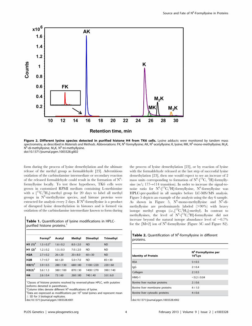
diamond-hydride column. This chromatographic system resolved
N6-acetyllysine, mono-, di-, and tri-N6-methyllysines, as well as
N6-formyllysine and lysine, as shown in Figure 2. With isotopically
labeled internal standards added prior to protease digestion,
identification and quantification of amino acids were accom-
plished by HPLC-coupled to tandem quadrupole mass spectrom-
etry in positive ion mode, using multiple reaction monitoring
(MRM) transitions. With a 2% precision for technical replicates,
the limits of detection were found to be 1 fmol for N6-formyl- and
N6-acetyllysine, 10 fmol for lysine, and 50 fmol for each of N6-
mono-, di-, and tri-methyl lysine. Data for the various lysine
modifications are expressed here as proportions of the total
number of lysines in the sample.
To validate the new analytical method for lysine modifications,
we compared the frequency of N6-formyllysine among different
classes of histone proteins extracted from TK6 cells and resolved
by reversed-phase HPLC. As shown in Figure S1A, all of the
major histone classes were separated, with further resolution of
variant forms of histones H1 and H3 (SDS-PAGE verification in
Figure S1B), which is consistent with previous observations in
cultured human cells [10]. N6-Formyllysine was detected in all
histone classes at a frequency of 1–4 modifications per 104 lysines.
This 3- to 4-fold variation among histone classes stands in contrast
to the 10- to 100-fold variation in the frequency of other
modifications (Table 1). The data in Table 1 represent the first
absolute quantification of the various lysine acetyl and methyl
modifications in histone proteins, and are consistent with
published studies of relative quantities of histone modifications
using immunologic and radiolabeling techniques [10,24–26].
Histone modification-based signaling involves the location and
number of specific modification targets within a histone protein, as
well as the frequency of modification of a target among all copies
of a particular histone protein. Our data provide some insight into
this issue. For example, we observed low-level acetylation and
methylation in histone H1, which is consistent with studies using
radiolabeled acetate [24], while this low level of modification maps
to specific sites in the globular domain and N-terminal tail of
histone H1 [10]. This low-level of acetylation and methylation in
histone H1 stands in contrast to the high level of acetylation of H2,
H3 and H4 (Table 1), which is again supported by studies using
radiolabeled acetate [24].
The new analytical method was next applied to quantify N6-
formyllysine in non-histone proteins. We had previously observed
N6-formyllysine mainly in histone proteins [9], perhaps as a result
of biased proteolysis or subsequent steps in the technique.
However, using the new method, we are now able to detect N6-
formyllysine modifications in a variety of different proteins, as
shown in Table 2. Further, an analysis of proteins in nuclear,
cytosolic, and membrane compartments in bovine liver revealed
the presence of N6-formyllysine in all three locations (Table 2).
These observations are consistent with a source for N6-formylly-
sine other than the 39-formylphosphate residues of DNA oxidation
previously identified for histone proteins [9].
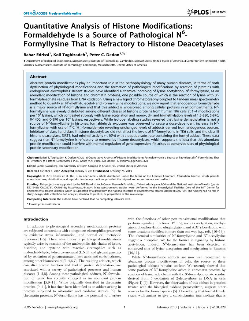
Formaldehyde as a source of N6-formyllysineOne alternative to 39-formylphosphate residues as a source of
N6-formyllysine is oxidation of the carbinolamine intermediate in
the reaction of formaldehyde with side chain amine of lysine
(Figure 1; N6-(hydroxymethyl)-lysine). To test this hypothesis, we
performed a series of experiments, starting with an in vitro reaction
Author Summary
Oxidative stress and inflammation lead to the generationof a multitude of electrophiles in cells that in turn reactwith nucleophilic macromolecules such as DNA, RNA,polyunsaturated fatty acids, and proteins, leading toprogression of a variety of disorders and diseases.Emerging evidence points to widespread modification ofcellular proteins by N6-formylation of lysine as a result ofadventitious reactions with endogenous electrophiles. N6-Formyllysine is a chemical homolog of the biologicallyimportant N6-acetyllysine and thus may interfere withacetylation signaling in cells. While N6-formyllysine ad-ducts are now well recognized as abundant proteinmodifications in cells, the source of these pathologicaladducts remains unclear. Our previous study proposed N6-formylation of lysine in histone proteins occurred byreaction of lysine with 39-formylphosphate residues arisingfrom DNA oxidation. Here, we investigate additionalsources as well as the fate of this abundant pathologicalprotein modification. Our results reveal that endogenousformaldehyde is a major source of N6-formyllysine and thatthis adduct is widely distributed among proteins in all cellcompartments. We also demonstrate for the first time thatN6-formyllysine modifications do not undergo appreciableremoval by histone deacetylases, which suggests that theypersist in proteins and possibly interfere with the signalingfunctions at conserved lysine positions in histone proteins.
Source and Fate of N6-Formyllysine in Proteins
PLOS Genetics | www.plosgenetics.org 2 February 2013 | Volume 9 | Issue 2 | e1003328
of L-lysine with different concentrations of formaldehyde and
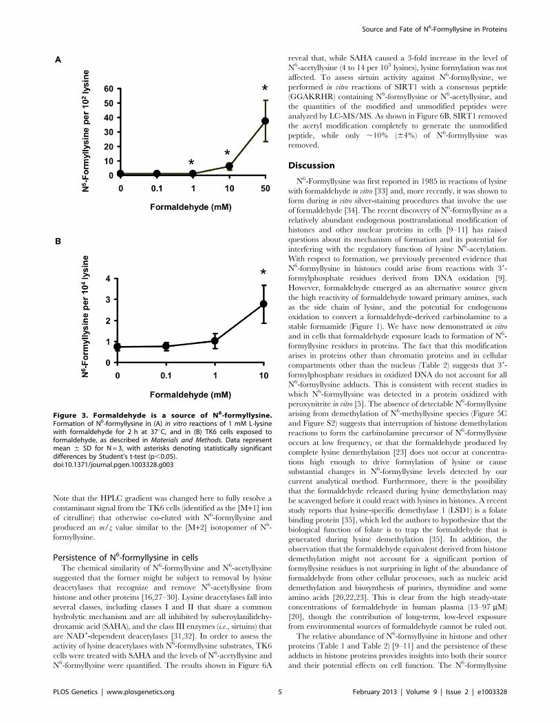
quantification of N6-formyllysine. As shown in Figure 3A, there
was a concentration-dependent formation of N6-formyllysine in
reactions with formaldehyde, presumably as a result of oxidation
of the carbinolamine adduct by the background of reactive oxygen
species generated by trace metals and dissolved oxygen in the
solution. The oxygen dependence of formaldehyde-induced N6-
formyllysine was verified by bubbling 100% oxygen (4 h) into the
solution of 1 mM lysine and 10 mM formaldehyde, which caused
a 2.2 (60.4)-fold increase in the level of N6-formyllysine after 12 h
of incubation at 37uC. The dose-response relationship for
formaldehyde-induced N6-formyllysine formation was also ob-
served in histone proteins extracted from TK6 cells exposed to
formaldehyde for 2 h at 37uC (Figure 3B), with 10 mM
formaldehyde producing roughly the same fold-change of N6-
formyllysine in both in vitro and cellular studies.
The relatively high endogenous levels of N6-formyllysine in
histone and other proteins (Table 1 and Table 2) raised the
question of the contribution of exogenous formaldehyde exposures
to the total load of N6-formyllysine in the cells. To address this
issue, we exposed TK6 cells to [13C,2H2]-labeled formaldehyde,
which led to the formation of N6-[13C,2H]-formyllysine that is 2
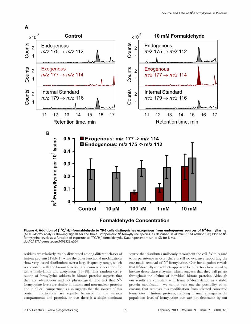
mass units heavier than the endogenous adducts (Figure 4A).
Following extraction of the histone proteins from formaldehyde-
treated TK6 cells (2 h, 37uC), both endogenous and exogenous
N6-formyllysine were quantified by monitoring the transitions m/z
175R112 and m/z 177R114, respectively (Figure 4A), with a
third transition (m/z 179R116) for the 4,4,5,5-[2H]-N6-formylly-
sine internal standard. As shown in Figure 4B, levels of
endogenous (unlabeled) N6-formyllysine remained constant at all
concentrations of [13C,2H2]-formaldehyde, while N6-[13C,2H]-
formyllysine increased as a function of the concentration of labeled
formaldehyde.
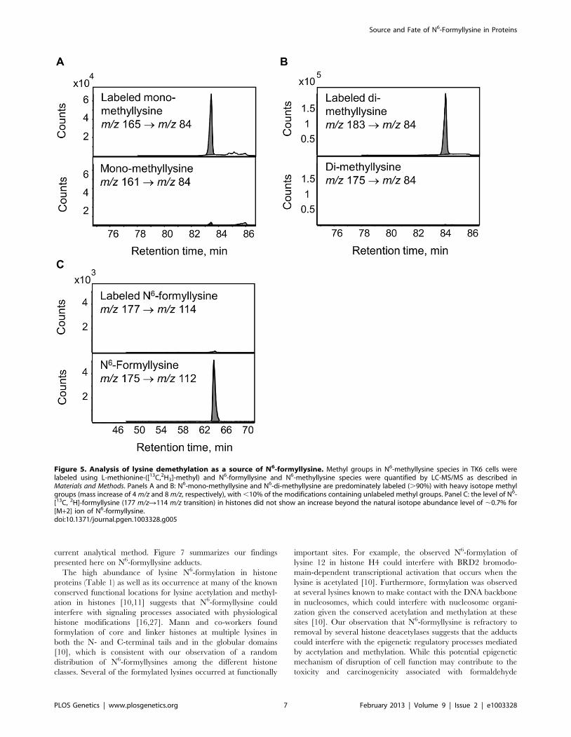
Lysine demethylation as a source of N6-formyllysineThe enzymatic demethylation of N6-methyllysine modifications
represents another possible source of N6-formyllysine in histone
proteins, given both the carbinolamine intermediate known to
Figure 1. Sources of N6-formyllysine. The adduct can be generated in chromatin proteins from reaction of lysine with 39-formylphosphateresidue derived from 59-oxidation of 2-deoxyribose in DNA or from reaction of lysine with endogenous or exogenous formaldehyde. Formaldehydereacts with amines to give a carbinolamine intermediate (N6-(hydroxymethyl)-lysine) that is in equilibrium with a Schiff base and that is one oxidationstate away from the formamide functional group of N6-formyllysine.doi:10.1371/journal.pgen.1003328.g001
Source and Fate of N6-Formyllysine in Proteins
PLOS Genetics | www.plosgenetics.org 3 February 2013 | Volume 9 | Issue 2 | e1003328
form during the process of lysine demethylation and the ultimate
release of the methyl group as formaldehyde [23]. Adventitious
oxidation of the carbinolamine intermediate or secondary reaction
of the released formaldehyde could result in the formation of N6-
formyllysine locally. To test these hypotheses, TK6 cells were
grown in customized RPMI medium containing L-methionine
with a [13C,2H3]-methyl group for 20 days to label all methyl
groups in N6-methyllysine species, and histone proteins were
extracted for analysis every 2 days. If N6-formyllysine is a product
of disrupted lysine demethylation in histones and is formed via
oxidation of the carbinolamine intermediate known to form during
the process of lysine demethylation [23], or by reaction of lysine
with the formaldehyde released at the last step of successful lysine
demethylation [23], then one would expect to see an increase of 2
mass units corresponding to formation of N6-[13C, 2H]-formylly-
sine (m/z 177R114 transition). In order to increase the signal-to-
noise ratio for N6-[13C,2H]-formyllysine, N6-formyllysine was
HPLC-pre-purified in all samples before LC-MS/MS analysis.
Figure 5 depicts an example of the analysis using the day 6 sample.
As shown in Figure 5, N6-mono-methyllysine and N6-di-
methyllysine are predominately labeled (.90%) with heavy
isotope methyl groups (i.e.,[13C,2H3]-methyl). In contrast to
methyllysines, the level of N6-[13C,2H]-formyllysine did not
increase beyond the natural isotope abundance level of ,0.7%
for the [M+2] ion of N6-formyllysine (Figure 5C and Figure S2).
Figure 2. Different lysine species detected in purified histone H4 from TK6 cells. Lysine adducts were monitored by tandem massspectrometry, as described in Materials and Methods. Abbreviations: FK, N6-formyllysine; AK, N6-acetyllysine; K, lysine; MK, N6-mono-methyllysine; M2K,N6-di-methyllysine; M3K, N6-tri-methyllysine.doi:10.1371/journal.pgen.1003328.g002
Table 1. Quantification of lysine modifications in HPLC-purified histone proteins.1
Formyl2 Acetyl Methyl Dimethyl Trimethyl
H1 (1)1 1.560.33 1.660.2 8.062.0 ND ND
H1 (2)1 1.260.2 1.560.3 7.062.0 ND ND
H2A 2.760.2 26620 2068.0 60630 ND
H2B 1.760.7 66620 5.067.0 ND 85630
H3(1)1 3.960.5 2806130 680680 11006220 220660
H3(2)1 3.661.3 3806100 870630 14006270 3906140
H4 2.660.4 73660 260680 740640 3.066.0
1Classes of histone proteins resolved by reversed-phase HPLC, with putativeisoforms denoted in parentheses.2Column titles denote different N6-modifications of lysine.3Data are expressed as modifications per 104 total lysines and represent mean6 SD for 3 biological replicates.doi:10.1371/journal.pgen.1003328.t001
Table 2. Quantification of N6-formyllysine in differentproteins.
Identity of ProteinN6-Formyllysine per104Lys
BSA 560.5
IgG 260.4
Collagen 260.5
HMG-1 ,0.260.04
Bovine liver nuclear proteins 260.6
Bovine liver membrane proteins 461.0
Bovine liver cytosolic proteins 460.6
doi:10.1371/journal.pgen.1003328.t002
Source and Fate of N6-Formyllysine in Proteins
PLOS Genetics | www.plosgenetics.org 4 February 2013 | Volume 9 | Issue 2 | e1003328
Note that the HPLC gradient was changed here to fully resolve a
contaminant signal from the TK6 cells (identified as the [M+1] ion
of citrulline) that otherwise co-eluted with N6-formyllysine and
produced an m/z value similar to the [M+2] isotopomer of N6-
formyllysine.
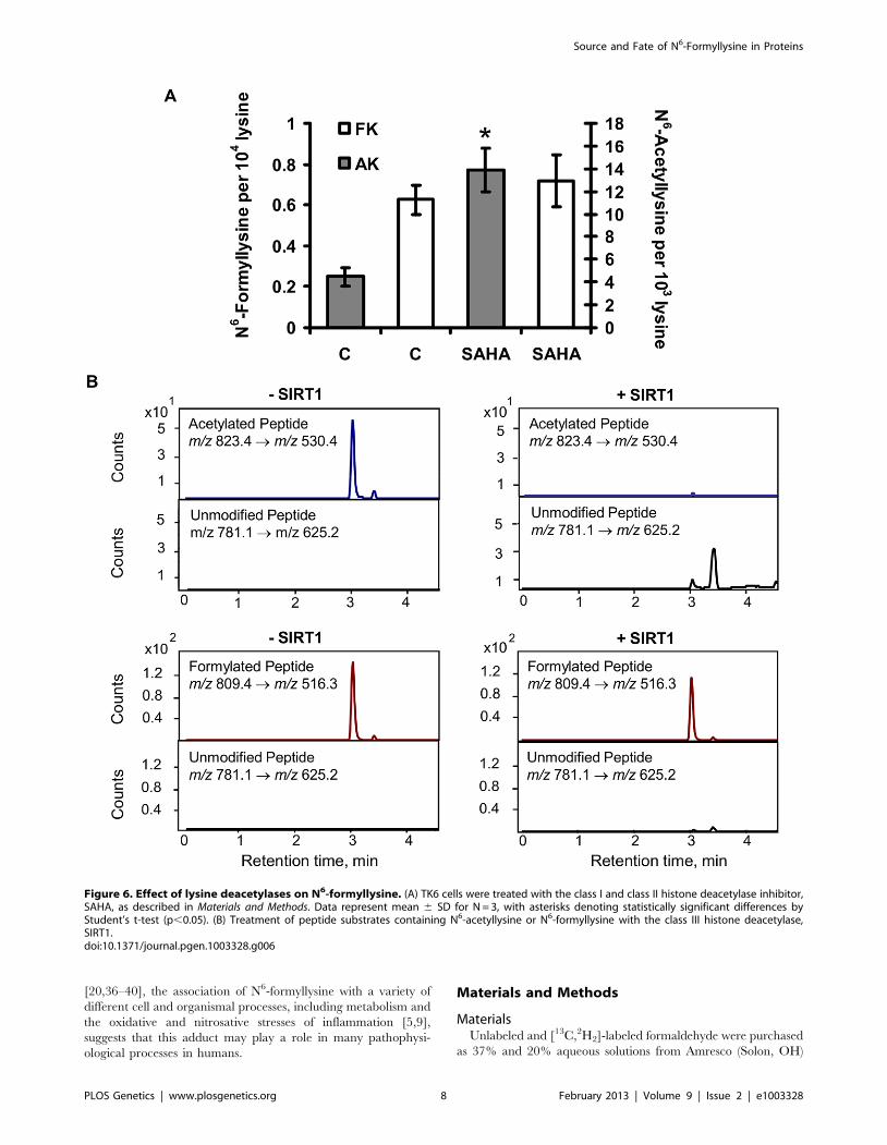
Persistence of N6-formyllysine in cellsThe chemical similarity of N6-formyllysine and N6-acetyllysine
suggested that the former might be subject to removal by lysine
deacetylases that recognize and remove N6-acetyllysine from
histone and other proteins [16,27–30]. Lysine deacetylases fall into
several classes, including classes I and II that share a common
hydrolytic mechanism and are all inhibited by suberoylanilidehy-
droxamic acid (SAHA), and the class III enzymes (i.e., sirtuins) that
are NAD+-dependent deacetylases [31,32]. In order to assess the
activity of lysine deacetylases with N6-formyllysine substrates, TK6
cells were treated with SAHA and the levels of N6-acetyllysine and
N6-formyllysine were quantified. The results shown in Figure 6A
reveal that, while SAHA caused a 3-fold increase in the level of
N6-acetyllysine (4 to 14 per 103 lysines), lysine formylation was not
affected. To assess sirtuin activity against N6-formyllysine, we
performed in vitro reactions of SIRT1 with a consensus peptide
(GGAKRHR) containing N6-formyllysine or N6-acetyllysine, and
the quantities of the modified and unmodified peptides were
analyzed by LC-MS/MS. As shown in Figure 6B, SIRT1 removed
the acetyl modification completely to generate the unmodified
peptide, while only ,10% (64%) of N6-formyllysine was
removed.
Discussion
N6-Formyllysine was first reported in 1985 in reactions of lysine
with formaldehyde in vitro [33] and, more recently, it was shown to
form during in vitro silver-staining procedures that involve the use
of formaldehyde [34]. The recent discovery of N6-formyllysine as a
relatively abundant endogenous posttranslational modification of
histones and other nuclear proteins in cells [9–11] has raised
questions about its mechanism of formation and its potential for
interfering with the regulatory function of lysine N6-acetylation.
With respect to formation, we previously presented evidence that
N6-formyllysine in histones could arise from reactions with 39-
formylphosphate residues derived from DNA oxidation [9].
However, formaldehyde emerged as an alternative source given
the high reactivity of formaldehyde toward primary amines, such
as the side chain of lysine, and the potential for endogenous
oxidation to convert a formaldehyde-derived carbinolamine to a
stable formamide (Figure 1). We have now demonstrated in vitro
and in cells that formaldehyde exposure leads to formation of N6-
formyllysine residues in proteins. The fact that this modification
arises in proteins other than chromatin proteins and in cellular
compartments other than the nucleus (Table 2) suggests that 39-
formylphosphate residues in oxidized DNA do not account for all
N6-formyllysine adducts. This is consistent with recent studies in
which N6-formyllysine was detected in a protein oxidized with
peroxynitrite in vitro [5]. The absence of detectable N6-formyllysine
arising from demethylation of N6-methyllysine species (Figure 5C
and Figure S2) suggests that interruption of histone demethylation
reactions to form the carbinolamine precursor of N6-formyllysine
occurs at low frequency, or that the formaldehyde produced by
complete lysine demethylation [23] does not occur at concentra-
tions high enough to drive formylation of lysine or cause
substantial changes in N6-formyllysine levels detected by our
current analytical method. Furthermore, there is the possibility
that the formaldehyde released during lysine demethylation may
be scavenged before it could react with lysines in histones. A recent
study reports that lysine-specific demethylase 1 (LSD1) is a folate
binding protein [35], which led the authors to hypothesize that the
biological function of folate is to trap the formaldehyde that is
generated during lysine demethylation [35]. In addition, the
observation that the formaldehyde equivalent derived from histone
demethylation might not account for a significant portion of
formyllysine residues is not surprising in light of the abundance of
formaldehyde from other cellular processes, such as nucleic acid
demethylation and biosynthesis of purines, thymidine and some
amino acids [20,22,23]. This is clear from the high steady-state
concentrations of formaldehyde in human plasma (13–97 mM)
[20], though the contribution of long-term, low-level exposure
from environmental sources of formaldehyde cannot be ruled out.
The relative abundance of N6-formyllysine in histone and other
proteins (Table 1 and Table 2) [9–11] and the persistence of these
adducts in histone proteins provides insights into both their source
and their potential effects on cell function. The N6-formyllysine
Figure 3. Formaldehyde is a source of N6-formyllysine.Formation of N6-formyllysine in (A) in vitro reactions of 1 mM L-lysinewith formaldehyde for 2 h at 37uC, and in (B) TK6 cells exposed toformaldehyde, as described in Materials and Methods. Data representmean 6 SD for N = 3, with asterisks denoting statistically significantdifferences by Student’s t-test (p,0.05).doi:10.1371/journal.pgen.1003328.g003
Source and Fate of N6-Formyllysine in Proteins
PLOS Genetics | www.plosgenetics.org 5 February 2013 | Volume 9 | Issue 2 | e1003328
residues are relatively evenly distributed among different classes of
histone proteins (Table 1), while the other functional modifications
show very biased distributions over a large frequency range, which
is consistent with the known function and conserved locations for
lysine methylation and acetylation [16–18]. This random distri-
bution of formyllysine adducts in histone proteins suggests that
they are adventitious and not physiological. The fact that N6-
formyllysine levels are similar in histone and non-nuclear proteins
and in all cell compartments also suggests that the sources of this
protein modification are equally balanced in the various
compartments and proteins, or that there is a single dominant
source that distributes uniformly throughout the cell. With regard
to its persistence in cells, there is still no evidence supporting the
enzymatic removal of N6-formyllysine. Our investigation reveals
that N6-formyllysine adducts appear to be refractory to removal by
histone deacetylase enzymes, which suggests that they will persist
throughout the lifetime of individual histone proteins. Although
our results are consistent with lysine N6-formylation as a stable
protein modification, we cannot rule out the possibility of an
enzyme that removes this modification from selected conserved
lysine sites in histone proteins, resulting in small changes in the
population level of formyllysine that are not detectable by our
Figure 4. Addition of [13C,2H2]-formaldehyde to TK6 cells distinguishes exogenous from endogenous sources of N6-formyllysine.(A) LC-MS/MS analysis showing signals for the three isotopomeric N6-formyllysine species, as described in Materials and Methods. (B) Plot of N6-formyllysine levels as a function of exposure to [13C,2H2]-formaldehyde. Data represent mean 6 SD for N = 3.doi:10.1371/journal.pgen.1003328.g004
Source and Fate of N6-Formyllysine in Proteins
PLOS Genetics | www.plosgenetics.org 6 February 2013 | Volume 9 | Issue 2 | e1003328
current analytical method. Figure 7 summarizes our findings
presented here on N6-formyllysine adducts.
The high abundance of lysine N6-formylation in histone
proteins (Table 1) as well as its occurrence at many of the known
conserved functional locations for lysine acetylation and methyl-
ation in histones [10,11] suggests that N6-formyllysine could
interfere with signaling processes associated with physiological
histone modifications [16,27]. Mann and co-workers found
formylation of core and linker histones at multiple lysines in
both the N- and C-terminal tails and in the globular domains
[10], which is consistent with our observation of a random
distribution of N6-formyllysines among the different histone
classes. Several of the formylated lysines occurred at functionally
important sites. For example, the observed N6-formylation of
lysine 12 in histone H4 could interfere with BRD2 bromodo-
main-dependent transcriptional activation that occurs when the
lysine is acetylated [10]. Furthermore, formylation was observed
at several lysines known to make contact with the DNA backbone
in nucleosomes, which could interfere with nucleosome organi-
zation given the conserved acetylation and methylation at these
sites [10]. Our observation that N6-formyllysine is refractory to
removal by several histone deacetylases suggests that the adducts
could interfere with the epigenetic regulatory processes mediated
by acetylation and methylation. While this potential epigenetic
mechanism of disruption of cell function may contribute to the
toxicity and carcinogenicity associated with formaldehyde
Figure 5. Analysis of lysine demethylation as a source of N6-formyllysine. Methyl groups in N6-methyllysine species in TK6 cells werelabeled using L-methionine-([13C,2H3]-methyl) and N6-formyllysine and N6-methyllysine species were quantified by LC-MS/MS as described inMaterials and Methods. Panels A and B: N6-mono-methyllysine and N6-di-methyllysine are predominately labeled (.90%) with heavy isotope methylgroups (mass increase of 4 m/z and 8 m/z, respectively), with ,10% of the modifications containing unlabeled methyl groups. Panel C: the level of N6-[13C, 2H]-formyllysine (177 m/zR114 m/z transition) in histones did not show an increase beyond the natural isotope abundance level of ,0.7% for[M+2] ion of N6-formyllysine.doi:10.1371/journal.pgen.1003328.g005
Source and Fate of N6-Formyllysine in Proteins
PLOS Genetics | www.plosgenetics.org 7 February 2013 | Volume 9 | Issue 2 | e1003328
[20,36–40], the association of N6-formyllysine with a variety of
different cell and organismal processes, including metabolism and
the oxidative and nitrosative stresses of inflammation [5,9],
suggests that this adduct may play a role in many pathophysi-
ological processes in humans.
Materials and Methods
MaterialsUnlabeled and [13C,2H2]-labeled formaldehyde were purchased
as 37% and 20% aqueous solutions from Amresco (Solon, OH)
Figure 6. Effect of lysine deacetylases on N6-formyllysine. (A) TK6 cells were treated with the class I and class II histone deacetylase inhibitor,SAHA, as described in Materials and Methods. Data represent mean 6 SD for N = 3, with asterisks denoting statistically significant differences byStudent’s t-test (p,0.05). (B) Treatment of peptide substrates containing N6-acetyllysine or N6-formyllysine with the class III histone deacetylase,SIRT1.doi:10.1371/journal.pgen.1003328.g006
Source and Fate of N6-Formyllysine in Proteins
PLOS Genetics | www.plosgenetics.org 8 February 2013 | Volume 9 | Issue 2 | e1003328
and Isotec (Miamisburg, OH), respectively. 4,4,5,5-[2H]-Lysine
was purchased from Cambridge Isotope Laboratories (Andover,
MA). 4,4,5,5-[2H]-N6-Formyllysine was synthesized from 4,4,5,5-
[2H]-lysine according to Jiang et al. [9]. 3,3,4,4,5,5,6,6-[2H]-N6-
Acetyllysine were obtained from CDN Isotopes (Pointe-Claire,
Quebec, Canada). L-Methionine-([13C,2H3]-methyl) was obtained
from Isotec (Miamisburg, OH). L-Lysine, N6-formyllysine, N6-
acetyllysine, bovine serum albumin, human recombinant HMG-1,
human IgG, Streptomyces griseus protease, and protease inhibitor
cocktail (for use with mammalian cell and tissue extracts) were
obtained from Sigma-Aldrich (St. Louis, MO). N6-Mono-methyl-
lysine, N6-di-methyllysine, and N6-tri-methyllysine were pur-
chased from Bachem Bioscience Inc. (King of Prussia, PA).
Nonidet P-40 was from Roche Diagnostic Corporation (Indianap-
olis, IN). Suberoylanilidehydroxamic acid (SAHA) and SIRT1
(human recombinant) enzyme were purchased from Cayman
chemical (Ann Arbor, MI). Peptide substrates for SIRT1
(GGAKRHR and its lysine-acetylated and -formylated forms)
were synthesized at Massachusetts Institute of Technology
Biopolymers Laboratory. The human lymphoblastoid TK6 cell
line was a generous gift of Prof. Gerald Wogan (Massachusetts
Institute of Technology).
TK6 cell culture, exposure, and labelingTK6 cells were cultured in RPMI 1640 medium (Cellgro,
Manassas, VA) supplemented with 10% heat-inactivated horse
serum (Atlanta Biologicals, Lawrenceville, GA), 10,000 U penicil-
lin/ml and 10,000 mg streptomycin/ml (Lonza, Walkersville,
MD), and 2 mM L-glutamine (Lonza, Walkersville, MD) at
37uC in a 5% CO2 atmosphere. For formaldehyde exposure
studies, TK6 cells were pelleted, washed, and resuspended in
RPMI 1640 medium without any supplements, prior to addition of
formaldehyde to the medium. Following addition of formalde-
hyde, cells were incubated at 37uC for 2 h with occasional mixing
prior to extracting chromatin proteins. Histones were extracted
from ,107 cells after exposure and the quantity of formyllysine, as
a percentage of total lysine, was measured as described below. For
lysine demethylation studies, TK6 cells were grown in a
customized RPMI-1640 medium identical to the traditional
medium (e.g., supplemented with horse serum, antibiotics, and L-
glutamine), except for the presence of labeled methionine (L-
methionine-([13C,2H3]-methyl)) instead of non-labeled methio-
nine. Histones (from ,107 cells) were extracted every 2 d for 20 d
in order to investigate the formation of N6-[13C,2H]-formyllysine.
For histone deacetylase studies, TK6 cells were incubated with the
histone deacetylase inhibitor, suberoylanilidehydroxamic acid
(SAHA), for 20 h at 37uC in a 5% CO2 atmosphere prior to
histone extraction. SAHA was dissolved in a 50:50 solution of
DMSO:PBS prior to addition to cell media. Control cells (,107)
were treated with the DMSO:PBS vehicle.
Histone extraction from TK6 cells and subcellular proteinfractionation from tissue
Extraction of histones was performed according to Boyne et al.
[41], with modifications. Cells (,107 per sample) were pelleted by
centrifugation at 10006 g for 5 min at 4uC and the pellet was
washed once with PBS. Cell pellets were then lysed by
resuspension in ice-cold lysis buffer (15 mM Tris-HCl, pH 7.5,
15 mM NaCl, 60 mM KCl, 1 mM CaCl2, 5 mM MgCl2,
250 mM sucrose, 1 mM dithiothreitol, 10 mM sodium butyrate)
containing a 100:1 v:v dilution of protease inhibitor cocktail in the
presence of 0.03% Nonidet P-40 and incubation on ice for 5 min
with occasional gentle mixing. Nuclei were pelleted by centrifu-
gation at 6006g for 5 min at 4uC, and the pellet was washed twice
with ice-cold lysis buffer without Nonidet P-40. Histones were
extracted by addition of ice-cold 0.4 N H2SO4 and incubation
overnight on ice. The solution was centrifuged at 30006 g for
5 min and proteins in supernatant were precipitated by addition of
20% v/v trichloroacetic acid and overnight incubation at 4uC.
Samples were then centrifuged at 140006 g for 10 min at 4uC,
washed once with ice-cold acetone containing 0.1% HCl, and
once with ice-cold acetone. The extracts were air-dried and stored
at 220uC until use. For collecting membrane, cytosolic, and
nuclear fractions, 20 mg of bovine liver tissue was cut into small
pieces and washed with PBS, and proteins were fractionated using
the Subcellular Protein Fractionation Kit from Thermo Scientific
(Waltham, MA) and a Kontes all-glass Dounce homogenizer (10
strokes with a type B pestle). Proteins in subcellular extracts were
precipitated by addition of 20% v/v trichloroacetic acid and
overnight incubation at 4uC. Samples were then centrifuged at
140006 g for 10 min at 4uC, washed once with ice-cold acetone
containing 0.1% HCl, and once with ice-cold acetone. The
extracts were air-dried and stored at 220uC until use.
Purification of individual histonesHPLC purification of total histones was performed according to
Boyne et al. [41] with modifications. Total histones (#50 mg) were
dissolved in 0.1% trifluoroacetic acid (TFA) in water and
fractionated by HPLC on an Agilent 1100 series instrument
(Agilent Technologies, Santa Clara, CA), using a Source 5RPC
C18 reversed-phase column (4.66150 mm, 5 mm particles; GE
Healthcare Life Sciences). The mobile phase flow rate was 1 mL/
min and the solvent system was 0.1% TFA in water (A) and
0.094% TFA in acetonitrile (B) with the elution starting at 0% B,
linearly increasing to 28% B over 28 min, reaching 37% B at
70 min, 38% B at 100 min, 60% B at 150 min, and finally 100%
B at 151 min, before the column was re-equilibrated to 0% B for
10 min. Protein elution was monitored by UV absorbance at
214 nm and histones in each fraction were tentatively identified by
resolution on a 13% SDS-PAGE gel with Coomassie Blue staining
(see Figure S1).
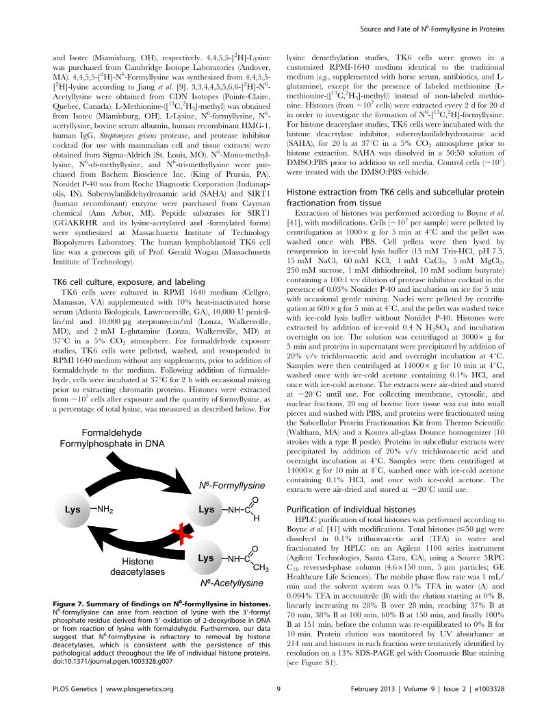
Figure 7. Summary of findings on N6-formyllysine in histones.N6-formyllysine can arise from reaction of lysine with the 39-formylphosphate residue derived from 59-oxidation of 2-deoxyribose in DNAor from reaction of lysine with formaldehyde. Furthermore, our datasuggest that N6-formyllysine is refractory to removal by histonedeacetylases, which is consistent with the persistence of thispathological adduct throughout the life of individual histone proteins.doi:10.1371/journal.pgen.1003328.g007
Source and Fate of N6-Formyllysine in Proteins
PLOS Genetics | www.plosgenetics.org 9 February 2013 | Volume 9 | Issue 2 | e1003328
Enzymatic hydrolysis of proteinsHistones extracted from TK6 cells and other protein samples
were dissolved in 50 mL of 100 mM ammonium bicarbonate
buffer (pH 8.5), 4,4,5,5-[2H]-lysine (2 nmol),4,4,5,5-[2H]-N6-for-
myllysine (1 pmol), and 3,3,4,4,5,5,6,6-[2H]-N6-acetyl lysine
(10 pmol) were added as internal standards, and the proteins
hydrolyzed by addition of S. griseus protease (freshly prepared
solution each time) with incubation at 37uC for $16 h. Streptomyces
griseus was used at a ratio of 1 mg of enzyme per 10 mg of protein.
Samples were then dried under vacuum and resuspended in 50 mL
of water before mass spectrometry analysis.
Quantification of amino acidsN6-Formyllysine and other amino acids were quantified as a
percentage of the total quantity of lysine, by liquid chromatogra-
phy-coupled mass spectrometry (LC-MS/MS). HPLC was per-
formed with an Agilent 1100 series instrument. Adducts of interest
in the resuspended protein hydrolysates were separated using an
aqueous normal phase Cogent diamond hydride column
(2.16150 mm, 4 mm) from MicroSolv Technology Corporation
(Eatontown, NJ). The mobile phase flow rate was 400 mL/min,
and the column temperature was maintained at 20uC. The solvent
system was 0.1% acetic acid in water (A) and 0.1% acetic acid in
acetonitrile (B), with the elution starting at 100% B, the gradient
linearly decreased to 25% B over 30 min, stayed at 25% B for 3
additional min before the column was re-equilibrated at 100% B
for 7 min. In order to separate a co-eluting contaminant from pre-
purified lysine demethylation study samples, an extended chro-
matography run time was used, with the elution starting at 100%
B, the gradient linearly decreased to 75% B over 75 min, further
decreased to 25% B over the next 3 min, reached 15% by 83 min
before the column was re-equilibrated at 100% B for 7 min. The
species of interest were then analyzed using Agilent 6410 triple
quadrupole mass spectrometer (MS/MS) equipped with an
electrospray ionization (ESI) source operated in positive ion mode.
The operating parameters were as follows: ESI capillary voltage,
4000 V; gas temperature, 350uC; drying gas flow, 12 L/min; and
nebulizer pressure, 30 psi. Selected reaction monitoring (SRM)
transitions are summarized in Table 3. Note that in addition to
chromatographic separation (Figure 2) and presence of internal
standards, the unique product ions of 112 m/z and 126 m/z for
formyl and acetyl lysines, respectively, distinguish them from their
isobaric compounds di- and tri-methyl lysines. The fragmentor
voltage and collision energy were optimized in order to maximize
the signal of each product ion monitored (see Figure S3) and are
summarized in Table 3. 4,4,5,5-[2H]-Lysine, 4,4,5,5-[2H]-N6-
formyllysine, and 3,3,4,4,5,5,6,6-[2H]-N6-acetyl lysine were used
as internal standards. Calibration curves for the labeled and
unlabeled forms of these analytes were constructed by plotting the
ratios of the areas of the MS signals for the labeled and unlabeled
forms against their corresponding concentration ratios (Figure S4).
N6-Methylated lysine species were quantified using the deuterated
acetyl lysine internal standard signal (Figure S4).
SIRT1 peptide experimentSIRT1 peptide substrates (GGAKRHR, GGAKacetylRHR, and
GGAKformylRHR) were HPLC purified on an Agilent 1100 series
instrument using Vydac 218TP52 C18 reverse-phase column
(2.16250 mm, 5 mm) from Grace Vydac (Hesperia, CA). The
mobile phase flow rate was 200 mL/min, and the column
temperature was maintained at 30uC. The solvent system was
0.05% trifluoroacetic acid in water (A) and 0.05% trifluoroacetic
acid in 80% acetonitrile (B), with the isocratic elution of 5% B for
the first 5 min, then a linear increase to 42% B over 25 min,
reaching 100% B at 31 min followed by column re-equilibration at
5% B for 10 min. Each purified SIRT1 peptide substrate
(100 pmol) were incubated overnight (12 h) at 25uC with 1 mg
SIRT1, in 50 mM Tris-HCl (pH 8) buffer containing 137 mM
NaCl, 2.7 mM KCl, 1 mM MgCl2, and 6 mM NAD+. The
removal of acetyl and formyl groups from SIRT1 peptide
substrates was determined using liquid chromatography-coupled
mass spectrometry. HPLC was performed on an Agilent 1100
series instrument using Agilent Exclipse XDB C18 reverse-phase
column (4.66150 mm, 5 mm). The mobile phase flow rate was
200 mL/min, and the column temperature was maintained at
40uC. The solvent system was 0.1% acetic acid in water (A) and
0.1% acetic acid in acetonitrile (B), with the elution starting at
20% B, the gradient linearly increased to 50% B over 5 min,
reached 100% B at 6 min, and kept at 100% B for 9 minutes
before the column was re-equilibrated at 20% B for 10 min. The
species of interest were then analyzed using the Agilent 6410 MS/
MS system equipped with an electrospray ionization (ESI) source
operated in positive ion mode. The operating parameters were as
follows: ESI capillary voltage, 3500 V; gas temperature, 345uC;
drying gas flow, 8 L/min; and nebulizer pressure, 30 psi. Multiple
reaction monitoring (MRM) transitions were as follow:
GGAKRHR peptide, m/z 781.1R625.2; GGAKformylRHR pep-
tide, m/z 809.4R516.3; and GGAKacetylRHR peptide, m/z
823.4R530.4. The fragmentor voltage and collision energy were
200 V and 40 V for GGAKRHR peptide, respectively; 100 V and
46 V for GGAKformylRHR peptide; and 100 V and 52 V for
GGAKacetylRHR peptide.
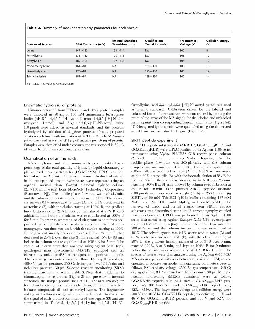
Table 3. Summary of mass spectrometry parameters for each species.
Species of Interest SRM Transition (m/z)Internal StandardTransition (m/z)
Qualifier IonTransition (m/z)
FragmentorVoltage (V)
Collision Energy(V)
Lysine 147R130 151R134 NA 100 8
Formyllysine 175R112 179R116 NA 105 10
Acetyllysine 189R126 197R134 NA 105 10
Mono-methyllysine 161R84 NA 161R130 100 10
Di-methyllysine 175R84 NA 175R130 100 14
Tri-methyllysine 189R84 NA 189R130 100 14
doi:10.1371/journal.pgen.1003328.t003
Source and Fate of N6-Formyllysine in Proteins
PLOS Genetics | www.plosgenetics.org 10 February 2013 | Volume 9 | Issue 2 | e1003328
Supporting Information
Figure S1 Reversed-phase HPLC fractionation of histone
proteins. (A) HPLC elution profile for histones extracted from
TK6 cells, as described in Materials and Methods. (B) SDS/PAGE
analysis of the HPLC-fractionated proteins shown in panel A.
Lanes 1 and 2 are molecular weight markers, while lanes 3 and 4
refer to total histones from TK6 cells and calf thymus, respectively.
(TIF)
Figure S2 Lysine demethylation is not a source of N6-
formyllysine in histones. By culturing TK6 cells in customized
RPMI medium containing L-Methionine-([13C,2H3]-methyl) for
20 days, it was shown that in contrast to predominant heavy
isotope labeling of mono-methyllysines (.90%), even as early as
day 2, the level of N6-[13C, 2H]-formyllysine did not show an
increase beyond the natural isotope abundance level (,0.7% for
[M+2] ion of N6-formyllysine), for any day.
(TIF)
Figure S3 Examples of product ions for each lysine species
monitored, after optimization. In all cases, the highest count was
used as the product ion for MRM in triple quadruple mass
spectrometer, as described in Materials and Methods. An exception
was lysine, for which the 130 m/z ion was selected due to lysine’s
high abundance compared to other species monitored.
(TIF)
Figure S4 Examples of calibration curves for the isotope-
dilution LC-MS/MS analysis of modified lysine species, as
described in Materials and Methods. Abbreviations: FK, N6-
formyllysine; DFK, deuterium-labeled N6-formyllysine; AK, N6-
acetyllysine; DAK, deuterium-labeled N6-acetyllysine; K, lysine;
DK, deuterium-labeled lysine; MK, N6-mono-methyllysine; M2K,
N6-di-methyllysine; M3K, N6-tri-methyllysine.
(TIF)
Acknowledgments
We thank Ms. Laura Trudel for her help with cell culture studies.
Author Contributions
Conceived and designed the experiments: BE PCD. Performed the
experiments: BE KT. Analyzed the data: BE KT PCD. Wrote the paper:
BE KT PCD.
References
1. Levine RL (2002) Carbonyl modified proteins in cellular regulation, aging, and
disease. Free Radic Biol Med 32: 790–796.
2. Jacobs AT, Marnett LJ (2010) Systems analysis of protein modification and
cellular responses induced by electrophile stress. Acc Chem Res 43: 673–683.
3. Thornalley PJ (2008) Protein and nucleotide damage by glyoxal and
methylglyoxal in physiological systems–role in ageing and disease. Drug
Metabol Drug Interact 23: 125–150.
4. Dedon PC (2008) The chemical toxicology of 2-deoxyribose oxidation in DNA.
Chem Res Toxicol 21: 206–219.
5. Vana L, Kanaan NM, Hakala K, Weintraub ST, Binder LI (2011) Peroxynitrite-
induced nitrative and oxidative modifications alter tau filament formation.
Biochemistry 50: 1203–1212.
6. Codreanu SG, Zhang B, Sobecki SM, Billheimer DD, Liebler DC (2009) Global
analysis of protein damage by the lipid electrophile 4-hydroxy-2-nonenal. Mol
Cell Proteomics 8: 670–680.
7. Tallman KA, Kim HY, Ji JX, Szapacs ME, Yin H, et al. (2007) Phospholipid-
protein adducts of lipid peroxidation: synthesis and study of new biotinylated
phosphatidylcholines. Chem Res Toxicol 20: 227–234.
8. Prasad A, Bekker P, Tsimikas S (2012) Advanced Glycation Endproducts and
Diabetic Cardiovascular Disease. Cardiol Rev 20:177–183.
9. Jiang T, Zhou X, Taghizadeh K, Dong M, Dedon PC (2007) N-formylation of
lysine in histone proteins as a secondary modification arising from oxidative
DNA damage. Proc Natl Acad Sci U SA 104: 60–65.
10. Wisniewski JR, Zougman A, Mann M (2008) Nepsilon-formylation of lysine is a
widespread post-translational modification of nuclear proteins occurring at
residues involved in regulation of chromatin function. Nucleic Acids Res 36:
570–577.
11. LeRoy G, Weston JT, Zee BM, Young NL, Plazas-Mayorca MD, et al. (2009)
Heterochromatin protein 1 is extensively decorated with histone code-like post-
translational modifications. Mol Cell Proteomics 8: 2432–2442.
12. Pesavento JJ, Kim YB, Taylor GK, Kelleher NL (2004) Shotgun annotation of
histone modifications: a new approach for streamlined characterization of
proteins by top down mass spectrometry. J Am Chem Soc 126: 3386–3387.
13. Felsenfeld G, Groudine M (2003) Controlling the double helix. Nature 421:
448–453.
14. Chi P, Allis CD, Wang GG (2010) Covalent histone modifications–miswritten,
misinterpreted and mis-erased in human cancers. Nat Rev Cancer 10: 457–469.
15. Rando OJ (2012) Combinatorial complexity in chromatin structure and
function: revisiting the histone code. Curr Opin Genet Dev 22: 148–55.
16. Kouzarides T (2007) Chromatin modifications and their function. Cell 128:
693–705.
17. Siuti N, Roth MJ, Mizzen CA, Kelleher NL, Pesavento JJ (2006) Gene-specific
characterization of human histone H2B by electron capture dissociation.
J Proteome Res 5: 233–239.
18. Garcia BA, Barber CM, Hake SB, Ptak C, Turner FB, et al. (2005)
Modifications of human histone H3 variants during mitosis. Biochemistry 44:
13202–13213.
19. Lu K, Moeller B, Doyle-Eisele M, McDonald J, Swenberg JA (2011) Molecular
dosimetry of N2-hydroxymethyl-dG DNA adducts in rats exposed to
formaldehyde. Chem Res Toxicol 24: 159–161.
20. Zhang L, Freeman LE, Nakamura J, Hecht SS, Vandenberg JJ, et al. (2010)
Formaldehyde and leukemia: epidemiology, potential mechanisms, and
implications for risk assessment. Environ Mol Mutagen 51: 181–191.
21. Le Curieux F, Pluskota D, Munter T, Sjoholm R, Kronberg L (2000)
Identification of fluorescent 29-deoxyadenosine adducts formed in reactions of
conjugates of malonaldehyde and acetaldehyde, and of malonaldehyde and
formaldehyde. Chem Res Toxicol 13: 1228–1234.
22. Begley TJ, Samson LD (2003) AlkB mystery solved: oxidative demethylation of
N1-methyladenine and N3-methylcytosine adducts by a direct reversal
mechanism. Trends Biochem Sci 28: 2–5.
23. Shi Y, Whetstine JR (2007) Dynamic regulation of histone lysine methylation by
demethylases. Mol Cell 25: 1–14.
24. Pasqualini JR, Mercat P, Giambiagi N (1989) Histone acetylation decreased by
estradiol in the MCF-7 human mammary cancer cell line. Breast Cancer Res
Treat 14: 101–105.
25. Byvoet P, Barber M, Amidei K, Lowell N, Trudeau W (1986) Effect of
exogenous histone H5 on integration of histone H1 in rat liver chromatin.
Correlations with aberrant epsilon-N-methylation of histone H1. Biochim
Biophys Acta 867: 163–175.
26. Wu M, Allis CD, Richman R, Cook RG, Gorovsky MA (1986) An intervening
sequence in an unusual histone H1 gene of Tetrahymena thermophila. Proc Natl
Acad Sci USA 83: 8674–8678.
27. Strahl BD, Allis CD (2000) The language of covalent histone modifications.
Nature 403: 41–45.
28. de Ruijter AJ, van Gennip AH, Caron HN, Kemp S, van Kuilenburg AB (2003)
Histone deacetylases (HDACs): characterization of the classical HDAC family.
Biochem J 370: 737–749.
29. Hildmann C, Riester D, Schwienhorst A (2007) Histone deacetylases–an
important class of cellular regulators with a variety of functions. Appl Microbiol
Biotechnol 75: 487–497.
30. Hake SB, Xiao A, Allis CD (2004) Linking the epigenetic ‘language’ of covalent
histone modifications to cancer. Br J Cancer 90: 761–769.
31. Chen L (2011) Medicinal chemistry of sirtuin inhibitors. Curr Med Chem 18:
1936–1946.
32. Dokmanovic M, Clarke C, Marks PA (2007) Histone deacetylase inhibitors:
overview and perspectives. Mol Cancer Res 5: 981–989.
33. Tyihak E, Trezl L, Kolonits P (1985) The isolation of Nepsilon-formyl-L-lysine
from the reaction between formaldehyde and L-lysine and its identification by
OPLC and NMR spectroscopy. J Pharm Biomed Anal 3: 343–349.
34. Oses-Prieto JA, Zhang X, Burlingame AL (2007) Formation of epsilon-formyllysine
on silver-stained proteins: implications for assignment of isobaric dimethylation sites
by tandem mass spectrometry. Mol Cell Proteomics 6: 181–192.
35. Luka Z, Moss F, Loukachevitch LV, Bornhop DJ, Wagner C (2011) Histone
demethylase LSD1 is a folate-binding protein. Biochemistry 50: 4750–4756.
36. Humans IWGotEoCRt (2006) Formaldehyde, 2-butoxyethanol and 1-tert-
butoxypropan-2-ol. IARC Monogr Eval Carcinog Risks Hum 88: 1–478.
37. Zhang L, Tang X, Rothman N, Vermeulen R, Ji Z, et al. (2010) Occupational
exposure to formaldehyde, hematotoxicity, and leukemia-specific chromosome
changes in cultured myeloid progenitor cells. Cancer Epidemiol Biomarkers
Prev 19: 80–88.
Source and Fate of N6-Formyllysine in Proteins
PLOS Genetics | www.plosgenetics.org 11 February 2013 | Volume 9 | Issue 2 | e1003328
38. Monticello TM, Swenberg JA, Gross EA, Leininger JR, Kimbell JS, et al. (1996)
Correlation of regional and nonlinear formaldehyde-induced nasal cancer withproliferating populations of cells. Cancer Res 56: 1012–1022.
39. Swenberg JA, Kerns WD, Mitchell RI, Gralla EJ, Pavkov KL (1980) Induction
of squamous cell carcinomas of the rat nasal cavity by inhalation exposure toformaldehyde vapor. Cancer Res 40: 3398–3402.
40. Kerns WD, Pavkov KL, Donofrio DJ, Gralla EJ, Swenberg JA (1983)
Carcinogenicity of formaldehyde in rats and mice after long-term inhalationexposure. Cancer Res 43: 4382–4392.
41. Boyne MT, 2nd, Pesavento JJ, Mizzen CA, Kelleher NL (2006) Precise
characterization of human histones in the H2A gene family by top down massspectrometry. J Proteome Res 5: 248–253.
Source and Fate of N6-Formyllysine in Proteins
PLOS Genetics | www.plosgenetics.org 12 February 2013 | Volume 9 | Issue 2 | e1003328


![Histone modifications and a choice of variant: a language that … · 2016-05-25 · histone variants [21,22], and this sophisticated language is evolving with implications in diverse](https://img.pdfslide.us/doc/110x75/5f192345e6ed625d5d60c77d/histone-modifications-and-a-choice-of-variant-a-language-that-2016-05-25-histone.jpg)
![Histone Deacetylase Inhibitors in Clinical Studies as ......reverse activities of HATs and HDA Cs regulate gene expression thr ough chromatin modifications [4,5]. Histone acetylation](https://img.pdfslide.us/doc/110x75/60ceab9bacd7766c844c979d/histone-deacetylase-inhibitors-in-clinical-studies-as-reverse-activities.jpg)