Embed Size (px)
Citation preview
Histone fold modifications control nucleosomeunwrapping and disassemblyMarek Simona, Justin A. Northa, John C. Shimkob,c, Robert A. Fortiesa, Michelle B. Ferdinandb, Mridula Manoharb,Meng Zhangd, Richard Fishela,d,e, Jennifer J. Ottesenb,c,1, and Michael G. Poiriera,b,c,d,e,1
aDepartment of Physics, bDepartment of Biochemistry, cThe Ohio State Biochemistry Program, and dThe Ohio State Biophysics Graduate Program, TheOhio State University, Columbus, OH 43210; and eDepartment of Molecular Virology, Immunology, and Medical Genetics, The Ohio State UniversityMedical Center, Columbus, OH 43210
Edited by Steven Henikoff, Fred Hutchinson Cancer Research Center, Seattle, WA, and approved June 14, 2011 (received for review April 19, 2011)
Nucleosomes are stable DNA–histone protein complexes that mustbe unwrapped and disassembled for genome expression, replica-tion, and repair. Histone posttranslational modifications (PTMs) aremajor regulatory factors of these nucleosome structural changes,but the molecular mechanisms associated with PTM function re-mains poorly understood. Here we demonstrate that histone PTMswithin distinct structured regions of the nucleosome directly reg-ulate the inherent dynamic properties of the nucleosome. PrecisePTMs were introduced into nucleosomes by chemical ligation. Sin-gle molecule magnetic tweezers measurements determined thatonly PTMs near the nucleosome dyad increase the rate of histonerelease in unwrapped nucleosomes. In contrast, FRET and restric-tion enzyme analysis reveal that only PTMs throughout the DNAentry–exit region increase unwrapping and enhance transcriptionfactor binding to nucleosomal DNA. These results demonstrate thatPTMs in separate structural regions of the nucleosome control dis-tinct dynamic events, where the dyad regulates disassembly whilethe DNA entry–exit region regulates unwrapping. These studiesare consistent with the conclusion that histone PTMs may indepen-dently influence nucleosome dynamics and associated chromatinfunctions.
histone acetylation ∣ chromatin dynamics ∣ native chemical ligation
Eukaryotic DNA is wrapped 1.65 times around histone proteinoctamers (1) (Fig. 1 A–C) to form nucleosomes: highly stable
DNA–protein complexes with 14 separate DNA binding domains(2) and a net free energy of approximately 40 kBT (3). This freeenergy poses a significant barrier to the nucleosome unwrappingand disassembly events that must occur during DNA replication,transcription, and repair (4, 5). Inherent changes to the nucleo-some through histone posttranslational modifications (PTMs) arehypothesized to impact these events in cooperation with externalfactors such as chromatin remodeling machinery that are knownto mediate nucleosome alterations (6). However, the mechanismsthat underpin the regulation of nucleosome unwrapping anddisassembly are not well understood.
Histone PTMs are located throughout the DNA–histone inter-face in several distinct regions, and genetic studies in budding yeastillustrate that amino acids at and near these modification sitesinfluence transcriptional regulation and DNA repair (7). HistoneH3 lysines 115 and 122 are simultaneously acetylated [H3(K115acK122ac)] in the dyad region (8) and together reduceDNA–histone binding affinity and increase nucleosome sliding(9), and the acetyllysine mimics H3(K115Q) and H3(K122Q) in-fluence transcription and DNA repair (10, 11). Mutations near thedyad result in SWI/SNF chromatin remodeling independent (SIN)transcription (12), increase nucleosome mobility (13, 14), accessi-bility (15), and decrease chromatin higher-order structure (16).These studies suggest histone alterations near the dyad may desta-bilize the nucleosome to facilitate transcription (17).
Histone H3 lysine 56 is located in the DNA entry–exit region(8, 18) and its acetylation [H3(K56ac)] is essential for DNA re-plication (18), repair (19), and transcriptional activation (20). H3
(K56ac) lowers H3-H4 binding affinity to DNA (21), increasesDNAunwrapping (22), and enhances transcription factor bindingwithin the nucleosome (23). These alterations in nucleosomedynamics may function at least in part to facilitate binding ofDNA processing machinery.
Histone H4 lysines 77 and 79 are simultaneously acetylated[H4(K77ac,K79ac)] in the DNA–histone interface about 35 basepairs into the nucleosome (8) (Fig. 1). The acetyllysine mimics H4(K77Q) and H4(K79Q) alter telomeric and ribosomal DNA(rDNA) silencing (24), whereas mutations at and near H4(K79)induce loss of rDNA silencing (LRS) (24). However, these LRSmutants do not result in a SIN phenotype nor influence chroma-tin higher-order structure (17), indicating that DNA–histoneinterface alterations outside of the dyad influence transcriptiondifferently than dyad alterations (17).
Together, these results suggested to us that histone PTMsthroughout the DNA–histone interface could impact nucleosomedynamics and potentially influence disassembly and unwrapping.To investigate the effect of modifications in distinct structuralregions on these dynamic nucleosome processes, we preparednucleosomes with the precise histone PTMs: H3(K56ac) in theentry–exit region, H4(K77ac,K79ac) 35 base pairs into the nu-cleosome, and H3(K115ac,K122ac) in the dyad region. We thenexplored the impact of these modifications using biochemical andbiophysical assays. H3(K56ac) was constructed by sequentialnative chemical ligation (23), whereas H3(K115ac,K122ac) andH4(K77ac,K79ac) were constructed by expressed protein ligation(9). Magnetic tweezers mechanical measurements of nucleosomearrays containing these histone PTMs revealed that only H3(K115ac,K122ac) enhanced histone dissociation followingmechanical unwrapping. Separately, FRET measurements andrestriction enzyme digestion studies determined that only H3(K56ac) and H4(K77ac,K79ac) enhance nucleosome unwrappingand protein binding to a transcription factor site embedded innucleosomal DNA.
These results reveal that the nucleosome dyad and its PTMsfacilitate nucleosome disassembly without impacting partial DNAunwrapping from the histone octamer, whereas the nucleosomeDNA entry–exit region, which extends 35 base pairs into thenucleosome, regulates partial DNA unwrapping without directlyimpacting nucleosome disassembly. Furthermore, our observa-tions combine to indicate that the nucleosome structure hasdecoupled the DNA–histone interactions that influence DNAun-
Author contributions: J.J.O. and M.G.P. designed research; M.S., J.A.N., and M.Z.performed research; J.C.S., M.B.F., and M.M. contributed new reagents/analytic tools;M.S., J.A.N., and R.A.F. analyzed data; and M.S., J.A.N., R.F., J.J.O., and M.G.P. wrotethe paper.
The authors declare no conflict of interest.
This article is a PNAS Direct Submission.1To whom correspondence may be addressed. E-mail: [email protected] or [email protected].
This article contains supporting information online at www.pnas.org/lookup/suppl/doi:10.1073/pnas.1106264108/-/DCSupplemental.
www.pnas.org/cgi/doi/10.1073/pnas.1106264108 PNAS ∣ August 2, 2011 ∣ vol. 108 ∣ no. 31 ∣ 12711–12716
BIOPH
YSICSAND
COMPU
TATIONALBIOLO
GY
wrapping from nucleosome disassembly, which allows histonePTMs to independently influence nucleosome disassembly andunwrapping.
ResultsPreparation of Nucleosome Arrays with Precise PTMs. To investigatethe influence of histone PTMs on nucleosome disassembly andunwrapping, we prepared semi- and fully synthetic histones con-taining specific PTMs and incorporated them into single nucleo-some and nucleosome arrays. H3(K115ac,K122ac) and H4(K77ac,K79ac) were prepared by expressed protein ligation(EPL) (9), whereas H3(K56ac) was prepared by sequential nativechemical ligation (NCL) (23) (Figs. S1–S3). We used ligation fol-lowed by desulfurization (25) for the syntheses of H3(K56ac) andH4(K77ac,K79ac) such that the cysteines introduced by ligationcould be converted to native alanines. We reconstituted nucleo-some arrays (26) with modified or unmodified purified histoneoctamers (27) and a 3,060 bpDNAmolecule that contains 17 high-affinity nucleosome positioning sequences (NPS) and was endlabeled with biotin and digoxigenin for single molecule analysis(28, 29) (Figs. S4 and S5). The arrays were purified on sucrosegradients and analyzed by atomic force microscopy (AFM) (Fig. 1DandE and Fig. S5) and electrophoretic mobility shift assays (Fig. S6),which confirmed saturation with 17 nucleosomes (Fig. S7).
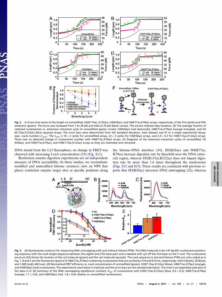
Magnetic Tweezers Measurements Quantify the Number of Nucleo-somes Within a Single DNA Molecule. We used magnetic tweezersmeasurements to determine nucleosome retention within singlenucleosome arrays while subjected to an external force (30).Nucleosomes release approximately 25 nm of DNA length whenexposed to forces above 15 pN (31, 32), so we use the number ofsteps to measure the number of nucleosomes within the array.The arrays were extended by increasing the force to 29 pN over100 s and then held at 29 pN for 130 s prior to relaxation. The firstand fifth extensions are shown in Fig. 2A. Steps are observed withan average extension of 25 nm (Fig. S8) and the step number islargely preserved for five stretching cycles (Fig. 2 A and E), aspreviously reported (31). We confirmed full nucleosome unwrap-ping by observing the array extended to its contour length of 1 μmand verified the observed step number as the force is increasedand held at 29 pN is representative of the nucleosome number byfitting the array’s force response up to 7 pN to a polymer model(Fig. S9 and SI Text). We studied 31 separate unmodified nucleo-some arrays with 399 nucleosomes by extending and retractingeach over five cycles. The fraction of retained nucleosomes perstretch cycle fits to an exponential decay with a characteristiccycle number of 16, which converts to a 40-min dissociation time.
PTMs near the Nucleosome Dyad Facilitate Nucleosome Disassembly.To investigate the influence of histone PTMs on nucleosomestability, we extended and retracted single nucleosome arrayscontaining H3(K56ac) (25 arrays with 310 nucleosomes), H4(K77ac,K79ac) (31 arrays with 372 nucleosomes), and H3(K115ac,K122ac) (41 arrays with 476 nucleosomes) for five cycles(Fig. 2 B–D). We found the decay time to be 22 cycles or 55 minfor H3(K56ac) nucleosome disassembly, and we observed no lossof H4(K77ac,K79ac) nucleosomes (Fig. 2E). In contrast, thenumber of observed steps in H3(K115ac,K122ac) nucleosome ar-rays dramatically reduced over five cycles, indicating that histoneoctamers dissociated at a rate of 3.4 cycles or 8.5 min (Fig. 2E)—afivefold increase relative to unmodified nucleosomes. Theseresults indicate that PTMs from the DNA entry–exit region to 35base pairs into the nucleosome do not facilitate nucleosome dis-assembly following DNA unwrapping, whereas the dyad histone–DNA contacts disrupted by H3(K115ac,K122ac) are importantfor the maintenance of partially unwrapped nucleosomes.
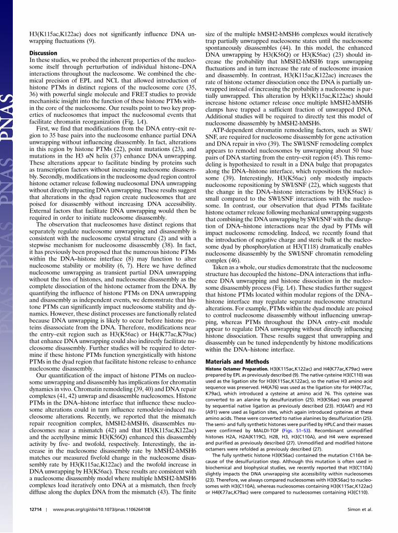
PTMs Within the DNA Entry–Exit Region of the Nucleosome FacilitateDNA Unwrapping and Transcription Factor Binding. We investigatedthe influence of histone PTMs within the DNA–histone interfaceon nucleosomal DNA unwrapping and transcription factor bind-ing using FRET (Fig. 3 and Figs. S10 and S11) and restrictionenzyme digestion experiments (Figs. S12 and S13). The FRETsystem was designed based on previous studies (23, 33) such thatthe 20 base pair target sequence for a model transcription factor,LexA, is located within the nucleosome between the eighth andthe 27th base pair (Fig. 3A). Cy3 and Cy5 fluorophores areattached to the nucleosome to monitor DNA unwrapping that istrapped as LexA is titrated in increasing concentration and bindsto its target sequence (Fig. 3 B and C). To quantify DNA unwrap-ping, we used the unwrapping equilibrium constant, Keq, whichwe define as the concentration ratio of partially wrapped nucleo-some states that LexA can bind, to the nucleosome states thatLexA cannot bind (see SI Materials and Methods). We determinedthe Keq of nucleosomes containing H3(K56ac), H4(K77ac,K79ac), and H3(K115ac,K122ac) relative to unmodified nucleo-somes (Fig. 3E). We find that H3(K56ac) and H4(K77ac,K79ac)increase Keq by twofold, a biologically significant increase as inthe case for dosage compensation and haploinsufficiency dis-eases. In contrast, H3(K115ac,K122ac) does not alter Keq, whichindicates that the dyad region does not impact partial DNAunwrapping.
To confirm that these results are due to DNAunwrapping ratherthan nucleosome sliding or repositioning, we repeated these ex-periments with the LexA target site on the opposite end of the
Fig. 1. A–C show the face, top, and bottom view of the nucleosome crystal structure (52). Acetylation sites are residues H3(K56) (red), H3(K115) (light blue), H3(K122) (blue), H4(K77) (yellow), and H4(K79) (orange). Circles inA show two functionally distinct regions of the nucleosome. The dyad region of the nucleosome(top circle) regulates the final release of the histone octamer without influencing DNA unwrapping, whereas the DNA entry–exit region (bottom left and rightcircles) regulates the DNA unwrapping without influencing histone octamer release. (D and E) AFM images of unmodified and H3(K115ac,K122ac) nucleosomearrays, respectively. The images’ width are 300 nm.
12712 ∣ www.pnas.org/cgi/doi/10.1073/pnas.1106264108 Simon et al.
DNA strand from the Cy3 fluorophore; no change in FRET wasobserved with increasing LexA concentration (33) (Fig. S11).
Restriction enzyme digestion experiments are an independentmeasure of DNA accessibility. In these studies, we reconstitutemodified and unmodified histone octamers onto an NPS thatplaces restriction enzyme target sites at specific positions along
the histone–DNA interface (34). H3(K56ac) and H4(K77ac,K79ac) increase digestion rate by threefold near the DNA entry–exit region, whereas H3(K115ac,K122ac) does not impact diges-tion rate by more than 1.6 times throughout the nucleosome(Figs. S12 and S13). These results are consistent with previous re-ports that H3(K56ac) increases DNA unwrapping (22), whereas
Fig. 2. A–D are time series of the length of unmodified, H3(K115ac, K122ac), H3(K56ac), and H4(K77ac,K79ac) arrays, respectively, of the first (pink) and fifthextension (green). The force was increased from 7 to 29 pN and held at 29 pN (black curves). The arrows indicate step locations. (E) The average fraction ofretained nucleosomes vs. extension–retraction cycle of unmodified (green circles), H3(K56ac) (red diamonds), H4(K77ac,K79ac) (orange triangles), and H3(K115ac,K122ac) (blue squares) arrays. The error bars were determined from the standard deviation. Each dataset was fit to a single exponential decay:exp½−ðcycle numberÞ∕tcycle�. The tcycle is 16� 2 cycles for unmodified arrays, 22� 3 cycles for H3(K56ac) arrays, and 3.4� 0.3 for H3(K115ac,K122ac) arrays.There was no detected change in nucleosome number with H4(K77ac,K79ac) arrays. (F) Diagrams of the extension–retraction cycles of unmodified, H3(K56ac), and H3(K77ac,K79ac), and H3(K115ac,K122ac) arrays as they are extended and retracted.
Fig. 3. (A) Nucleosome construct for measuring DNA unwrapping with and without histone PTMs. The DNA molecule is the 147 bp 601 nucleosome position-ing sequence with the LexA target sequence between the eighth and 27th base pairs and is labeled with cy3 off the first base on the 5′ end. The nucleosomestructure (52) shows the location of the cy3 molecule (green) and the cy5 molecules (purple). The LexA sequence is teal and histone PTMs are color coded as inFig. 1. B and C are the fluorescent spectra of H4(K77ac,K79ac) containing nucleosomes that are excited by 510 and 610 nm, respectively, with 0 (black), 30 (blue),and 1,000 (red) mM LexA. (D) Normalized FRET efficiency vs. LexA concentration of unmodified (green), H3(K115ac,K122ac) (blue), H4(K77ac,K79ac) (orange),and H3(K56ac) (red) nucleosomes. The experiments were done in triplicate and the error bars are the standard deviation. The inset is an expanded scale plot ofthe data in D. (E) Summary of the DNA unwrapping equilibrium constant, Keq, of nucleosomes with H3(K115ac,K122ac) (blue; 0.9� 0.2), H4(K77ac,K79ac)(orange; 1.7� 0.4), and H3(K56ac) (red: 1.8� 0.4) relative to unmodified nucleosomes.
Simon et al. PNAS ∣ August 2, 2011 ∣ vol. 108 ∣ no. 31 ∣ 12713
BIOPH
YSICSAND
COMPU
TATIONALBIOLO
GY
H3(K115ac,K122ac) does not significantly influence DNA un-wrapping fluctuations (9).
DiscussionIn these studies, we probed the inherent properties of the nucleo-some itself through perturbation of individual histone–DNAinteractions throughout the nucleosome. We combined the che-mical precision of EPL and NCL that allowed introduction ofhistone PTMs in distinct regions of the nucleosome core (35,36) with powerful single molecule and FRET studies to providemechanistic insight into the function of these histone PTMs with-in the core of the nucleosome. Our results point to two key prop-erties of nucleosomes that impact the nucleosomal events thatfacilitate chromatin reorganization (Fig. 1A).
First, we find that modifications from the DNA entry–exit re-gion to 35 base pairs into the nucleosome enhance partial DNAunwrapping without influencing disassembly. In fact, alterationsin this region by histone PTMs (22), point mutations (23), andmutations in the H3 αN helix (37) enhance DNA unwrapping.These alterations appear to facilitate binding by proteins suchas transcription factors without increasing nucleosome disassem-bly. Secondly, modifications in the nucleosome dyad region controlhistone octamer release following nucleosomal DNA unwrappingwithout directly impacting DNAunwrapping. These results suggestthat alterations in the dyad region create nucleosomes that arepoised for disassembly without increasing DNA accessibility.External factors that facilitate DNA unwrapping would then berequired in order to initiate nucleosome disassembly.
The observation that nucleosomes have distinct regions thatseparately regulate nucleosome unwrapping and disassembly isconsistent with the nucleosome crystal structure (2) and with astepwise mechanism for nucleosome disassembly (38). In fact,it has previously been proposed that the numerous histone PTMswithin the DNA–histone interface (8) may function to alternucleosome stability or mobility (6, 7). Here we have definednucleosome unwrapping as transient partial DNA unwrappingwithout the loss of histones, and nucleosome disassembly as thecomplete dissociation of the histone octamer from the DNA. Byquantifying the influence of histone PTMs on DNA unwrappingand disassembly as independent events, we demonstrate that his-tone PTMs can significantly impact nucleosome stability and dy-namics. However, these distinct processes are functionally relatedbecause DNA unwrapping is likely to occur before histone pro-teins disassociate from the DNA. Therefore, modifications nearthe entry–exit region such as H3(K56ac) or H4(K77ac,K79ac)that enhance DNA unwrapping could also indirectly facilitate nu-cleosome disassembly. Further studies will be required to deter-mine if these histone PTMs function synergistically with histonePTMs in the dyad region that facilitate histone release to enhancenucleosome disassembly.
Our quantification of the impact of histone PTMs on nucleo-some unwrapping and disassembly has implications for chromatindynamics in vivo. Chromatin remodeling (39, 40) and DNA repaircomplexes (41, 42) unwrap and disassemble nucleosomes. HistonePTMs in the DNA–histone interface that influence these nucleo-some alterations could in turn influence remodeler-induced nu-cleosome alterations. Recently, we reported that the mismatchrepair recognition complex, hMSH2-hMSH6, disassembles nu-cleosomes near a mismatch (42) and that H3(K115ac,K122ac)and the acetyllysine mimic H3(K56Q) enhanced this disassemblyactivity by five- and twofold, respectively. Interestingly, the in-crease in the nucleosome disassembly rate by hMSH2-hMSH6matches our measured fivefold change in the nucleosome disas-sembly rate by H3(K115ac,K122ac) and the twofold increase inDNA unwrapping by H3(K56ac). These results are consistent witha nucleosome disassembly model where multiple hMSH2-hMSH6complexes load iteratively onto DNA at a mismatch, then freelydiffuse along the duplex DNA from the mismatch (43). The finite
size of the multiple hMSH2-hMSH6 complexes would iterativelytrap partially unwrapped nucleosome states until the nucleosomespontaneously disassembles (44). In this model, the enhancedDNA unwrapping by H3(K56Q) or H3(K56ac) (23) should in-crease the probability that hMSH2-hMSH6 traps unwrappingfluctuations and in turn increase the rate of nucleosome invasionand disassembly. In contrast, H3(K115ac,K122ac) increases therate of histone octamer dissociation once the DNA is partially un-wrapped instead of increasing the probability a nucleosome is par-tially unwrapped. This alteration by H3(K115ac,K122ac) shouldincrease histone octamer release once multiple hMSH2-hMSH6clamps have trapped a sufficient fraction of unwrapped DNA.Additional studies will be required to directly test this model ofnucleosome disassembly by hMSH2-hMSH6.
ATP-dependent chromatin remodeling factors, such as SWI/SNF, are required for nucleosome disassembly for gene activationand DNA repair in vivo (39). The SWI/SNF remodeling complexappears to remodel nucleosomes by unwrapping about 50 basepairs of DNA starting from the entry–exit region (45). This remo-deling is hypothesized to result in a DNA bulge that propagatesalong the DNA–histone interface, which repositions the nucleo-some (39). Interestingly, H3(K56ac) only modestly impactsnucleosome repositioning by SWI/SNF (22), which suggests thatthe change in the DNA–histone interactions by H3(K56ac) issmall compared to the SWI/SNF interactions with the nucleo-some. In contrast, our observation that dyad PTMs facilitatehistone octamer release following mechanical unwrapping suggeststhat combining the DNAunwrapping by SWI/SNF with the disrup-tion of DNA–histone interactions near the dyad by PTMs willimpact nucleosome remodeling. Indeed, we recently found thatthe introduction of negative charge and steric bulk at the nucleo-some dyad by phosphorylation at H3(T118) dramatically enablesnucleosome disassembly by the SWI/SNF chromatin remodelingcomplex (46).
Taken as a whole, our studies demonstrate that the nucleosomestructure has decoupled the histone–DNA interactions that influ-ence DNA unwrapping and histone dissociation in the nucleo-some disassembly process (Fig. 1A). These studies further suggestthat histone PTMs located within modular regions of the DNA–
histone interface may regulate separate nucleosome structuralalterations. For example, PTMs within the dyad module are poisedto control nucleosome disassembly without influencing unwrap-ping, whereas PTMs throughout the DNA entry–exit moduleappear to regulate DNA unwrapping without directly influencinghistone dissociation. These results suggest that unwrapping anddisassembly can be tuned independently by histone modificationswithin the DNA–histone interface.
Materials and MethodsHistone Octamer Preparation. H3(K115ac,K122ac) and H4(K77ac,K79ac) wereprepared by EPL as previously described (9). The native cysteine H3(C110) wasused as the ligation site for H3(K115ac,K122ac), so the native H3 amino acidsequence was preserved. H4(A76) was used as the ligation site for H4(K77ac,K79ac), which introduced a cysteine at amino acid 76. This cysteine wasconverted to an alanine by desulfurization (25). H3(K56ac) was preparedby sequential native ligation as previously described (23). H3(A47) and H3(A91) were used as ligation sites, which again introduced cysteines at theseamino acids. These were converted to native alanines by desulfurization (25).The semi- and fully synthetic histones were purified by HPLC and their masseswere confirmed by MALDI-TOF (Figs. S1–S3). Recombinant unmodifiedhistones H2A, H2A(K119C), H2B, H3, H3(C110A), and H4 were expressedand purified as previously described (27). Unmodified and modified histoneoctamers were refolded as previously described (27).
The fully synthetic histone H3(K56ac) contained the mutation C110A be-cause of the desulfurization step. Although this mutation is often used inbiochemical and biophysical studies, we recently reported that H3(C110A)slightly impacts the DNA unwrapping site accessibility within nucleosomes(23). Therefore, we always compared nucleosomes with H3(K56ac) to nucleo-somes with H3(C110A), whereas nucleosomes containing H3(K115ac,K122ac)or H4(K77ac,K79ac) were compared to nucleosomes containing H3(C110).
12714 ∣ www.pnas.org/cgi/doi/10.1073/pnas.1106264108 Simon et al.
Nucleosome Array Preparation. All nucleosome arrays were reconstituted witha 17-mer tandem repeat of a 148 bp variant of the 601 positioning sequencewith 30 bp of linker DNA (29), which was cut out of the pUC19 plasmid withEcoRI and SphI. Biotin was attached to the tandem repeat by ligating theannealed pair of oligonucleotides: biotin-AGCTAGCTTTCAATAGCTCG andAATTCGAGCTATTGAAAGCTAGCT to the EcoRI overhang, whereas digoxigen-in was attached to the tandem repeat by ligating the pair of annealed oli-gonucleotides: GGGCGGCGACCT-dig and AGGTCGCCGCCCCATG to the SphIoverhang. The ligations were done simultaneously with excess concentrationof annealed oligonucleotides to ensure that the tandem repeat did not ligateinto multimers. The excess annealed oligonucleotides were purified awaywith a S-400 HR spin column (GE Healthcare). The remaining half of the plas-mid was further digested with DdeI into seven pieces of various lengthsranging from 166 to 636 bp, which served as buffering DNA for nucleosomereconstitution.
Unmodified and modified nucleosomal arrays were reconstituted bysalt double-dialysis (47) in 60 μL with 3 μg of plasmid DNA containing labeled17-mer array, 15 μg of core particle DNA, 12 μg of purified histone octamer,2 M NaCl, 1 mM benzamidine hydrochloride (BZA), 5 mM Tris (pH 8.0), and0.5 mM EDTA. Recovered reconstituted nucleosome arrays were thenpurified on 5–40% sucrose gradients. Purified nucleosome arrays were char-acterized by electrophoretic mobility shift assay with a 2% polyacrylamideand 1% agarose gel in 90 mM Tris–borate and by AFM.
AFM Imaging of Nucleosome Arrays. Freshly cleaved mica surface was rinsedwith ultrapure water and vigorously dried with nitrogen. Poly-D-lysine (50 μL,Sigma) at 10 ng∕μL was deposited on the mica surface, incubated for 90 s,washed with 200 μL ultrapure water, and gently dried with nitrogen. Fiftymicroliters of purified nucleosome arrays at 0.5–1 nM in 0.2× TE (2 mM Tris,0.2 mM EDTA, pH 8.0) were deposited on the poly-D-lysine-treated mica sur-face, incubated for 5 min, washed with 200 μL ultrapure water, and gentlydried with nitrogen. The nucleosome arrays were then immediately imagedwith a Dimension 3000 Scanning ProbeMicroscope (Veeco Instruments) usingetched silicon nitride tips (PPP-NCH, Nanosensor) with a scan rate of 1 Hz andan amplitude set point of 0.9–1.2 V, which varied from tip to tip. The numberof nucleosomes within each imaged array was determined manually.
Single Molecule Magnetic Tweezers Nucleosome Counting Experiments. Allforce-extension measurements were done with single nucleosome arraystethered to antidigoxigenin-coated cover glass slides and streptavidin-coatedmagnetic beads (DynabeadM280, Invitrogen) in lab-built flow cells on amag-netic tweezers apparatus (30, 48, 49) (see SI Text for details). We determinedthe number of nucleosomes within a single array by detecting the number ofsteps in the array extension as the force is increased from 7.5 to 29 pN. Theforce was initially increased to 7.5 pN over 20 s, then increased from 7.5 to29 pN over 100 s, and then held at 29 pN for 130 s. The tension was relaxed to0.1 pN and held at this force for 4min. This cycle was repeated four additionaltimes. All bead heights were measured relative to a nearby bead fixed to thesurface.
The step number and the step size were determined by Matlab analysis ofeach time series of the nucleosome array length. The steps were detected by
calculating the convolution of a 31-point step function with 31 data points(1 s) centered about the time point of interest. This calculation resulted in atime series with a peak centered about each step. The number of peaks wasdetermined and the center of each peak was located. The step size was thendetermined from the difference between the average of the 10 points beforeand after the step. The two adjacent points before and after the step wereignored when calculating step sizes.
FRET Measurements of Nucleosomal DNA Unwrapping. FRET measurements ofDNA unwrapping were performed as previously described (33). The DNAmo-lecules 601-LexA-left (Fig. 3A), and 601-LexA-right (Fig. S11A) were preparedby PCR with Cy3-labeled oligonucleotides from a plasmid containing the601 positioning sequence with LexA binding site at bases 8–27 or 121–140,respectively (33). Oligonucleotides were labeled with a Cy3-NHS ester (GEhealthcare) at a 5′ amino group then purified by RP-HPLC. The oligos usedto amplify 601-LexA-L were Cy3-CTGGAGATACTGTATGAGCATACAGTACAA-TTGGTC and ACAGGATGTATATATCTGACACGTGCCTGGAGACTA; 601-LexA-Rwere Cy3-CTGGAGAATCCCGGTGCCGA and CTCCATACTGTATGCTCATACAG-TAATCCTGT.
H2A(K119C) was labeled before or after histone octamer refolding withCy5-maleamide (GE Healthcare) as previously described (23). Nucleosomeswere reconstituted by salt double dialysis (47) with 7 μg of DNA and 5 μg ofhistone octamer in 50 μL of 0.5× TE (pH 8.0), 2 M NaCl, and 1 mM BZA.Reconstituted nucleosomes were purified by 5–30% sucrose gradient. LexAprotein was expressed and purified from the pJWL288 plasmid (gift fromJonathan Widom, Northwestern University, Evanston, IL) as previously de-scribed (50).
The equilibrium constants for site accessibility were determined from thereduction in FRET efficiency as LexA binds to its target site buried within thenucleosome (33) (Fig. 3A). FRET efficiency measurements were determinedby the ðratioÞA method as previously described (51) (see SI Text for details).LexA was titrated from 0 to 3 μM with 5 nM Cy3/Cy5-labeled nucleosomes in0.5× TE. The FRET efficiency was determined by the ðratioÞA method intriplicate for each LexA concentration. The average FRET efficiency vs. LexAconcentration was fit to a noncooperative binding isotherm: E ¼ EFþðE0 − EFÞ∕ð1þ ½LexA�∕S0.5Þ, where E is the FRET efficiency, E0 is the FRET effi-ciency without LexA, EF is the FRET efficiency at high LexA concentration,and S0.5-nuc is the LexA concentration at which the FRET efficiency has beenreduced by half [i.e., E ¼ ðE0 þ EFÞ∕2]. The relative equilibrium constant be-tween the unmodified and modified nucleosome was determined as follows:Relative Keq ¼ S0.5-nuc-modified∕S0.5-nuc-unmodified.
ACKNOWLEDGMENTS. We are grateful to Karin Musier-Forsyth for access to aTyphoon Trio fluorescence scanner and a fluorescence plate reader and toDongping Zhong for access to an FPLC system. We acknowledge supportfrom American Heart Association Predoctoral Fellowship 0815460D (toJ.A.N.); National Institutes of Health GM083055 (to M.G.P. and J.J.O.);National Institutes of Health GM080176 (to R.F.); National Science Founda-tion (NSF) CAREER MCB0845696 (to J.J.O.), a Career Award from TheBurroughs Wellcome Fund (M.G.P.), and seed funding from NSF MaterialsResearch Science and Engineering Center 0820414 (to M.G.P.).
1. Kornberg RD (1974) Chromatin structure: A repeating unit of histones and DNA.
Science 184:868–871.
2. Luger K, Mader AW, Richmond RK, Sargent DF, Richmond TJ (1997) Crystal structure of
the nucleosome core particle at 2.8 A resolution. Nature 389:251–260.
3. Ranjith P, Yan J, Marko JF (2007) Nucleosome hopping and sliding kinetics determined
from dynamics of single chromatin fibers in Xenopus egg extracts. Proc Natl Acad Sci
USA 104:13649–13654.
4. Morrison AJ, Shen X (2009) Chromatin remodelling beyond transcription: the INO80and SWR1 complexes. Nat Rev Mol Cell Biol 10:373–384.
5. Maier VK, Chioda M, Becker PB (2008) ATP-dependent chromatosome remodeling.
Biol Chem 389:345–352.
6. Cosgrove MS, Boeke JD, Wolberger C (2004) Regulated nucleosome mobility and the
histone code. Nat Struct Mol Biol 11:1037–1043.
7. Mersfelder EL, Parthun MR (2006) The tale beyond the tail: Histone core domain mod-
ifications and the regulation of chromatin structure. Nucleic Acids Res 34:2653–2662.
8. Zhang L, Eugeni EE, Parthun MR, Freitas MA (2003) Identification of novel histonepost-translational modifications by peptide mass fingerprinting. Chromosoma
112:77–86.
9. Manohar M, et al. (2009) Acetylation of histone H3 at the nucleosome dyad alters
DNA–histone binding. J Biol Chem 284:23312–23321.
10. English CM, Adkins MW, Carson JJ, Churchill ME, Tyler JK (2006) Structural basis for the
histone chaperone activity of Asf1. Cell 127:495–508.
11. Hyland EM, et al. (2005) Insights into the role of histone H3 and histone H4 core
modifiable residues in Saccharomyces cerevisiae. Mol Cell Biol 25:10060–10070.
12. Kruger W, et al. (1995) Amino acid substitutions in the structured domains of histonesH3 and H4 partially relieve the requirement of the yeast SWI/SNF complex for tran-scription. Genes Dev 9:2770–2779.
13. Flaus A, Rencurel C, Ferreira H, Wiechens N, Owen-Hughes T (2004) Sin mutations alterinherent nucleosome mobility. EMBO J 23:343–353.
14. Muthurajan UM, et al. (2004) Crystal structures of histone Sin mutant nucleosomesreveal altered protein-DNAinteractions. EMBO J 23:260–271.
15. Kurumizaka H, Wolffe AP (1997) Sin mutations of histone H3: Influence on nucleo-some core structure and function. Mol Cell Biol 17:6953–6969.
16. Horn PJ, Crowley KA, Carruthers LM, Hansen JC, Peterson CL (2002) The SIN domainof the histone octamer is essential for intramolecular folding of nucleosomal arrays.Nat Struct Biol 9:167–171.
17. Fry CJ, Norris A, Cosgrove M, Boeke JD, Peterson CL (2006) The LRS and SIN domains:Two structurally equivalent but functionally distinct nucleosomal surfaces requiredfor transcriptional silencing. Mol Cell Biol 26:9045–9059.
18. Xu F, Zhang K, Grunstein M (2005) Acetylation in histone H3 globular domain regu-lates gene expression in yeast. Cell 121:375–385.
19. Chen CC, et al. (2008) Acetylated lysine 56 on histone H3 drives chromatin assemblyafter repair and signals for the completion of repair. Cell 134:231–243.
20. Williams SK, TruongD, Tyler JK (2008) Acetylation in the globular core of histone H3 onlysine-56 promotes chromatin disassembly during transcriptional activation. Proc NatlAcad Sci USA 105:9000–9005.
21. Andrews AJ, Chen X, Zevin A, Stargell LA, Luger K (2010) The histone chaperoneNap1 promotes nucleosome assembly by eliminating nonnucleosomal histone DNAinteractions. Mol Cell 37:834–842.
Simon et al. PNAS ∣ August 2, 2011 ∣ vol. 108 ∣ no. 31 ∣ 12715
BIOPH
YSICSAND
COMPU
TATIONALBIOLO
GY
22. Neumann H, et al. (2009) Amethod for genetically installing site-specific acetylation inrecombinant histones defines the effects of H3 K56 acetylation. Mol Cell 36:153–163.
23. Shimko JC, North JA, Bruns AN, Poirier MG, Ottesen JJ (2011) Preparation of fullysynthetic histone H3 reveals that acetyl-lysine 56 facilitates protein binding withinnucleosomes. J Mol Biol 408:187–204.
24. Park JH, Cosgrove MS, Youngman E, Wolberger C, Boeke JD (2002) A core nucleosomesurface crucial for transcriptional silencing. Nat Genet 32:273–279.
25. Wan Q, Danishefsky SJ (2007) Free-radical-based, specific desulfurization of cysteine:A powerful advance in the synthesis of polypeptides and glycopolypeptides. AngewChem Int Ed Engl 46:9248–9252.
26. Dorigo B, Schalch T, Bystricky K, Richmond TJ (2003) Chromatin fiber folding: Require-ment for the histone H4 N-terminal tail. J Mol Biol 327:85–96.
27. Luger K, Rechsteiner TJ, Richmond TJ (1999) Preparation of nucleosome core particlefrom recombinant histones. Methods Enzymol 304:3–19.
28. Lowary PT, Widom J (1998) New DNA sequence rules for high affinity binding to his-tone octamer and sequence-directed nucleosome positioning. J Mol Biol 276:19–42.
29. Poirier MG, BussiekM, Langowski J, Widom J (2008) Spontaneous access to DNA targetsites in folded chromatin fibers. J Mol Biol 379:772–786.
30. Strick TR, Allemand JF, Bensimon D, Bensimon A, Croquette V (1996) The elasticity of asingle supercoiled DNA molecule. Science 271:1835–1837.
31. Brower-Toland BD, et al. (2002) Mechanical disruption of individual nucleosomes re-veals a reversible multistage release of DNA. Proc Natl Acad Sci USA 99:1960–1965.
32. Claudet C, Angelov D, Bouvet P, Dimitrov S, Bednar J (2005) Histone octamer instabilityunder single molecule experiment conditions. J Biol Chem 280:19958–19965.
33. Li G, Widom J (2004) Nucleosomes facilitate their own invasion. Nat Struct Mol Biol11:763–769.
34. Polach KJ, Widom J (1995) Mechanism of protein access to specific DNA sequences inchromatin: A dynamic equilibriummodel for gene regulation. J Mol Biol 254:130–149.
35. Chatterjee C, Muir TW (2010) Chemical approaches for studying histonemodifications.J Biol Chem 285:11045–11050.
36. Allis CD, Muir TW (2011) Spreading chromatin into chemical biology. Chembiochem12:264–279.
37. Ferreira H, Somers J, Webster R, Flaus A, Owen-Hughes T (2007) Histone tails andthe H3 alphaN helix regulate nucleosome mobility and stability. Mol Cell Biol27:4037–4048.
38. Bohm V, et al. (2010) Nucleosome accessibility governed by the dimer/tetramer inter-face. Nucleic Acids Res 39:3093–3102.
39. Clapier CR, Cairns BR (2009) The biology of chromatin remodeling complexes. AnnuRev Biochem 78:273–304.
40. Gangaraju VK, Bartholomew B (2007) Mechanisms of ATP dependent chromatinremodeling. Mutat Res 618:3–17.
41. Dupaigne P, et al. (2008) Rad51 polymerization reveals a new chromatin remodelingmechanism. PLoS One 3:e3643.
42. Javaid S, et al. (2009) Nucleosome remodeling by hMSH2-hMSH6. Mol Cell36:1086–1094.
43. Jeong C, et al. (2011) MutS switches between two fundamentally distinct clampsduring mismatch repair. Nat Struct Mol Biol 18:379–385.
44. Forties RA, et al. (2011) A quantitative model of nucleosome dynamics. Nucleic AcidsRes, in press.
45. Dechassa ML, et al. (2008) Architecture of the SWI/SNF-nucleosome complex. Mol CellBiol 28:6010–6021.
46. North JA, et al. (2011) Phosphorylation of histone H3(T118) alters nucleosomedynamics and remodeling. Nucleic Acids Res, 10.1128/MCB.00693-08.
47. ThastromA, Lowary PT, Widom J (2004) Measurement of histone-DNA interaction freeenergy in nucleosomes. Methods 33:33–44.
48. Skoko D, Wong B, Johnson RC, Marko JF (2004) Micromechanical analysis of thebinding of DNA-bending proteins HMGB1, NHP6A, and HU reveals their ability to formhighly stable DNA–protein complexes. Biochemistry 43:13867–13874.
49. Strick TR, Allemand JF, Bensimon D, Croquette V (1998) Behavior of supercoiled DNA.Biophys J 74:2016–2028.
50. Little JW, et al. (1994) Cleavage of LexA repressor. Methods Enzymol 244:266–284.51. Clegg RM (1992) Fluorescence resonance energy transfer and nucleic acids. Methods
Enzymol 211:353–388.52. Richmond TJ, Davey CA (2003) The structure of DNA in the nucleosome core. Nature
423:145–150.
12716 ∣ www.pnas.org/cgi/doi/10.1073/pnas.1106264108 Simon et al.


![Computational biology: deep learning...from DNA sequence, RNA polymerase binding, nucleosome positioning and transcriptional data [16], as well as gene expression from histone modifications](https://img.pdfslide.us/doc/110x75/61487fc62918e2056c22ba9d/computational-biology-deep-learning-from-dna-sequence-rna-polymerase-binding.jpg)