Embed Size (px)
Citation preview
1
Pulmonary Journal Club December 2020 (Articles from November 2020) Presented by
Dr. Anja C. Roden, Professor of Pathology and Laboratory Medicine Dr. Lacey J. Schrader, Pulmonary Pathology Fellow
Mayo Clinic Rochester, MN Articles for Discussion Page
4 Ren HZ. C-MET immunohistochemistry for differentiating malignant mesothelioma from benign mesothelial proliferations Hum Patholy. 2020;105:31-36.
5 Yang C et al. Lung-only melanoma: UV mutational signature supports origin from occult cutaneous primaries and argues against the concept of primary pulmonary melanoma. Mod Pathol. 2020; 33:2244-55.
6 Dall’Olio FG et al. Comparison of Sequential Testing and Next Generation Sequencing in advanced Lung Adenocarcinoma patients – A single centre experience. Lung Cancer 2020; 149:5-9.
7 Borczuk AC et al. COVID-19 pulmonary pathology: a multi-institutional autopsy cohort from Italy and New York City. Mod Pathol. 2020. 33:2156-68.
Articles for Notation – Neoplastic 8 Argani P et al. EWSR1/FUS–CREB fusions define a distinctive malignant epithelioid
neoplasm with predilection for mesothelial-lined cavities. Mod Pathol. 2020. 33:2233-43.
9 Hung YP et al. ARID1A mutations and expression loss in non-small cell lung carcinomas: clinicopathologic and molecular analysis. Mod Pathol. 2020; 33:2256-68.\
11 Takeda-Miyata N et al. Prognostic significance of spread through air spaces in pulmonary metastases from colorectal cancer. Lung Cancer 2020;149:61-7
12 Gao Q et al. Clinical and histopathological features of pulmonary sclerosing pneumocytoma with dense spindle stromal cells and lymph node metastasis. Histopathol. 2020;77:718-27.
13 Gui H et al. Primary Pulmonary Myxoid Sarcoma and Myxoid Angiomatoid Fibrous Histiocytoma. A Unifying Continuum With Shared and Distinct Features. Am J Surg Pathol. 2020; 44:1535-40.
2
14 Zhao J et al. Identification of a novel gene expression signature associated with overall survival in patients with lung adenocarcinoma: A comprehensive analysis based on TCGA and GEO databases. Lung Cancer. 2020; 149: 90-6.
15 Dacic S et al. Whole exome sequencing reveals BAP1 somatic abnormalities in mesothelioma in situ. Lung Cancer 2020; 149:1-4
16 Hung YP et al. Molecular characterization of diffuse malignant peritoneal mesothelioma. Mod Pathol 2020; 33:2269-79.
17 Wolf JL et al. Interobserver variation in the classification of thymic lesions including biopsies and resection specimens in an international digital microscopy panel. Histopathology 2020; 77:734-41.
18 Prabhakaran S et al. The potential utility of GATA binding protein 3 for diagnosis of malignant pleural mesotheliomas. Hum Pathol 2020; 105:1-8.
19 Frank MS et al. Re-biopsy after first line treatment in advanced NSCLC can reveal changes in PD-L1 expression. Lung Cancer 2020; 149:23-32.
Articles for Notation - Non-Neoplastic
20 Furusawa H et al. Chronic Hypersensitivity Pneumonitis, an Interstitial Lung Disease with Distinct Molecular Signatures. Am J Respir Crit Care Med. 2020; 10:1430–44.
21 Schaefer IM et al. In situ detection of SARS-CoV-2 in lungs and airways of patients with COVID-19. Mod Pathol 2020; 33:2104-14.
22 Prieto-Perez L et al Histiocytic hyperplasia with hemophagocytosis and acute alveolar damage in COVID-19 infection. Mod Pathol 2020; 33:2139-46
23 Kheir F et al. Using Bronchoscopic Lung Cryobiopsy and a Genomic Classifier in the Multidisciplinary Diagnosis of Diffuse Interstitial Lung Diseases. Chest 2020; 158:2015-25.
24 Kheir F et al. Using Bronchoscopic Lung Cryobiopsy and a Genomic Classifier in the Multidisciplinary Diagnosis of Diffuse Interstitial Lung Diseases. Chest 2020; 158:2015-25
Letters, Brief Communications, Case Reports
24 Asahina M et al. Identification of CTNNB1-PLAG1 gene rearrangement in a patient with pulmonary pleomorphic adenoma. Virch Arch 2020. 477:739-42
3
25 Zeng Z et al. Pulmonary pathology of early-phase COVID-19 pneumonia in a patient with a benign lung lesion. Histopathology 2020; 77:823-31
25 Pernazza A et al. Early histologic findings of pulmonary SARS-CoV-2 infection detected in a surgical specimen. Virchows Archive 2020; 477:743-8.
26 Calabrese F et al. Two Sorts of Microthrombi in a Patient With Coronavirus Disease 2019 and Lung Cancer. J Thorac Oncol; 15:1782-5.
Reviews
26 Primer DS et al. Overview of Pathologic Findings of Vaping in the Context of an Autopsy Patient With Chronic Injury. Arch Pathol Lab Med 2020; 144:1408-13
26 Friedlaender A et al. The METeoric Rise of MET in Lung Cancer. Cancer 2020; 126:4826-37.
27 Bruce-Brand C et al. Rosai-Dorfman Disease: an overview. J Clin Pathol 2020; 73:697-705.
27 Chen M. Special issue on diagnosis of primary hematolymphoid disease of the lung and pleura. Sem Diagn Pathol 2020; 37:
27 Churg A et al. The Separation of Benign and Malignant Mesothelial Proliferations. New Markers and How to Use Them. Am J Surg Pathol. 2020; 44:e100-112.
28 Pollack SB et al. A systematic review of pathological findings in COVID-19: a pathophysiological timeline and possible mechanisms of disease progression. Mod Pathol 2020; 33:2128-38.
4
Articles for Discussion
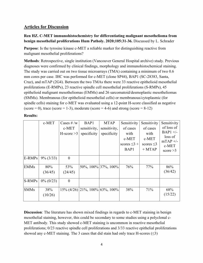
Ren HZ. C-MET immunohistochemistry for differentiating malignant mesothelioma from benign mesothelial proliferations Hum Patholy. 2020;105:31-36. Discussed by L. Schrader
Purpose: Is the tyrosine kinase c-MET a reliable marker for distinguishing reactive from malignant mesothelial proliferations?
Methods: Retrospective, single institution (Vancouver General Hospital archive) study. Previous diagnoses were confirmed by clinical findings, morphology and immunohistochemical staining. The study was carried out on two tissue microarrays (TMA) containing a minimum of two 0.6 mm cores per case. IHC was performed for c-MET (clone SP44), BAP1 (SC-28383, Santa, Cruz), and mTAP (2G4). Between the two TMAs there were 33 reactive epithelioid mesothelial proliferations (E-RMPs), 23 reactive spindle cell mesothelial proliferations (S-RMPs), 45 epithelioid malignant mesotheliomas (EMMs) and 26 sarcomatoid/desmoplastic mesotheliomas (SMMs). Membranous (for epithelioid mesothelial cells) or membranous/cytoplasmic (for spindle cells) staining for c-MET was evaluated using a 12-point H-score classified as negative (score = 0), trace (score = 1-3), moderate (score = 4-6) and strong (score = 8-12)
Results:
c-MET Cases # /w c-MET
H-score >3
BAP1 sensitivity, specificity
MTAP sensitivity, specificity
Sensitivity of cases
with c-MET
scores ≤3 + BAP1
Sensitivity of cases
with c-MET
scores ≤3 + MTAP
Sensitivity of loss of BAP1 +/-
loss of mTAP +/-
c-MET score >3
E-RMPs 9% (3/33) 0
EMMs 80% (36/45)
53% (24/45)
50%, 100% 37%, 100% 76% 77% 86% (36/42)
S-RMPs 0% (0/23) 0
SMMs 38% (10/26)
15% (4/26) 21%, 100% 63%, 100% 38% 71% 68% (15/22)
Discussion: The literature has shown mixed findings in regards to c-MET staining in benign mesothelial staining, however, this could be secondary to some studies using a polyclonal c-MET antibody. This study showed c-MET staining is uncommon in reactive mesothelial proliferations; 0/23 reactive spindle cell proliferations and 3/33 reactive epithelial proliferations showed any c-MET staining. The 3 cases that did stain had only trace H-scores (≤3)
5
Comment: c-MET is a useful tool in distinguishing reactive versus malignant mesothelial proliferation in certain morphologic context (epithelial vs spindle cell). The application of the H-scores described in this paper may not be a practical application for most practices unless there is a high volume of mesothelioma cases; however, an easily visible moderate or strong membrane staining may serve instead as an indicator of malignancy.
Yang C et al. Lung-only melanoma: UV mutational signature supports origin from occult cutaneous primaries and argues against the concept of primary pulmonary melanoma. Mod Pathol. 2020; 33:2244-55.
Background:
• Primary pulmonary melanomas (PPM); defined by WHO: +IHC or EM, exclusively involving lung, no current or previous primary cutaneous, uveal, mucosal melanoma despite exhaustive clinical eval; solitary lesion with central location adjacent to major airways – considered characteristic of PPM given presumed origin from intrabronchial melanocytes
• Controversial - no native melanocytes in lung & possible regression of cutaneous melanomas
Aim:
• To investigate clinicopathologic and genomic features of lung-only melanomas to clarify their site of origin
Methods:
• Retrospective review of lung-only melanomas with NGS (468 cancer genes, MSK-IMPACT) • Controls: 605 lung adenoCa, 549 cutaneous melanomas
Results:
• 10 lung only melanomas; 4 met criteria of PPM; all male, mean age 63.8 yo (range, 47-75) • Solitary lesions (N=4), mean size, 5.1 cm (range, 3-10.1), 3 central, peribronchial; 2-3 lesions
(N=4); >10 lesions (N=2) • Micro: entire/predominant epithelioid (N=8); spindle cell (N=2), melanin pigment (N=4);
bronchial epithelium involved (3/6), not diagnostic of melanoma in situ; no subepithelial nevi • IHC: 2-4 melanocytic markers+ in 10/10; all +SOX10, S100; pan-keratin weak/focal + in 1/5 • NGS – mean of 43.6 non-synonymous mutations (range, 2-113) • Mean TMB 42.6/Mb (range, 1.8-126); frequent mutations: BRAF (N=4), NRAS (N=1), NF1
(N=7), KIT (N=1), KRAS (N=1) = genomic profile typical of UV-associated cutaneous melanoma; other oncogenic alterations commonly encountered in cutaneous melanoma: TP53 (N=5), CDKN2A (N=1)
• Mutational signature (>20 mutations; N=8) - all had dominant UV signature including all 3 evaluable solitary lesions; 2 non-evaluable cases: BRAFV600E mutation (commonly found in cutaneous and mucosal melanomas) (N=2) and GG>AA substitution in TERT promotor
6
(N=1) - highly specific for UV-mutagenesis (almost exclusively found in cutaneous melanomas and cutaneous squamous cell carcinomas)
• Controls: 97% (470/486) of cutaneous melanomas - dominant UV signature; not in any Lung adenoCa (N=291)
• Mean f/u 27.1 months (range, 5-43): 9/10 received anti-PD-1 and/or anti-CTLA-4 inhibitors; most patients with 1-3 lesions had resection; extrapulm mets in 7/10 patients (brain, bone, pancreas, liver), 3 died of disease; median OS 40 months; patients with solitary lesion (N=4) – 1 had metastatic progression at extra-pulmonary site; no primary melanoma found
Conclusions:
• Lung-only melanomas should be considered as likely metastatic even in the absence of a known primary melanoma elsewhere and even they meet the strict criteria of PPM
• Be aware of epithelioid morphology of these melanomas that can closely mimic NSCLC
Dall’Olio FG et al. Comparison of Sequential Testing and Next Generation Sequencing in advanced Lung Adenocarcinoma patients – A single centre experience. Lung Cancer 2020; 149:5-9.
Background
• Prognostic and theranostic markers in NSCLC - performed sequentially or as one-shot NGS
Aims:
• To compare a sequential single-gene testing algorithm to NGS in lung adenoCa (effectiveness, costs, tissue consumption, TAT)
Methods
• Retrospective; advanced lung adenoCa, homogeneous Caucasian population in Northern Italy • Sequential approach (1/2014-2/2019): EGFR (RT-PCR) → KRAS (pyrosequencing) →
molecular testing (only if EGFR, KRAS neg) “physician driven” driven by availability of drugs or clinical trials including ALK (since 2012 with crizotinib approval, FISH, IHC) → ROS1 (since 2016, FISH) → BRAF (pyrosequ), MET, HER2, RET
• NGS (2/2019-1/2020): 35 hotspot mutations, 19 amplifications, 23 rearrangements • Price: EGFR RT-PCR 380€; KRAS pyrosequencing 160€, single gene Sanger sequencing
(BRAF, MET) 160€, FISH (RET, MET, HER2) 330€ each; IHC (ALK, ROS1, NTRK) 200€ each; NGS 770€
• NGS 25µm; 3µm slides for IHC or FISH • TAT – appr. 10 days for NGS; 5 days for RT-PCR, 5 days for FISH, 2 days for IHC
Results
• 1758 consecutive patients; 1221 sequential algorithm, 537 NGS
7
• Prevalence of EGFR, ALK, KRAS alterations similar between sequential and NGS (16.5 vs 14.3%, 6.3 vs 6.3%, 36 vs 33.5%);
• Sequential: 50% finished after first round (EGFR, KRAS) – with ALK testing – 6.3% positive → ROS1 testing (4.7% +) → 286 patients left for other testing
• ROS-1 rearrangements, MET amplification, MET mutations, HER2 amplification, and mutations, BRAFV600E mutations prevalence higher in sequential vs NGS (4.7 vs 0.7%, 11.2 vs 2.2%, 9 vs 2.4%, 3.3 vs 1.9%, 9.8 vs 3%, 4.5 vs 1.5%)
• NGS: other mutations found in 26.3% of cases; concurrent mutations in 24.4% • Cost for sequential testing 1375€ vs 770 € for NGS; • Sequential testing estimated 33.3µm slide vs 25µm slide for NGS • Mean TAT – sequential 13.15 days vs 10 days for NGS
Take Home Points:
• NGS – less expensive, potentially overall faster TAT for lung adenoCa biomarker testing • Sequential testing - faster in detecting EGFR (5 days), ALK, ROS1 (2 days) and probably
also KRAS, the more common molecular alterations and those with a higher impact on current clinical practice – still viable alternative specifically in critically ill patients
• Evidence from other studies did not show a survival benefit when using NGS
\
Borczuk AC et al. COVID-19 pulmonary pathology: a multi-institutional autopsy cohort from Italy and New York City. Mod Pathol. 2020. 33:2156-68.
Background:
• Mortality of SARS-CoV-2, etiologic agent of COVID-19, dominated by ARDS
Aim:
• To evaluate large # of autopsies from 3 institutions in heavily hit areas
Methods:
• Autopsies from 2 US (covering Manhattan, Queens, Brooklyn) and 1 Italian (covering part of the Veneto area) hospital
• Detailed eval of several lung compartments (airways, alveolar walls, airspaces, vasculature) • All patients confirmed COVID-19 by nasopharyngeal PCR • IHC for SARS-CoV-2 spike protein (1A9); RNAish-sequence for spike protein (ACD)
Results
• 68 consecutive autopsies of COVID-19 patients, 47 (69%) males, median age 73 yo (range, 30-96)
• Comorbidities in 97%; 60% had ≥3; most common hypertension, diabetes
8
• 5% smokers, 32% former smokers • Days of disease, median, 11 (Q1-Q3; 8-19), 40% intubated • Median of 19 lung tissue blocks including bronchi and trachea (range, 8-30) • Associated or superimposed non-viral (bacterial, fungal) pneumonia by ante-or postmortem
cultures or bug stains 26.5% • Pathology: 92% had combined lung weight of >1300 g and consolidations patchy and diffuse • Tracheobronchitis frequent; independent of intubation or superimposed pneumonia; often
associated with aphthous ulcers, 2-3 mm • DAD – 87%; later phases less frequent, correlated with longer duration of disease • DAD seen together with OP, interstitial fibroblasts, squamous metaplasia – likely ongoing
viral-induced injury of type 2 pneumocytes • 42% had large vessel thrombi; microthrombi at least focal in 84% • In some cases with rapid progression to severe respiratory failure w/o evidence for
superinfection and ventilator-associated pneumonia- neutrophils seen together with alveolar wall injury, necrosis, microthrombi
• EM: small vessels with basal membrane reduplication, endothelial swelling with cytoplasmic vacuolization c/w endothelial cell injury; virus particles in type 2 pneumocytes
• IHC and RNAish (N=23) and 5 neg controls; in COVID patients positive in tracheal epithelium, hyaline membranes, atypical type 2 pneumocytes; most COVID-lungs positive in first 2 weeks of disease in hyaline membranes and adjacent cells; often patchy; beyond 2 weeks positivity less frequent. Overall 13 of the 23 cases were positive by IHC and RNAish (56.5%) including 75% of cases within 1 week, 70% within 2 weeks, 50% within 3 and 4 weeks, none within 5 weeks.
• All findings were similar between the 3 hospitals
Take Home Points:
• COVID-19 pneumonia – heterogeneous disease (tracheobronchitis, DAD, vascular injury in patients with frequent comorbidities, specifically hypertension and diabetes)
• Disease appears to begin in airways extending to alveolar zones
Articles for Notation
Neoplastic Disease
Argani P et al. EWSR1/FUS–CREB fusions define a distinctive malignant epithelioid neoplasm with predilection for mesothelial-lined cavities. Mod Pathol. 2020. 33:2233-43.
Background:
9
• EWSR1-CREB-hallmark of angiomatoid fibrous histiocytoma (AFH-EWSR1-CREB1), soft tissue and GI clear cell sarcoma (CCS-EWSR1-ATF1), primary pulmonary myxoid sarcoma (PPMS- EWSR1-CREB1), hyalinizing clear cell carcinoma of salivary gland (EWSR1-ATF1), subset of mesothelioma in young adults, and others
Aim:
• To investigate previously unclassified malignant epithelioid neoplasms frequently showing epithelioid phenotype and marked predilection to peritoneal cavity, defined by EWSR1/FUS-CREB fusions
Methods
• Consult cases with malignant epithelioid phenotype and EWSR1/FUS-CREB fusions • Various RNA-sequencing platforms (N=9) or FISH (N=4) • Meso markers WT-1, calretinin, and various other markers
Results
• N=13 (6 males; mean age, 36, range, 9-63) • 10 intra-abdominal (often involving or spreading along peritoneal surface, omentum,
mesentery; 5 arising in mesocolon or perirectal/rectovaginal pouch; 4 involving other organs: stomach, adrenal, cecum, kidney), 1 pleural cavity (large, solid-cystic pleural-based mass), 1 upper and 1 lower limb soft tissue;
• Most well circumscribed, thick tumor capsule, tumor size, 2-15 cm (mean, 8) • All at least focal epithelioid (predominant epithelioid [8; with focal rhabdoid features, 5/8],
mixed epithelioid/spindled [2] or epithelioid/round cell [3]) associated with cystic or microcystic changes in all cases, variable lymphoid cuffing (intermixed or peripheral)
• Most low mitoses (1-2/10 HPF [9]; 5-6/10 HPF [2], 20/10 HPF [1]), necrosis rare (N=2) • IHC pos: EMA and/or CK (N=12), WT-1 (N=5) with 4 CK+/WT1+, ALK+ (N=1); neg:
S100, calretinin; BAP1, INI-1 preserved • EM (N=1) no features of mesothelioma • EWSR1-CREM (N=7), FUS-CREM (N=4), EWSR1-ATF1 (N=2) • Metastases at presentation or later (N=7), multiple recurrences (N=1), no death • Diff diagn: AFH (CK-), mesothelioma (lack of lymphoid cuffing, localized nature to
abdomen), however EWSR1/FUS-ATF1 fusions can be found in mesos in young patients
Take Home Points
• Authors view as “entity” with hybrid features between AFH (cystic growth, lymphoid cuffing) and mesothelioma (location, epithelioid, CK and WT-1 co-expression)
• Seems to have better outcome than meso • Authors did not eval other meso markers; expression of BAP1 does not argue against meso
10
• I might have signed as mesothelioma if CK+ and WT-1+ (CK5/6, D2-40, GATA3 status unknown)
Hung YP et al. ARID1A mutations and expression loss in non-small cell lung carcinomas: clinicopathologic and molecular analysis. Mod Pathol. 2020; 33:2256-68.
Background:
• AT-rich interacting domain containing protein 1A (ARID1A, SMARCF1)-component of SWI/SNF complex, implicated in modulating response in immunotherapy in various tumors; mutation in subset of NSCLC; functional significance of ARID1A-context dependent – tumor suppressor in ovarian clear cell and endometrioid Ca for instance
• In NSCLC-evidence suggests that ARID1A mutation status may predict improved response to durvalumab (anti-PD-L1) plus tremelimumab (anti-CTLA4)
Aims:
• To characterize the spectrum of ARID1A mutations and expression in NSCLC
Methods:
• Retrospective targeted NGS of consecutive NSCLC • IHC for ARID1A (polyclonal Ab) on all ARID1A-mutated NSCLC and 40 wt NSCLC; %
tumor cells expressed, expression pattern; nuclear staining
Results:
• 2440 consecutive NSCLC (80% adenoCa, 13% SQCC, 6% NOS, 0.5% sarcomatoid Ca, 0.5% adenosquam Ca; 59% female, median age 67 yo (range, 11-96)
• 184/2440 (7.5%) of NSCLC harbored ARID1A mutations including adenoCa (75%), SQCC (18%), others (7%); of these 184 – 69% were loss-of-function mutations
• IHC in 139 ARID1A-mutated NSCLC: aberrant expression in 64 (46%) with complete loss in 13 (9%), diffuse diminished in 17 (12%), heterogeneous loss in 34 (25%); all 40 wt NSCLC had intact expression
• Relationship between mutations and expression status appears complex – same mutation shows various protein expression patterns; Complete loss of expression correlated with premature truncating mutations with biallelic inactivation
• Complete loss / diminished ARID1A expression - associated with loss-of-function mutations (88% vs 55% in ARID1A intact) and biallelic inactivation (59% vs 5% in ARID1A intact)
• Presence of multiple ARID1A mutations alone – no association with presence of aberrant expression (20% vs 9%); positions of loss-of-function mutation within the gene did not explain expression pattern
11
• NSCLC with complete loss of expression (N=13) - all had one or multiple loss-of-function mutations, all truncal
• ARID1A mutations and aberrant expression correlated with lack of EGFR mutations (9% vs 20% in ARID1A intact), frequent TP53 mutations (69% vs 52%), increased TMB
• No mutations in other SWI/SNF members except PBRM1 • Similar OS between ARID1A-mutated and wt tumors; among mutated tumors, aberrant
expression of ARID1A correlated with worse OS • Diffuse loss of ARID1A expression – predominantly adenoCa, poorly differentiated, almost
exclusively smokers, enriched for MMR deficiency → Loss of ARID1A expression in <2% of NSCLC but associated with distinct clinicopathologic features
Conclusions:
• ARID1A mutational status is unreliable in predicting expression status but no protein loss in the absence of a mutation
• What is a better biomarker - mutation or expression status?
Takeda-Miyata N et al. Prognostic significance of spread through air spaces in pulmonary metastases from colorectal cancer. Lung Cancer 2020;149:61-7
Background:
• Resection of pulmonary metastases from colorectal cancer → prolonged relapse-free survival, standard therapy; surgical margin relapse occurs sometimes
Aims:
• To evaluate prognostic significance of STAS for pulmonary mets from colorectal cancer • To assess whether STAS can be predicated based on preop clinical info
Methods:
• Retrospective • STAS definition according to Shiono: tumor nests within air spaces ≥0.5 mm from edge of
main tumor; tumor nests composed of >5 cells
Results:
• 96 pulmonary mets from colorectal cancer in 37 patients (14 men); median # of resected mets 2 (range, 1-9); median size of mets, 9.5 mm (range, 2.3-36.6)
• Median distance from margin, 10 mm (range, 5-42) • STAS present in 24/37 (64.9%) patients; 40/96 lesions (41.6%), median # of STAS clusters,
3 (range, 1-15)
12
• Median distance main tumor edge and farthest STAS, 1.5 mm (range, 0.5-11 mm) • 26 patients – synchronous multiple mets – STAS consistently found among multiple lesions
from same patient, N=12 • 5-yr OS after metastasectomy 48.3%, recurrence in 22 patients (59.5%) • Surgical margin relapse in 8.3% of lesions (N=8) from 7 patients (18.9%); only 5
histologically confirmed (others just radiologic) • Patients with STAS – worse OS (5-yr OS 30.3% with STAS vs 76.9% w/o STAS) • STAS in metastasis, mucinous adenoCa in primary cancer – higher risk of death on
multivariable analysis • Surgical margin relapse correlates with higher # of STAS clusters and larger distance
between STAS and main tumor • Distance of STAS – independent risk factor for surgical margin relapse • Elevated presurgical serum CEA independently correlated with occurrence of STAS
Take Home Points:
• STAS in metastatic colorectal cancer – poor prognosis and higher risk for surgical margin relapse – presence might trigger closer surveillance and early treatment of radiologic changes in previous surgical field
• STAS might be left behind despite seemingly negative margin • May need to be included in pathology report ?
Gao Q et al. Clinical and histopathological features of pulmonary sclerosing pneumocytoma with dense spindle stromal cells and lymph node metastasis. Histopathol. 2020;77:718-27.
Backgound:
• Pulmonary sclerosing pneumocytoma (PSP) – reclassified as adenoma • LN mets rarely reported
Aims:
• To determine clinicopathologic features of PSP with spindle cells or PSP cells in LNs
Methods
• Retrospective study with re-review of all cases
Results:
• 239 PSP cases; 7 are described including 5 with dense stromal spindle cells one of which with LN met and 2 add PSP with LN met
• Tumor size, mean, 3.1 cm (range, 0.9-10 cm)
13
• Female predominance (6/7), median age, 53 yo (range, 24-68) • Imaging: unifocal solid tumors; 1 was lobulated with uneven densities • 5 cases - mainly solid regions of diffuse stromal spindle cells – 1/5 almost all stromal cells
spindled, 4/5 spindled stromal cells together with round and polygonal cells • IHC – spindle cells: +TTF1, vimentin, EMA (weak), ER (partial) • 2 additional cases with LN met did not show spindled stromal cells • LN mets – metastatic cells were stromal cells • Mean f/u 31 months (range, 2-82 months), no new mets • In entire study population metastatic rate 2/234 PSP with f/u (0.8%); met in 1/5 PSP with
spindle cells
Take Home Points:
• Stromal cells in PSP might be of spindled cytology rather than round cells • 1.3% of PSP have mets at time of resection • PSP with spindle cells might be more prone to met, however, study number low • Based on literature review, male patients are more likely to develop mets • LN mets do not affect long-term outcome • Recommend that LN are removed if enlarged or if spindle cells are a component of the
stromal cells • Diff diagn: SFT
Gui H et al. Primary Pulmonary Myxoid Sarcoma and Myxoid Angiomatoid Fibrous Histiocytoma. A Unifying Continuum With Shared and Distinct Features. Am J Surg Pathol. 2020; 44:1535-40.
Background:
• Primary pulmonary myxoid sarcoma (PPMS) – rare low grade neoplasm, reticular/lace-like growth of spindle to epithelioid cells in myxoid matrix; morphologic overlap with myxoid variant of angiomatoid fibrous histiocytoma (AFH) – both harbor EWSR1-CREB1 fusion; EWSR1-ATF1 only reported in AFH
Aim:
• To present a case of a primary pulmonary low-grade myxoid spindle cell tumor with morphologic and IHC features of PPMS but EWSR1-ATF1 fusion gene (case 1)
• To present a case of endobronchial AFH with EWSR1-CREB1 translocation and focal morphologic features of PPMS (case 2)
Methods
• FISH, NGS
14
Results
• Case 1: 49 yo man, non-smoker, cough, hemoptysis for 1 month, FDG-avid lung mass, 3.8cm o RLL – lobectomy – gross: tan yellow, rubbery, 3.8 cm mass extending into bronchus; o Morphology: well circumscribed, lobules of spindle, stellate, and epithelioid cells
arranged in cords, strands, reticular patterns; abundant myxoid stroma; rare mites, no necrosis, occasional cytologic atypia
o IHC: weak focal EMA; neg for keratins, S100, other markers o Molecular: EWSR1-ATF1 fusion gene
• Case 2: 41yo man, 1 yr intractable wheezing, now worsening SOB, left main bronchus mass. o Gross: well circumscribed, 2.5 cm nodule, centered around bronchus o Morphology: extensively involving submucosa, lymphoid cuff, oval mononucleated
cells loosely arranged in nodular, vaguely whorled, and storiform pattern, scattered lymphoplasmacytic infiltrate, fibrous stroma; focal morphologic features of PPMS (tumor cells spindled in short lace pattern in myxoid stroma); no significant atypia or mites
o IHC: desmin+, neg: EMA, keratins, S100, CD34, others o Molecular: EWSR1-CREB1 translocation o No recurrence after 2-5 years f/u
Take Home Message:
• Findings support concept that PPMS and myxoid variant of AFH represent a continuum with overlapping histologic, IHC, and genetic features
Zhao J et al. Identification of a novel gene expression signature associated with overall survival in patients with lung adenocarcinoma: A comprehensive analysis based on TCGA and GEO databases. Lung Cancer. 2020; 149: 90-6.
Background: • Gene signatures of lung adenoCa are available but have not yet improved survival
Aims: • To identify novel molecular markers associated with lung adenoCa prognosis
Methods: • RNA sequencing data of lung adenoCa and paired normal tissue from TCGA– differentially
expressed (DE) identified by comparison gene expression between early stage tumors and normal and between advanced stage and early stage tumors
• Risk score used weighted linear combination of individual dysregulated protein-coding genes • Microarray data from the Gene Expression Omnibus (GEO) database
Results:
15
• 513 treatment-naïve lung adenoCa including 395 early-stage (stages I, II) and 110 advanced-stage (stages III, IV), 59 normal tissues from TCGA
• 68 DE genes; 19 protein-coding DE genes were individually associated with OS • Risk score calculated for each patient, normalizing for these 19 genes = 19-gene signature • Lung adenoCa patients with low risk score – better survival than high risk score patients • Multivariate analysis (adjusted for age, sex, clinical stage, smoking history, treatments) –
patients with low risk score had 81% decreased risk for death compared with high risk score patients
• Findings validated in 3 independent GEO datasets which showed significant results in 1 of the datasets in multivariate analysis with similar trends in the other 2 datasets.
Take Home Points: • Prognostic signatures might facilitate risk-stratified disease management
Dacic S et al. Whole exome sequencing reveals BAP1 somatic abnormalities in mesothelioma in situ. Lung Cancer 2020; 149:1-4
Background: • Mesothelioma in situ (MIS) – defined as pure surface population of mesothelial cells that
show loss of BAP1 in patients with unexplained recurrent effusion and no clinically/radiologically mesothelial tumor – high propensity for invasive mesothelioma
Aims: • To investigate genetic events including driver mutations and copy number alterations that
might lead to mesothelioma in situ
Methods: • Whole exome sequencing in paired neoplastic and normal FFPE tissue
Results: • 2 cases – 1 pleura, 1 peritoneum • Somatic alterations only in BAP1 gene • Pleural MIS – 67 yo man; MIS incidentally found during surgery for lung cancer - copy
number loss and LOH in BAP1 locus on chr. 3; CDKN2A intact • Peritoneal MIS – 68 yo woman - 7 years of unexplained ascites - BAP1 somatic splice site
mutation involving intron5-exon6 boundary (A126_splice) with an allelic fraction of 10% and BAP1 copy number loss
• No germline mutations in normal samples → BAP1 mutations somatic in both cases
Conclusions: • BAP1 mutation/deletion might represent a very early event in development of mal meso
16
Hung YP et al. Molecular characterization of diffuse malignant peritoneal mesothelioma. Mod Pathol 2020; 33:2269-79.
Background:
• Besides recurring BAP1 mutations molecular characteristics of peritoneal mesothelioma are not well studied
Aim:
• To examine molecular features of diffuse peritoneal mesos and to compare them with localized peritoneal mesos and WDPM with invasive foci
Methods:
• NGS
Results
• N=26 – diffuse (23 epithelioid, 3 biphasic, 16 women, median OS, 4.1 years, 50% of tumors showed loss of BAP1 expression, 31% deletion of CDKN2A, 25% deletion of NF2)
• N=1 – localized • N=1 – WDPM with invasive foci • Various categories of peritoneal mesos identified:
o Group 1: N=18 (69%), BAP alterations, 31% > 1 BAP1 alteration, concomitant
alterations in PBRM1 (46%) or SETD2 (35%); all mesos with loss of BAP1 expression harbored BAP1 molecular alterations
PBRM1 alterations enriched in BAP1-altered cohort Frequent CN loss of BAP1, ARID1B, PRDM1, PBRM1, SETD2, NF2,
CDKN2A o Group 2:
N=8 (31%), BAP1-wt, TP53 mutations (N=2; one with evidence of genomic near-haploidization with LOH of all chromosomes except 5, 7, 16, 20), TRAF7 activating mutation (N=1), SUZ12 inactivating mutation (N=1), ALK rearrangements (N=3)
o Localized peritoneal meso (N=1, woman <40yo; alive w/o disease >13 yo, intact BAP1 expression)– nonsense CHEK2 mutation
o WDPM with invasive foci (N=1; woman, repeated resection since over 12 years, intact BAP1 expression) – no variants
• BAP1 genomic alterations – associated with older age, worse outcome
Take Home Points:
• Results implicate DNA repair, epigenetics, cell cycle regulation in the pathogenesis of perit. mesos; those tumors are also genetically heterogeneous
17
• Some alterations might represent potential therapeutic targets • Loss of CDKN2A less common in perit mesos (vs pleural meso), possibly because less
sarcomatoid mesos in peritoneum • ALK rearrangements only identified in perit mesos so far – might be treatable with TKIs?
Wolf JL et al. Interobserver variation in the classification of thymic lesions including biopsies and resection specimens in an international digital microscopy panel. Histopathology 2020; 77:734-41.
Background
• Diagnosis of thymic lesions can be challenging despite well-defined WHO categories due to rarity and diversity of tumors
Aim:
• To investigate the reproducibility of the WHO classification among a large group of international pathologists with expertise in thymic pathology using WSI
Methods:
• WSI • 2015 WHO classification
Results:
• 305 tumors (215 resections, 90 biopsies), all consecutively submitted to thymoma panel of the Netherlands (2011-2018); 13 pathologists (Netherlands, USA, Germany, UK)
• 70% thymoma (type AB most common); 10.8% thymic carcinoma; 4.6% benign; 26% no consensus (<7 reviewers agreed on diagnosis)
• Overall agreement: total agreement 20.3% (95-100% reviewers agreed), majority 31.5% (75-94%), consensus 33.8% (50-74%), trend 14.1% (25-49%), lack of agreement, 0.3% (<25%)
• Overall agreement substantial k=0.68 (resection, k=0.73; biopsies, k=0.59) • Thymomas only: B2/B3 vs A, AB, B1, k=0.55; B3 vs A, AB, B1, B2, k=0.49 • Thymoma vs thymic carcinoma and thymic carcinoma vs metastasis was difficult, no kappas • Distinction between various B categories was also difficult (B1 vs B2; B2 vs B3), no kappas
Take Home Points:
• Use of current WHO criteria can lead to substantial agreement, but still difficult areas • Dig path provides a great multinational resource for reviewing rare tumors
Prabhakaran S et al. The potential utility of GATA binding protein 3 for diagnosis of malignant pleural mesotheliomas. Hum Pathol 2020; 105:1-8.
18
Background
• GATA3 suggested as diagnostic marker for sarcomatoid mesos; but no info in regards to usefulness for prognosis and clinical and pathologic correlations in various subtypes of meso
Aims:
• To investigate GATA3, BAP1, and Ki-67 LI in 3 major histologic subtypes of meso
Methods
• Malignant pleural mesos (MPM) and fibrous pleuritis • GATA3 clone L50-823 • 2 pathologists scored independently GATA3 as % tumor cell nuclei staining (4-tier system) • TMA included 100 patients, whole tissue sections in additional 49 patients
Results
• 149 MPM (87 epithelioid, 40 sarcomatoid with 8 being desmoplastic, 22 biphasic); 10 fibrous pleuritis
• GATA3 neg in all fibrous pleuritis • GATA3 + in 50% mesos; significantly highest in sarcomatoid (73%) vs biphasic (50%) vs
epithelioid (40%) and all 8 desmoplastic mesos; k=0.82 between pathologists • Biphasic mesos – discordant GATA3 staining between epithelioid and sarcomatoid
component in 23% (80% showed expression in sarcomatoid and no expression in epithelioid) • Loss of BAP1 expression in 59% (epithelioid), 12% (sarcomatoid), 32% (biphasic mesos);
discordant in 18% of biphasic (75% had loss in epithelioid, retained in sarcomatoid) • Discordant Ki-67 LI in 2 biphasic mesos (expression only in epithelioid component) • Loss of BAP1 expression correlated with absence of expression of GATA3 (not evaluated in
multivariate analysis adjusting for histologic subtype) • Sarcomatoid mesos had worse survival than other subtypes (mean OS for epithelioid,
biphasic and sarcomatoid, 19, 12, 8.6 months, respectively); no association between GATA3 expression and survival
• No association between expression of GATA3, BAP1 or KI-67 LI • Multivariate analysis – only sarcomatoid and biphasic subtype were indicators of shorter OS
Take Home Points:
• GATA3 useful for sarc mesos including desmoplastic subtype • Discordance in GATA3 and BAP1 labeling of epithelioid and sarcomatoid components in
biphasic subtypes is not uncommon • GATA3 – not specific – be careful with angiosarcs, monophasic synovial sarcs, rare
sarcomatoid carcinomas of lung as those can be keratin and GATA3 positive • Might also be useful in distinction of sarcomatoid meso from chronic pleuritis
19
Frank MS et al. Re-biopsy after first line treatment in advanced NSCLC can reveal changes in PD-L1 expression. Lung Cancer 2020; 149:23-32.
Aims: • To investigate the feasibility, risk of complications, and clinical relevance of systematically
performed re-biopsies of NSCLC
Methods: • NSCLC patients with advanced, non-targetable disease (no EGFR mutation, no ALK
rearrangement) who receive first line systemic therapy (pembrolizumab or chemotherapy, based on eligibility)
• Prospective, single center • Biopsy at baseline and at time of progression • PD-L1 TPS (clone 22C3) • Clinical relevance of re-biopsy defined as a potential of changing treatment or follow up, due
to new histological evidence, change in PD-L1 TPS from negative (<1%) to positive (>1%) or vice versa
Results: • 51 patients (45.1% male, median age, 60, range 50-84) • adenoCa (54.5%), SQCC (19.6%), NOS (5.9%); stage IIIB - IVB • original specimen – cytology (74%) or biopsy (26%) • PD-L1 score at initial bx: <1% (33.3%), 1- <5% (2%), 5-<50% (15.7%), 50-100% (49%) • Rebiopsy – cytology (31%) or biopsy (31%) • Complication rate of re-biopsy – 6% (pneumothorax, hydro-pneumothorax, pneumonia) • PD-L1 (N=46) – change in 33% including 1 case which was not really a change as it changed
from group >10-50% to 5-10%; 7 cases had higher TPS, 8 had lower TPS in re-biopsy; in 17% (of 46) – change in PD-L1 expression was potentially clinically relevant; change in PD-L1 TPS was significantly higher in chemotherapy-treated patients in which it was more frequent in adenoCa than SQCC
• In patients with change of TPS (N=15), in 73% the re-biopsy was taken from different location – this % was similar to % of location change in patients without change in TPS
• Overall 17% of patients likely benefited from rebiopsy as TPS changed negative → positive. • In 3 cases TPS changed positive → negative; 2 had received pembro before progression • In 5 cases TPS changed negative → positive including from negative to >50 in some cases • In 3 re-biopsies no malignancy identified
Take Home Message: • Re-biopsy might show change in TPS from negative to positive; specifically in patients
treated with chemo
20
Non-Neoplastic Disease
Furusawa H et al. Chronic Hypersensitivity Pneumonitis, an Interstitial Lung Disease with Distinct Molecular Signatures. Am J Respir Crit Care Med. 2020; 10:1430–44.
Background:
• Some patients with CHP have UIP-like features and are clinically similar to IPF; molecular relationship between the 2 entities relatively unknown
• MUC5B promoter variant rs35705950 – strongest risk factor for IPF, also associated with risk of CHP
Aims:
• To determine common and unique molecular features of CHP and IPF
Methods:
• Retrospective analysis; SLB or explants • Pathology re-review with structured path report, confidence of HP diagnosis • RNA sequencing; MUC5B rs35705950 genotyping; weighted gene coexpression analysis • Adjusting for sex, race, age, smoking
Results:
• CHP (N=82), IPF (N=103), unaffected controls (N=103) • Frequency of MUC5B promoter variant rs35705950 increased in CHP and IPF (vs controls),
comparable between CHP and IPF; CHP patients with MUC5B variant were older and more often smokers
• CHP – explant patients had worse PFT, lower exposure to feather antigens, higher exposure to unknown antigens, more common steroid or immunosuppressant use than SLB patients
• CHP: 413 upregulated, 317 downregulated genes when compared with controls • IPF: 861 upregulated, 322 downregulated genes when compared with controls • 109 genes were upregulated and 150 downregulated only in CHP, not IPF • Genes that were concordantly up- or downregulated in CHP and IPF: Upregulated: related to
collagen catabolic processes, collagen fibril organization, cell adhesion, epithelial development; downregulated: calcium ion transmembrane transport, angiogenesis
• CHP o Genes specific to CHP (differentially expressed in CHP): Upregulated: related to
chemokine-mediated signaling and immune responsiveness; downregulated: steroid metabolic processes, positive regulation of angiogenesis
o Weighted gene coexpression network analysis - genes involved in adaptive immunity and epithelial development associated with improved and reduced lung function, respectively
21
o Higher MUC5B mRNA expression associated with lower %DLCO, morphologic presence of lung fibrosis and honeycombing
o 71 cases with available histology: Fibrosis and lymphocyte inflammation (>80% of cases), giant cells, granulomas, airway-centered inflammation, or fibroblast foci (>50%), HC (48%), OP (28%), GC (21%)
o HRCT: GGO, irregular reticulation, traction bronchiectasis (>80%), HC (30%), mosaic attenuation (57%), LA (54%)
Take Home Points:
• Some gene expression profiles are unique to CHP; some overlap between IPF and CHP
Schaefer IM et al. In situ detection of SARS-CoV-2 in lungs and airways of patients with COVID-19. Mod Pathol 2020; 33:2104-14.
Background
• Only few reports on expression of SARS-CoV-2 in FFPE tissue
Aims:
• To examine IHC expression of SARS-CoV-2 in airways and lung from autopsies from patients who had confirmed COVID-19 by RT-PCR
Methods:
• Rabbit polyclonal antibody to nucleocapsid protein; also double stains for TTF-1/SARS, PU.1-SARS, p63/SARS, MUC5AC/SARS, FOXJ1/SARS; red for SARS
• Staining of 3 representative lung tissues and 1 upper airway section/patient • Stains were reviewed by 4 pathologists (not clear whether they reviewed all slides)
Results:
• 7 autopsies (5 males, median age, 66 years, range, 50-77; 3 non-smoker, 1 current smoker, 2 former smoker); COVID-19 confirmed by RT-PCR
• Median time from onset of symptoms to death, 9 days (range, 6-31 days); from positive RT-PCR test to death, 7 days (0-18)
• Imaging: diffuse airspace disease (N=7) • Morphology:
o DAD including acute (N=5; 4 also had foci of organization) and organizing (N=2) o diffuse inflammatory infiltrates with interstitial and peribronchial lymphocytes and
intraalveolar macrophages, N=7 o no definite viral cytopathic changes o pulmonary embolism or in situ thrombi, N=5 o superimposed bacterial lobar pneumonia, N=3
22
o preexisting chronic Aspergillus lung abscess, N=1 • IHC:
o Patients with acute-phase DAD (≤7 days from onset of respiratory failure) – SARS-CoV-2 detected in pneumocytes and ciliated airway cells (N=5) and upper airway epithelium (N=2); also weak within hyaline membranes, edema, cell debris in alveolar spaces (N=5)
o Staining varied regionally; double staining confirmed SARS localized in pneumocytes but only in rare macrophages; no definite staining in endothelial cells
o Patients with org-phase DAD (>14 days from onset of respiratory failure), no virus detected in lungs or airways
Take Home Points:
• Virus expression might only be seen in acute phase of infection in epithelial cells of airway and lung parenchyma; not seen in endothelial cells
• Findings suggest viral migration • No interobserver reproducibility tested
Prieto-Perez L et al Histiocytic hyperplasia with hemophagocytosis and acute alveolar damage in COVID-19 infection. Mod Pathol 2020; 33:2139-46
Aims:
• To characterize the spectrum of histologic features corresponding to clinical ARDS and macrophage activation syndrome (MAS) that occur in COVID-19
Methods
• Postmortem lung and bone marrow biopsies, 33 patients; only 16 cases had both – only those included in study; 3 had confirmed hemophagocytic lymphohistiocytosis (HLH); lung – percutaneous core biopsy with 14 gauge needle in random areas
• COVID-19 IHC test, clone BSB-134, detects spike protein • Bone marrow biopsies from living patients, N=3
Results
• N=33; median age 79 years (range, 53-98), 64% male; most had underlying health conditions • Imaging: Interstitial infiltrates with centrilobular nodules (N=13), GGOs (N=6), ill-defined
consolidation (N=14) • Bone marrow: lesions of histiocytic hyperplasia with hemophagocytosis (16/17) postmortem
and all 3 living patients- mimicked hemophagocytic histiocytosis • Lungs: hyaline membranes (N=20); numerous fibrinous microthromboemboli (N=12),
predominantly in small vessels
23
• COVID-IHC performed in 1 case – scattered positive pneumocytes
Take Home Points:
• COVID-19 infection triggers systemic immune-inflammatory disease – might allow for specific therapies
Kheir F et al. Using Bronchoscopic Lung Cryobiopsy and a Genomic Classifier in the Multidisciplinary Diagnosis of Diffuse Interstitial Lung Diseases. Chest 2020; 158:2015-25
Background
• Genomic classifier (GC) may be able to distinguish UIP from non-UIP • Is there a benefit to use both, cryobiopsy and GC in addition to MDD for diagnosis of ILD?
Aims:
• To study the impact of sequentially presented data from cryobiopsy and GC on diagnostic confidence of MDD in diagnosing fibrosing ILD
Methods:
• 2 MDD teams; MDD1 reviewed clinical findings→ imaging→cryobx→ GC; MDD2 reviewed GC→cryobiopsy
• MDD diagnosis and confidence level recorded at each step • Inclusion: patients with ILD w/o definite UIP pattern on HRCT; all ≥40 yo old • GC: 5 pieces from TBBx – evaluated by Veracyte through early access program
Results:
• 24 patients, 54.2% male\, mean age 65.7 yo • MDD1 – significant increase in confidence (43% to 93%) in patients with probable UIP after
addition of GC to cryobiopsy • MDD2 - non-significant increase in confidence (27% to 73%) after addition of cryobiopsy to
GC • Concordance coefficient and % agreement of IPF vs non-IPF diagnosis: GC vs MDD1: 0.92,
96%; GC vs MDD2: 0.83, 92%; cryobiopsy 1 vs MDD1: 0.67, 83%; cryobiopsy 2 vs MDD2: 0.66, 83%
• Proportion of high-confidence diagnosis before histology or GC: MDD1 (13%), MDD2 (17%)
• Overall agreement between 2 chest radiologists: 79%, kappa=0.64 • Histology - % non-diagnostic 42% (expert pathologist 1), 33% (expert pathologist 2), overall
agreement 83%, kappa=0.77

24
• MDD1: clinic/imaging + cryobiopsy: high-confidence diagnosis increased from 13 to 46%, with GC increased to 75%
• MDD2: clinic/imaging + GC: high-confidence diagnosis increased from 17 to 29%, with histology increased to 67%
• Overall agreement in final diagnosis of MDD1 and MDD2 was 73%; 100% for categorical diagnosis of IPF vs non-IPF
• Cases with final diagnosis of IPF - GC positive in all, histology showed UIP or favored UIP in 69% in both MDDs
• In MDD1 – concordance between GC and histology for UIP vs non-UIP – 79%, kappa 0.6 (4/5 discordant cases had non-diagnostic histology, 1/5 was diagnosed as CHP)
• In MDD2 – concordance was 75%, kappa 0.53 (3/6 non-diagnostic histology; 3/6 showed aspiration, OP, CHP)
• MDD1 – agreement between final diagnosis before providing GC and GC results – 88%; between histology and final diagnosis 83%
• MDD2 – agreement between final diagnosis before providing GC and GC results – 92%; between histology and final diagnosis 83%
• Interpretation: GC significantly impacted diagnostic confidence of clinicians; high level of agreement between positive GC for UIP and final MDD diagnosis in patients with HRCT interpreted as probable for UIP suggests that GC alone might be used in that setting. Cryobiopsy most useful with a non-UIP pattern on HRCT
Take Home Points:
• Probably more to come on the importance of GC
Letters, Brief Communications, Case Reports
Asahina M et al. Identification of CTNNB1-PLAG1 gene rearrangement in a patient with pulmonary pleomorphic adenoma. Virch Arch 2020. 477:739-42
• Pulmonary pleomorphic adenoma (PA) occurs predominantly in prox airway • Rearrangement of pleomorphic adenoma gene 1 (PLAG1) and high-mobility groupAT-hook
2 gene (HMGA2) – most frequent genetic event in PAs of salivary gland • Report of pulmonary PA • Middle lobar bronchus, 54-yo male, non-smoker, Japanese • Incidental finding; • RML lobectomy – 2.3 cm solid nodule with well-defined pushing margins, glossy cut
surface; firmly adherent to middle lobar bronchus, partially protruding into bronchial lumen • No mitoses, necrosis or cytologic atypia
25
• Myoepithelial cells tended to show plasmacytoid features; +S100, -p63; a finding previously shown in PLAG1-rearranged PAs of salivary glands
• IHC: + S100, p63; -TTF1; no entrapped pneumocytes • CTNNB1-PLAG1 gene fusion by RT-PCR • PLAG1 expression in nucleus of all tumor cells by IHC
Zeng Z et al. Pulmonary pathology of early-phase COVID-19 pneumonia in a patient with a benign lung lesion. Histopathology 2020; 77:823-31
• 55-yo female from Wuhan; underwent RLL lobectomy for benign pulmonary nodule; developed fever shortly after surgery; on day 4 postop nasopharyngeal swab positive for COVID-19; CT now showed GGO and consolidation
• RT-PCR and RNAish positive in FFPE tissue of lobectomy specimen • Lung: exudative inflammation; more prominent towards periphery including monocytes and
lymphocytes • Perivascular inflammation (mainly T cells, which were predominantly CD4+ helper T cells;
some B cells and plasma cells), intra-alveolar multinucleated giant cells, pneumocyte hyperplasia, intracytoplasmic viral-like inclusion bodies
• No hyaline membranes or fibrinous exudate • Abnormal accumulation of CD4+ helper T cells and CD163+ M2 macrophages in lung tissue
Pernazza A et al. Early histologic findings of pulmonary SARS-CoV-2 infection detected in a surgical specimen. Virchows Archive 2020; 477:743-8.
• 61 yo male smoker, lung adenoCa; also MALT lymphoma 6 months prior-complete remission; lobectomy
• Post-surgery – progressive lymphopenia; fever on 5th postop day; COVID-19 confirmed by nasopharyngeal swab
• CT-bilateral, peripheral, ill-defined GGOs; mainly lower lobe • Histology: lung adenoCa; surrounded by diffuse hemorrhage and clusters of alveolar
macrophages, occasional multinucleated giant cells; diffuse pneumocyte loss, reactive hyperplasia, focal pneumocytes with nuclear inclusions, no hyaline membranes
• Mild interstitial inflammatory infiltrate, mainly cytotoxic CD8+ T cells • Focal OP • Diffuse aspects of neutrophilic margination within small arterioles
26
Calabrese F et al. Two Sorts of Microthrombi in a Patient With Coronavirus Disease 2019 and Lung Cancer. J Thorac Oncol; 15:1782-5.
• 77-yo male, died of COVID-19, lung squamous cell carcinoma with lymphatic and vascular tumor thromboemboli in the lung
• DAD and OP • Small vessel inflammation with endotheliitis and microthrombi. Microthrombi mainly in
small and medium-sized vessels • EM of non-neoplastic lung: severe endothelial changes with diffuse reduplication of
basement membrane, swollen endothelial cells with cytoplasmic vacuolization, viral-like particles in type II pneumocytes and endothelial cells
Reviews
Primer DS et al. Overview of Pathologic Findings of Vaping in the Context of an Autopsy Patient With Chronic Injury. Arch Pathol Lab Med 2020; 144:1408-13
• Autopsy case of patient died of vaping-induced injury after prolonged symptomatology and review of the literature
• 2711 reported cases of VALI and 60 deaths according to CDC (as of 1/2020); 82% of cases were patients who reported use of THC-containing products with 33% of total reporting exclusive use of THC-containing products
• Case: 65-yo woman with various comorbidities, long time smoker, opioid dependency; cannabinoid vaporizer used almost daily in the year prior to death; lung bx acute and org DAD; foamy histiocytes scattered in alveoli; subtle areas of interstitial chronic inflammation with scattered eos; same findings in autopsy (36 days after initial presentation), in addition dense interstitial fibrosis; focal alveolar hemorrhage and hemosiderosis
• Review: Discussion of pathogenesis of VALI; clinical characteristics; imaging findings • Pathology: acute and chronic lung injury including DAD, OP, intraalveolar foamy
macrophages, airway-centered acute fibrinous pneumonitis, chronic interstitial inflammation with or w/o rare eos, fibrinous exudate in airspaces, peribronchiolar and/or interstitial granulomatous inflammation; also reported: “bronchiolitis”, parenchymal hemorrhage
• None of the findings are specific
Friedlaender A et al. The METeoric Rise of MET in Lung Cancer. Cancer 2020; 126:4826-37.
• Review of the biology of MET, how to diagnose clinically relevant MET alterations (amplifications, copy numbers, exon 14 skipping), clinical importance of these alterations

27
given the increase in targeted therapies in the context of MET including targeting MET, MET as a resistance mechanism
• Dysregulated activation of MET tyrosine kinase receptor gained importance due to development of effective treatments
• MET dysregulation (by gene amplification, overexpression of the receptor and/or its ligand hepatocyte growth factor, acquisition of activating mutations) leads to prolonged activation of cellular MET (c-MET or MET) receptor and downstream proliferation pathways
• 3-5% of NSCLC harbor MET mutations – mainly adenoCa; overrepresented in sarcomatoid subtype
• 1-5% of NSCLC have de novo MET amplifications, predominantly adenoCa
Bruce-Brand C et al. Rosai-Dorfman Disease: an overview. J Clin Pathol 2020; 73:697-705.
• Classified as part of the “R group” of histiocytosis by the Histiocyte Society in 2016; except for cutaneous RDD which is a separate entity and belongs to the “C group” of histiocyoses
• Clonality in a subset of cases raising the possibility of neoplasm. • Possible association with IgG4-related disease still controversial • Sporadic or familial • Review of pathogenesis, classification, clinical features, pathologic findings, differential
diagnosis, current treatment and prognosis
Chen M. Special issue on diagnosis of primary hematolymphoid disease of the lung and pleura. Sem Diagn Pathol 2020; 37:257-8. – There are several articles devoted to hematolymphoid neoplasms in the lung including
• Kelemen K et al. Primary pulmonary B-cell lymphoma. pp 259-67 • Rosado F et al. Hematolymphoid neoplasms with a plasma cell phenotype. pp 268-72 • T-cell and NK-cell lymphomas in the lung. pp 273-82 • Ngyuen H et al. Mimickers of pulmonary lymphomas. pp 283-95 • Myeloid diseases in the lung and pleura. pp 296-302 • Application of flowcytometry in the analysis of lymphoid disease in the lung and pleural
space. pp 303-320
Churg A et al. The Separation of Benign and Malignant Mesothelial Proliferations. New Markers and How to Use Them. Am J Surg Pathol. 2020; 44:e100-112.
• Established markers: BAP1, mTAP by IHC; CDKN2A (p16) FISH
28
• Potentially useful markers that require more study: EZH2, cyclin D1, PD-L1, CD47, 5HMC by IHC, NF2 FISH
• Not useful: NF2 IHC, LATS 1/2, YAP/TAZ • Discussion about application of these markers in epithelioid vs sarcomatoid mesotheliomas,
mesothelioma in situ and cytology preparartions
Pollack SB et al. A systematic review of pathological findings in COVID-19: a pathophysiological timeline and possible mechanisms of disease progression. Mod Pathol 2020; 33:2128-38.
• 3 stages of infection – viral infection, pulmonary involvement with inflammation, fibrosis • Low and high elastance phenotypes – distinguished in mechanically ventilated patient –
presumed differences in underlying pathophysiology • Review focused on pathophysiology of COVID-19 constructing a timeline and correlating
histologic findings with clinical stages • Lungs: 3 main histologic patterns: epithelial with reactive changes and DAD (85%), vascular
with microvascular damage and (micro)thrombi and AFOP (59%), fibrotic with interstitial fibrosis (22%); epithelial and vascular patterns can present in all stages of symptomatic COVID-19; fibrotic pattern presents starting at about 3 weeks
• Reviews also imaging findings and morphologic findings in other organs