Embed Size (px)
Citation preview
ECHO IN PULMONARY
HYPERTENSION AND
PULMONARY EMBOLISM
T E R E S A S . M . T S A N G , M D , F R C P C , FA C C FA S E
D I R E C T O R O F E C H O , VA N C O U V E R G E N E R A L
H O S P I TA L A N D U N I V E R S I T Y O F B R I T I S H
C O L U M B I A H O S P I TA L
P R O F E S S I O N O F M E D I C I N E
J A N U A R Y 1 9 , 2 0 1 6

No Relevant Disclosure

LEARNING OBJECTIVES
• Clinical contexts and etiologies of
pulmonary hypertension (PH)
• Current classification of PH
• Role of echo in pulmonary
hypertension
• Echo in pulmonary embolism
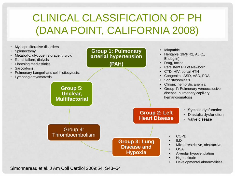
CLINICAL CLASSIFICATION OF PH
(DANA POINT, CALIFORNIA 2008)
Group 1: Pulmonary arterial hypertension
(PAH)
Group 2: Left Heart Disease
Group 3: Lung Disease and
Hypoxia
Group 4: Thromboembolism
Group 5: Unclear,
Multifactorial
• Idiopathic
• Heritable (BMPR2, ALK1,
Endoglin)
• Drug, toxins
• Persistent PH of Newborn
• CTD, HIV, portal HTN
• Congenital: ASD, VSD, PDA
• Schistosomiasis
• Chronic hemolytic anemia
• Group 1’: Pulmonary venoocclusive
disease, pulmonary capillary
hemangiomatosis
• Systolic dysfunction
• Diastolic dysfunction
• Valve disease
• COPD
• ILD
• Mixed restrictive, obstructive
• OSA
• Alveolar hypoventilation
• High altitude
• Developmental abnormalities
• Myeloproliferative disorders
• Splenectomy
• Metabolic: glycogen storage, thyroid
• Renal failure, dialysis
• Fibrosing mediastinitis
• Sarcoidosis,
• Pulmonary Langerhans cell histiocytosis,
• Lymphagiomyomatosis
Simonnereau et al. J Am Coll Cardiol 2009;54: S43–54
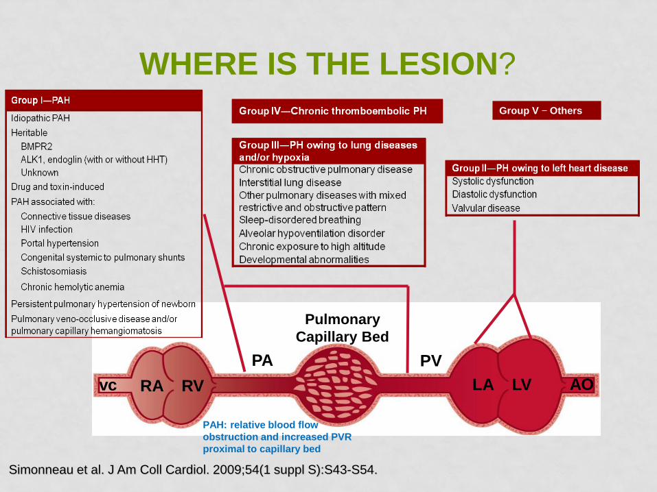
WHERE IS THE LESION?
Simonneau et al. J Am Coll Cardiol. 2009;54(1 suppl S):S43-S54.
vc RA RV
Pulmonary
Capillary Bed
PA PV
LA LV AO
Group V Others
PAH: relative blood flow
obstruction and increased PVR
proximal to capillary bed
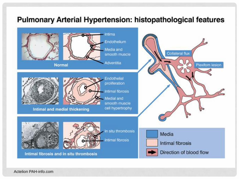
Actelion PAH-info.com
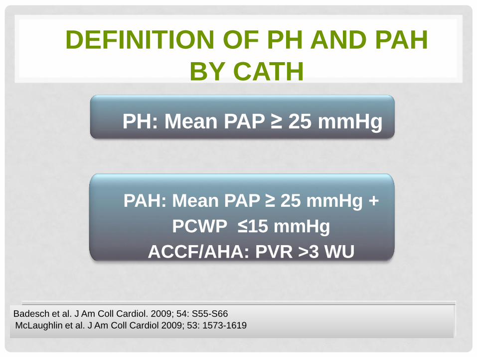
Badesch et al. J Am Coll Cardiol. 2009; 54: S55-S66
McLaughlin et al. J Am Coll Cardiol 2009; 53: 1573-1619
DEFINITION OF PH AND PAH
BY CATH
PH: Mean PAP ≥ 25 mmHg
PAH: Mean PAP ≥ 25 mmHg +
PCWP ≤15 mmHg
ACCF/AHA: PVR >3 WU
PULMONARY VASCULAR RESISTANCE
PULMONARY WEDGE PRESSURE
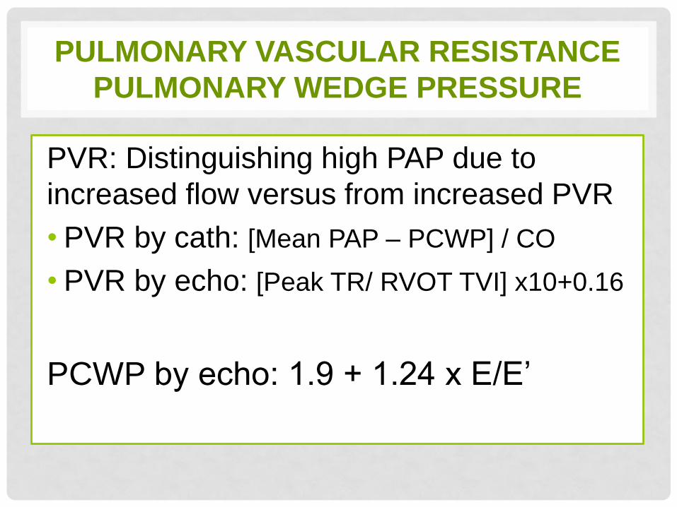
PVR: Distinguishing high PAP due to
increased flow versus from increased PVR
• PVR by cath: [Mean PAP – PCWP] / CO
• PVR by echo: [Peak TR/ RVOT TVI] x10+0.16
PCWP by echo: 1.9 + 1.24 x E/E’
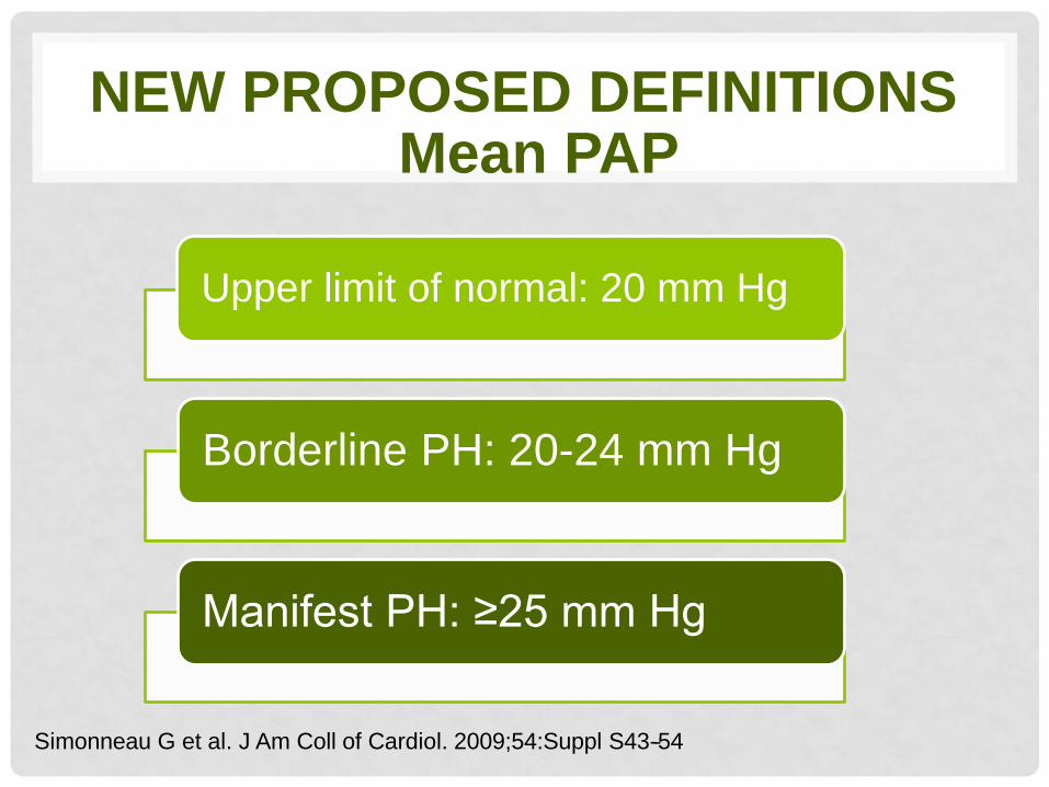
NEW PROPOSED DEFINITIONS
Upper limit of normal: 20 mm Hg
Borderline PH: 20-24 mm Hg
Manifest PH: ≥25 mm Hg
Mean PAP
Simonneau G et al. J Am Coll of Cardiol. 2009;54:Suppl S43-‐54
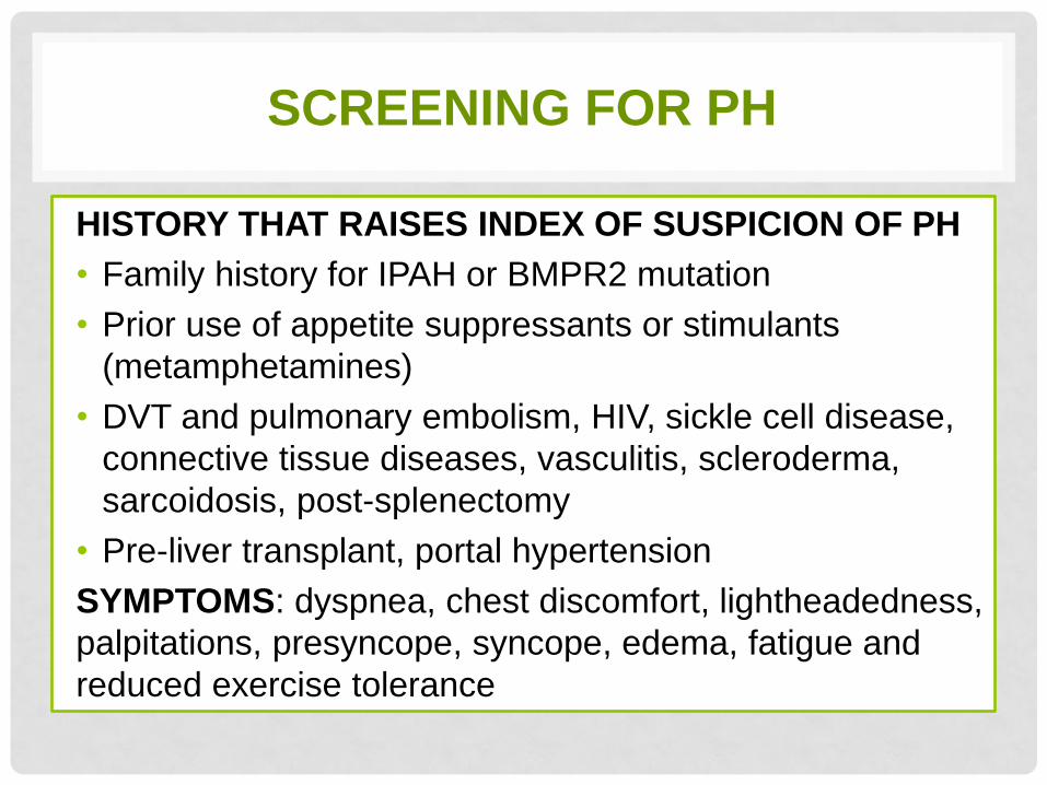
SCREENING FOR PH
HISTORY THAT RAISES INDEX OF SUSPICION OF PH
• Family history for IPAH or BMPR2 mutation
• Prior use of appetite suppressants or stimulants
(metamphetamines)
• DVT and pulmonary embolism, HIV, sickle cell disease,
connective tissue diseases, vasculitis, scleroderma,
sarcoidosis, post-splenectomy
• Pre-liver transplant, portal hypertension
SYMPTOMS: dyspnea, chest discomfort, lightheadedness,
palpitations, presyncope, syncope, edema, fatigue and
reduced exercise tolerance
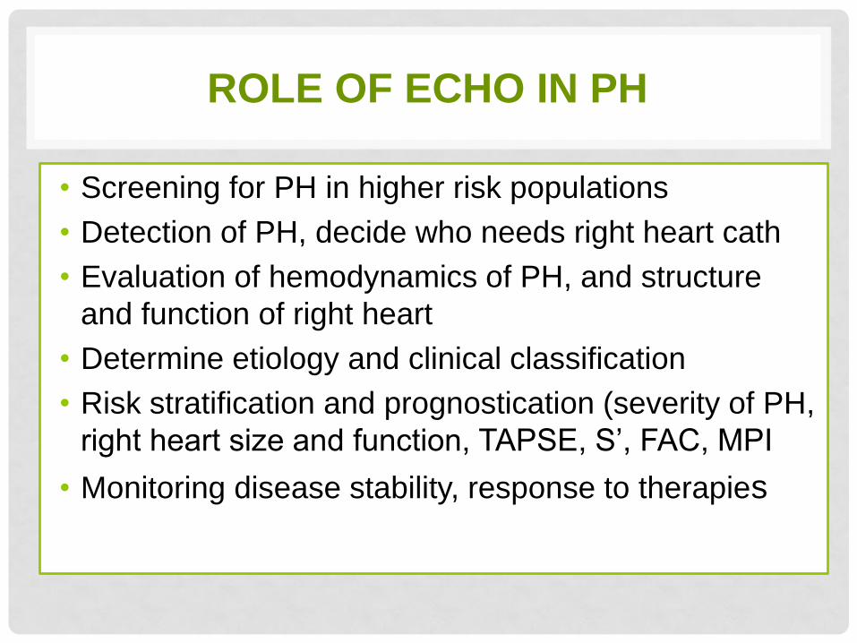
ROLE OF ECHO IN PH
• Screening for PH in higher risk populations
• Detection of PH, decide who needs right heart cath
• Evaluation of hemodynamics of PH, and structure
and function of right heart
• Determine etiology and clinical classification
• Risk stratification and prognostication (severity of PH,
right heart size and function, TAPSE, S’, FAC, MPI
• Monitoring disease stability, response to therapies
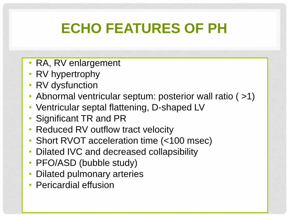
ECHO FEATURES OF PH
• RA, RV enlargement
• RV hypertrophy
• RV dysfunction
• Abnormal ventricular septum: posterior wall ratio ( >1)
• Ventricular septal flattening, D-shaped LV
• Significant TR and PR
• Reduced RV outflow tract velocity
• Short RVOT acceleration time (<100 msec)
• Dilated IVC and decreased collapsibility
• PFO/ASD (bubble study)
• Dilated pulmonary arteries
• Pericardial effusion
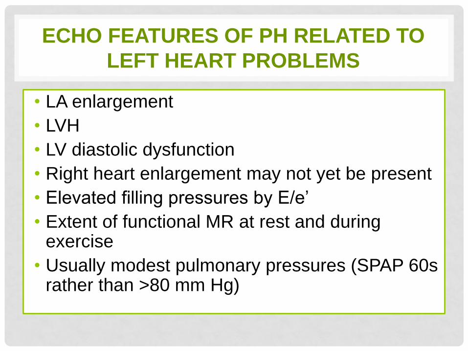
ECHO FEATURES OF PH RELATED TO
LEFT HEART PROBLEMS
• LA enlargement
• LVH
• LV diastolic dysfunction
• Right heart enlargement may not yet be present
• Elevated filling pressures by E/e’
• Extent of functional MR at rest and during exercise
• Usually modest pulmonary pressures (SPAP 60s rather than >80 mm Hg)
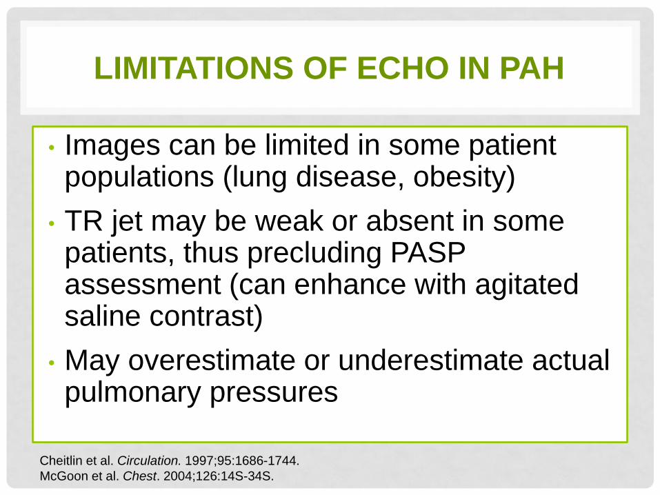
LIMITATIONS OF ECHO IN PAH
• Images can be limited in some patient populations (lung disease, obesity)
• TR jet may be weak or absent in some patients, thus precluding PASP assessment (can enhance with agitated saline contrast)
• May overestimate or underestimate actual pulmonary pressures
Cheitlin et al. Circulation. 1997;95:1686-1744.
McGoon et al. Chest. 2004;126:14S-34S.
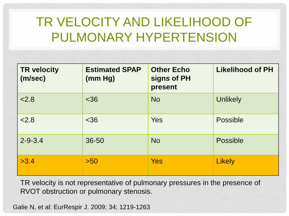
TR VELOCITY AND LIKELIHOOD OF
PULMONARY HYPERTENSION
Galie N, et al: EurRespir J. 2009; 34; 1219-1263
TR velocity
(m/sec)
Estimated SPAP
(mm Hg)
Other Echo
signs of PH
present
Likelihood of PH
<2.8 <36 No Unlikely
<2.8 <36 Yes Possible
2-9-3.4 36-50 No Possible
>3.4 >50 Yes Likely
TR velocity is not representative of pulmonary pressures in the presence of
RVOT obstruction or pulmonary stenosis.
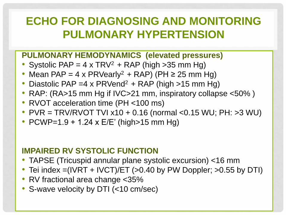
ECHO FOR DIAGNOSING AND MONITORING
PULMONARY HYPERTENSION
PULMONARY HEMODYNAMICS (elevated pressures)
• Systolic PAP = 4 x TRV2 + RAP (high >35 mm Hg)
• Mean PAP = 4 x PRVearly2 + RAP) (PH ≥ 25 mm Hg)
• Diastolic PAP =4 x PRVend2 + RAP (high >15 mm Hg)
• RAP: (RA>15 mm Hg if IVC>21 mm, inspiratory collapse <50% )
• RVOT acceleration time (PH <100 ms)
• PVR = TRV/RVOT TVI x10 + 0.16 (normal <0.15 WU; PH: >3 WU)
• PCWP=1.9 + 1.24 x E/E’ (high>15 mm Hg)
IMPAIRED RV SYSTOLIC FUNCTION
• TAPSE (Tricuspid annular plane systolic excursion) <16 mm
• Tei index =(IVRT + IVCT)/ET (>0.40 by PW Doppler; >0.55 by DTI)
• RV fractional area change <35%
• S-wave velocity by DTI (<10 cm/sec)
PULMONARY HEMODYNAMICS
ECHO ASSESSMENTS
•Pulmonary pressures
•Pulmonary vascular resistance
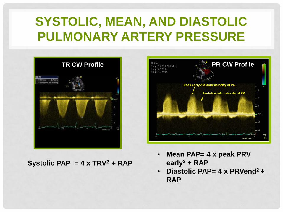
SYSTOLIC, MEAN, AND DIASTOLIC
PULMONARY ARTERY PRESSURE
• Mean PAP= 4 x peak PRV
early2 + RAP
• Diastolic PAP= 4 x PRVend2 +
RAP
Systolic PAP = 4 x TRV2 + RAP
TR CW Profile PR CW Profile
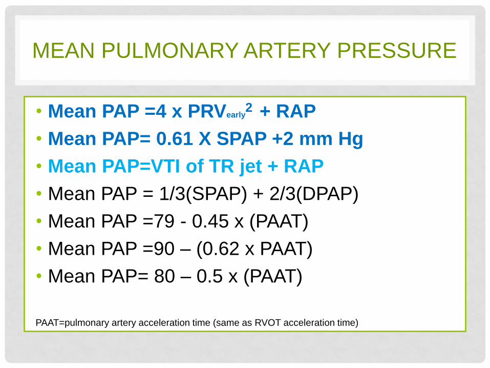
MEAN PULMONARY ARTERY PRESSURE
• Mean PAP =4 x PRVearly2 + RAP
• Mean PAP= 0.61 X SPAP +2 mm Hg
• Mean PAP=VTI of TR jet + RAP
• Mean PAP = 1/3(SPAP) + 2/3(DPAP)
• Mean PAP =79 - 0.45 x (PAAT)
• Mean PAP =90 – (0.62 x PAAT)
• Mean PAP= 80 – 0.5 x (PAAT)
PAAT=pulmonary artery acceleration time (same as RVOT acceleration time)
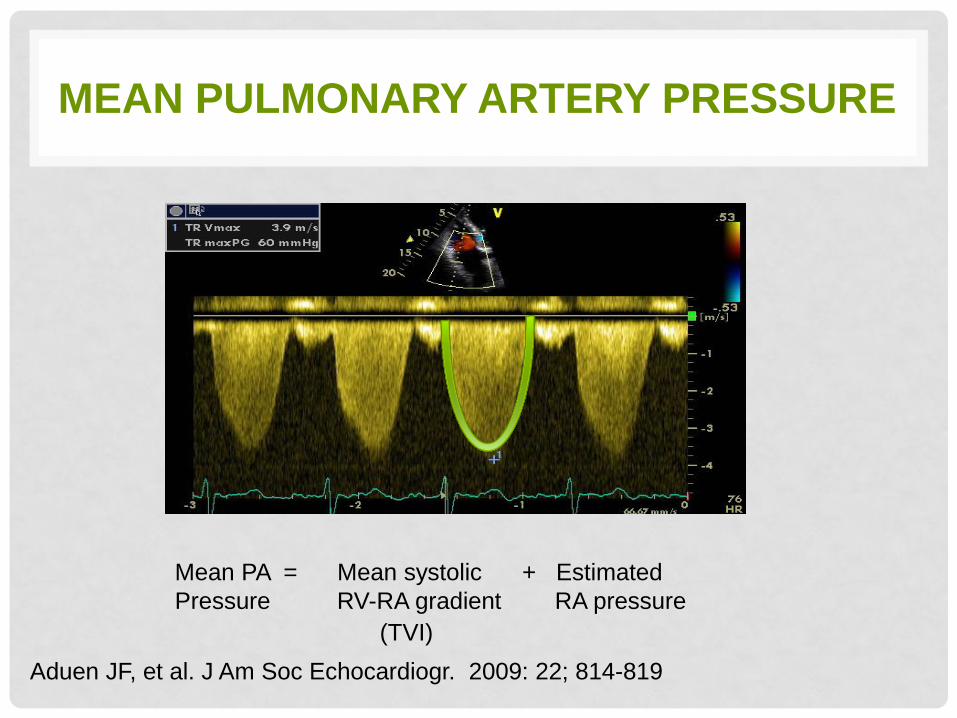
MEAN PULMONARY ARTERY PRESSURE
Mean PA = Mean systolic + Estimated
Pressure RV-RA gradient RA pressure
Aduen JF, et al. J Am Soc Echocardiogr. 2009: 22; 814-819
(TVI)
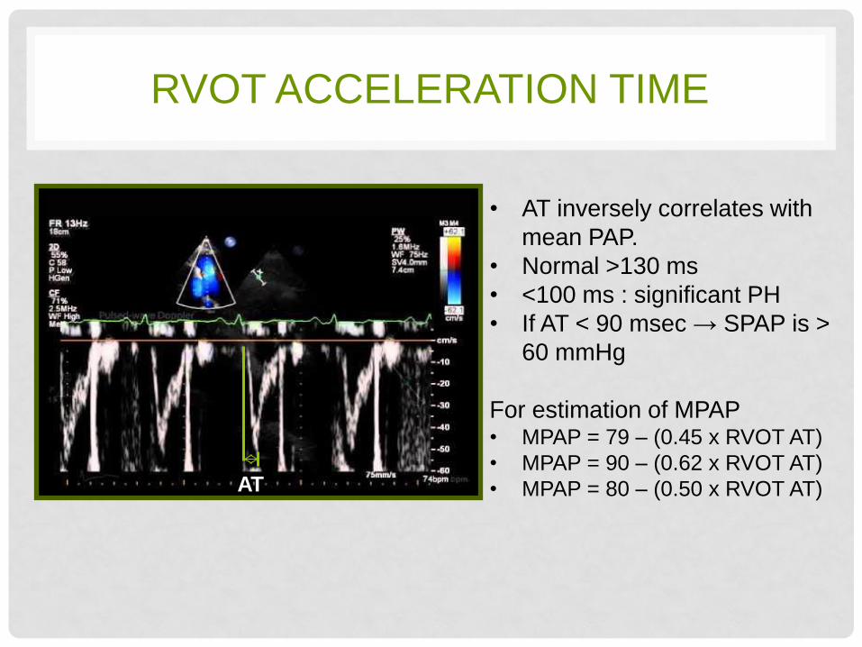
RVOT ACCELERATION TIME
AT
• AT inversely correlates with
mean PAP.
• Normal >130 ms
• <100 ms : significant PH
• If AT < 90 msec → SPAP is >
60 mmHg
For estimation of MPAP • MPAP = 79 – (0.45 x RVOT AT)
• MPAP = 90 – (0.62 x RVOT AT)
• MPAP = 80 – (0.50 x RVOT AT)
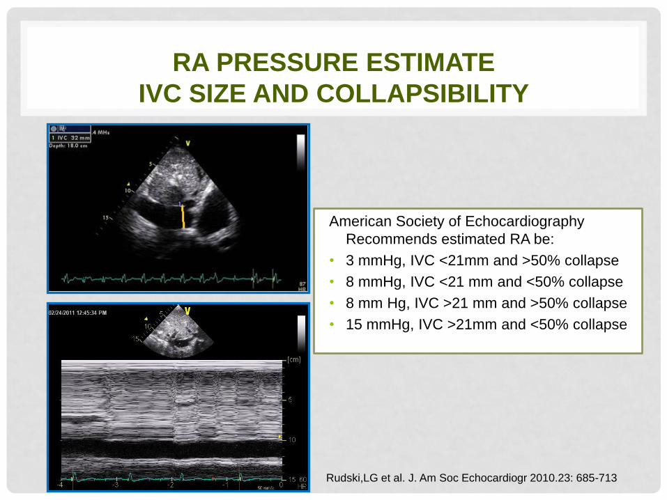
RA PRESSURE ESTIMATE
IVC SIZE AND COLLAPSIBILITY
American Society of Echocardiography
Recommends estimated RA be:
• 3 mmHg, IVC <21mm and >50% collapse
• 8 mmHg, IVC <21 mm and <50% collapse
• 8 mm Hg, IVC >21 mm and >50% collapse
• 15 mmHg, IVC >21mm and <50% collapse
Rudski,LG et al. J. Am Soc Echocardiogr 2010.23: 685-713
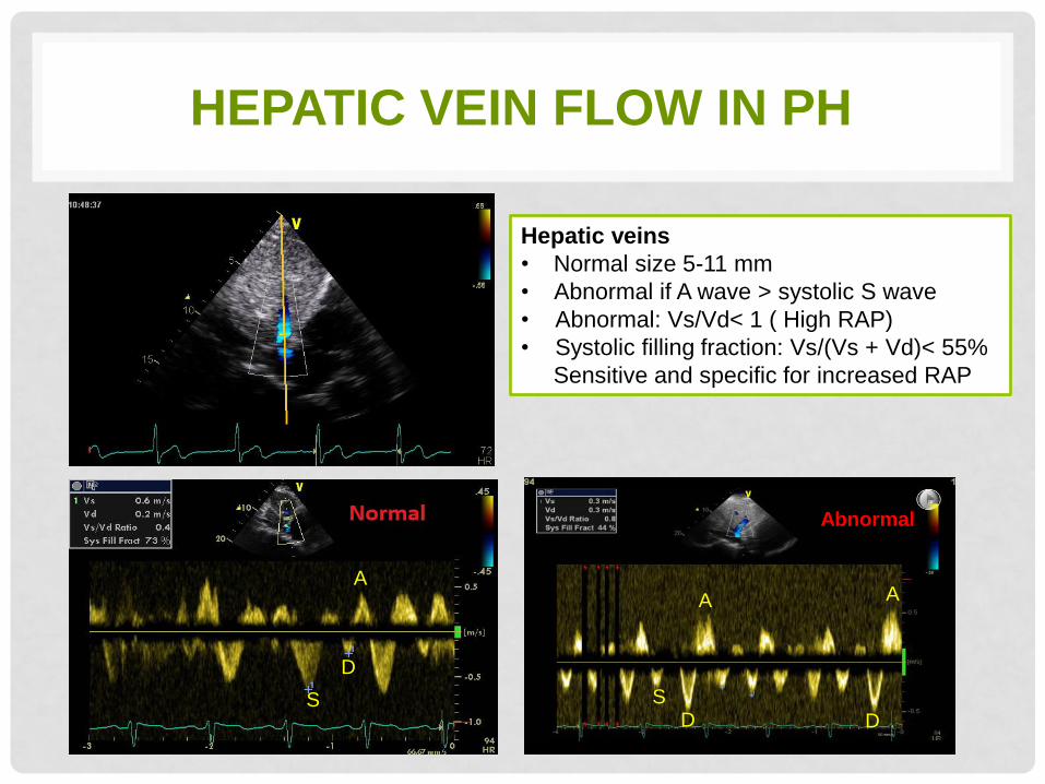
HEPATIC VEIN FLOW IN PH
Hepatic veins
• Normal size 5-11 mm
• Abnormal if A wave > systolic S wave
• Abnormal: Vs/Vd< 1 ( High RAP)
• Systolic filling fraction: Vs/(Vs + Vd)< 55%
Sensitive and specific for increased RAP
S
A
D
A A
D D
S
Abnormal
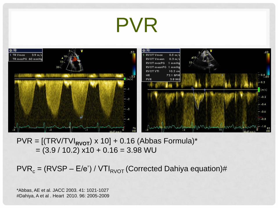
PVR
PVR = [(TRV/TVIRVOT) x 10] + 0.16 (Abbas Formula)*
= (3.9 / 10.2) x10 + 0.16 = 3.98 WU
PVRc = (RVSP – E/e’) / VTIRVOT (Corrected Dahiya equation)#
*Abbas, AE et al. JACC 2003. 41: 1021-1027
#Dahiya, A et al . Heart 2010. 96: 2005-2009

RV IMPACT
ECHO ASSESSMENT
US/DS/MAR11/001
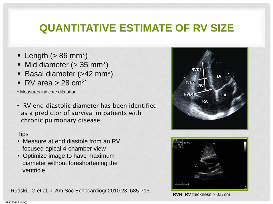
QUANTITATIVE ESTIMATE OF RV SIZE
Length (> 86 mm*)
Mid diameter (> 35 mm*)
Basal diameter (>42 mm*)
RV area > 28 cm2*
* Measures indicate dilatation
Tips
• Measure at end diastole from an RV
focused apical 4-chamber view
• Optimize image to have maximum
diameter without foreshortening the
ventricle
Rudski,LG et al. J. Am Soc Echocardiogr 2010.23: 685-713
RVH: RV thickness > 0.5 cm
• RV end-diastolic diameter has been identified
as a predictor of survival in patients with
chronic pulmonary disease
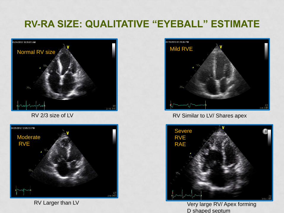
RV-RA SIZE: QUALITATIVE “EYEBALL” ESTIMATE
Severe
RVE
RAE
Mild RVE
Moderate
RVE
RV 2/3 size of LV
Very large RV/ Apex forming
D shaped septum
Normal
RV Similar to LV/ Shares apex
RV Larger than LV
Normal RV size
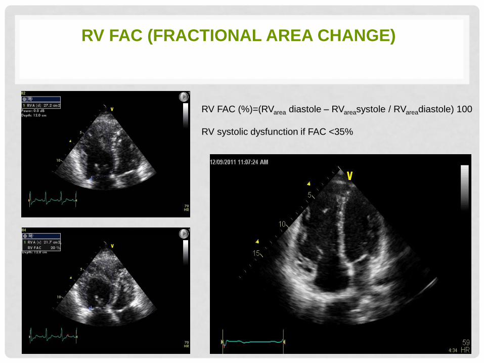
RV FAC (FRACTIONAL AREA CHANGE)
RV FAC (%)=(RVarea diastole – RVareasystole / RVareadiastole) 100
RV systolic dysfunction if FAC <35%
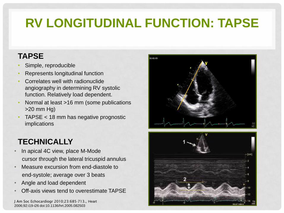
TAPSE • Simple, reproducible
• Represents longitudinal function
• Correlates well with radionuclide
angiography in determining RV systolic
function. Relatively load dependent.
• Normal at least >16 mm (some publications
>20 mm Hg)
• TAPSE < 18 mm has negative prognostic
implications
TECHNICALLY • In apical 4C view, place M-Mode
cursor through the lateral tricuspid annulus
• Measure excursion from end-diastole to
end-systole; average over 3 beats
• Angle and load dependent
• Off-axis views tend to overestimate TAPSE
RV LONGITUDINAL FUNCTION: TAPSE
J Am Soc Echocardiogr 2010;23:685-713., Heart
2006;92:i19-i26 doi:10.1136/hrt.2005.082503
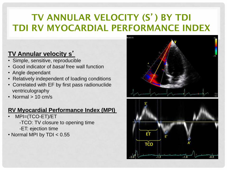
TV ANNULAR VELOCITY (S’) BY TDI
TDI RV MYOCARDIAL PERFORMANCE INDEX
TV Annular velocity s’ • Simple, sensitive, reproducible
• Good indicator of basal free wall function
• Angle dependant
• Relatively independent of loading conditions
• Correlated with EF by first pass radionuclide
ventriculography
• Normal > 10 cm/s
RV Myocardial Performance Index (MPI) • MPI=(TCO-ET)/ET
-TCO: TV closure to opening time
-ET: ejection time
• Normal MPI by TDI < 0.55
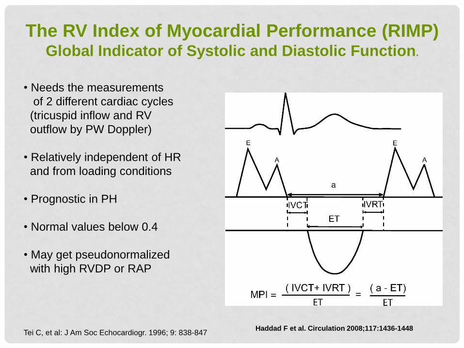
The RV Index of Myocardial Performance (RIMP) Global Indicator of Systolic and Diastolic Function.
Haddad F et al. Circulation 2008;117:1436-1448
• Needs the measurements
of 2 different cardiac cycles
(tricuspid inflow and RV
outflow by PW Doppler)
• Relatively independent of HR
and from loading conditions
• Prognostic in PH
• Normal values below 0.4
• May get pseudonormalized
with high RVDP or RAP
Tei C, et al: J Am Soc Echocardiogr. 1996; 9: 838-847
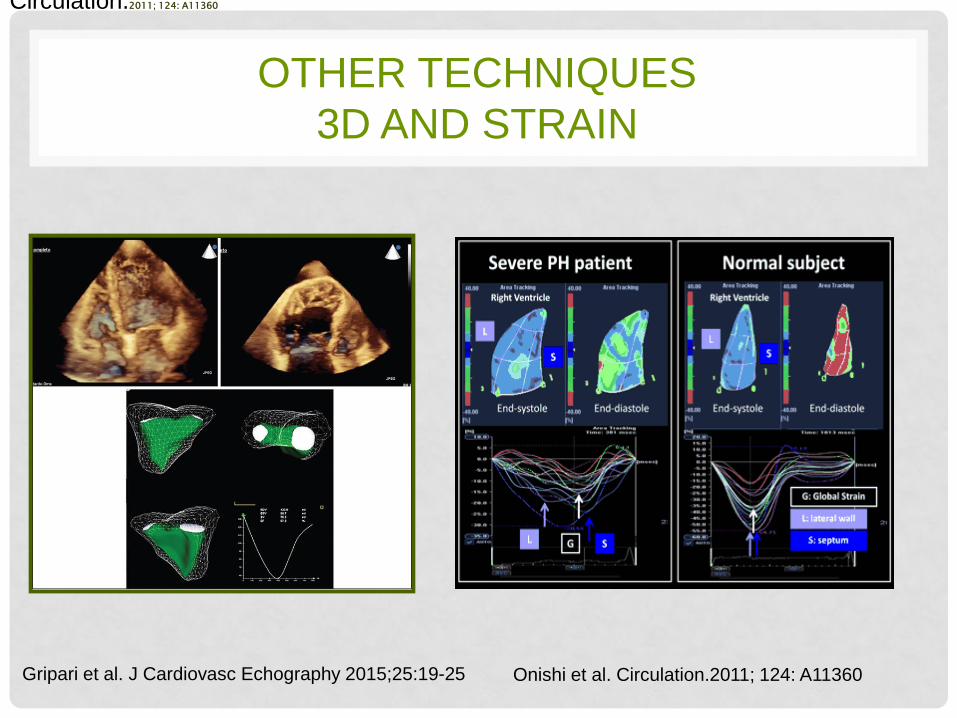
OTHER TECHNIQUES
3D AND STRAIN
Gripari et al. J Cardiovasc Echography 2015;25:19-25
Circulation.2011; 124: A11360
Onishi et al. Circulation.2011; 124: A11360
INDIRECT SIGNS OF
PULMONARY HYPERTENSION
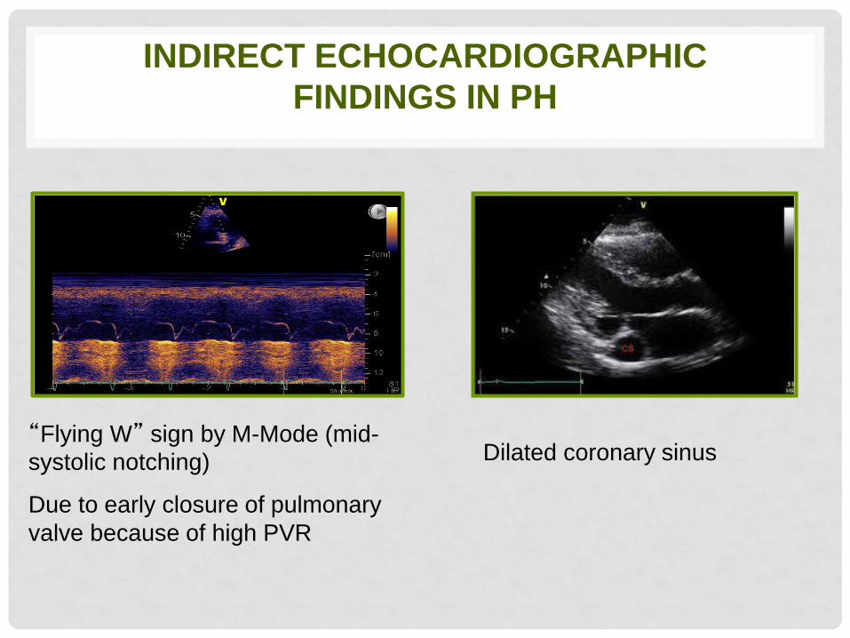
INDIRECT ECHOCARDIOGRAPHIC
FINDINGS IN PH
“Flying W” sign by M-Mode (mid-
systolic notching)
Due to early closure of pulmonary
valve because of high PVR
Dilated coronary sinus
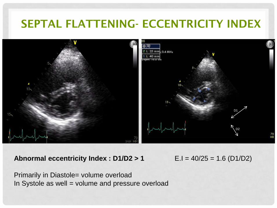
SEPTAL FLATTENING- ECCENTRICITY INDEX
E.I = 40/25 = 1.6 (D1/D2)
D1
D2
Abnormal eccentricity Index : D1/D2 > 1
Primarily in Diastole= volume overload
In Systole as well = volume and pressure overload
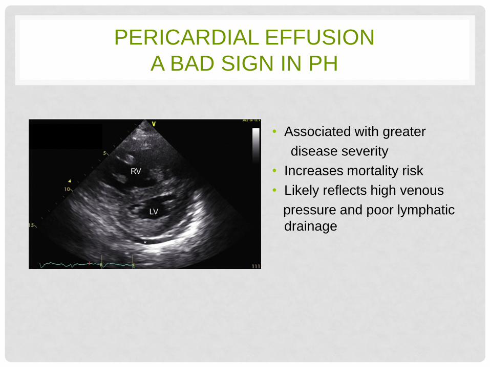
PERICARDIAL EFFUSION
A BAD SIGN IN PH
• Associated with greater
disease severity
• Increases mortality risk
• Likely reflects high venous
pressure and poor lymphatic
drainage
DETERMINANTS OF PROGNOSIS
IN PAH
39
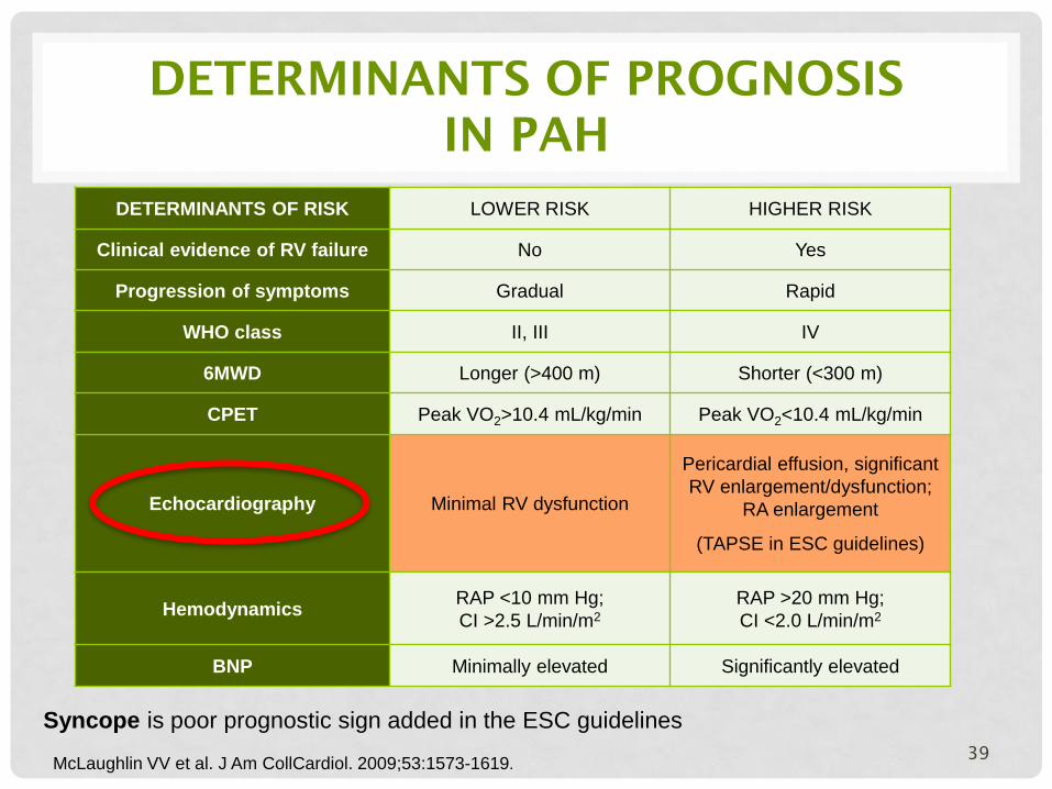
DETERMINANTS OF RISK LOWER RISK HIGHER RISK
Clinical evidence of RV failure No Yes
Progression of symptoms Gradual Rapid
WHO class II, III IV
6MWD Longer (>400 m) Shorter (<300 m)
CPET Peak VO2>10.4 mL/kg/min Peak VO2<10.4 mL/kg/min
Echocardiography Minimal RV dysfunction
Pericardial effusion, significant
RV enlargement/dysfunction;
RA enlargement
(TAPSE in ESC guidelines)
Hemodynamics RAP <10 mm Hg;
CI >2.5 L/min/m2
RAP >20 mm Hg;
CI <2.0 L/min/m2
BNP Minimally elevated Significantly elevated
McLaughlin VV et al. J Am CollCardiol. 2009;53:1573-1619.
Syncope is poor prognostic sign added in the ESC guidelines
PROGNOSTIC VALUE OF ECHO PARAMETERS
IN PULMONARY HYPERTENSION
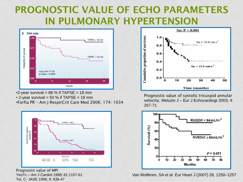
•2-year survival = 88 % if TAPSE > 18 mm
• 2-year survival = 50 % if TAPSE < 18 mm
•Forfia PR – Am J RespirCrit Care Med 2006; 174: 1034
Prognostic value of MPI
YeoTc – Am J Cardiol 1998; 81:1157-61
Tei, C- JASE 1996; 9: 838-47
Prognostic value of systolic tricuspid annular
velocity. Meluzin J – Eur J Echocardiogr 2003; 4:
267-71
Van Wolferen, SA et al. Eur Heart J (2007) 28, 1250–1257
ECHOCARDIOGRAPHIC PREDICTORS
OF OUTCOMES
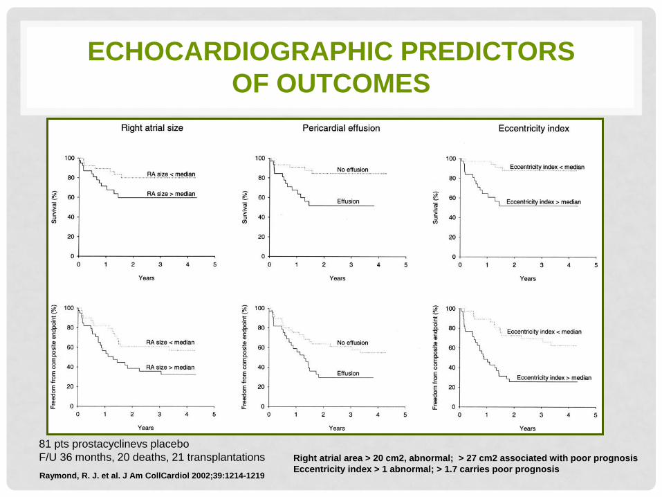
Raymond, R. J. et al. J Am CollCardiol 2002;39:1214-1219
Right atrial area > 20 cm2, abnormal; > 27 cm2 associated with poor prognosis
Eccentricity index > 1 abnormal; > 1.7 carries poor prognosis
81 pts prostacyclinevs placebo
F/U 36 months, 20 deaths, 21 transplantations
EXERCISE INDUCED PULMONARY
HYPERTENSION
• Slight increase in pulmonary pressures with
exercise appeared normal SPAP <40-45 mm Hg
• Athletes SPAP <55-60 mm Hg (due to
substantial increase in pulmonary flow)
• Competitive athletes who exercise at high levels
of CO and PAP because of an intrinsically steep
pressure-flow relationship may be exposed in
the long term to RV remodeling and subsequent arrhythmias
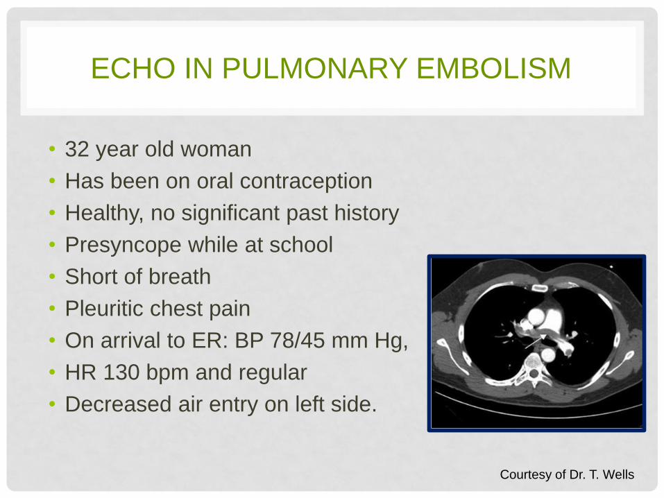
ECHO IN PULMONARY EMBOLISM
• 32 year old woman
• Has been on oral contraception
• Healthy, no significant past history
• Presyncope while at school
• Short of breath
• Pleuritic chest pain
• On arrival to ER: BP 78/45 mm Hg,
• HR 130 bpm and regular
• Decreased air entry on left side.
Courtesy of Dr. T. Wells
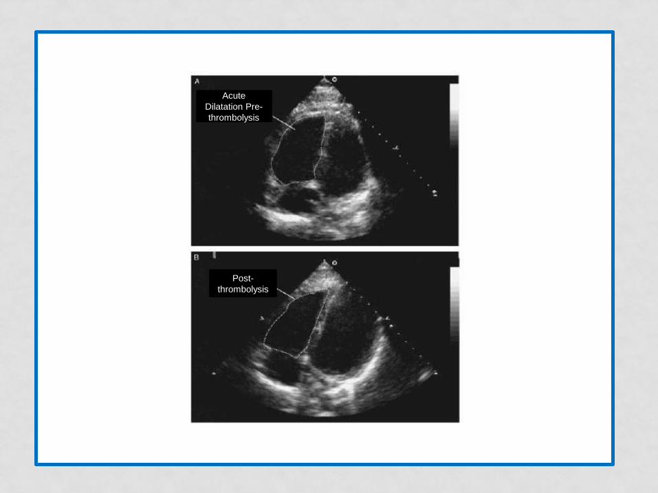
Acute
Dilatation Pre-
thrombolysis
Post-
thrombolysis
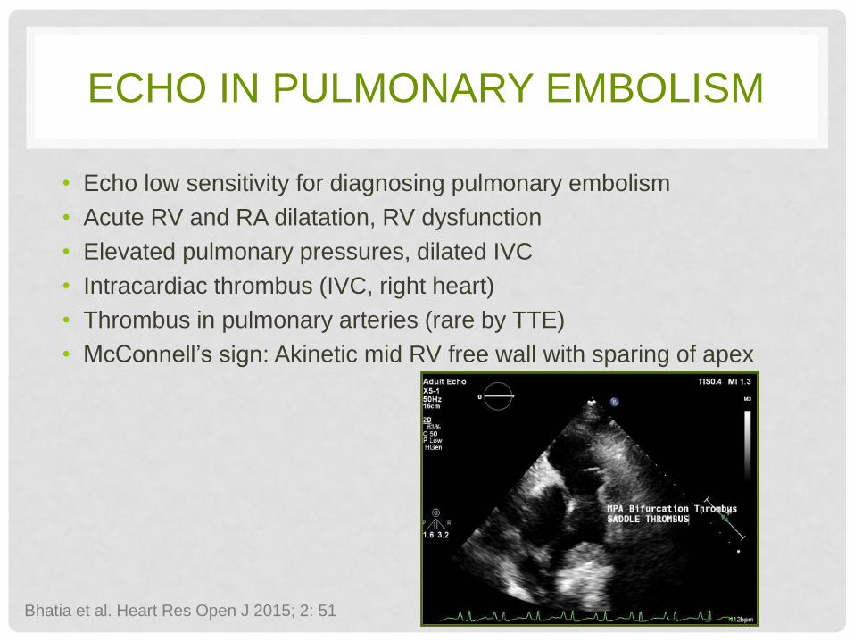
ECHO IN PULMONARY EMBOLISM
• Echo low sensitivity for diagnosing pulmonary embolism
• Acute RV and RA dilatation, RV dysfunction
• Elevated pulmonary pressures, dilated IVC
• Intracardiac thrombus (IVC, right heart)
• Thrombus in pulmonary arteries (rare by TTE)
• McConnell’s sign: Akinetic mid RV free wall with sparing of apex
Bhatia et al. Heart Res Open J 2015; 2: 51
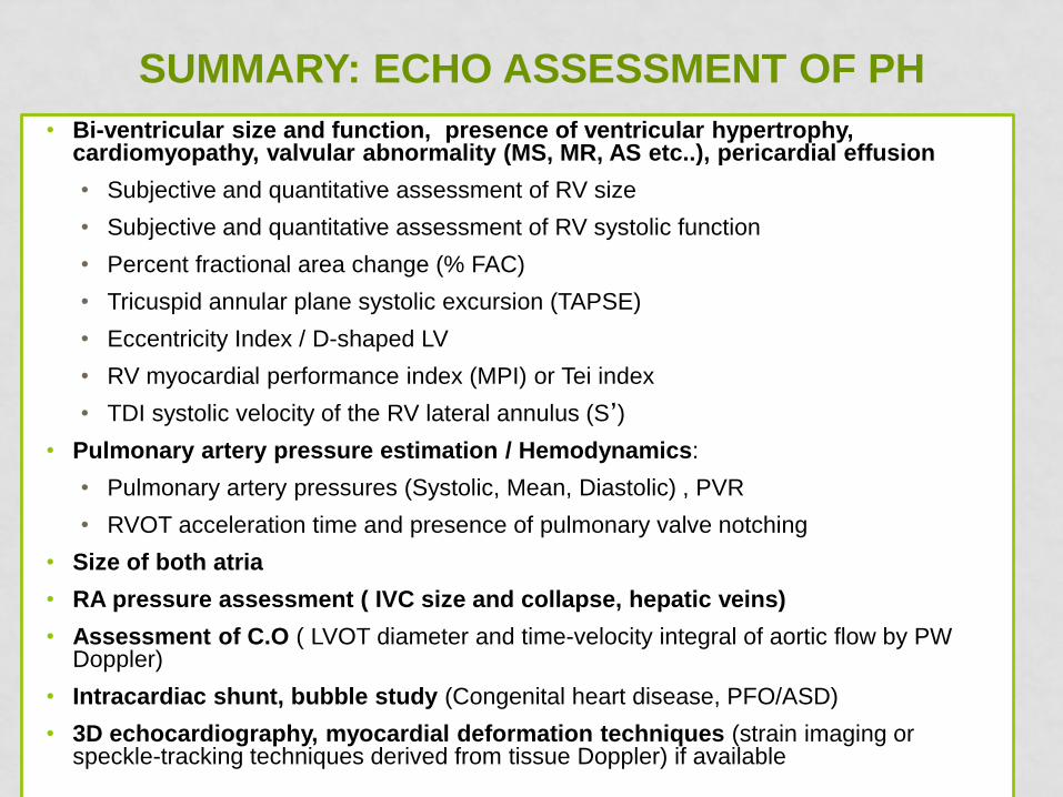
SUMMARY: ECHO ASSESSMENT OF PH
• Bi-ventricular size and function, presence of ventricular hypertrophy, cardiomyopathy, valvular abnormality (MS, MR, AS etc..), pericardial effusion
• Subjective and quantitative assessment of RV size
• Subjective and quantitative assessment of RV systolic function
• Percent fractional area change (% FAC)
• Tricuspid annular plane systolic excursion (TAPSE)
• Eccentricity Index / D-shaped LV
• RV myocardial performance index (MPI) or Tei index
• TDI systolic velocity of the RV lateral annulus (S’)
• Pulmonary artery pressure estimation / Hemodynamics:
• Pulmonary artery pressures (Systolic, Mean, Diastolic) , PVR
• RVOT acceleration time and presence of pulmonary valve notching
• Size of both atria
• RA pressure assessment ( IVC size and collapse, hepatic veins)
• Assessment of C.O ( LVOT diameter and time-velocity integral of aortic flow by PW Doppler)
• Intracardiac shunt, bubble study (Congenital heart disease, PFO/ASD)
• 3D echocardiography, myocardial deformation techniques (strain imaging or speckle-tracking techniques derived from tissue Doppler) if available
SUMMARY: ROLE OF ECHO IN PH
Echo: Key for initial diagnosis and follow up
evaluation of a patient with PH.
Despite its limitations, it is the most clinically
useful noninvasive test for the assessment of the
pulmonary circulation
Echo is reasonably accurate estimate of the RV,
and PA pressures and hemodynamics.
Echo is important for prognostication, monitor
progression and response to therapy,
independent of clinical and right heart cath data