Embed Size (px)
Citation preview
Pulmonary Patterns
VMA 976
PULMONARY PATTERNS
Which pulmonary patterns are commonly described in veterinary medicine?
PULMONARY PATTERNSNormalAlveolarInterstitial
Structured/NodularUnstructured
Bronchial“Mixed”
BronchointerstitialVascular
NORMAL PATTERNCan be the most difficult pattern to identify!Normal is NOT synonymous with UNSTRUCTURED INTERSTITIALUNSTRUCTURED INTERSTITIAL !!!!!!
Patient caseload at NCSU-CVM makes unstructured interstitial a common pattern but it is NOT normal
Normal means that there is no radiographic evidence of pulmonary pathology
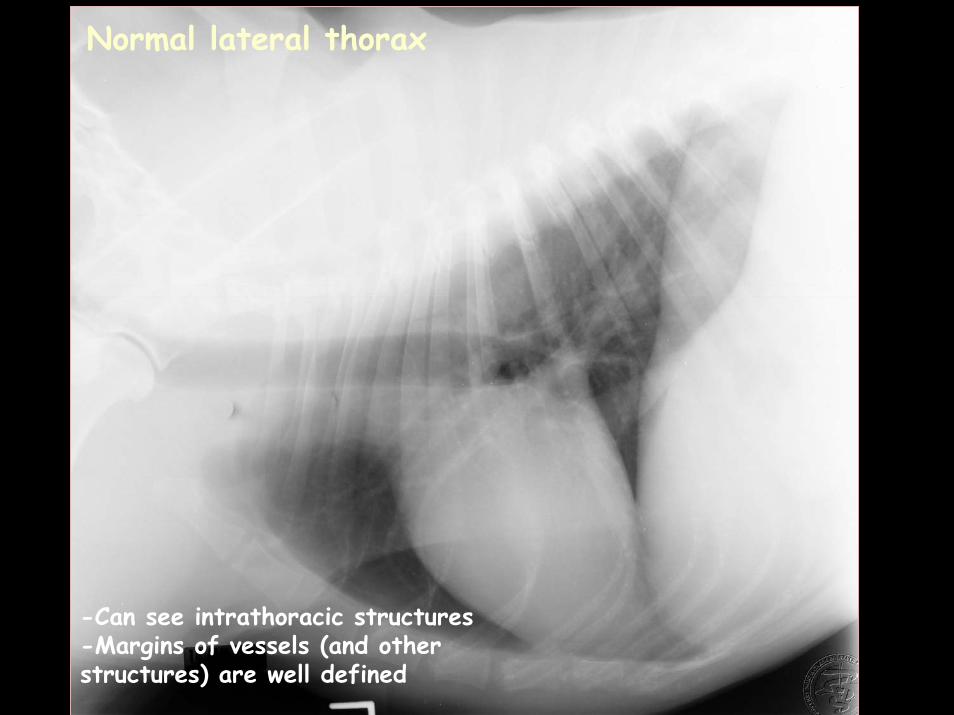
Normal lateral thorax
-Can see intrathoracic structures-Margins of vessels (and other structures) are well defined
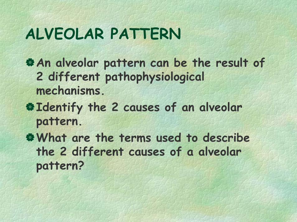
ALVEOLAR PATTERN
Radiographic SignsWhat are 4 radiographic signs of an alveolar pattern?
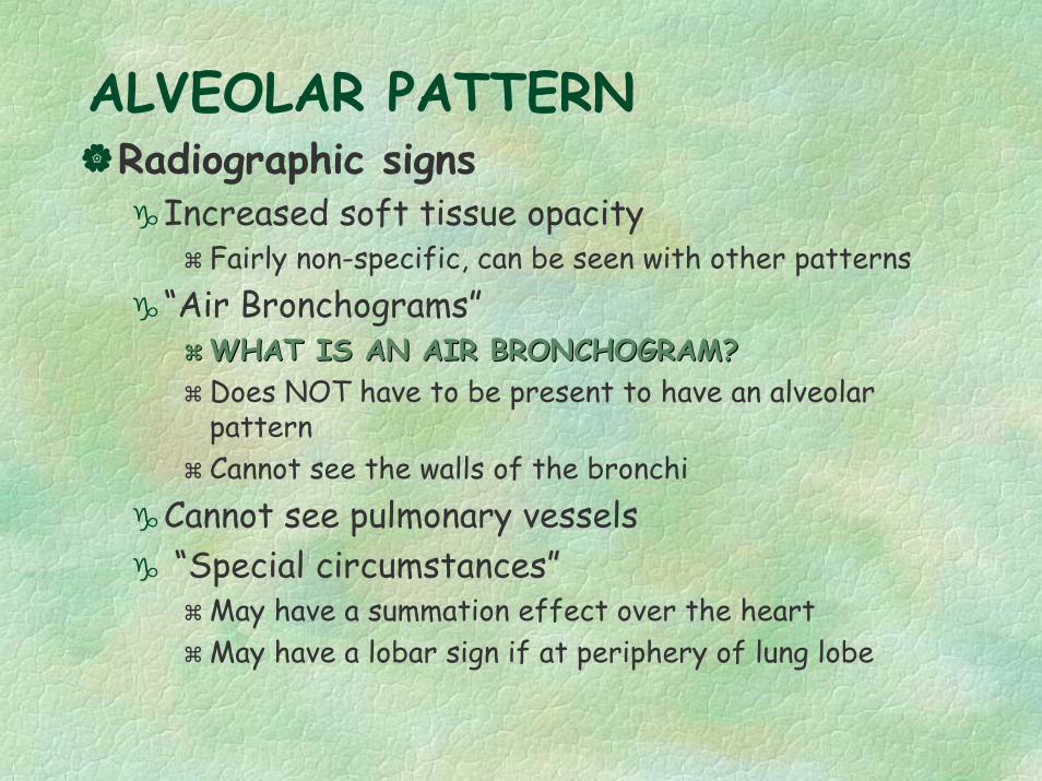
ALVEOLAR PATTERNRadiographic signs
Increased soft tissue opacityFairly non-specific, can be seen with other patterns
“Air Bronchograms”WHAT IS AN AIR BRONCHOGRAM?WHAT IS AN AIR BRONCHOGRAM?Does NOT have to be present to have an alveolar patternCannot see the walls of the bronchi
Cannot see pulmonary vessels“Special circumstances”
May have a summation effect over the heartMay have a lobar sign if at periphery of lung lobe
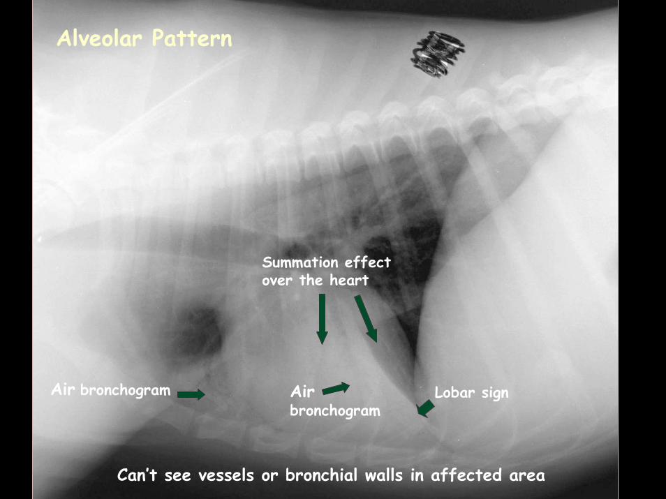
Lobar sign
Summation effect over the heart
Air bronchogram Airbronchogram
Can’t see vessels or bronchial walls in affected area
Alveolar Pattern
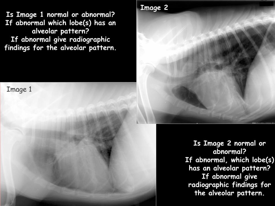
Image 2
Image 1
Is Image 1 normal or abnormal?If abnormal which lobe(s) has an
alveolar pattern?If abnormal give radiographic
findings for the alveolar pattern.
Image 1
Is Image 2 normal or abnormal?
If abnormal, which lobe(s) has an alveolar pattern?
If abnormal give radiographic findings for
the alveolar pattern.
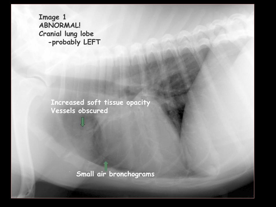
Image 1 ABNORMAL!Cranial lung lobe
-probably LEFT
Small air bronchograms
Increased soft tissue opacityVessels obscured
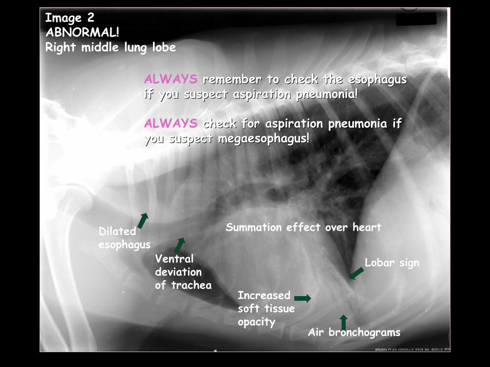
Image 2ABNORMAL!Right middle lung lobe
Dilated esophagus
Ventral deviation of trachea
Lobar sign
ALWAYS remember to check the esophagus remember to check the esophagus if you suspect aspiration pneumonia!if you suspect aspiration pneumonia!
ALWAYS check for aspiration pneumonia if check for aspiration pneumonia if you suspect you suspect megaesophagusmegaesophagus!!
Air bronchograms
Summation effect over heart
Increased soft tissue opacity
ALVEOLAR PATTERN
An alveolar pattern can be the result of 2 different pathophysiological mechanisms. Identify the 2 causes of an alveolar pattern. What are the terms used to describe the 2 different causes of a alveolar pattern?
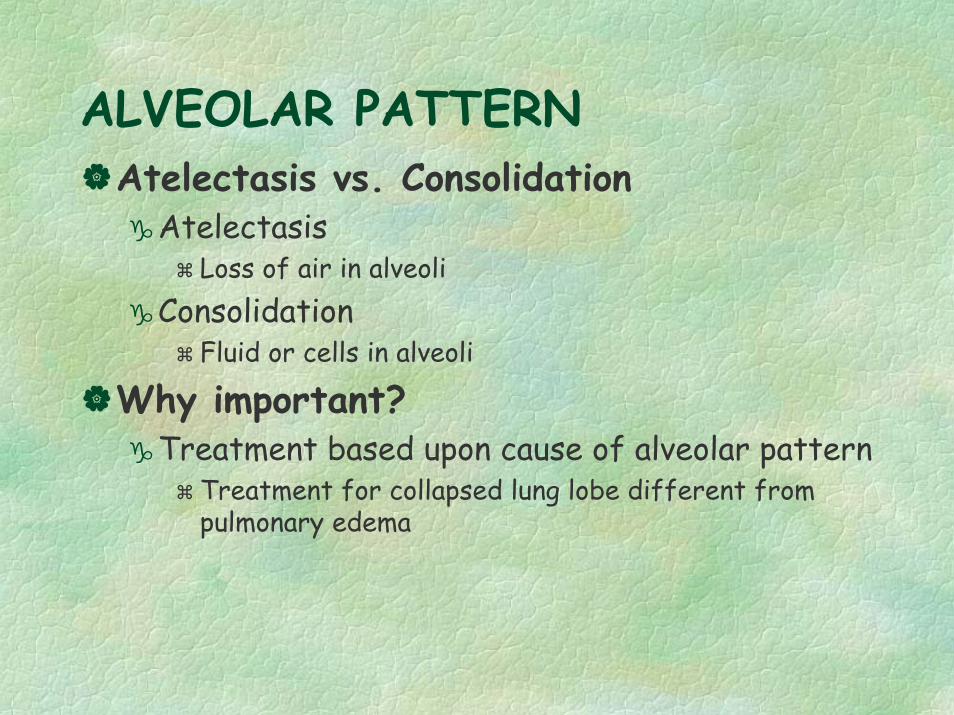
ALVEOLAR PATTERN Atelectasis vs. Consolidation
AtelectasisLoss of air in alveoli
ConsolidationFluid or cells in alveoli
Why important?Treatment based upon cause of alveolar pattern
Treatment for collapsed lung lobe different from pulmonary edema
ALVEOLAR PATTERN
What are the radiographic signs of atelectasis vs consolidation?
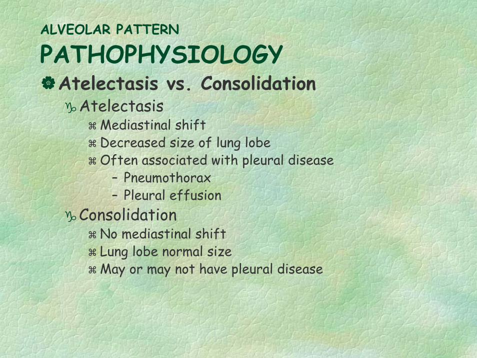
ALVEOLAR PATTERN
PATHOPHYSIOLOGYAtelectasis vs. Consolidation
AtelectasisMediastinal shiftDecreased size of lung lobeOften associated with pleural disease
– Pneumothorax– Pleural effusion
ConsolidationNo mediastinal shiftLung lobe normal sizeMay or may not have pleural disease
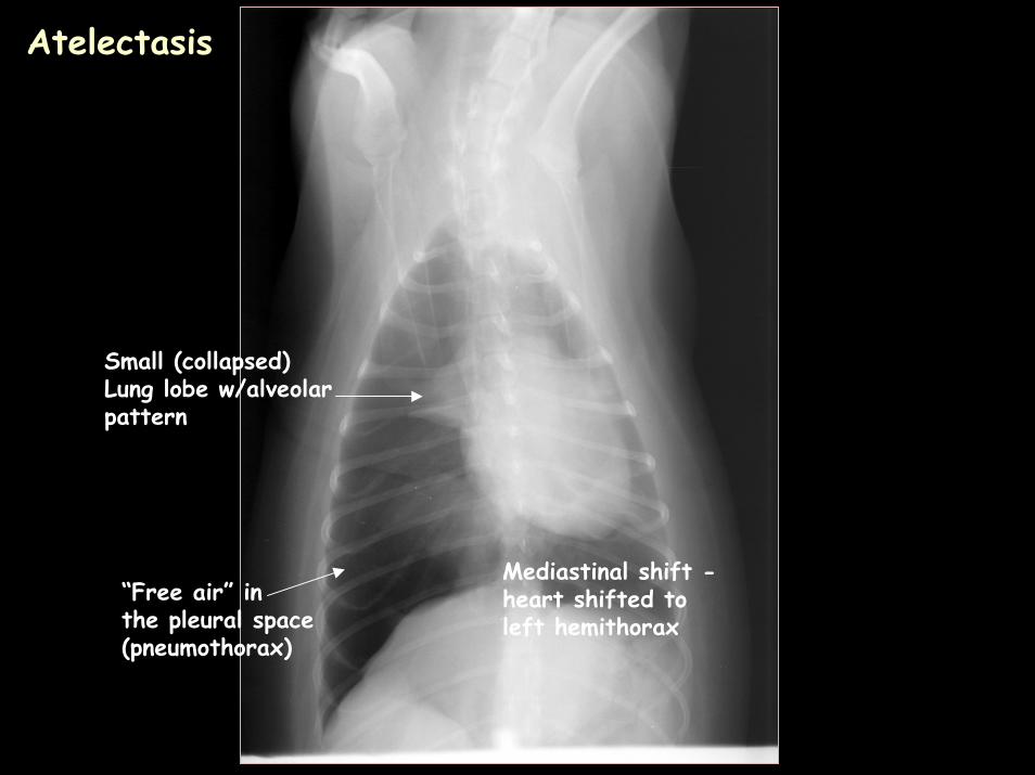
“Free air” in the pleural space(pneumothorax)
Small (collapsed)Lung lobe w/alveolarpattern
Mediastinal shift -heart shifted to left hemithorax
Atelectasis
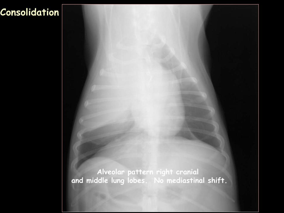
Alveolar pattern right cranial and middle lung lobes. No mediastinal shift.
Consolidation
ALVEOLAR PATTERN
List differentials for an alveolar patternInclude any specific distributions of the pattern that may influence making that differential designation
ALVEOLAR PATTERN
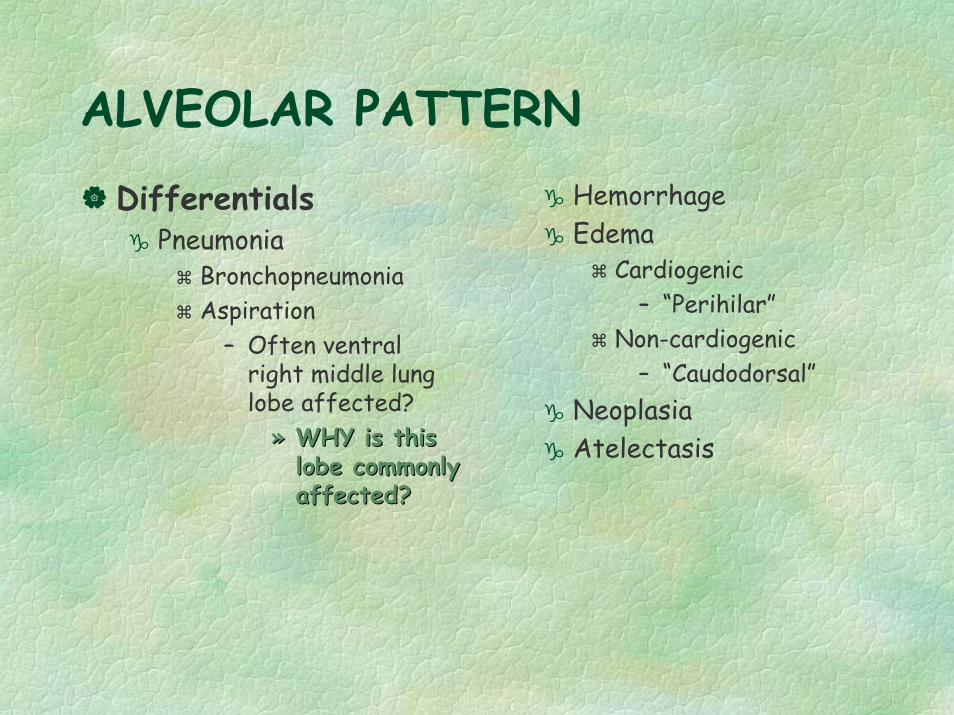
DifferentialsPneumonia
BronchopneumoniaAspiration
– Often ventral right middle lung lobe affected?
»» WHY is this WHY is this lobe commonly lobe commonly affected?
HemorrhageEdema
Cardiogenic– “Perihilar”
Non-cardiogenic– “Caudodorsal”
NeoplasiaAtelectasis
affected?

STRUCTURED INTERSTITIAL PATTERN
What are the 2 main categories for a structured interstitial pattern?
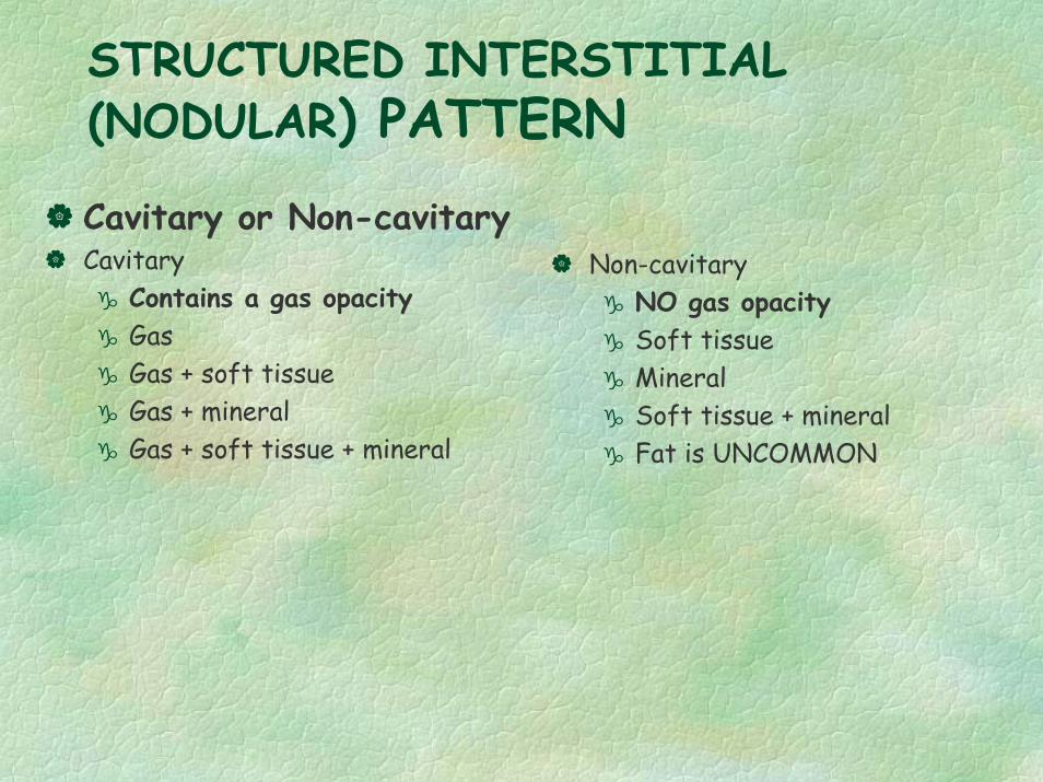
STRUCTURED INTERSTITIAL (NODULAR) PATTERN
Cavitary or Non-cavitaryCavitary
Contains a gas opacityGasGas + soft tissueGas + mineralGas + soft tissue + mineral
Non-cavitaryNO gas opacitySoft tissueMineralSoft tissue + mineralFat is UNCOMMON
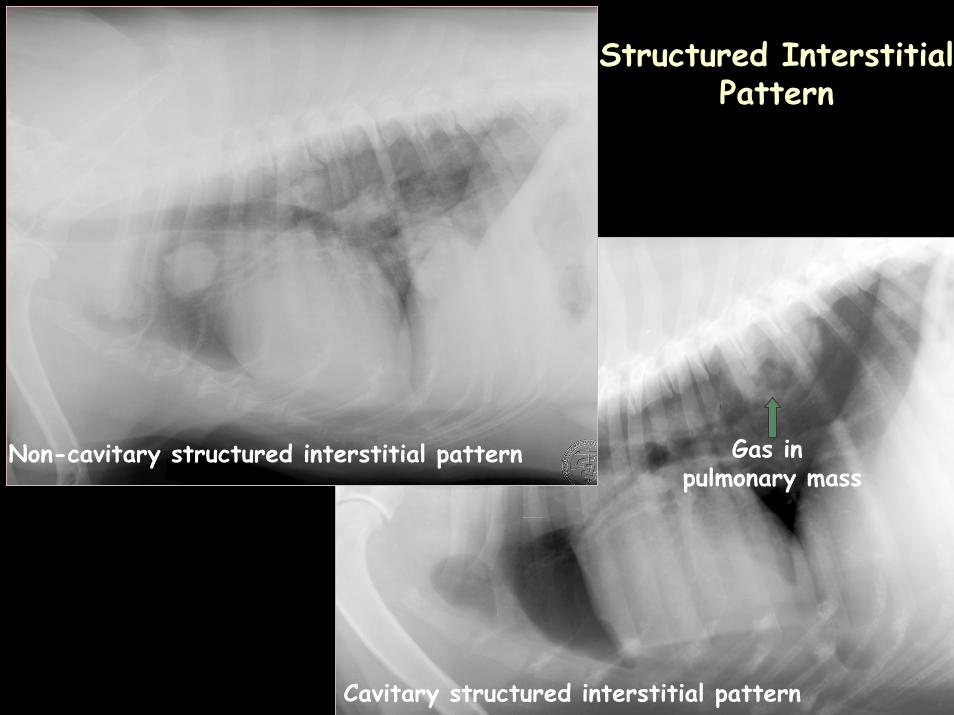
Non-cavitary structured interstitial pattern
Cavitary structured interstitial pattern
Gas in pulmonary mass
Structured Interstitial Pattern
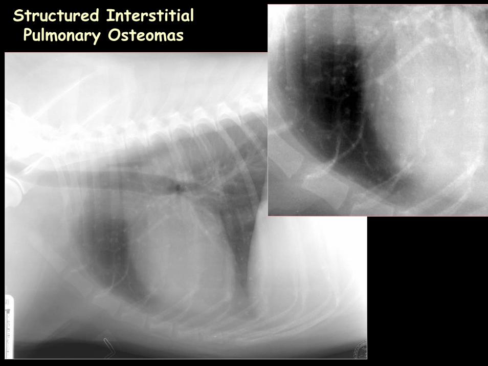
Structured InterstitialPulmonary Osteomas
STRUCTURED INTERSTITIAL PATTERN
How do you tell an end on vessel from a pulmonary nodule?
List 6 ways to identify an end on vessel
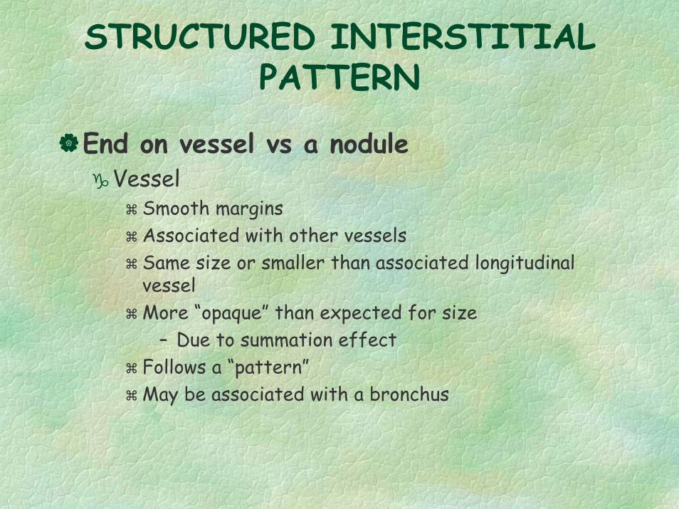
STRUCTURED INTERSTITIAL PATTERN
End on vessel vs a noduleVessel
Smooth marginsAssociated with other vesselsSame size or smaller than associated longitudinal vesselMore “opaque” than expected for size
– Due to summation effectFollows a “pattern”May be associated with a bronchus
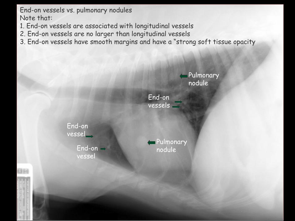
Pulmonary nodule
End-onvessel
End-onvessel
Pulmonary nodule
End-onvessels
End-on vessels vs. pulmonary nodulesNote that: 1. End-on vessels are associated with longitudinal vessels2. End-on vessels are no larger than longitudinal vessels3. End-on vessels have smooth margins and have a “strong soft tissue opacity
STRUCTURED INTERSTITIAL PATTERN
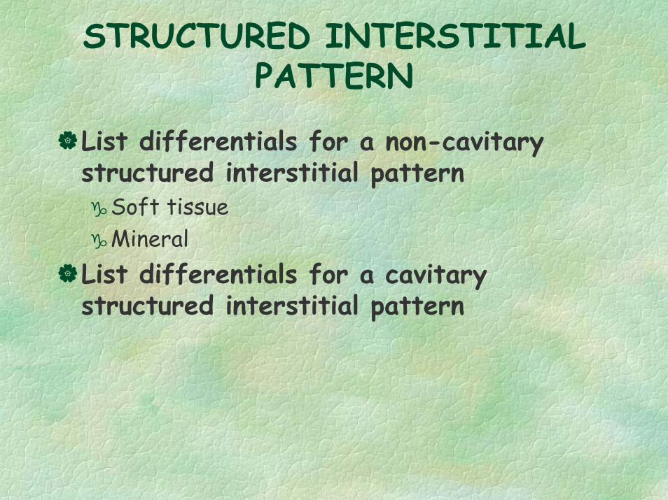
List differentials for a non-cavitary structured interstitial pattern
Soft tissueMineral
List differentials for a cavitary structured interstitial pattern
STRUCTURED INTERSTITIAL PATTERN
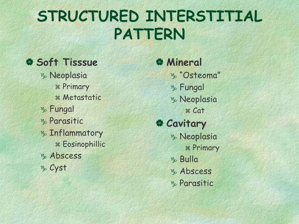
Soft TisssueNeoplasia
PrimaryMetastatic
FungalParasiticInflammatory
EosinophillicAbscessCyst
Mineral“Osteoma”FungalNeoplasia
Cat
CavitaryNeoplasia
PrimaryBullaAbscessParasitic
UNSTRUCTURED INTERSTITIAL PATTERN
What technical factors can influence the determination of the presence of an unstructured interstitial pattern?
UNSTRUCTURED INTERSTITIAL PATTERN
Technical FactorsPhase of respiration
Expiratory– Heart & diaphragm are in close contact
Fat animalMotionUnderexposure
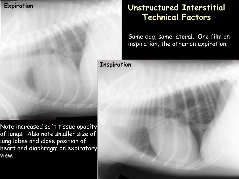
Expiration
Inspiration
Same dog, same lateral. One film on inspiration, the other on expiration.
Note increased soft tissue opacityof lungs. Also note smaller size oflung lobes and close position of heart and diaphragm on expiratoryview.
Unstructured InterstitialTechnical Factors
UNSTRUCTURED INTERSTITIAL PATTERN
Radiographic signsWhat are 4 radiographic signs of an unstructured interstitial pattern?
UNSTRUCTURED INTERSTITIAL PATTERN
Radiographic signsIncreased soft tissue opacityFuzzy, hazy, lacy appearanceCan still see vesselsPeribronchial enhancement
Margins of bronchi are easier to see because of increased soft tissue opacity between bronchus and alveoli
Similar to looking through a screenCan see all structures but a fine pattern between you and the outside
UNSTRUCTURED INTERSTITIAL PATTERN
What is the pathophysiology behind an unstructured interstitial pattern?
“Active”“Old dog”
UNSTRUCTURED INTERSTITIAL PATTERN
PathophysiologyFluid, cells or fibrosis in the interstitial space“Active” vs “Old dog”Active
Fluid or cells in the interstitial space– Tends to look more “fuzzy”
Old dogFibrosis in the interstitial space
– Tends to look more linear
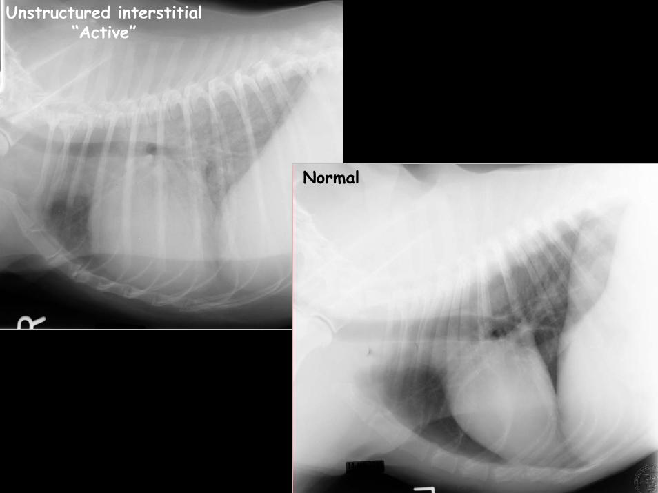
Normal
Unstructured interstitial“Active”
BRONCHIAL PATTERN
Radiographic SignsName 4 radiographic signs of a bronchial pattern
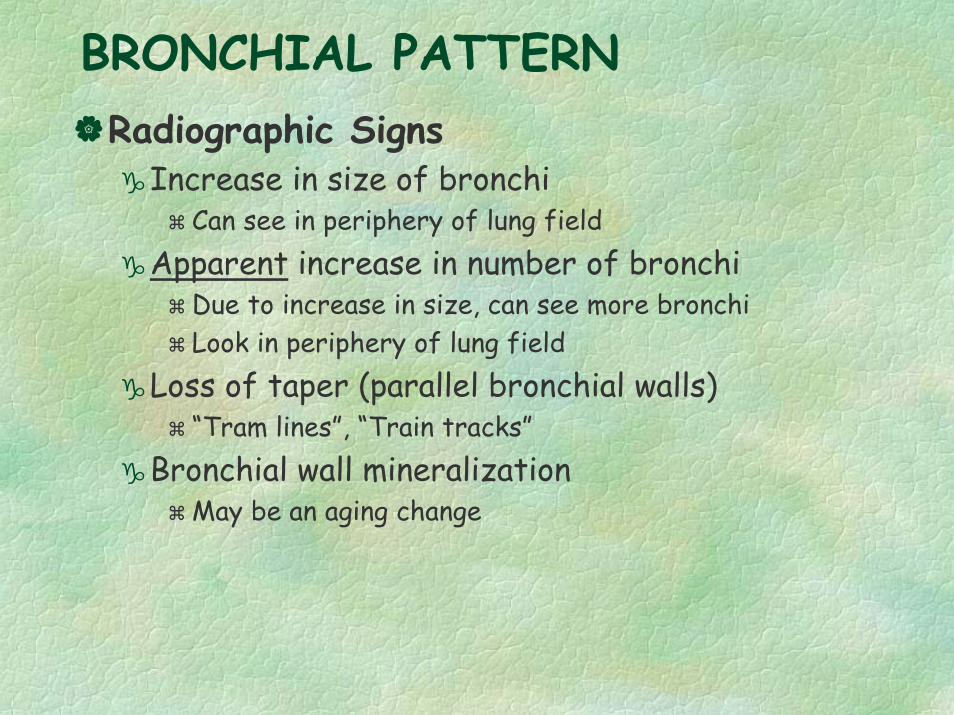
BRONCHIAL PATTERNRadiographic Signs
Increase in size of bronchiCan see in periphery of lung field
Apparent increase in number of bronchiDue to increase in size, can see more bronchiLook in periphery of lung field
Loss of taper (parallel bronchial walls)“Tram lines”, “Train tracks”
Bronchial wall mineralizationMay be an aging change
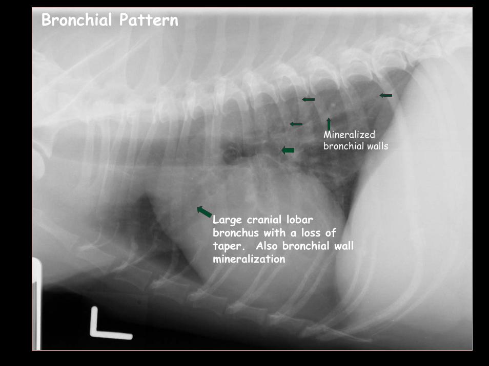
Large cranial lobar bronchus with a loss of taper. Also bronchial wall mineralization
Mineralized bronchial walls
Bronchial Pattern