Embed Size (px)
Citation preview
JSM Dentistry
Cite this article: Theobaldo JD, de Oliveira Lima M, Vieira-Junior WF, do Carmo Aguiar Jordão Mainardi M, Ferraz LN, et al. (2016) Management and Treat-ment of Dentin Hypersensitivity not Associated with a Significant Loss of Tooth Structure. JSM Dent 4(4): 1074.
Central
*Corresponding authorFlávio Henrique Baggio Aguiar, Department of Restorative Dentistry, Piracicaba Dental School, P.O. BOX 52 - University of Campinas –UNICAMP; 13414-903, Piracicaba, SP, Brazil, Tel: 55 19 2101 5340; Email:
Submitted: 12 July 2016
Accepted: 08 Novemeber 2016
Published: 10 Novemeber 2016
ISSN: 2333-7133
Copyright© 2016 Baggio Aguiar et al.
OPEN ACCESS
Keywords•Dentin sensitivity•Toothpastes•Dentin•Lasers•Adhesives
Review Article
Management and Treatment of Dentin Hypersensitivity not Associated with a Significant Loss of Tooth StructureJéssica Dias Theobaldo, Michele de Oliveira Lima, Waldemir Francisco Vieira-Junior, Maria do Carmo Aguiar Jordão Mainardi, Laura Nobre Ferraz, Débora Alves Nunes Leite Lima, and Flávio Henrique Baggio Aguiar*Department of Restorative Dentistry, University of Campinas, Brazil
Abstract
Dentin hypersensitivity (DH) is viewed by individuals as an important health problem and it is reported by the patient as a sharp pain caused by different external stimuli in dentin exposure.
Objective: To describe the best approaches for DH treatment, mainly in cases with no clinically significant loss of tooth structure.
Review: Several different approaches (in office and at home) have been proposed to control DH, including root coverage surgery, lasers application, and toothpaste and desensitizer application. The current review explores these treatments, especially in relation to their efficacy, limitation and safety.
Conclusion: The association of at home and in office treatment must be performed. At home treatment promotes maintenance and biodisponibility of desensitizing agents in oral environment.
ABBREVIATIONSDH: Dentin Hypersensitivity
INTRODUCTIONDentin hypersensitivity (DH) is characterized by pain arising
from exposed dentin in response to thermal, evaporative, osmotic, tactile, or chemical stimulus [1,2]. Clinically, DH is described as a brief and sharp pain that affects one or multiple teeth simultaneously [3]. This exposure may be due to enamel loss on the cervical region, as well as gingival recession with cementum loss. Dentin exposure, loss of dental structure and DH is often related to noncarious lesions such as attrition, abrasion, and erosion and most of the time because of the association of these factors [4]. Regardless of dentin exposure etiology, there is a close relation between the outer environment and the odontoblastic cells caused by the opened dentin tubules [5] even in initial lesions.
Nowadays, the most widely accepted biological mechanism for DH is the hydrodynamic theory [6,7]. The theory asserts that the dentinal fluid flow induced by external stimulus may activate
pulpal nociceptor, resulting in pain [6,8-10]. These external stimuli increase outward fluid flow within tubules, inducing shear stress on the receptor nerves in the tubule, causing hypersensitivity [6,7]. In this direction, treatment protocols should be discussed (Table 1), and performed especially in the cases where the loss of structure is not huge but able to make the patient uncomfortable.
The best treatment for DH has been widely discussed, and a number of clinical protocols have been reported. Restorative materials are indicated when there is loss of dental structure [2]. In this way, glass ionomer, resin-reinforced glass ionomer cements, and resin composite are considered the best choices of materials, once they provide a physical barrier against stimulus from the outer environment, decreasing the fluid motion inside the dentin tubules [2].
Several treatment protocols for DH are managed before any significant loss of dental structure occurs, including root coverage surgery, NdYAG laser application, use of toothpaste, and in office desensitizer application. Treatment (at home or in
Baggio Aguiar et al. (2016)Email:
JSM Dent 4(4): 1074 (2016) 2/5
Central
office) depends on the etiology of the lesion, as well its size and symptoms. In this sense, this literature review aimed to explore and discuss the best therapeutic protocols to treat DH, mainly in the cases with no significant loss of dental structure.
METHODSA search in the databases Pub Med, Scielo, and MEDLINE was
conducted and limited to dental journals in English language, using the following search terms: dentin sensitivity, toothpastes, dentifrice, root coverage surgery, gingival surgery, dentin, lasers and adhesives. Titles, abstracts, and articles were reviewed, and the papers in accordance to scientific evidence were selected.
Root coverage surgery
Periodontal plastic surgeries such as root coverage have been used to treat DH caused by gingival recession [11,12] aimed at decreasing the exposed dentin. The principal objective of root coverage surgery is complete coverage of the defect in the cervical region, promoting good appearance and minimal probing depth after healing [13-15]. To predict the results of this procedure, the clinical conditions of the patient should be analyzed with caution. For the indication of root coverage procedure, it is very important to evaluate the height of the interdental periodontal support, including clinical attachment and alveolar bone levels [16]. Thus, the success of the root coverage surgery is directly related to the clinical conditions of each patient, and a limitation of this technique is that the recession of area initially protected with the surgery procedure can occur after some time, leading to dentin exposure again.
Laser application
Laser is a device that transforms light of different frequencies into a chromatic radiation in the infrared, ultraviolet, and visible regions. The waves in these phases are capable of mobilizing immense heat and power when focused at close range [17]. Low frequency lasers act on nerve transmission through the depolarization of C-afferent fibers [18]. It induces changes in the neural transmission inside the pulp rather than acting directly on the exposed dentin area. On other hand, high frequency laser can occlude dentin tubules by a fusion mechanism, It causes a dissolution and remineralization of hydroxyapatite crystals, forming a remineralized layer, which is responsible to eliminate/
decrease the DH for a long period of time [19,20], as it occludes the opened dentin tubules.
Studies regarding the application of diode lasers have shown that they act directly in the pulp, increasing the metabolic activity of odontoblastic cells that occludes the dentin tubules, and further increases the tertiary dentin formation [21,22]. However, the major limitation of the laser technique is the need of a skilled professional for the use of laser apparatus, in addition to its high treatment cost.
Toothpaste
Dentifrices are the first choice for the treatment of DH, especially in cases not associated with a significant loss of tooth structure. Toothpaste promotes maintenance of desensitizing agents in oral environment. Therefore, the present manuscript focuses on the different modes of action of dentifrices in treating DH, as reported below. All modes can decrease or eliminate the DH.
Disrupting the neural response to external stimulus: Potassium salts are the only dentifrice compounds capable of blocking the neural response, and the most common potassium salts used in dentifrices are potassium nitrate, potassium chloride, and potassium citrate. These salts reduce the excitability of pulp nerve fibers and their prolongations, thus blocking the neural response to painful stimulus [23,24].
Several studies have shown the therapeutic efficacy of potassium salts to reduce the DH [25-30]. It is a consensus that the desensitizing effect starts after 2 weeks of dentifrice use, with significant reduction within 4 to 8 weeks. However, the desensitizing effect stops as soon as the dentifrice use is interrupted. The treatment of DH using dentifrices is not indicated to patients with acute hypersensitivity, as it does not provide immediate and long-lasting relieve of pains.
Occlusion of open dentin tubules: The principle of occluding the exposed and open dentin tubules is quite interesting, once it blocks both the stimulus and symptoms. It can occlude the tubules and avoid the hydrodynamic mechanism inside the dentin tubules through two modes of action: 1) precipitation of a thin particle layer over the exposed dentin [31], which is provided by strontium- or stannous fluoride-containing dentifrices; 2) use
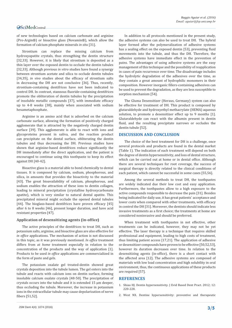
Table 1: Treatment approaches of dentin hypersensitivity and mechanisms of action.
Treatment Management Mechanism of Action
Pro-Argin® At home– dentifrice In office - paste Occlusion of dentin tubules by the precipitation of saliva ions and glycoprotein
Novamin® At home– dentifrice In office - paste Occlusion of dentin tubules by the precipitation of minerals rich in calcium and phosphate
Strontium salts At home– dentifrice Occlusion of dentin tubules by the precipitation of a thin mineral layer
Potassium salts At home – dentifrice In office - gel Celular depolarization blocking the neural response
Stannous fluoride At home– dentifrice Occlusion of dentin tubules by the precipitation of metallic ions
Oxalate In office Reaction with calcium ions present on dentin surface forming calcium oxalate insoluble crystals
Laser In office Nerve fibers depolarization and/or occlusion of dentin tubules
Root coverage surgery In office Coverage of exposed dentin with surrounding gingival
Adhesive system In office Seal the dentin tubules through the formed hybrid layer
Baggio Aguiar et al. (2016)Email:
JSM Dent 4(4): 1074 (2016) 3/5
Central
of new technologies based on calcium carbonate and arginine (Pro-Argin®) or bioactive glass (Novamin®), which allow the formation of calcium phosphate minerals in situ [31].
Strontium can replace the missing calcium from hydroxyapatite crystals, thus strengthing the dentin structure [32,33]. However, it is likely that strontium is deposited as a thin layer over the exposed dentin to occlude the dentin tubules [32,33]. Although previous in vitro studies have found a synergy between strontium acetate and silica to occlude dentin tubules [34,35], in vivo studies about the efficacy of strontium salts in decreasing the DH are not conclusive [36]. Thus, recently, strontium-containing dentifrices have not been indicated to control DH. In contrast, stannous fluoride-containing dentifrices promote the obliteration of dentin tubules by the precipitation of insoluble metallic compounds [37], with immediate efficacy up to 4-8 weeks [38], mainly when associated with sodium hexametaphosphate.
Arginine is an amino acid that is adsorbed on the calcium carbonate surface, allowing the formation of positively charged agglomerate that is attracted by the negatively charged dentin surface [39]. This agglomerate is able to react with ions and glycoproteins present in saliva, and the reaction product can precipitate on the dental surface, obliterating the dentin tubules and thus decreasing the DH. Previous studies have shown that arginine-based dentifrices reduce significantly the DH immediately after the direct application, and the patient is encouraged to continue using this toothpaste to keep its effect against DH [40-42].
Bioactive glass is a material able to bond chemically to dental tissues. It is composed by calcium, sodium, phosphorous, and silica, in amounts that provides the bioactivity to the material [43]. The great bioavailability of calcium, phosphorous, and sodium enables the attraction of these ions to dentin collagen, leading to mineral precipitation (crystalline hydroxycarbonate apatite), which is very similar to natural dental apatite. This precipitated mineral might occlude the opened dental tubules [44]. The bioglass-based dentifrices have proven efficacy [45] after 6 to 8 weeks [46], present longer duration, and have acid resistant properties [47].
Application of desensitizing agents (in-office)
The active principles of the dentifrices to treat DH, such as potassium salts, arginine, and bioactive glass are also effective for in office applications. The mechanism of action is not discussed in this topic, as it was previously mentioned. In office treatment differs from at home treatment especially in relation to the concentration of the products and the way of application [1]. Products to be used in office applications are commercialized in the form of paste and gels.
The potassium oxalate gel treated-dentin showed great crystals deposition into the tubule lumen. The gel enters into the tubule and reacts with calcium ions on dentin surface, forming insoluble calcium oxalate crystals [48-50]. The precipitation of crystals occurs into the tubule and it is extended 15 µm deeper, thus occluding the tubule. Moreover, the increase in potassium ions in the extracellular tubule may cause depolarization of nerve fibers [51,52].
In addition to all protocols mentioned in the present study, the adhesive systems can also be used to treat DH. The hybrid layer formed after the polymeralization of adhesive systems has a sealing effect on the exposed dentin [53], preventing fluid movements into the tubule, and thus the DH. Therefore, the adhesive systems have immediate effect in the prevention of pains. The advantages of using adhesive systems are the easy management of this technique and the possibility of reapplication in cases of pain recurrence over time. The disadvantage includes the hydrolytic degradation of the adhesives over the time, as they contain a great amount of hydrophilic monomers in their composition. However inorganic fillers-containing adhesives can be used to prevent the degradation, as they are less susceptible to sorption mechanism [54].
The Gluma Desensitizer (Heraus, Germany) system can also be effective for treatment of DH. This product is composed by glutaraldehyde and hydroxyethyl methacrylate (HEMA) aqueous solution, to promote a desensitizer effect up to 9 months [1]. Glutaraldehyde can react with the albumin present in dentin fluid, and the resulting precipitate narrows or occludes the dentin tubule [52].
DISCUSSION AND CONCLUSIONThe choice of the best treatment for DH is a challenge, once
several protocols and products are found in the dental market (Table 1). The indication of each treatment will depend on both the degree of dentin hypersensitivity, and loss of dental structure, which can be carried out at home or in dental office. Although there are several techniques for root coverage, the success of surgical therapy is directly related to the clinical conditions of each patient, which cannot be successful in some cases [55,56].
Among the several methods to treat DH, the toothpastes are widely indicated due their low cost and easy application. Furthermore, the toothpastes allow to a high exposure to the active compounds responsible to decrease the pain [31]. Besides being indicated for daily use, it has great patients` acceptance and lower costs when compared with other treatments, with efficacy in control the DH [31]. Moreover, the dentists should indicate less invasive treatments as a first choice; the treatments at home are considered noninvasive and should be preferred.
When treatment with toothpastes in not effective, other treatments can be indicated, however, they may not be yet effective. The laser therapy is a technique that requires skilled professional and equipment, leading to high costs of treatment, thus limiting patient access [17,21]. The application of adhesive or desensitizer compounds have proven to be effective [50,52,53], however its duration decreases over time. In relation to the desensitizing agents (in-office), there is a short contact with the affected area [12]. The adhesive systems are composed of materials with low load concentration and high solubility in oral environment, thus, the continuous applications of these products are required [57].
REFERENCES1. Shiau HJ. Dentin hypersensitivity. J Evid Based Dent Pract. 2012; 12:
220-228.
2. West NX. Dentine hypersensitivity: preventive and therapeutic
Baggio Aguiar et al. (2016)Email:
JSM Dent 4(4): 1074 (2016) 4/5
Central
approaches to treatment. Periodontol 2000. 2008; 48: 31-41.
3. Orchardson R, Gillam DG. Managing dentin hypersensitivity. J Am Dent Assoc. 2006; 137: 990-998.
4. Grippo JO, Simring M, Schreiner S. Attrition, abrasion, corrosion and abfraction revisited: a new perspective on tooth surface lesions. J Am Dent Assoc. 2004; 135: 1109-1118.
5. Bartold PM. Dentinal hypersensitivity: a review. Aust Dent J. 2006; 51: 212-218.
6. Brännström M, Aström A. The hydrodynamics of the dentine; its possible relationship to dentinal pain. Int Dent J. 1972; 22: 219-227.
7. Brännström M, Johnson G, Nordenvall KJ. Transmission and control of dentinal pain: resin impregnation for the desensitization of dentin. J Am Dent Assoc. 1979; 99: 612-618.
8. Brännström M, Lindén LA, Aström A. The hydrodynamics of the dental tubule and of pulp fluid. A discussion of its significance in relation to dentinal sensitivity. Caries Res. 1967; 1: 310-317.
9. Brännström M, Johnson G. The sensory mechanism in human dentin as revealed by evaporation and mechanical removal of dentin. J Dent Res. 1978; 57: 49-53.
10. Braennstroem M, Astroem A. A study on the Mechanism of Pain Elicited From The Dentin. J Dent Res. 1964; 43: 619-625.
11. Miller PD Jr. Using periodontal plastic surgery techniques. J Am Dent Assoc. 1990; 121: 485-488.
12. Al-Sabbagh M, Brown A, Thomas MV. In-office treatment of dentinal hypersensitivity. Dent Clin North Am. 2009; 53: 47-60.
13. Cairo F, Pagliaro U, Nieri M. Treatment of gingival recession with coronally advanced flap procedures: a systematic review. J Clin Periodontol. 2008; 35: 136-162.
14. Clauser C, Nieri M, Franceschi D, Pagliaro U, Pini-Prato G. Evidence-based muco gingival therapy. Part 2: ordinary and individual patient data meta-analyses of surgical treatment of recession using complete root coverage as the outcome variable. J Periodontol. 2003; 74: 741-756.
15. Roccuzzo M, Bunino M, Needleman I, Sanz M. Periodontal plastic surgery for treatment of localized gingival recessions: a systematic review. J Clin Periodontol. 2002; 29: 178-194.
16. Miller PD Jr. A classification of marginal tissue recession. Int J Periodontics Restorative Dent. 1985; 5: 8-13.
17. Kimura Y, Wilder-Smith P, Yonaga K, Matsumoto K. Treatment of dentine hypersensitivity by lasers: a review. J Clin Periodontol. 2000; 27: 715-721.
18. Wakabayashi H, Hamba M, Matsumoto K, Tachibana H. Effect of irradiation by semiconductor laser on responses evoked in trigeminal caudal neurons by tooth pulp stimulation. Lasers Surg Med. 1993; 13: 605-610.
19. Dilsiz A, Canakci V, Ozdemir A, Kaya Y. Clinical evaluation of Nd:YAG and 685-nm diode laser therapy for desensitization of teeth with gingival recession. Photomed Laser Surg. 2009; 27: 843-848.
20. Lopes AO, Aranha AC. Comparative evaluation of the effects of Nd:YAG laser and a desensitizer agent on the treatment of dentin hypersensitivity: a clinical study. Photomed Laser Surg. 2013; 31: 132-138.
21. Ladalardo TC, Pinheiro A, Campos RA, Brugnera Júnior A, Zanin F, Albernaz PL, et al. Laser therapy in the treatment of dentine hypersensitivity. Braz Dent J. 2004; 15: 144-150.
22. Gerschman JA, Ruben J, Gebart-Eaglemont J. Low level laser therapy
for dentinal tooth hypersensitivity. Aust Dent J. 1994; 39: 353-357.
23. Markowitz K, Kim S. The role of selected cations in the desensitization of intradental nerves. Proc Finn Dent Soc. 1992; 88: 39-54.
24. Markowitz K1. The original desensitizers: strontium and potassium salts. J Clin Dent. 2009; 20: 145-151.
25. Tarbet WJ, Silverman G, Stolman JM, Fratarcangelo PA. Clinical evaluation of a new treatment for dentinal hypersensitivity. J Periodontol. 1980; 51: 535-540.
26. Silverman G, Berman E, Hanna CB, Salvato A, Fratarcangelo P, Bartizek RD, et al. Assessing the efficacy of three dentifrices in the treatment of dentinal hypersensitivity. J Am Dent Assoc. 1996; 127: 191-201.
27. Chesters RK, Kaufman HW, Huntington E, Kleinberg I. Use of multiple sensitivity measurements and logit statistical analysis to assess the effectiveness of a potassium citrate-containing dentifrice in reducing dentinal hypersensitivity . J Clin Periodontol. 1992; 19: 256-261.
28. Hu D, Zhang YP, Chaknis P, Petrone ME, Volpe AR, DeVizio W. Comparative investigation of the desensitizing efficacy of a new dentifrice containing 5.5% potassium citrate: an eight-week clinical study. J Clin Dent. 2004; 15: 6-10.
29. Salvato AR, Clark GE, Gingold J, Curro FA. Clinical effectiveness of a dentifrice containing potassium chloride as a desensitizing agent. Am J Dent. 1992; 5: 303-306.
30. Silverman G, Gingold J, Curro FA. Desensitizing effect of a potassium chloride dentifrice. Am J Dent. 1994; 7: 9-12.
31. Cummins D1. Dentin hypersensitivity: from diagnosis to a breakthrough therapy for everyday sensitivity relief. J Clin Dent. 2009; 20: 1-9.
32. Banfield N, Addy M. Dentine hypersensitivity: development and evaluation ofamodel in situ to study tubulepatency. J Clin Periodontol. 2004; 31: 325-335.
33. Mason S, Hughes N, Layer T. Considerations for the development of over-the-counter dentifrices for the treatment and relief of dentin sensitivity. J Clin Dent. 2009; 20: 167-173.
34. Claydon NC, Addy M, MacDonald EL, West NX, Maggio B, Barlow A, et al. Development of an in situ methodology for the clinical evaluation of dentine hypersensitivity occlusion ingredients. J Clin Dent. 2009; 20: 158-166.
35. Earl JS, Ward MB, Langford RM. Investigation of dentinal tubule occlusion using FIB-SEM milling and EDX. J Clin Dent. 2010; 21: 37-41.
36. Bae JH, Kim YK, Myung SK. Desensitizing toothpaste versus placebo for dentin hypersensitivity: a systematic review and meta-analysis. J Clin Periodontol. 2015; 42: 131-141.
37. Walters PA. Dentin hypersensitivity: A review. J Contemp Dent Pract. 2005; 6: 107-117.
38. Schiff T, He T, Sagel L, Baker R. Efficacy and safety of a novel stabilized stannous fluoride and sodium hexametaphosphate dentifrice for dental hypersensitivity. J Contemp Dent Pract. 2006; 7: 1-10.
39. Kleinberg I. SensiStat. A new saliva-based composition for simple and effective treatment of dentinal sensitivity pain. Dent Today. 2002; 21: 42-7.
40. Ayad F, Ayad N, Delgado E, Zhang YP, DeVizio W, Cummins D, et. al. Comparing the efficacy in providing instant relief of dentin hypersensitivity of a new toothpaste containing 8% arginine, calcium carbonate and 1450 ppm fluoride to a sensitive toothpaste containing 2% potassium ion and 1450 ppm fluoride, and to a control toothpaste with 1450 ppm fluoride: A three-day clinical study in Mississauga, Canada . J Clin Dent. 2009; 20: 115-122.
Baggio Aguiar et al. (2016)Email:
JSM Dent 4(4): 1074 (2016) 5/5
Central
Theobaldo JD, de Oliveira Lima M, Vieira-Junior WF, do Carmo Aguiar Jordão Mainardi M, Ferraz LN, et al. (2016) Management and Treatment of Dentin Hyper-sensitivity not Associated with a Significant Loss of Tooth Structure. JSM Dent 4(4): 1074.
Cite this article
41. Nathoo S, Delgado E, Zhang YP, DeVizio W, Cummins D, Mateo LR. Comparing the efficacy in providing instant relief of dentin hypersensitivity of a new toothpaste containing 8% arginine, calcium carbonate and 1450 ppm fluoride relative to a sensitive toothpaste containing 2% potassium ion and 1450 ppm fluoride, and a control toothpaste with 1450 ppm fluoride: A three-day clinical study in New Jersey, USA. J Clin Dent. 2009; 20: 123-130.
42. Schiff T, Delgado E, Zhang YP, DeVizio W, Cummins D, Mateo LRThe clinical effect of a single direct topical application of a dentifrice containing 8.0% arginine, calcium carbonate, and 1450 ppm fluoride on dentin hypersensitivity: the use of a cotton swab applicator versus the use of a fingertip. J Clin Dent. 2009; 20: 131-136.
43. Yli-Urpo H, Vallittu PK, Närhi TO, Forsback AP, Väkiparta M. Release of silica, calcium, phosphorus, and fluoride from glass ionomer cement containing bioactive glass. J Biomater Appl. 2004; 19: 5-20.
44. Burwell A, Jennings D, Muscle D, Greenspan DC. NovaMin and dentin hypersensitivity--in vitro evidence of efficacy. J Clin Dent. 2010; 21: 66-71.
45. Wang Z, Sa Y, Sauro S, Chen H, Xing W, Ma X, et al. Effect of desensitising toothpastes on dentinal tubule occlusion: a dentine permeability measurement and SEM in vitro study. J Dent. 2010; 38: 400-10.
46. Du MQ, Bian Z, Jiang H, Greenspan DC, Burwell AK, Zhong J, et. al. Clinical evaluation of a dentifrice containing calcium sodium phosphosilicate (NovaMin) for the treatment of dentin hypersensitivity. Am J Dent. 2008; 21: 210-221.
47. Parkinson CR, Willson RJ. A comparative in vitro study investigating the occlusion and mineralization properties of commercial toothpastes in a four-day dentin disc model. J Clin Dent. 2011; 22: 74-81.
48. Gillam DG, Mordan NJ, Sinodinou AD, Tang JY, Knowles JC, Gibson IR. The effects of oxalate-containing products on the exposed dentine surface: an SEM investigation. J Oral Rehabil. 2001; 28: 1037-1044.
49. Kolker JL, Vargas MA, Armstrong SR, Dawson DV. Effect of desensitizing agents on dentin permeability and dentin tubule occlusion. J Adhes Dent. 2002; 4: 211-221.
50. Pashley DH, Carvalho RM, Pereira JC, Villanueva R, Tay FR. The use of oxalate to reduce dentin permeability under adhesive restorations. Am J Dent. 2001; 14: 89-94.
51. Markowitz K, Bilotto G, Kim S. Decreasing intradental nerve activity in the cat with potassium and divalent cations. Arch Oral Biol. 1991; 36: 1-7.
52. Arrais CA, Chan DC, Giannini M. Effects of desensitizing agents on dentinal tubule occlusion. J Appl Oral Sci. 2004; 12: 144-148.
53. Brännström M, Johnson G, Nordenvall KJ. Transmission and control of dentinal pain: resin impregnation for the desensitization of dentin. J Am Dent Assoc. 1979; 99: 612-618.
54. Oysaed H, Ruyter IE. Water sorption and filler characteristics of composites for use in posterior teeth. J Dent Res. 1986; 65: 1315-1318.
55. Zucchelli G, Mounssif I. Periodontal plastic surgery. Periodontol 2000. 2015; 68: 333-368.
56. Zucchelli G, De Sanctis M. Treatment of multiple recession-type defects in patients with esthetic demands. J Periodontol. 2000; 71: 1506-1514.
57. de Assis Cde A, Antoniazzi RP, Zanatta FB, Rösing CK. Efficacy of Gluma Desensitizer on dentin hypersensitivity in periodontally treated patients. Braz Oral Res. 2006; 20: 252-256.