Embed Size (px)
Citation preview
4428 Biochemistry 1995,34, 4428-4433
Proton Transfer in Cytochrome bo3 Ubiquinol Oxidase of Escherichia coli: Second-Site Mutations in Subunit I That Restore Proton Pumping in the Mutant
Asp 135-Asnt
J. Arturo Garcia-Horsman,tJ Anne Puustinen,§ Robert B. Gennis,* and MArten Wikstrom*.B
School of Chemical Sciences, University of Illinois at Urbana-Champaign, Urbana, Illinois 61801, and Helsinki Bioenergetics Group, Institute of Biomedical Sciences, Department of Medical Chemistry, University of Helsinki, P. 0. Box 8,
FI-00014 Helsinki, Finland
Received October 26, 1994; Revised Manuscript Received December 27, 1994@
ABSTRACT: The ubiquinol oxidase, cytochrome b03, of Escherichia coli is a member of the respiratory heme-copper oxidase family and conserves energy from the reduction of dioxygen to water by translocation of protons across the bacterial membrane. Mutation of an aspartic acid residue (Asp135) to asparagine in subunit I of this enzyme was previously found to impair proton translocation [Thomas et al. (1993) Biochemistry 32, 10923- 109281. This residue is located in an interhelical “loop” between transmembranous helices I1 and 111, which contains six well-conserved residues (Asn124, Pro128, Gly132, Asp135, Pro139, and Asn142). Site-directed mutagenesis was performed to study the function of this entire domain. Nonconservative mutations of Asn124 and Asn142 also resulted in a loss of proton translocation, whereas their conservative substitution to glutamine had no effect. Mutations in eight other positions within this domain did not affect proton translocation. Introduction of an acidic group at positions 139 or 142, but not at eight other tested positions, restored proton pumping in the Aspl35-Asn mutated protein. These results suggest that the C-terminal part of the domain may be a-helical and that the entire “loop” plays an important structural and functional role as part of an input channel of the proton translocation machinery.
Cytochrome bo3 is the primary membrane-bound terminal oxidase of Escherichia coli grown under high-aeration conditions. It catalyzes the oxidation of ubiquinol and the reduction of oxygen to water in a process coupled to translocation of protons across the cell membrane (Puustinen et al., 1989, 1991). This enzyme belongs to the family of heme-copper oxidases which includes aa3-type cytochrome c oxidases from bacteria and mitochondria (Garcia-Horsman et al., 1994; Saraste, 1990; Calhoun et al., 1994; Hosler et al., 1993). The members of this family are structurally and functionally related. They are characterized by a binuclear heme iron-copper center where the oxygen chemistry takes place and by a low-spin heme that transfers electrons from the substrate (quinol or cytochrome c ) to the binuclear center [for reviews, see Babcock and Wikstrom (1992), Garcia- Horsman et al. (1994), Calhoun et al. (1994), Trumpower and Gennis (1994)l. In cytochrome bo3 the low-spin heme is a protoheme but the oxygen-reactive heme is of type 0 (Puustinen & Wikstrom, 1991; Puustinen et al., 1992; Wu et al., 1992). Subunits I of this enzyme family possess a high degree of sequence homology. Mainly on the basis of extensive mutagenesis and analysis of the bacterial oxidases cytochrome aa3 from Rhodobacter sphaeroides and cyto-
+This work was supported by grants from the Sigrid Jushlius Foundation and the Academy of Finland (to M.W.) and the U.S. Department of Energy (DE-FG-02-87ER13716, to R.B.G.).
* To whom correspondence should be addressed. University of Illinois. Present address: Departamento de Microbiologia, Instituto de
Fisiologia Celular, Universidad Nacional Autonoma de Mexico, Ciudad Universitaria, Mexico D.F. 04510.
University of Helsinki. @ Abstract published in Advance ACS Abstracts, March 15, 1995.
chrome bo3 from E. coli, it has been shown that both the low-spin heme and the binuclear center are ligated by specific histidine residues in transmembrane helices of this subunit (Chepuri et al., 1990a; Hill et al., 1992; Calhoun et al., 1993a-c; Lemieux et al., 1992; Minagawa et al., 1992; Shapleigh et al., 1992a,b; Thomas et al., 1993a, 1994).
Although this work has provided a picture of the structural environment around the metal centers, there is still little information available on the structures involved in proton translocation. It has been shown that subunits I and I1 comprise the minimal unit of the enzyme sufficient for both electron transfer and proton pumping (Solioz et al., 1982; Fine1 & Wikstrom, 1986; Hendler et al., 1991). Due to the high degree of conservation, subunit I is therefore a logical target for analysis. Acidic amino acid residues have been shown to be essential for proton movements both in bacteriorhodopsin (Butt et al., 1989; Henderson et al., 1990; Krebs & Khorana, 1993) and in the bacterial photosynthetic reaction center (Rongey et al., 1993; Shinkarev et al., 1993; Takahashi & Wraight, 1991). Consequently, the five most conserved acidic amino acids in subunit 1 of cytochrome bo3 were mutated to the corresponding amide, but only one mutant, Asp1 35-Asn, was found to exhibit dramatically decreased proton translocation with little change in redox activity (Thomas et al., 1993b; Wikstrom et al., 1994). This aspartate is located in a “loop” predicted to reside on the cytoplasmic (Le., proton input) side of the membrane between transmembrane helices I1 and I11 (Chepuri & Gennis, 1990; Chepuri et al., 1990b). From sequence alignment of subunits I from over 80 different species, it has been shown that this loop, which comprises approximately 20 residues, contains six very well conserved residues: two asparagines, two
0006-2960/95/0434-4428$09.00/0 0 1995 American Chemical Society
Proton Transfer in Cytochrome bo3 from E. coli
prolines, a glycine, and an aspartate [see Garcia-Horsman et al. (1994)l.
Here we report a more systematic mutagenesis study of the 11-111 loop domain of cytochrome bo3 to further test its possible involvement in proton translocation. Mutations of the well-conserved residues As11124 and Asn142 cause decoupling of proton translocation from electron transfer, as previously reported for Aspl35-Asn (Thomas et al., 1993b). However, mutations in the three other well- conserved residues, Pro128, Gly132, and Prol39, show normal proton-pumping activity. Second-site mutations along with Asp1 35-Asn were introduced to place the acidic group in other positions of the domain. Two loci were found (at Pro139 and Asn142) where positioning of an acidic group reverses the loss of proton pumping due to the primary mutation at Asp135. These results suggest that the C- terminal segment of the 11-111 loop domain may be a-helical and that the entire domain may form part of a proton-uptake pathway of the proton-translocation machinery.
Biochemistry, Vol. 34, No. 13, 1995 4429
selective electrode (Radiometer) was used to measure potassium uptake. The reaction medium contained 100 mM NaS04, 1 mM MgS04, 15 mM MOPS, pH 7.2, and 5 mM sodium succinate. Cells were added to anaerobic medium to a concentration of 1-2 mg of p r o t e i d d and incubated anaerobically for 20-40 min to deplete internal ATP. Then 0.5 mM KCl and 10-15 pM valinomycin were added anaerobically. After a further equilibration period of 3-5 min, the reaction was started by a pulse of 2.58 nmol of 0 2
as air-saturated water (25 OC, 1 atm). Correction for potassium dilution was made by additions of argon-saturated water.
MATERIALS AND METHODS
Site-directed mutagenesis (T7-GEN mutagenesis kit; U.S. Biochemical, Cleveland, OH) and cloning were performed as reported previously (Lemieux et al., 1992). The used M13SEAH template is an M13 derivative containing the 2.18-kbp SulVEcoRI fragment from pMC3 1 (Lemieux et al., 1992). SaNEcoRI fragments containing mutations were subcloned into the expression plasmid pMC39 (Lemieux et al., 1992) or into pJT40 (i.e., pMC39 with the addition of an XhoI site at 3.38 kbp and a Hind111 site at 3.65 kbp). The mutant protein was expressed in the oxidase-deficient host strain RG 129 (Acyo,Acyd; Au & Gennis, 1987). For double mutations, M13SEAH with the primary mutation was used as a template for the second mutation. The Asp135--Lys/ Lys362-Asp double mutant was made by cloning the HindIIUNsiI fragment from pJT40Lys362-Asp to pJT40/ Aspl35-Lys. Both the M13 single-stranded templates and the double-stranded expression plasmids of the mutants were sequenced (Sequenase 2.0 kit; U.S. Biochemical, Cleveland, OH) to confirm the mutation. Complementation analysis of the mutant plasmid was done as described by Lemieux et al. (1992).
Ubiquinol oxidase activity measurements of the mutant and wild type enzymes in bacterial membranes, as well as determinations of cytochrome bo3 concentration, were done as noted previously (Thomas et al., 1993b). Proton trans- location was assayed as described by Puustinen et al. (1989, 1991) and Thomas et al. (1993b). In some cases cells were treated with 200 mM Tris-HC1, pH 8, and 0.5 mM EDTA, for 10 min instead of preparing spheroplasts with lysozyme treatment.
For electrical charge translocation measurements, cells were grown in succinate minimal medium and harvested in the middle of the exponential phase. Washed cells were treated with 200 mM Tris-HC1, pH 8, and 0.5 mM EDTA for 10 min, after which the cells were loaded with sodium, as described by Nakamura et al. (1982). The cells were washed with 100 mM NaS04,l mM MgSOd, 15 mM tricine, and 15 mM MOPS, pH 7.8, and then resuspended in the same medium and kept on ice. The oxygen pulse method used to measure proton translocation was employed (Puust- inen et al., 1989, 1991), but instead of H+ ejection a K+-
RESULTS
Figure 1 shows a two-dimensional model of subunit I of cytochrome bo3 of E. coli. The first set of mutants included changes in the six most conserved residues within the II- 111 “loop” domain (Table 1). The redox activity was high enough to support aerobic growth of the oxidase-deficient strain RG129 (cyo, cyd), except for Prol28-Asp that could not be further analyzed. Relative to the wild type, the redox activity ranges from 20% to 120% depending on the position of the mutation and the residue that it was changed to, but in all cases this activity was high enough to allow measure- ments of proton translocation (see Discussion). These measurements were carried out in spheroplasts, and the results are presented as H+/e- ratios in Table 1. For the wild type this ratio is near 2.0 (at pH 6-7), as has been reported previously (Puustinen et al., 1989, 1991; Verkhovskaya et al., 1992): 1 protodelectron is released extracellularly due to the oxidation of ubiquinol, and the second is vectorially pumped across the membrane from the cytoplasm. This yields an overall translocation of two electrical charge equivalents across the membrane per electron transferred from ubiquinol to dioxygen, as has been verified independently (Wikstrom et al., 1994). In some of the mutants described here, the q/e- ratio of charge trans- location was determined independently to verify the proton ejection measurements (see Materials and Methods).
As shown in Table 1, the Aspl35-Asn, Aspl35-Lys, Asn124--His, Asnl24-Asp, and Asn142-Val mutants are deficient in proton pumping. The observed decrease in the H+/e- ratio is not due to an increased proton permeability of the membrane in these mutants, since all of them show a slow protonic decay after proton ejection following an oxygen pulse [cf. Thomas et al. (1993b)l. On the other hand, the mutants Pro128-Ala, Gly132-Ala, Gly132-Arg, Prol39-Ala, and Prol39-Glu show wild type proton- translocation activity; Pro128-Asp does not complement growth and could, hence, not be tested. It is interesting to note that for all residues critical for vectorial proton translocation, Le., Asn124, Asp135, and Asn142, a conserva- tive change to a chemically analogous residue such as glutamine and glutamate, respectively, supports proton- translocating activity (Table 1). Moreover, Asnl42--Asp retains some proton-translocation activity whereas Asn142-Val does not, indicating the necessity of a polar residue at this site.
A second set of mutations (Table 2) is classified here as changes in less conserved residues. It is necessary to mention that the E. coli cytochrome b03 can be considered
4430 Biochemistry, Vol. 34, No. 13, 1995 Garcia-Horsman et al.
V V I VI1 Vlll IX x X I XI1 Xlll XIV 0 I II 111 IV
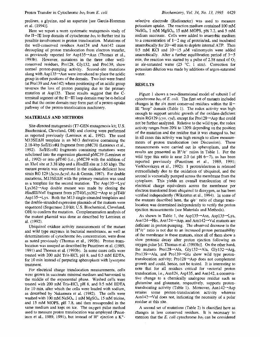
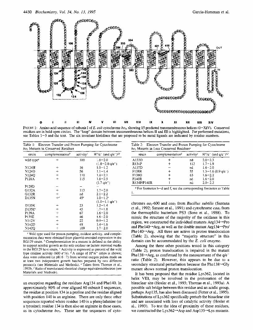
FIGURE 1: Amino acid sequence of subunit I of E. coli cytochrome bo,, showing 15 predicted transmembranous helices (0-XIV). Conserved residues are in bold open circles. The “loop” domain between transmembranous helices I1 and I11 is highlighted. For performed mutations, see Tables 1-3 and the text. The six invariant histidines that are proposed to be metal ligands are indicated by residue numbers.
Table 1: bo3 Mutants in Conserved Residues
Electron Transfer and Proton Pumping for Cytochrome
strain complementationb activity‘ H+/e- (and q/e-)df
Table 2: bo3 Mutants in Less Conserved Residues“
Electron Transfer and Proton Pumping for Cytochrome
strain complementationb activity‘ H+/e- (and q/e-)df wild type“
N124H N 124D N124Q P128A
P128D G132A G132R D135N
D135K D135E‘ P139A P139E N142V N142D N142Q
+ + + + L
- + + L e
100
16 56 110 115
- 113 nd 45‘
23 45 67 46 22 48 109
1.8-2.0 (1.8-2.0 q/e-) 1.0- 1.2 1.1 -1.4 1.8-2.1 1.8-2.5 ( 1.7 q/e-)
1.7-2.0 2.1-2.2 1.0-1.2‘ (1.0-1.1 q/e-) 1.2-1.4 1.7-1.8 1.6-2.0 1.6-2.0 0.9-1.2 1.4-1.6 1.7-2.0
-
a Wild type used for proton pumping, oxidase activity, and comple- mentation data were obtained from plasmid-encoded expression in the RG129 strain. Complementation in a mutant is defined as the ability to support aerobic growth as the sole oxidase on lactate minimal media in the RG129 host strain. Activity is expressed as percent of the wild type oxidase activity (600 e-ls). A range of H+/e- ratios is shown; data were collected (at pH 6-7) from several oxygen pulses made on at least two independent growth batches prepared by two different protocols (see Materials and Methods). e Taken from Thomas et al., 1993b. f Ratio of translocated electrical charge equivalents/electron (see Materials and Methods).
an exception regarding the residues Arg134 and Phel40. In approximately 90% of over aligned 80 subunit I sequences, the residue at position 134 is a proline and the residue aligned with position 140 is an arginine. There are only three other sequences reported where residue 140 is a phenylalanine (or a tyrosine); residue 134 is then also an arginine in these cases, as in cytochrome b03. These are the sequences of cyto-
A133D + nd 2.0-2.3 R134P + 112 1.7-1.9 A137D + nd 1.6-2.0
F138G + 63 1.6-2.2 F140R + nd 1.6-2.0
F138R + 55 1.3-1.6 (0.9 q/e-)
R 1 34PF 140R + nd 2.0-2.2 For footnotes b-d and f, see the corresponding footnotes to Table
1.
chromes aas-600 and caa3 from Bacillus subtilis (Santana et al., 1992; Saraste et al., 1991) and cytochrome caa3 from the thermophilic bacterium PS3 (Sone et al., 1988). To mimic the structure of the majority of the oxidases in this region, we constructed the individual mutants Argl34-Pro and Phel4WArg, as well as the double mutant Argl34-Pro/ Phel40-Arg. All three are active in proton translocation (Table 2), showing that the “majority structure” in this domain can be accommodated by the E. coli enzyme.
Among the three other positions tested in this category (Table 2), proton translocation is impaired in the mutant Phel38-Arg, as confirmed by the measurement of the q/e- ratio (Table 2). However, this appears to be due to a secondary structural perturbation because the Phel38-Gly mutant shows normal proton translocation.
It has been proposed that the residue Lys362, located in helix VIII, may be involved in the protonation of the binuclear site (Hosler et al., 1993; Thomas et al., 1993a). A possible salt bridge between this residue and an acidic group, perhaps Asp135, has also been discussed (Fetter et al., 1995). Substitutions of Lys362 specifically perturb the binuclear site and are associated with loss of catalytic activity (Hosler et al., 1993). To test the idea of proximity of these residues, we constructed the Lys362-Asp and Aspl35-Lys mutants.
Proton Transfer in Cytochrome bo3 from E. cwli Biochemistry, Vol. 34. No. 13, 1995 4431
A.
me "\ct
' I"--
-
i '
I24
B.
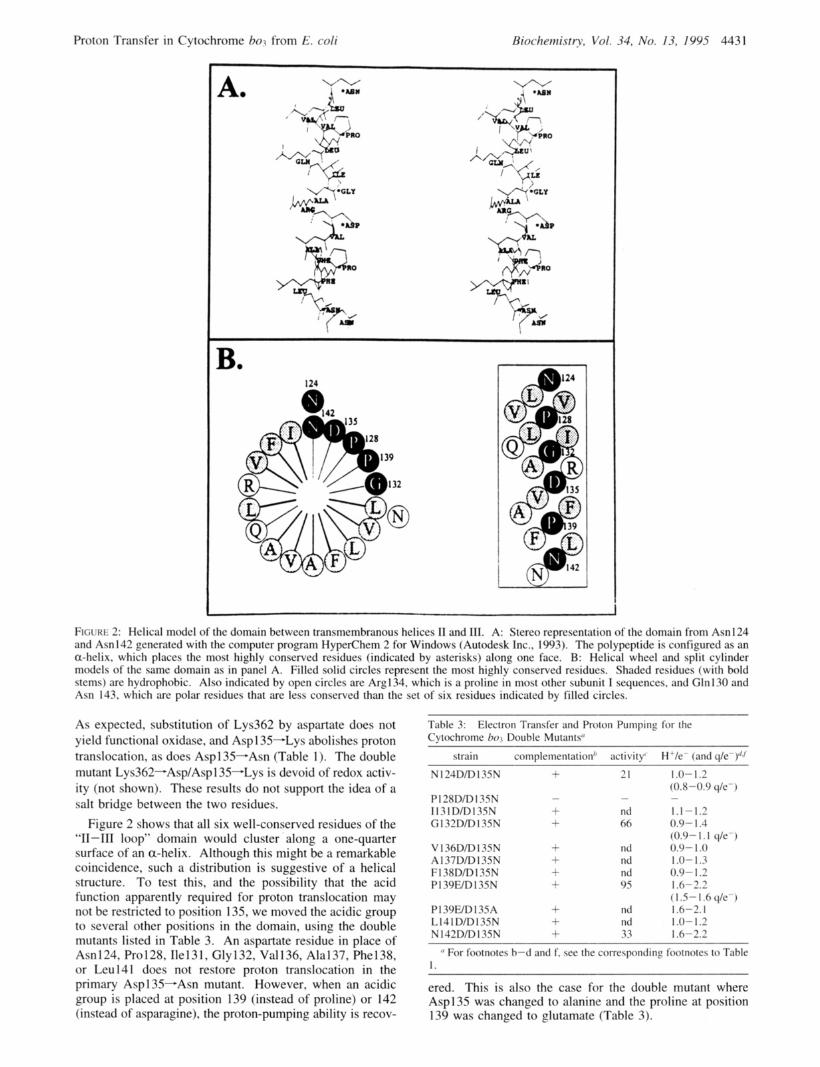
'I 1 FtcxaE 2: Helical model of the domain between transmembranous helices I1 and 111. A: Stereo representation of the domain from Asn 124 and Asn142 generated with the computer program HygerChem 2 for Windows (Autodesk Inc., 1993). The polypeptide is configured as an a-helix. which places the most highly conserved residues (indicated by asterisks) along one face. B: Helical wheel and split cylinder models of the same domain as in panel A. Filled solid circles represent the most highly conserved residues. Shadcd residues (with bold stems) are hydrophobic. Also indicated by open circles are Arg134. which is a proline in most other subunit I sequences. and Gln130 and Asn 143, which are polar residues that are less conserved than the set of six residues indicated by filled circles.
As expected, substitution of Lys362 by aspartate does not yield functional oxidase, and Asp 1 35-Lys abolishes proton translocation, as does Asp1 35-Asn (Table 1 ). The double mutant Lys362-Asp/Aspl35-Lys is devoid of redox activ- ity (not shown). These results do not support the idea of a salt bridge between the two residues.
Figure 2 shows that all six well-conserved residues of the "11-111 loop" domain would cluster along a one-quarter surface of an a-helix. Although this might be a remarkable coincidence, such a distribution is suggestive of a helical structure. To test this, and the possibility that the acid function apparently required for proton translocation may not be restricted to position 135, we moved the acidic group to several other positions in the domain, using the double mutants listed in Table 3. An aspartate residue in place of Asn 124, Pro 128, Ile 13 I , Gly 132, Val 136, Ala 137, Phe 138, or Leu141 does not restore proton translocation in the primary Asp 135-Asn mutant. However, when an acidic group is placed at position 139 (instead of proline) or 142 (instead of asparagine), the proton-pumping ability is recov-
~
Table 3: Cytochrome h o 7 Double Mutants"
Electron Transfer and Proton Pumping for the
strain complementation" activity' H'/e- (and q/e-)"J
N I24D/D I3SN
PI 28D/D I 3SN I 1 3 1 D/D I3SN G I32D/D I3SN
V 136D/D I3SN A I37D/D I3SN F I 38D/D 1 3SN P 1 39WD I 35 N
P139FID13SA L I4 I D/D I3SN N I 42D/D I 35 N
21
- nd 66
ntl nd nd 9s
nd nd 33
I .o- 1.2 (0.8-0.9 qk-) - 1.1-1.2 0.9- I .4 (0.9- I . 1 qk - ) 0.9- 1 .o 1 .o- 1.3 0.9- 1.2 I .6-2.2
( I 5- I .6 q/c-) 1.6-2.1 1 .o- I .2 1 .o-2.2
I' For footnotes h-d and f. sec the corrcspontlins footnotes to Table I .
ered. This is also the case for the double mutant where Asp135 was changed to alanine and the proline at position 139 was changed to glutamate (Table 3).
4432 Biochemistry, Vol. 34, No. 13, 1995 Garcia-Horsman et al.
DISCUSSION
The conclusion that a mutation has specifically affected proton translocation requires proton pumping to be affected more significantly than the linked redox reaction. This may be determined as a lowered H+/e- or q/e- ratio. However, the measured Hf/e- ratio may also be lowered artefactually if the redox reaction is slowed down to the extent that proton ejection can no longer compete kinetically with the natural proton permeability of the membrane. In the mutants studied here, in which proton translocation is impaired, respiration is generally also inhibited. But in all cases, electron transfer still occurs with a turnover time of <20 ms, which is almost 3 orders of magnitude faster than the characteristic proton conductance of the spheroplast membranes (21/2 > 10 s). We hence conclude that the reported mutations have primarily affected a proton transfer pathway. On the other hand, inhibition of proton transfer is, a priori, expected to cause inhibition of respiration as well, due to the linkage of the two functions. Therefore, the observed lowering of the H+/ e- ratio means that this linkage has been weakened or lost by perturbation of the structure. It is interesting that in many cases where a mutation inhibits both proton translocation and electron transfer (see Asn124 and Asn142 mutants, Table 1) both activities are restored by a mutation to an amino acid side chain chemically analogous to that in wild type enzyme.
It is difficult to obtain structural information about specific protein domains, or residues, involved in proton translocation by the heme-copper oxidases. Some insight was obtained recently from the Aspl 35-Asn mutant, where proton translocation is decoupled from electron transfer (Thomas et al., 1993b; Wikstrom et al., 1994). However, this finding is beset with the well-known difficulty associated with site- directed mutations in enzymes of unknown structure: a secondary structural effect could explain such an isolated observation, without direct functioning of the aspartate residue in proton transfer. To try to overcome this difficulty, we studied in some detail the entire ‘‘loop’’ domain between transmembrane helices I1 and I11 where Asp135 is located (Figure 1). This domain of about 20 amino acids contains six residues (Asn124, Pro128, Gly132, Asp135, F’ro139, and Asn142) that are conserved in virtually all respiratory oxidases (except those of the cytochrome cbbs-type; cf. below). Their distribution is suggestive of a-helical second- ary structure (Figure 2), even though these residues are not typical for helices, and computer-aided structure predictions (Crofts, 1992) yield a reasonably high score for a buried helix. Such a helix would also be amphiphatic: with few exceptions, the less conserved residues are nonpolar, making up the remaining three-quarter helical surface. It is also of interest to note that if this domain is truly a cytoplasmic “loop” (Figure l), it would lie far from the metal centers, which are near the opposite side of the membrane (Hosler et al., 1993).
In addition to the Aspl35-Asn reported earlier (Thomas et al., 1993b), mutations of Asn124 and As11142 also impaired proton pumping, except when substituted conser- vatively with glutamine. Mutations in less conserved residues lead to loss of proton translocation in one case, but this appeared to be due to a less specific structural perturba- tion (see Results and Table 2). The finding that three mutations in the “11-111 loop” domain specifically affect
proton translocation supports the notion (Thomas et al., 1993b) that this domain may indeed be of key importance for the proton-translocating function of the enzyme. How- ever, in the family of heme-copper oxidases, the polar II- I11 loop residues are not conserved in the cytochromes of cbb3-type (van der Oost et al., 1994), yet at least the cbb3- type enzymes in Paracoccus denitrificans (Raitio & Wik- strom, 1994) and Rh. sphaeroides (unpublished results) have been shown to pump protons. On the other hand, there are acidic residues in the cytoplasmic loops between transmem- brane helices IV and V and between VI11 and IX in subunit I of these enzymes. Hence, some variation in the structures responsible for proton channeling into the enzyme may occur, as has been shown for the bacterial photosynthetic reaction centers (Takahashi & Wraight, 1991).
Of the six well-conserved residues, the three with polar character (Asn124, Asp135, and Asn142) are apparently crucial. Yet, in each case the side chain can be lengthened by one methylene group without loss of proton-translocating activity. But most significantly, the carboxylic group, originally at position 135, can be moved to two other unique positions (139 and 142) without loss of function. Assuming a helical secondary structure, these positions would be on the same face as the 135 locus but displaced from it toward the C-terminus by one and two helical turns, respectively (Figure 2). In contrast, placing the acidic group at position 136, 137, 138, or 141, which would be on the opposite helical face, does not restore activity, nor did any of the tested relocations toward the N-terminus, even to position 132 which (in a helix) would lie on the same face as position 135 (Table 3, Figure 2). These findings strongly suggest that the C-terminal region of the “11-111 loop”, between residues 135 and 142, may indeed be a-helical. Interestingly, a recent structure prediction suggests that the transmembrane helix I11 might, in fact, start already at residue 136 in cytochrome bo3 from E. coli (Jones et al., 1994). This would place Aspl 35 on the membrane interface and include residues 136-142 as part of helix 111, which would be consistent with our results. On the other hand, the fact that a phenylalanine at position 140 is almost unique for the E. coli enzyme (see above), and is usually an arginine, may well change this prediction.
The 11-111 “loop” domain clearly requires both acidic and potentially hydrogen-bonding amide residues in order for proton translocation to occur normally. The C-terminal residues (in positions 135-142) may provide a hydrogen- bonded network for proton transfer along one polar side of a helix, which is likely to be in contact with some as yet unidentified proteinaceous structure of the enzyme. This network might well include bound water molecules as found in the bacterial photosynthetic reaction center (Ermler et al., 1994). The way in which the “loop 11-111” domain interacts with the rest of the subunit is obviously of greatest significance to understanding the function. For example, it might be folded into the membranous part of the enzyme, thus interacting with transmembrane helices. Work is in progress to create second-site revertants of mutations in this domain to gain answers to these intriguing questions.
REFERENCES
Au, D. C.-T., & Gennis, R. B. (1987) J. Bacteriol. 169, 3237- 3242.
Proton Transfer in Cytochrome bo3 from E. coli
Butt, H. J., Fendler, K., Bamberg, E., Tittor, J., & Oesterhelt, D. (1989) EMBO J. 8, 1657-1663.
Calhoun, M. W., Hill, J. J., Lemieux, L. J., Ingledew, W. J., Alben, J. O., & Gennis, R. B. (1993a) Biochemistry 32, 11524-1 1529.
Calhoun, M. W., Lemieux, L. J., Thomas, J. W., Hill, J. J., Goswitz, V. C., Alben, J. O., & Gennis, R. B. (1993b) Biochemistry 32, 13254- 13261.
Calhoun, M. W., Thomas, J. W., Hill, J. J., Hosler, J. P., Shapleigh, J. P., Tecklenburg, M. M. J., Ferguson-Miller, S . , Babcock, G. T., Alben, J. O., & Gennis, R. B. (1993~) Biochemistry 32, 10905- 109 1 1.
Calhoun, M. W., Thomas, J. W., & Gennis, R. B. (1994) Trends Biochem. Sci. 19, 325-330.
Chepuri, V., & Gennis, R. B. (1990) J . Biol. Chem. 265, 12978- 12986.
Chepuri, V., Lemieux, L. J., Au, D. C.-T., & Gennis, R. B. (1990a) J . Biol. Chem. 265, 11185-11192.
Chepuri, V., Lemieux, L., Hill, J., Alben, J. O., & Gennis, R. B. (1990b) Biochim. Biophys. Acta 1018, 124-127.
Crofts, A. R. (1992) pSAAhf for Windows. A Program for Protein Sequence Analysis and Modelling, University of Illinois, Urbana- Champaign, IL.
Ermler, U., Fritzsch, G., Buchanan, S . , & Michel, H. (1995) Structure (in the press).
Fetter, J. R., Shapleigh, J. P., Thomas, J. W., Garcia-Horsman, J. A., Georgiou, C., Shmidt, E., Hosler, J. P., Babcock, G. T., Gennis, R. B., & Ferguson-Miller, S . (1995) Proc. Natl. Acad. Sci. U S A . (in the press).
Finel, M., & Wikstrom, M. (1986) Biochim. Biophys. Acta 851, 99-108.
Garcia-Horsman, J. A., Barquera, B., Rumbley, J., Ma, J., & Gennis, R. B. (1994) J. Bacteriol. 176, 5587-5600.
Henderson, R., Baldwin, J. M., Ceska, T. A., Zemlii, F., Beckmann, E., & Downing, K. H. (1990) J. Mol. Biol. 213, 899-929.
Hendler, R. W., Pardhasaradhi, K., Reynafarje, B., & Ludwig, B. (1991) Biophys. J . 60, 415-423.
Hill, J., Goswitz, V. C., Calhoun, M. W., Garcia-Horsman, J. A., Lemieux, L., Alben, J. O., & Gennis, R. B. (1992) Biochemistry 31, 11435-11440.
Hosler, J. P., Ferguson-Miller, S . , Calhoun, M. W., Thomas, J. W., Hill, J. J., Lemieux, L., Ma, J., Georgiou, C., Fetter, J., Shapleigh, J., Tecklenburg, M. M. J., Babcock, G. T., & Gennis, R. B. (1993) J. Bioenerg. Biomembr. 25, 121-136.
Jones, D. T., Taylor, W. R., & Thornton, J. M. (1 994) Biochemistry 33, 3038-3049.
Krab, K., & Wikstrom, M. (1978) Biochim. Biophys. Acta 504, 200-2 14.
Krebs, M. P., & Khorana, H. G. (1993) J. Bacteriol. 175, 1555- 1560.
Lemieux, L. J., Calhoun, M. W., Thomas, J. W., Ingledew, W. J., & Gennis, R. B. (1992) J. Biol. Chem. 267, 2105-2113.
Minagawa, J., Mogi, T., Gennis, R. B., & Anraku, Y. (1992) J. Biol. Chem. 267, 2096-2104.
Minghetti, K. C., & Gennis, R. B. (1988) Biochem. Biophys. Res. Commun. 155, 243-248.
Minghetti, K. C., Goswitz, V. C., Gabriel, N. E., Hill, J. J., Barassi, C., Georgiou, C. D., Chan, S . I., & Gennis, R. B. (1992) Biochemistry 31, 6917-6924.
Biochemistry, Vol. 34, No. 13, 1995 4433
Nakamura, T., Tokuda, H., & Unemoto, T. (1982) Biochim. Biophys. Acta 692, 389-396.
Puustinen, A., & Wikstrom, M. (1991) Proc. Natl. Acad. Sci. U S A . 88, 6122-6126.
Puustinen, A., Finel, M., Virkki, M., & Wikstrom, M. (1989) FEBS Lett. 249, 163-167.
Puustinen, A., Finel, M., Haltia, T., Gennis, R. B., & Wikstrom, M. (1991) Biochemistry 30, 3936-3942.
Puustinen, A., Morgan, J. E., Verkhovsky, M., Thomas, J. W., Gennis, R. B., & Wikstrom, M. (1992) Biochemistry 31, 10363- 10369.
Raitio, M., & Wikstrom, M. (1994) Biochim. Biophys. Acta 1186, 100-106.
Rongey, S . H., Paddock, M. L., Feher, G., & Okamura, M. Y. (1993) Proc. Natl. Acad. Sci. USA 90, 1325-1329.
Santana, M., Kunst, F., Hullo, M. F., Rapoport, G., Danchin, A., & Glaser, P. (1992) J. Biol. Chem. 267, 10225-10231.
Saraste, M., Metso, T., Nakari, T., Jalli, T., Lauraeus, M., & van der Oost, J. (1991) Eur. J. Biochem. 195, 517-525.
Shapleigh, J. P., Hill, J. J., Alben, J. O., & Gennis, R. B. (1992a) J . Bacteriol. 174, 2338-2343.
Shapleigh, J. P., Hosler, J. P., Tecklenburg, M. M. J., Kim, Y., Babcock, G. T., Gennis, R. B., & Ferguson-Miller, S . (1992b) Proc. Natl. Acad. Sci. U.SA. 89, 4786-4790.
Shinkarev, V. P., Takahashi, E., & Wraight, C. A. (1993) Biochim. Biophys. Acta 1142, 214-216.
Solioz, M., Carafoli, E., & Ludwig, B. (1982) J. Biol. Chem. 257,
Sone, N., & Hinkle, P. C. (1982) J. Biol. Chem. 257, 12600-12604. Sone, N., Yokoi, F., Fu, T., Ohta, S . , Metso, T., Raitio, M., &
Saraste, M. (1988) J. Biochem. (Tokyo) 103, 606-610. Takahashi, E., & Wraight, C. A. (1991) Biochemistry 31, 855-
866. Thomas, J. W., Lemieux, L. J., Alben, J. O., & Gennis, R. B.
(1993a) Biochemistry 32, 11 173- 11 180. Thomas, J. W., Puustinen, A., Alben, J. O., Gennis, R. B., &
Wikstrom, M. (1993b) Biochemistry 32, 10923-10928. Thomas, J. W., Calhoun, M. W., Lemieux, L. J., Puustinen, A.,
Wikstrom, M., Alben, J. O., & Gennis, R. B. (1994) Biochemistry
Trumpower, B. L., & Gennis, R. B. (1994) Annu. Rev. Biochem.
Verkhovskaya, M., Verkhovsky, M., & Wikstrom, M. (1992) J.
Wikstrom, M. (1977) Nature 266, 271-273. Wikstrom, M. (1988) FEBS Lett. 231, 247-252. Wikstrom, M. (1989) Nature 338, 776-778. Wikstrom, M., & Krab, K. (1979) Biochim. Biophys. Acta 549,
177-222. Wikstrom, M., Bogachev, A., Finel, M., Morgan, J. E., Puustinen,
A., Raitio, M., Verkhovskaya, M., & Verkhovsky, M. I. (1994) Biochim. Biophys. Acta 1187, 106-111.
Wu, W., Chang, C. K., Varotsis, C., Babcock, G. T., Puustinen, A., & Wikstrom, M. (1992) J.Am. Chem. SOC. 114,1182-1187.
BI942499D
1579- 1582.
33, 13013-13021.
63, 675-716.
Biol. Chem. 267, 14559-14562.