Embed Size (px)
Citation preview
Proteins are not rigid structures:
Protein dynamics, conformational variability,
and thermodynamic stability
Dr. Andrew LeeUNC School of Pharmacy (Div. Chemical Biology and Medicinal Chemistry)
UNC Med School (Biochemistry and Biophysics)
[email protected] Marsico Hall
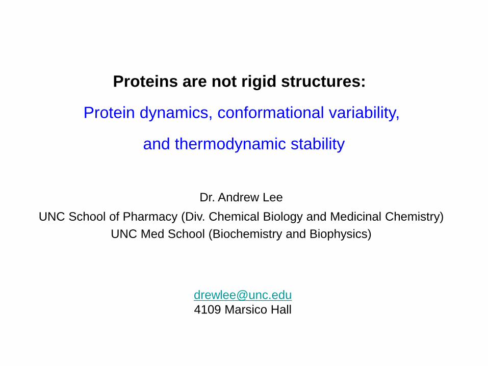
Proteins are long polypeptide chains of ~50 or more “residues”.
amino terminus carboxy terminus“N” “C”
“N”
“C”
globular fold
MAKRNIVTATTSKGEFTMLGVHDNVAILPTHASPGESIVIDGKE-VEILDAKALEDQAGTNLEITIITLKRNEKFRDIRPHIPTQITETNDG-VLIVNTSKYPNMYVPVGAVTEQGYLNLGGRQTARTLMYNFPTRAGQ….
• Protein sequences are given as linear sequences of their one-letter amino acids:
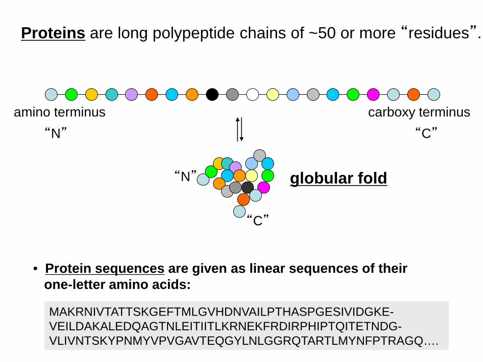
Proteins are typically “globular” in shape
myoglobin(17 kDa)
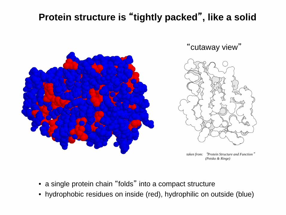
Protein structure is “tightly packed”, like a solid
• a single protein chain “folds” into a compact structure• hydrophobic residues on inside (red), hydrophilic on outside (blue)
“cutaway view”
taken from: “Protein Structure and Function”(Petsko & Ringe)
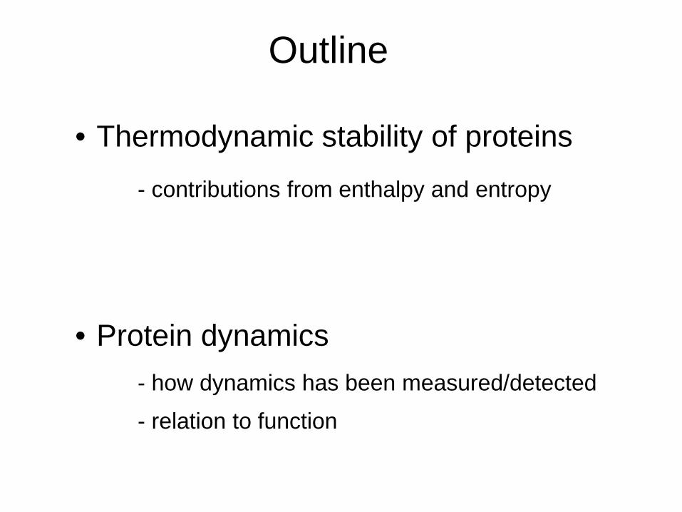
Outline
• Thermodynamic stability of proteins
• Protein dynamics
- contributions from enthalpy and entropy
- how dynamics has been measured/detected
- relation to function
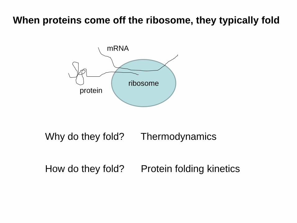
When proteins come off the ribosome, they typically fold
Why do they fold? Thermodynamics
How do they fold? Protein folding kinetics
ribosome
mRNA
protein
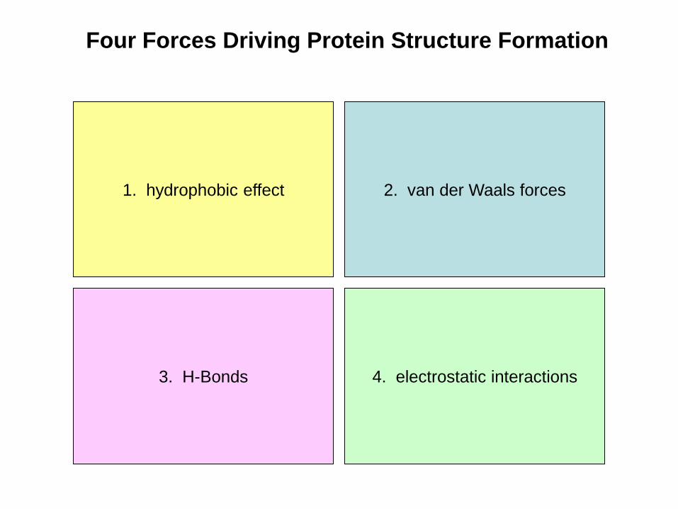
Four Forces Driving Protein Structure Formation
3. H-Bonds
2. van der Waals forces1. hydrophobic effect
4. electrostatic interactions
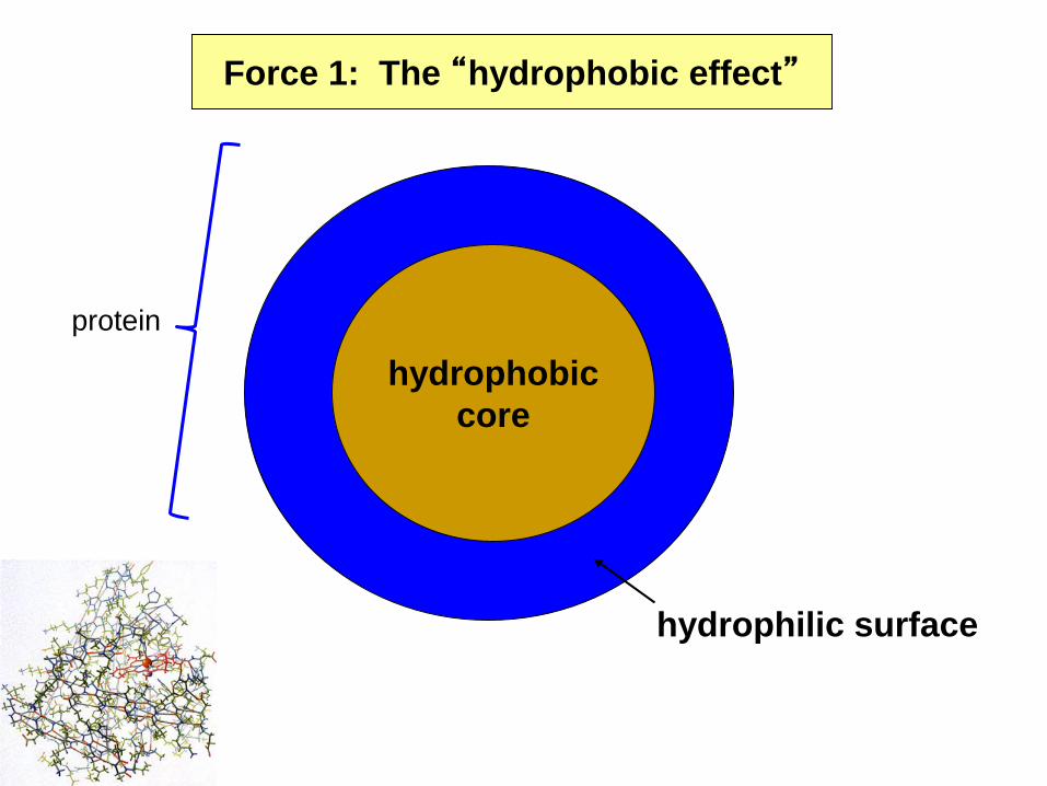
Force 1: The “hydrophobic effect”
hydrophobiccore
hydrophilic surface
protein
“greasy”molecule
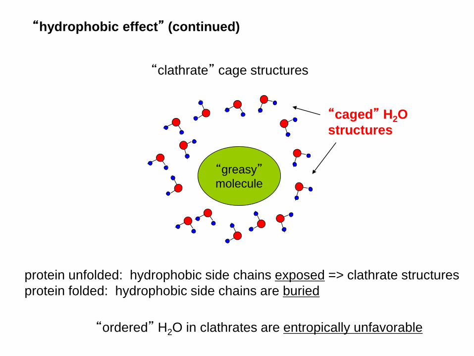
“clathrate” cage structures
protein unfolded: hydrophobic side chains exposed => clathrate structuresprotein folded: hydrophobic side chains are buried
“ordered” H2O in clathrates are entropically unfavorable
“caged” H2Ostructures
“hydrophobic effect” (continued)
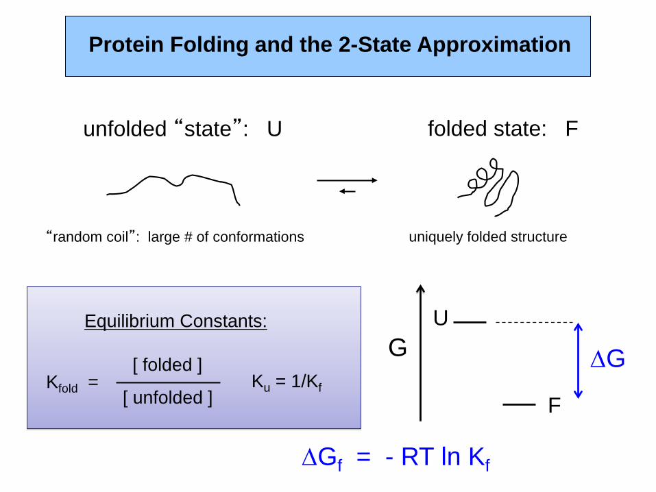
Protein Folding and the 2-State Approximation
unfolded “state”: U folded state: F
“random coil”: large # of conformations uniquely folded structure
∆Gf = - RT ln Kf
Kfold =[ folded ]
[ unfolded ]Ku = 1/Kf
Equilibrium Constants: U
F
G ∆G
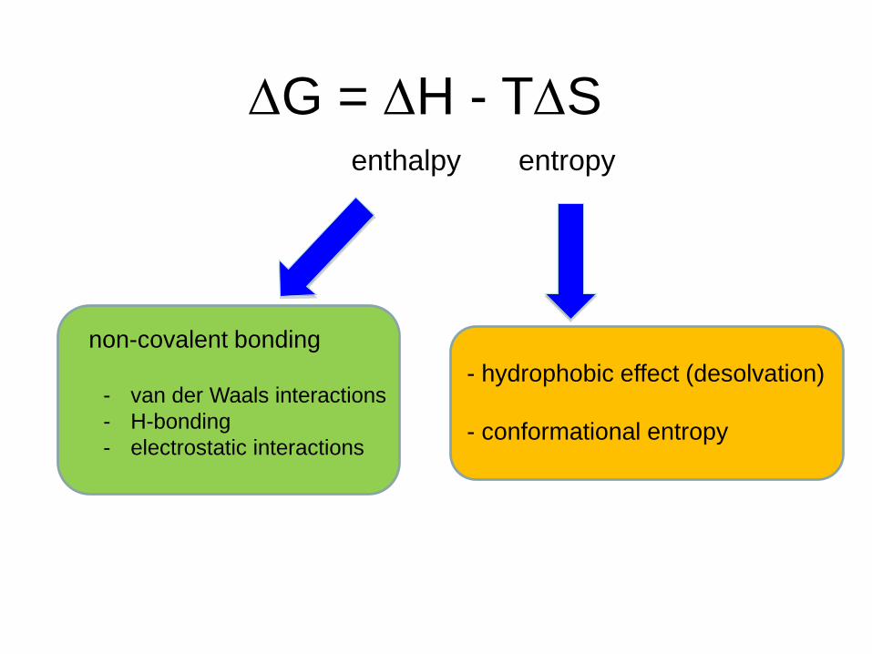
∆G = ∆H - T∆S enthalpy entropy
non-covalent bonding- hydrophobic effect (desolvation)
- conformational entropy- van der Waals interactions- H-bonding- electrostatic interactions
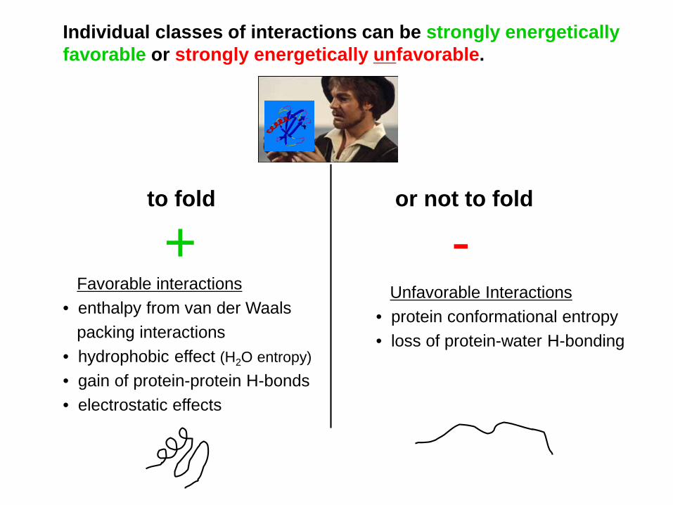
Individual classes of interactions can be strongly energeticallyfavorable or strongly energetically unfavorable.
to fold or not to fold
Favorable interactions• enthalpy from van der Waals
packing interactions• hydrophobic effect (H2O entropy)• gain of protein-protein H-bonds• electrostatic effects
Unfavorable Interactions• protein conformational entropy• loss of protein-water H-bonding
+ -
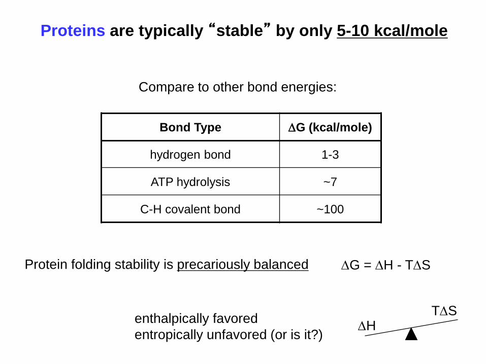
Proteins are typically “stable” by only 5-10 kcal/mole
Protein folding stability is precariously balanced
enthalpically favoredentropically unfavored (or is it?)
∆G = ∆H - T∆S
Bond Type ∆G (kcal/mole)
hydrogen bond 1-3
ATP hydrolysis ~7
C-H covalent bond ~100
Compare to other bond energies:
∆HT∆S
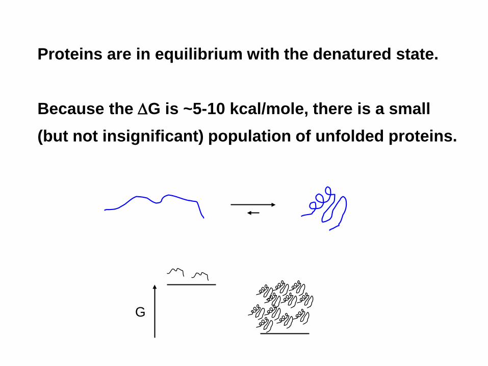
Proteins are in equilibrium with the denatured state.
Because the ∆G is ~5-10 kcal/mole, there is a small(but not insignificant) population of unfolded proteins.
G
Protein Dynamics
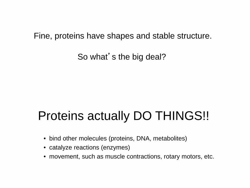
Fine, proteins have shapes and stable structure.
Proteins actually DO THINGS!!
So what’s the big deal?
• bind other molecules (proteins, DNA, metabolites)• catalyze reactions (enzymes)• movement, such as muscle contractions, rotary motors, etc.
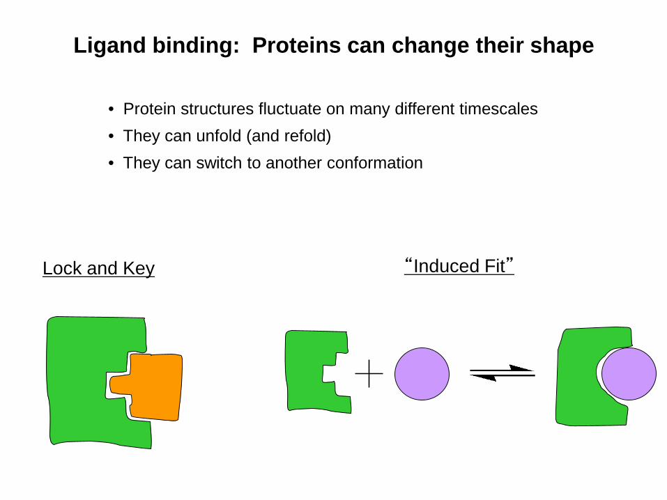
Ligand binding: Proteins can change their shape
• Protein structures fluctuate on many different timescales• They can unfold (and refold)• They can switch to another conformation
Lock and Key “Induced Fit”
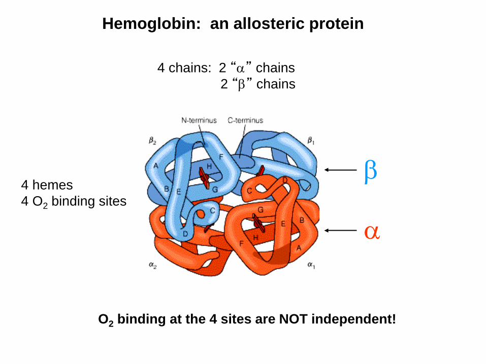
Hemoglobin: an allosteric protein
4 chains: 2 “α” chains2 “β” chains
4 hemes4 O2 binding sites
O2 binding at the 4 sites are NOT independent!
α
β
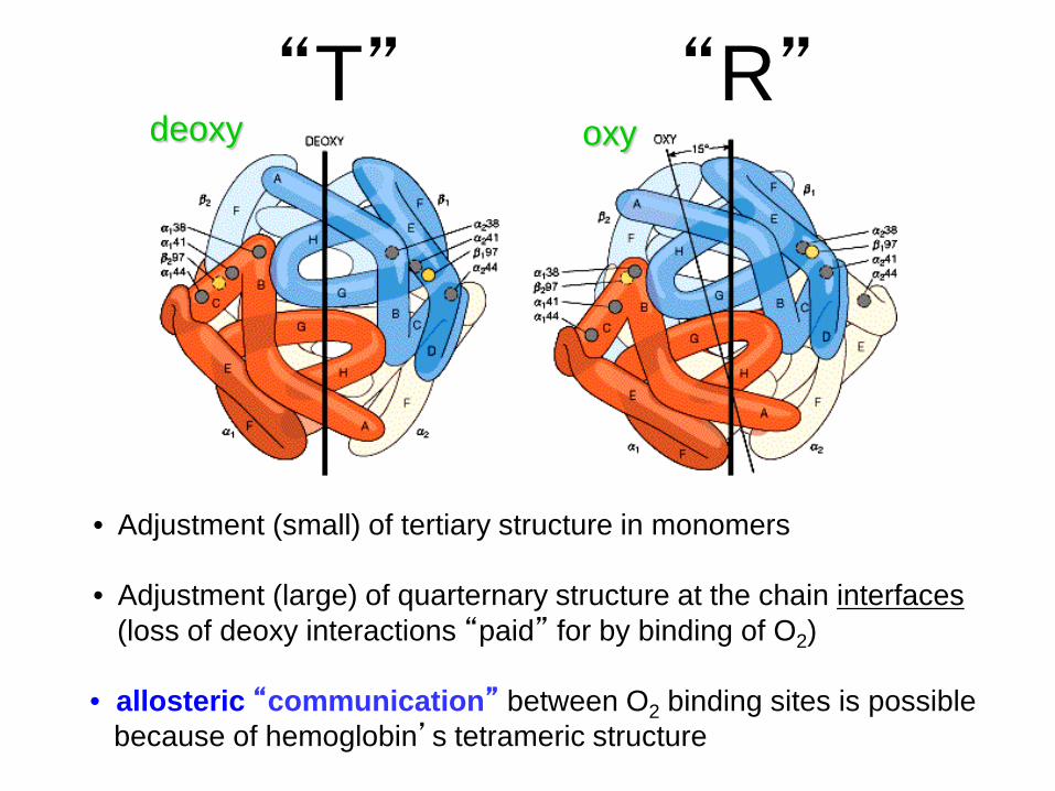
“T” “R”
• Adjustment (small) of tertiary structure in monomers
• Adjustment (large) of quarternary structure at the chain interfaces(loss of deoxy interactions “paid” for by binding of O2)
deoxy oxy
• allosteric “communication” between O2 binding sites is possiblebecause of hemoglobin’s tetrameric structure
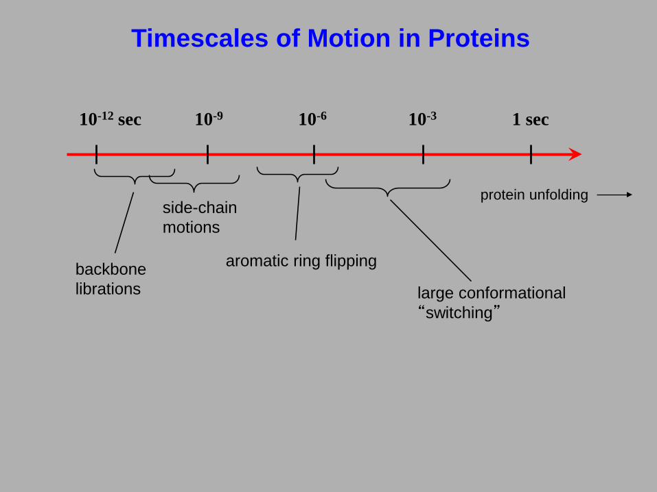
Timescales of Motion in Proteins
10-12 sec 10-9 10-6 10-3 1 sec
large conformational “switching”
protein unfolding
aromatic ring flipping
side-chain motions
backbonelibrations
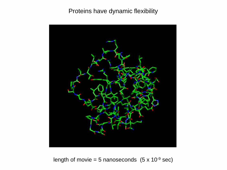
Proteins have dynamic flexibility
length of movie = 5 nanoseconds (5 x 10⁻⁹ sec)
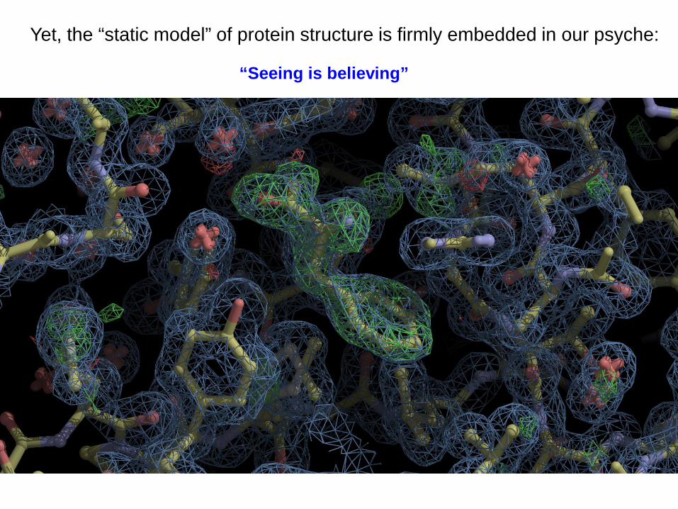
Yet, the “static model” of protein structure is firmly embedded in our psyche:
“Seeing is believing”
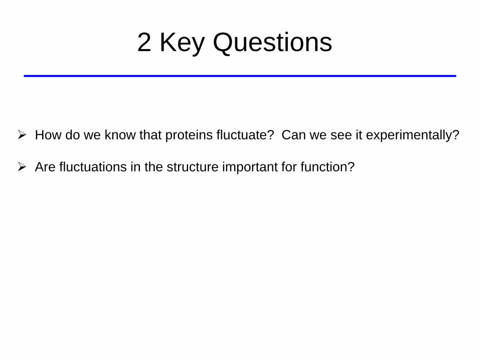
How do we know that proteins fluctuate? Can we see it experimentally?
Are fluctuations in the structure important for function?
2 Key Questions
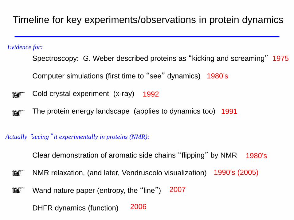
Timeline for key experiments/observations in protein dynamics
Spectroscopy: G. Weber described proteins as “kicking and screaming” 1975
Computer simulations (first time to “see” dynamics) 1980’s
Cold crystal experiment (x-ray)
The protein energy landscape (applies to dynamics too)
Clear demonstration of aromatic side chains “flipping” by NMR
NMR relaxation, (and later, Vendruscolo visualization)
Wand nature paper (entropy, the “line”)
DHFR dynamics (function)
Evidence for:
Actually “seeing” it experimentally in proteins (NMR):
1980’s
1990’s (2005)
2007
2006
1992
1991
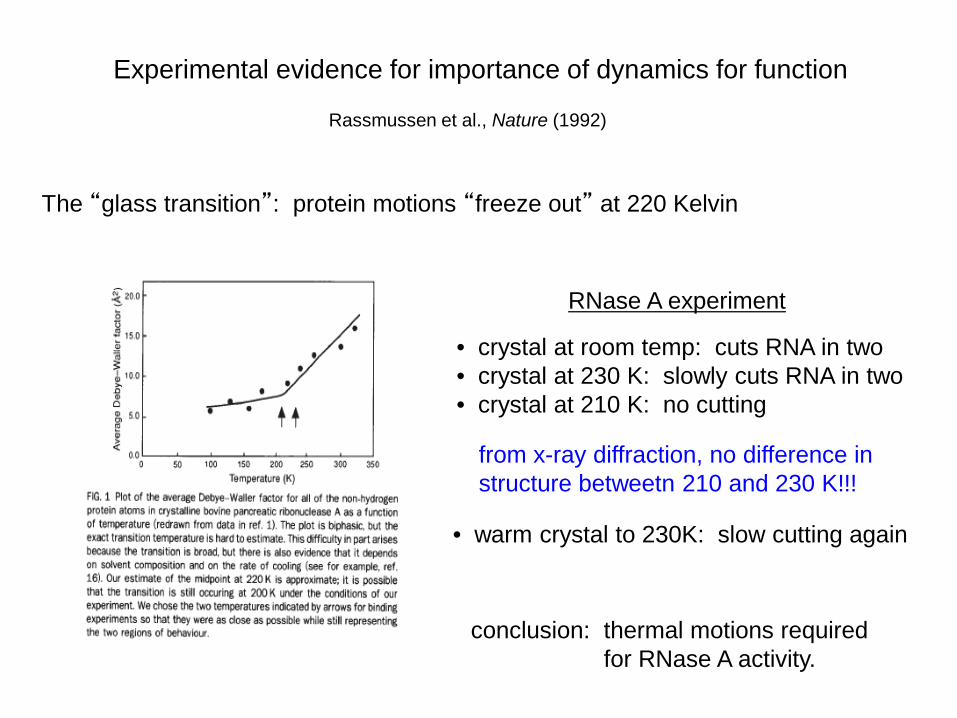
Rassmussen et al., Nature (1992)
Experimental evidence for importance of dynamics for function
The “glass transition”: protein motions “freeze out” at 220 Kelvin
RNase A experiment
• crystal at room temp: cuts RNA in two• crystal at 230 K: slowly cuts RNA in two• crystal at 210 K: no cutting
• warm crystal to 230K: slow cutting again
from x-ray diffraction, no difference instructure betweetn 210 and 230 K!!!
conclusion: thermal motions requiredfor RNase A activity.
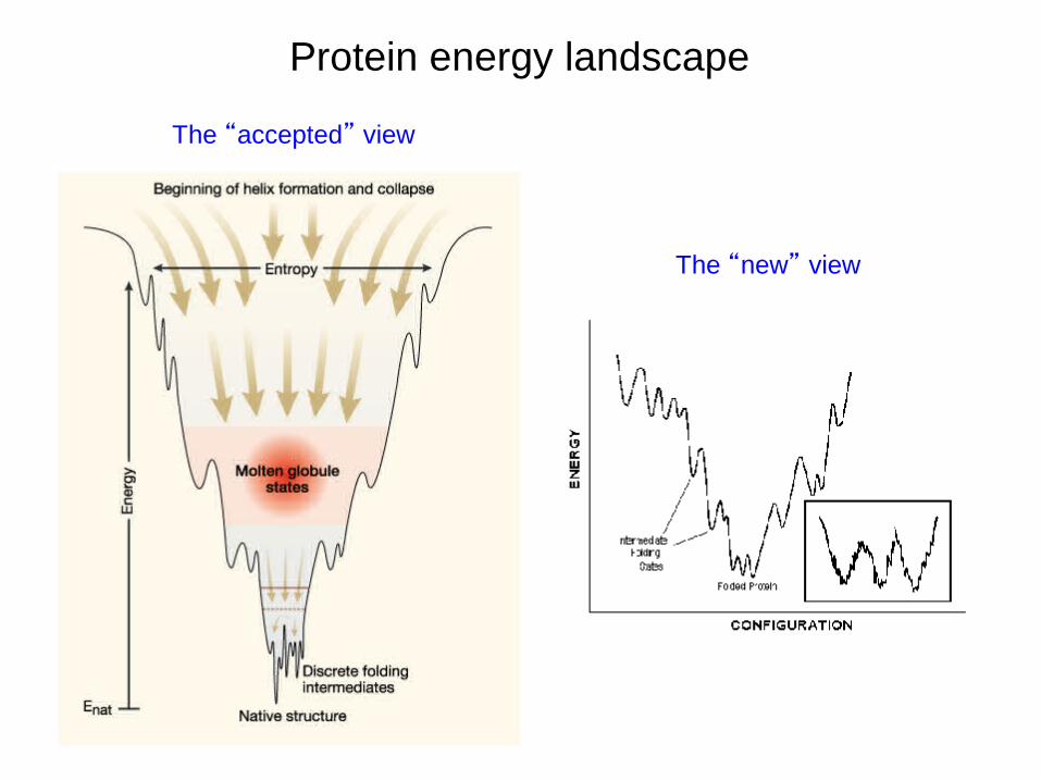
The “accepted” view
The “new” view
Protein energy landscape
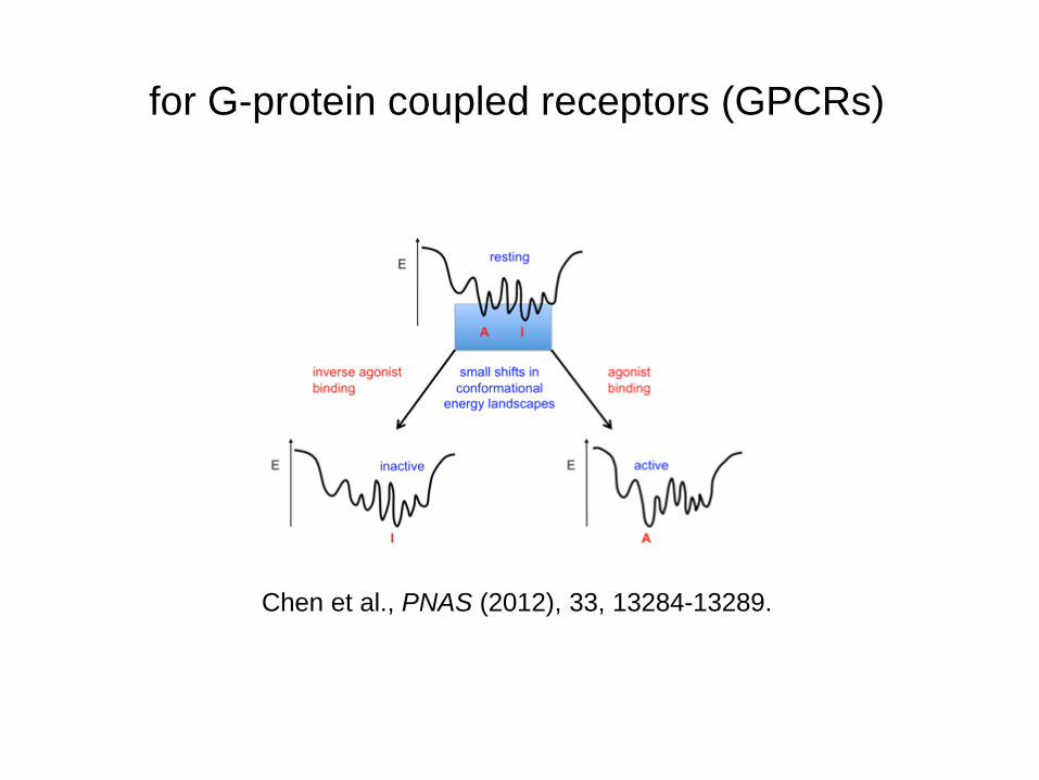
Chen et al., PNAS (2012), 33, 13284-13289.
for G-protein coupled receptors (GPCRs)
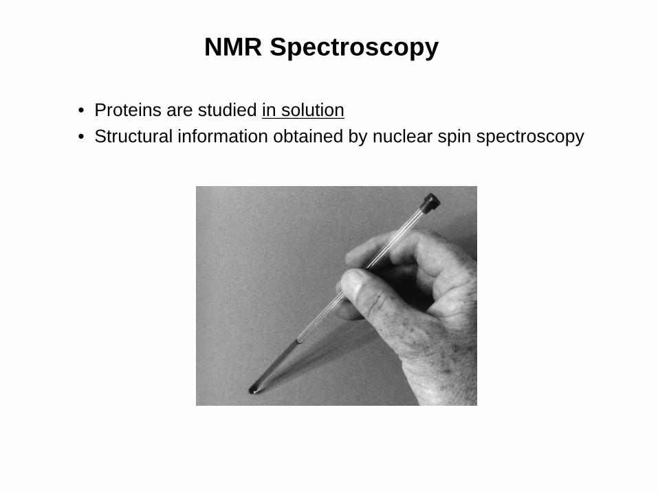
NMR Spectroscopy
• Proteins are studied in solution• Structural information obtained by nuclear spin spectroscopy
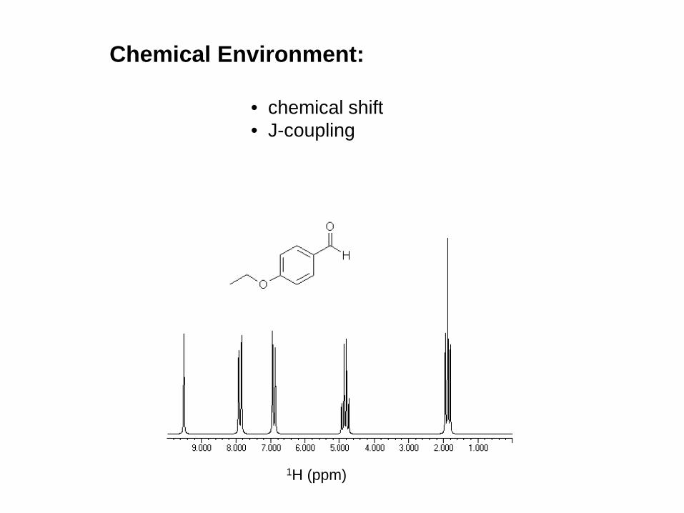
Chemical Environment:
• chemical shift• J-coupling
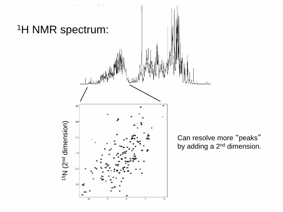
1H (ppm)
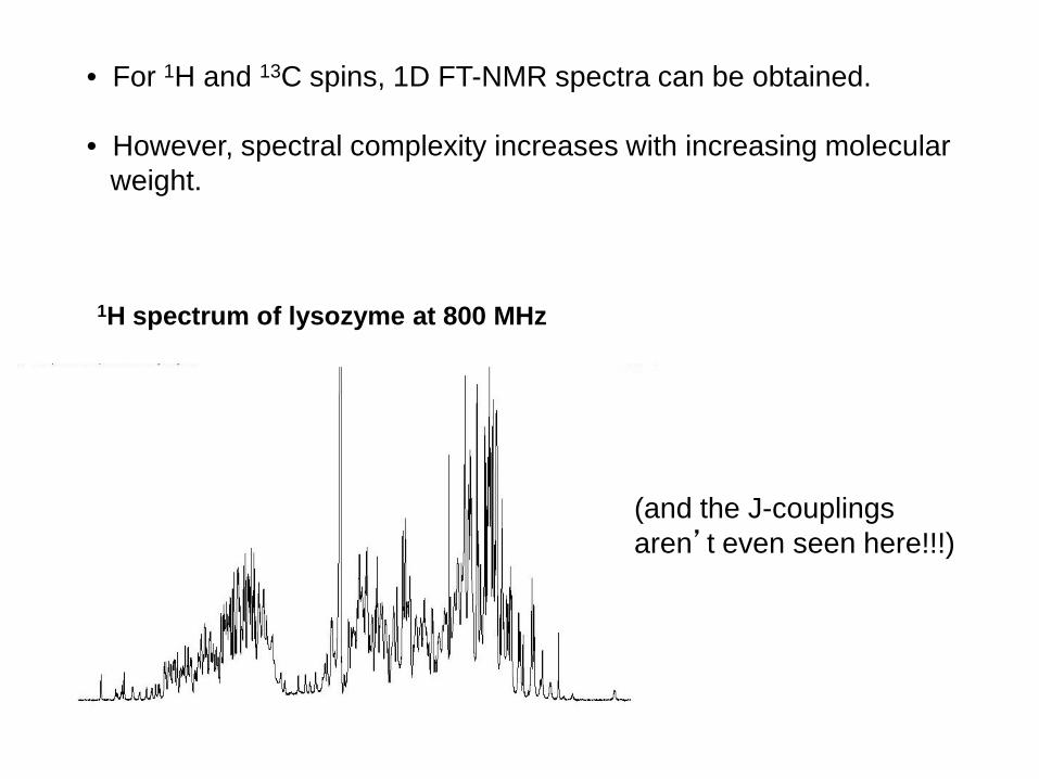
• For 1H and 13C spins, 1D FT-NMR spectra can be obtained.
• However, spectral complexity increases with increasing molecularweight.
1H spectrum of lysozyme at 800 MHz
(and the J-couplingsaren’t even seen here!!!)
15N
(2nd
dim
ensi
on)
1H NMR spectrum:
Can resolve more “peaks”by adding a 2nd dimension.
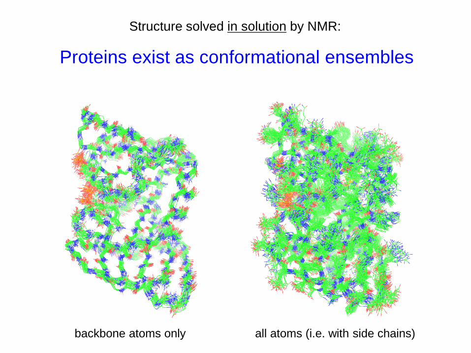
Structure solved in solution by NMR:
Proteins exist as conformational ensembles
backbone atoms only all atoms (i.e. with side chains)
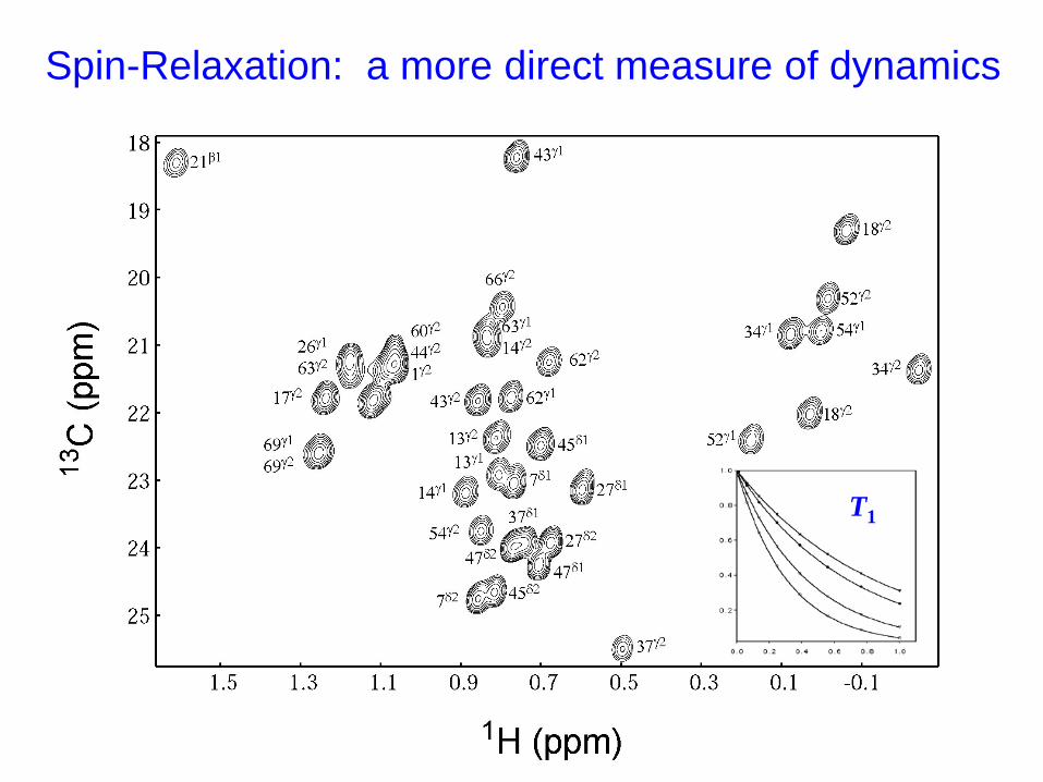
Spin-Relaxation: a more direct measure of dynamics
T1
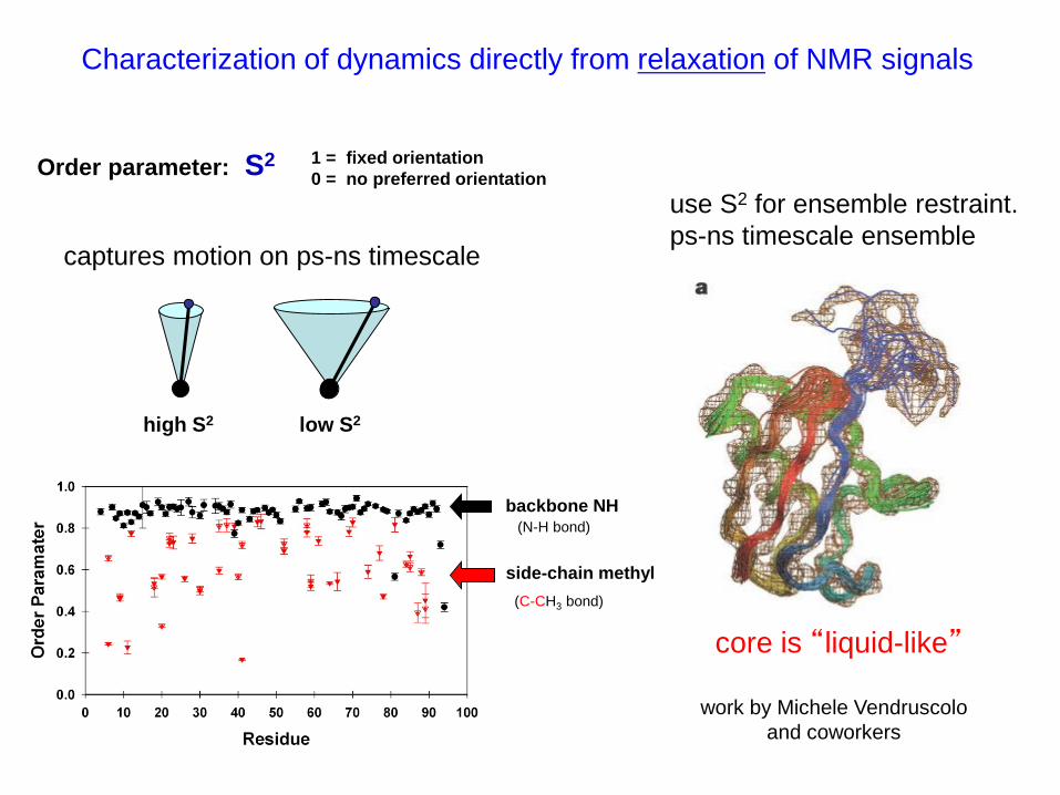
Characterization of dynamics directly from relaxation of NMR signals
Order parameter: S2 1 = fixed orientation0 = no preferred orientation
captures motion on ps-ns timescale
backbone NH
side-chain methyl(C-CH3 bond)
(N-H bond)
low S2high S2
use S2 for ensemble restraint.ps-ns timescale ensemble
work by Michele Vendruscoloand coworkers
core is “liquid-like”
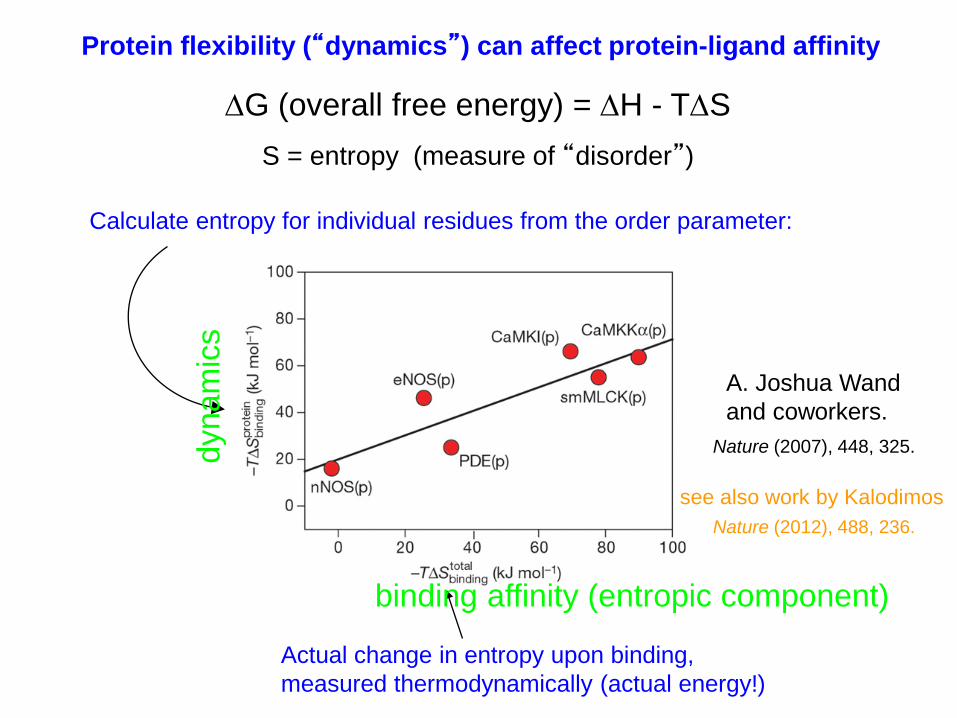
Calculate entropy for individual residues from the order parameter:
Actual change in entropy upon binding,measured thermodynamically (actual energy!)
S = entropy (measure of “disorder”)
∆G (overall free energy) = ∆H - T∆S
A. Joshua Wandand coworkers.
Protein flexibility (“dynamics”) can affect protein-ligand affinity
binding affinity (entropic component)
dyna
mic
s
Nature (2007), 448, 325.
see also work by KalodimosNature (2012), 488, 236.
• “Protein dynamics” is still a relatively new field of research
• Complementary to static structure determination (i.e. x-ray diffraction)
• Motions on order of 10 ns – 1 μs very hard to characterize in detail
• Computer simulated proteins provides the “clearest” picture(but computer power is an issue – hard to go longer than tens of μs)
• Dynamics important for function, but we are just beginning to understand why.
Where are we at now with our knowledge of protein dynamics?
Study Questions
2. Based on basic principles of physical chemistry, how can dynamics affectfree energy changes via entropy?
1. Would you expect a globular, folded protein to spontaneously unfold? If “no”, why? If “yes”, what would you expect to happen after that?
see optional question #3 on next slide……
4. For a protein that is stable at 3 kcal/mole (that is, ΔGfold = -3 kcal/mole),calculate the percentage of proteins that will be unfolded at 25 ºCat equilibrium?
Optional question:
3) You encounter a shrink ray that reduces you to a 10nm sized human being. Taking advantage of your small size, you decide to scuba dive into a beaker and observe protein folding and dynamics. This protein is 20kDa in size, has both hydrophobic and hydrophillic residues, and is known to be allosteric.
a) Given your knowledge of the driving forces for protein folding and unfolding, visually represent these forces in play for a protein in water. Label your diagram and include detailed explanations for what you see, considering all the complications discussed this week forprotein folding and dynamics. (E.g. Drawing representative amino acid residues within part of this protein that are known to be involved in van der Waals interactions and whetherthese (non-covalent “bonds”) are stronger or weaker than other types of bonds, what forces are at play with these bonds, how these forces do or do not contribute to folding, ΔH, or ΔS.)
b) The scientist whose bench you have invaded adds adequate ligand to bind to both binding sites on the allosteric protein. Again, visually represent what happens to the protein and the forces at play. Include in your discussion the dynamics of the protein and how it would affect ligand binding, conformational selection vs. induced fit models, and which model you think is more likely to represent your allosteric protein.