Embed Size (px)
Citation preview
Received: 24 April 2019 Revised: 11 June 2019 Accepted: 13 July 2019
DOI: 10.1002/ptr.6469
R E V I EW
Protective effects of Ginkgo biloba L. against natural toxins,chemical toxicities, and radiation: A comprehensive review
Seyedeh Farzaneh Omidkhoda1 | BiBi Marjan Razavi1,2 | Hossein Hosseinzadeh1,3
1Department of Pharmacodynamics and
Toxicology, School of Pharmacy, Mashhad
University of Medical Sciences, Mashhad, Iran
2Targeted Drug Delivery Research Center,
Pharmaceutical Technology Institute, Mashhad
University of Medical Sciences, Mashhad, Iran
3Pharmaceutical Research Center,
Pharmaceutical Technology Institute, Mashhad
University of Medical Sciences, Mashhad, Iran
Correspondence
Hossein Hosseinzadeh, Pharmaceutical
Research Center, Pharmaceutical Technology
Institute, Mashhad University of Medical
Sciences, Mashhad, Iran.
Email: [email protected]
Abbreviation: AA, arachidonic acid; AChE, acetylcholine e
aminotransferase; Apaf‐1, apoptotic protease activating fac
Bcl‐2, B‐cell lymphoma 2; Bcl2‐Xl, B‐cell lymphoma‐extr
cPLA2α, cytosolic phospholipase A2; Cr, creatinine; ERK,
oxygenase‐1; IκB, IκB kinase; IL‐6, Interleukin‐6; IL‐1β, intLPS, lipopolysaccharide; MAPK, mitogen‐activated protein
mPGES‐1, microsomal prostaglandin E synthase‐1; MPO,
factor kappa beta; NO, nitric oxide; Nrf‐2, nuclear factor (
peroxisome proliferator‐activated receptor α; ROS, reactiv
SREBP‐1c, sterol regulatory element binding protein 1c; TB
beta; TLR‐4, toll‐like receptor‐4; TNF‐α, tumour necrosis
ligand‐1; TTP, tristetraprolin; 5‐FU, 5‐fluorouracil
Phytotherapy Research. 2019;1–20.
Nowadays in our developing and industrial world, humans' health or even their life is
threatened by exposure to poisons. In this situation, detecting a protective compound
could be helpful and interesting. In the present article, we collected and reviewed all
studies, which have been conducted so far about the protective effects of Ginkgo
biloba L. (GB), one of the most ancient medicinal tree species, against toxicities
induced by chemical toxic agents, natural toxins, and also radiation. In overall, inves-
tigations showed that GB exerts the antioxidant, antiinflammatory, antiapoptotic, and
antigenotoxicity effects in different toxicities. There are also some special mecha-
nisms about its protective effects against some specific toxic agents, such as acetyl-
choline esterase inhibition in the aluminium neurotoxicity or membrane‐bond
phosphodiesterase activation in the triethyltin toxicity. Ginkgolide A was the most
investigated active ingredient of G. biloba leaf extract as a protective compound
against toxicities, which had the similar effects of total extract. A few clinical studies
have been conducted in this field, which demonstrated the beneficial effects of GB
against toxic agents. However, the promising effects of this valuable herbal extract
will practically remain useless without carrying out more clinical studies and proving
its effects on human beings.
KEYWORDS
antidote, Ginkgo biloba, natural toxin, radiation, toxic chemicals
1 | INTRODUCTION
Ginkgo biloba L. (GB; Ginkgoaceae family) is one of the most ancient
medicinal tree species with useful applications in health, food, and
supplements. It is native to China, Japan, and Korea and popularly
sterase; AD, Alzheimer's disease; AFB1
tor 1; AP‐1, activator protein‐1; AST, a
a large; BUN, blood urea nitrogen; CA
extracellular signal‐regulated kinase; G
erleukin‐1beta; iNOS, nitric oxide synth
kinase; MDA, malondialdehyde; MIP‐2,
myeloperoxidase; mTOR, mammalian ta
erythroid‐derived 2)‐like 2; PDE, phosp
e oxygen species; SAPK, stress‐activate
ARS, thiobarbituric acid reactive substa
factor; TNFR‐1, tissue necrosis factor
wileyonlinelibrary.com/
known as living fossil. Nowadays, it is cultivated worldwide for bene-
ficial using of its nuts and leaves (Mohanta, Tamboli, & Zubaidha,
2014). The main active components, which are thought to be respon-
sible for the pharmacological properties of GB, include flavonoids
(kaempferol, quercetin, myricetin, apigenin, isorhamnetin, luteolin,
, Aflatoxin B1; AKt, protein kinase B; Alb, albumin; ALP, alkaline phosphatase; ALT, alanine
spartate aminotransferase; Bad, Bcl‐2 agonist of cell death; Bax, Bcl2‐Associated X Protein;
T, catalase; CCl4, carbon tetrachloride; CO, carbon monoxide; COX‐2, cyclooxygenase‐2;
B, Ginkgo biloba L; GPX, Glutathione peroxidase; GSH, reduced glutathione; HO‐1, Heme
ase; JNK, jun N‐terminal kinase; LDH, lactate dehydrogenase; LPC, lysophosphatidylcholine;
macrophage inflammatory protein‐2; MMP‐9, matrix metalloproteinase‐9; MN, micronuclei;
rget of rapamycin; NADPH, nicotinamide adenine dinucleotide phosphate; NF‐ΚB, nuclearhdiesterase enzyme; PGE‐2, prostaglandin E2; PI3K, phosphatidylinositol‐3‐Kinase; PPAR‐α,d protein kinase; SGLT1, sodium‐glucose linked transporter‐1; SOD, superoxide dismutase;
nces; TET, triethyltin; TEWL, transepidermal water loss; TGF‐β1, transforming growth factor
receptor 1; TP, total protein; TRAIL‐1, tumour necrosis factor‐related apoptosis‐inducing
© 2019 John Wiley & Sons, Ltd.journal/ptr 1
2 OMIDKHODA ET AL.
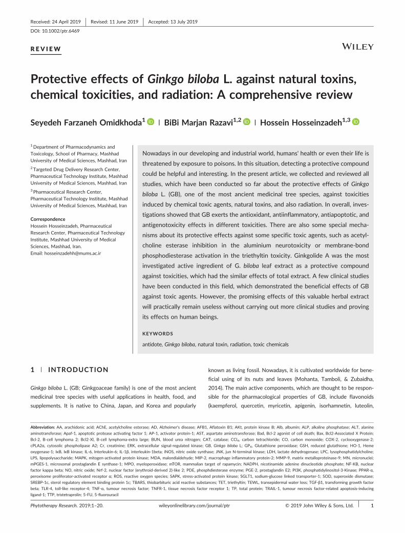
and tamarixetin), terpene trilactones (ginkgolide A, ginkgolide B,
ginkgolide C, ginkgolide J, ginkgolide M, ginkgolide K, ginkgolide L,
ginkgolide P, ginkgolide Q, and bilobalide; Figure 1) and
proanthocyanidins (van Beek and Montoro, 2009; Liao, Zheng, Li, &
Peng, 2011). Leaves and seeds of GB have been used in Chinese,
Japanese, and Indonesian traditional medicine for thousands of years
(Singh, Barreto, Aliev, & Echeverria, 2017). Recently, GB leaf extracts
(GBLEs) are being used very commonly as the phytomedicines all over
the world (Tang et al., 2017). In this regard, there are a lot of commer-
cial products of GBLEs that have been purchased and applied in
approximately all studies reviewed here, except those we especially
explained. These standardized forms that also named EGb 761 contain
24% of ginkgo flavone glycosides and 6% of terpene trilactones.
According to the modern pharmacological studies, GBLE has been
used in the treatment of several diseases including neurodegenerative
diseases such as Alzheimer's disease (AD; Ihl et al., 2011), cerebral dis-
orders (Saleem, Zhuang, Biswal, Christen, & Dore, 2008), vascular
problems (Keheyan, Dunn, & Hall, 2011), age‐related memory deficit
(Stackman et al., 2003), and oxidative stress (Bernatoniene et al.,
2011; Mohamed & Abd El‐Moneim, 2017).
The extract or active ingredients of some important plants, such as
Curcuma longa (Hosseini & Hosseinzadeh, 2018), Berberis vulgaris
(Mohammadzadeh, Mehri, & Hosseinzadeh, 2017), Nigella Sativa
(Tavakkoli, Ahmadi, Razavi, & Hosseinzadeh, 2017), Cinnamomum
zeylanicum (Dorri, Hashemitabar, & Hosseinzadeh, 2018), Silybum
Marianum (Fanoudi, Alavi, Karimi, & Hosseinzadeh, 2018), Camellia
sinensis (Rameshrad, Razavi, & Hosseinzadeh, 2017), Vitis vinifera
(Tabeshpour, Mehri, Shaebani, & Hosseinzadeh, 2018), and Crocus
sativus (Razavi & Hosseinzadeh, 2015) have shown the beneficial
effects against natural toxins and toxic chemicals. Several reviews
exist about the protective effects of GB mostly on dementia and neu-
rological disorders; however, no review has been written about its
neutralizing effects against toxins and toxicities so far, and in regard
to the various and useful applications and the effects of GB, which
have been established up to now, it was necessary to collect and
review the papers about the protective effects of this precious plant
on toxicities developed via the exposure to natural toxins, chemical
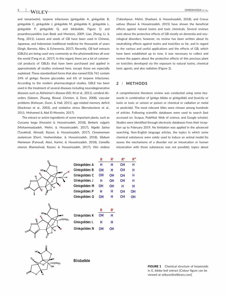
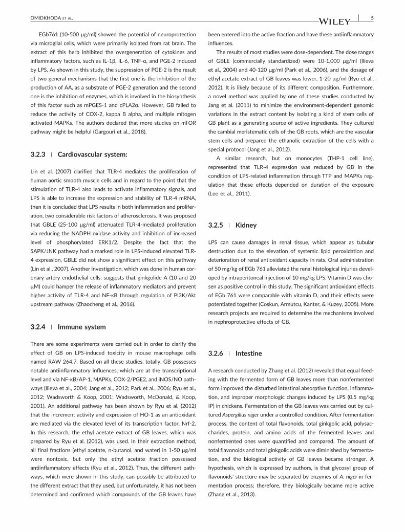
toxic agents, and also radiation (Figure 2).
2 | METHODS
A comprehensive literature review was conducted using some key-
words in combination of (ginkgo biloba or ginkgolide) and (toxicity or
toxin or toxic or venom or poison or chemical or radiation or metal
or pesticide). The most relevant titles were chosen among hundreds
of articles. Following scientific databases were used to search (last
accessed on: Scopus, PubMed, Web of science, and Google scholar).
Studies were identified through electronic databases from their incep-
tion up to February 2019. No limitation was applied in the advanced
searching. Non‐English language articles, the topics in which some
chemical substances were solely used to induce an animal model (to
assess the mechanisms of a disorder not an intoxication or human
intoxication with those substances was not possible), topics about
FIGURE 1 Chemical structure of terpenoidsin G. biloba leaf extract [Colour figure can beviewed at wileyonlinelibrary.com]
FIGURE 2 Protective effects of G. biloba extract against radiation, natural toxins and chemical toxicities [Colour figure can be viewed atwileyonlinelibrary.com]
OMIDKHODA ET AL. 3
the adverse reactions of nontoxic medications, and pathological disor-
ders such as Alzheimer were excluded. The flow chart of the search
process was shown in Figure 3. The contents were categorized into
three main headings: natural toxins, chemical toxicities, and radiation.
Figure 1 was drawn by ChemDraw software, ultra 7.0 version.
3 | NATURAL TOXINS
Natural toxins are chemicals that are naturally produced by living
organisms, including scorpion or snake venom, mycotoxins, bacterial,
and plant toxins. The antidotal effects of GBLE against natural toxins
have been shown in the several studies.
3.1 | Scorpion venoms
Leiurus quinquestriatus scorpion venom induces cellular damages,
which is associated to multiple organ failure (Amitai, 1998). In a study,
GBLE (150 mg/kg/day, orally, for 2 weeks before venom) and
aprotinin (nonselective protease inhibitor, 30 min before venom), sig-
nificantly reduced venom‐induced oxidative stress in the heart and
lung tissues of rats. Results indicated that combination of GBLE and
aprotinin potentiated the protective effects of GB on reduced gluta-
thione (GSH), malondialdehyde (MDA), and lactate dehydrogenase
(LDH) in the heart and lung tissues (Fatani et al., 2006). Therefore, a
direct conclusion is that oxidative stress and proteases are involved
in the venom‐induced cellular damages; however, it is better to evalu-
ate the protease inhibiting effects of GBLE in addition to its antioxi-
dant power.
3.2 | Lipopolysaccharide
3.2.1 | Lung
Lipopolysaccharide (LPS), which is found in the outer membrane of
Gram‐negative bacteria, provokes the activity of immune cells and
elicits inflammatory responses. The protective effects of GB against
LPS‐induced acute lung injury in mice (Huang et al., 2013; Lee et al.,
2014; Wu et al., 2016; Yao et al., 2015) and D‐galactose‐aged rats
(Sun, Zhang, Si, & Wang, 2002) have been shown.
To investigate the effect of GBLE on the acute lung injury induced
by LPS in D‐galactose‐aged rats, GBLE was given 7 days before the
experiment, once a day via intragastric administration. LPS‐induced
acute lung injury. In the LPS‐treated rats, more inflammatory cells in
the lung tissue were observed. Moreover, TNF‐α, LDH, and
myeloperoxidase (MPO) activities were increased in lung tissue of
LPS‐administrated rats compared with the control. GBLE could allevi-
ate all changes induced by LPS in these animals (Sun et al., 2002).
In another study EGb761 (0‐1000μg/kg) was administrated 30 min
before LPS in mice. After 6 hr, histopathological damages and arterial
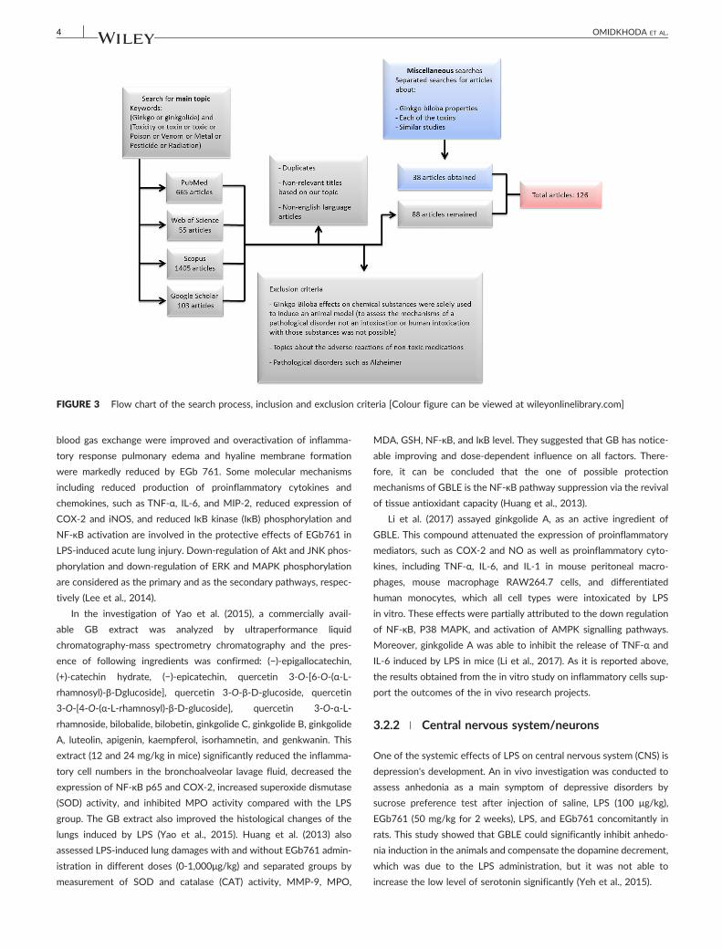
FIGURE 3 Flow chart of the search process, inclusion and exclusion criteria [Colour figure can be viewed at wileyonlinelibrary.com]
4 OMIDKHODA ET AL.
blood gas exchange were improved and overactivation of inflamma-
tory response pulmonary edema and hyaline membrane formation
were markedly reduced by EGb 761. Some molecular mechanisms
including reduced production of proinflammatory cytokines and
chemokines, such as TNF‐α, IL‐6, and MIP‐2, reduced expression of
COX‐2 and iNOS, and reduced IκB kinase (IκB) phosphorylation and
NF‐κB activation are involved in the protective effects of EGb761 in
LPS‐induced acute lung injury. Down‐regulation of Akt and JNK phos-
phorylation and down‐regulation of ERK and MAPK phosphorylation
are considered as the primary and as the secondary pathways, respec-
tively (Lee et al., 2014).
In the investigation of Yao et al. (2015), a commercially avail-
able GB extract was analyzed by ultraperformance liquid
chromatography‐mass spectrometry chromatography and the pres-
ence of following ingredients was confirmed: (−)‐epigallocatechin,
(+)‐catechin hydrate, (−)‐epicatechin, quercetin 3‐O‐[6‐O‐(α‐L‐
rhamnosyl)‐β‐Dglucoside], quercetin 3‐O‐β‐D‐glucoside, quercetin
3‐O‐[4‐O‐(α‐L‐rhamnosyl)‐β‐D‐glucoside], quercetin 3‐O‐α‐L‐
rhamnoside, bilobalide, bilobetin, ginkgolide C, ginkgolide B, ginkgolide
A, luteolin, apigenin, kaempferol, isorhamnetin, and genkwanin. This
extract (12 and 24 mg/kg in mice) significantly reduced the inflamma-
tory cell numbers in the bronchoalveolar lavage fluid, decreased the
expression of NF‐κB p65 and COX‐2, increased superoxide dismutase
(SOD) activity, and inhibited MPO activity compared with the LPS
group. The GB extract also improved the histological changes of the
lungs induced by LPS (Yao et al., 2015). Huang et al. (2013) also
assessed LPS‐induced lung damages with and without EGb761 admin-
istration in different doses (0‐1,000μg/kg) and separated groups by
measurement of SOD and catalase (CAT) activity, MMP‐9, MPO,
MDA, GSH, NF‐κB, and IκB level. They suggested that GB has notice-
able improving and dose‐dependent influence on all factors. There-
fore, it can be concluded that the one of possible protection
mechanisms of GBLE is the NF‐κB pathway suppression via the revival
of tissue antioxidant capacity (Huang et al., 2013).
Li et al. (2017) assayed ginkgolide A, as an active ingredient of
GBLE. This compound attenuated the expression of proinflammatory
mediators, such as COX‐2 and NO as well as proinflammatory cyto-
kines, including TNF‐α, IL‐6, and IL‐1 in mouse peritoneal macro-
phages, mouse macrophage RAW264.7 cells, and differentiated
human monocytes, which all cell types were intoxicated by LPS
in vitro. These effects were partially attributed to the down regulation
of NF‐κB, P38 MAPK, and activation of AMPK signalling pathways.
Moreover, ginkgolide A was able to inhibit the release of TNF‐α and
IL‐6 induced by LPS in mice (Li et al., 2017). As it is reported above,
the results obtained from the in vitro study on inflammatory cells sup-
port the outcomes of the in vivo research projects.
3.2.2 | Central nervous system/neurons
One of the systemic effects of LPS on central nervous system (CNS) is
depression's development. An in vivo investigation was conducted to
assess anhedonia as a main symptom of depressive disorders by
sucrose preference test after injection of saline, LPS (100 μg/kg),
EGb761 (50 mg/kg for 2 weeks), LPS, and EGb761 concomitantly in
rats. This study showed that GBLE could significantly inhibit anhedo-
nia induction in the animals and compensate the dopamine decrement,
which was due to the LPS administration, but it was not able to
increase the low level of serotonin significantly (Yeh et al., 2015).
OMIDKHODA ET AL. 5
EGb761 (10‐500 μg/ml) showed the potential of neuroprotection
via microglial cells, which were primarily isolated from rat brain. The
extract of this herb inhibited the overgeneration of cytokines and
inflammatory factors, such as IL‐1β, IL‐6, TNF‐α, and PGE‐2 induced
by LPS. As shown in this study, the suppression of PGE‐2 is the result
of two general mechanisms that the first one is the inhibition of the
production of AA, as a substrate of PGE‐2 generation and the second
one is the inhibition of enzymes, which is involved in the biosynthesis
of this factor such as mPGES‐1 and cPLA2α. However, GB failed to
reduce the activity of COX‐2, kappa B alpha, and multiple mitogen
activated MAPKs. The authors declared that more studies on mTOR
pathway might be helpful (Gargouri et al., 2018).
3.2.3 | Cardiovascular system:
Lin et al. (2007) clarified that TLR‐4 mediates the proliferation of
human aortic smooth muscle cells and in regard to the point that the
stimulation of TLR‐4 also leads to activate inflammatory signals, and
LPS is able to increase the expression and stability of TLR‐4 mRNA,
then it is concluded that LPS results in both inflammation and prolifer-
ation, two considerable risk factors of atherosclerosis. It was proposed
that GBLE (25‐100 μg/ml) attenuated TLR‐4‐mediated proliferation
via reducing the NADPH oxidase activity and inhibition of increased
level of phosphorylated ERK1/2. Despite the fact that the
SAPK/JNK pathway had a marked role in LPS‐induced elevated TLR‐
4 expression, GBLE did not show a significant effect on this pathway
(Lin et al., 2007). Another investigation, which was done in human cor-
onary artery endothelial cells, suggests that ginkgolide A (10 and 20
μM) could hamper the release of inflammatory mediators and prevent
higher activity of TLR‐4 and NF‐κB through regulation of PI3K/Akt
upstream pathway (Zhaocheng et al., 2016).
3.2.4 | Immune system
There are some experiments were carried out in order to clarify the
effect of GB on LPS‐induced toxicity in mouse macrophage cells
named RAW 264.7. Based on all these studies, totally, GB possesses
notable antiinflammatory influences, which are at the transcriptional
level and via NF‐κB/AP‐1, MAPKs, COX‐2/PGE2, and iNOS/NO path-
ways (Ilieva et al., 2004; Jang et al., 2012; Park et al., 2006; Ryu et al.,
2012; Wadsworth & Koop, 2001; Wadsworth, McDonald, & Koop,
2001). An additional pathway has been shown by Ryu et al. (2012)
that the increment activity and expression of HO‐1 as an antioxidant
are mediated via the elevated level of its transcription factor, Nrf‐2.
In this research, the ethyl acetate extract of GB leaves, which was
prepared by Ryu et al. (2012), was used. In their extraction method,
all final fractions (ethyl acetate, n‐butanol, and water) in 1‐50 μg/ml
were nontoxic, but only the ethyl acetate fraction possessed
antiinflammatory effects (Ryu et al., 2012). Thus, the different path-
ways, which were shown in this study, can possibly be attributed to
the different extract that they used, but unfortunately, it has not been
determined and confirmed which compounds of the GB leaves have
been entered into the active fraction and have these antiinflammatory
influences.
The results of most studies were dose‐dependent. The dose ranges
of GBLE (commercially standardized) were 10‐1,000 μg/ml (Ilieva
et al., 2004) and 40‐120 μg/ml (Park et al., 2006), and the dosage of
ethyl acetate extract of GB leaves was lower, 1‐20 μg/ml (Ryu et al.,
2012). It is likely because of its different composition. Furthermore,
a novel method was applied by one of these studies conducted by
Jang et al. (2011) to minimize the environment‐dependent genomic
variations in the extract content by isolating a kind of stem cells of
GB plant as a generating source of active ingredients. They cultured
the cambial meristematic cells of the GB roots, which are the vascular
stem cells and prepared the ethanolic extraction of the cells with a
special protocol (Jang et al., 2012).
A similar research, but on monocytes (THP‐1 cell line),
represented that TLR‐4 expression was reduced by GB in the
condition of LPS‐related inflammation through TTP and MAPKs reg-
ulation that these effects depended on duration of the exposure
(Lee et al., 2011).
3.2.5 | Kidney
LPS can cause damages in renal tissue, which appear as tubular
destruction due to the elevation of systemic lipid peroxidation and
deterioration of renal antioxidant capacity in rats. Oral administration
of 50 mg/kg of EGb 761 alleviated the renal histological injuries devel-
oped by intraperitoneal injection of 10 mg/kg LPS. Vitamin D was cho-
sen as positive control in this study. The significant antioxidant effects
of EGb 761 were comparable with vitamin D, and their effects were
potentiated together (Coskun, Armutcu, Kanter, & Kuzey, 2005). More
research projects are required to determine the mechanisms involved
in nephroprotective effects of GB.
3.2.6 | Intestine
A research conducted by Zhang et al. (2012) revealed that equal feed-
ing with the fermented form of GB leaves more than nonfermented
form improved the disturbed intestinal absorptive function, inflamma-
tion, and improper morphologic changes induced by LPS (0.5 mg/kg
IP) in chickens. Fermentation of the GB leaves was carried out by cul-
tured Aspergillus niger under a controlled condition. After fermentation
process, the content of total flavonoids, total ginkgolic acid, polysac-
charides, protein, and amino acids of the fermented leaves and
nonfermented ones were quantified and compared. The amount of
total flavonoids and total ginkgolic acids were diminished by fermenta-
tion, and the biological activity of GB leaves became stronger. A
hypothesis, which is expressed by authors, is that glycosyl group of
flavonoids' structure may be separated by enzymes of A. niger in fer-
mentation process; therefore, they biologically became more active
(Zhang et al., 2013).
6 OMIDKHODA ET AL.
3.2.7 | Ear
In an experimental study, LPS was directly applied into the ear of the
male guinea pigs to develop labyrinth toxicity. Degrees of hearing loss,
structural hair cell damages, increased extravasation, and decreased
cochlear blood flow were consequences of the LPS ototoxicity. About
20 μl of LPS in a concentration of 3 mg/ml was dropped
intratympanicly. The animals were treated with 100 mg/kg of GB for
3 days. The results indicated that this herbal extract strongly inhibited
the mentioned effects arose from LPS toxicity (Jang et al., 2011). The
specific cellular and molecular mechanisms involved in GB protection
against LPS‐ototoxicity have not been investigated.
3.2.8 | Eye
Ilieva et al. (2004) evaluated the effects of GB on uveitis caused by
LPS injection in rats, in addition to the cell culture assessment that it
has been mentioned before. In the in vivo part of the study, 24 hr after
200 μg (100 μg/footpad) of LPS and 1, 10, and 100 μg of GB admin-
istration, the aqueous humour of the animals was separated. Protein,
infiltrated cells, and NO level dramatically increased in LPS group
FIGURE 4 Summary of the mechanisms of G. biloba protection against Lirespectively, shows a significant decrease or increase, and × shows no signkinase B; AP‐1: activator protein‐1; CAT: catalase; COX‐2: cyclooxygenaseregulated kinase; GB: Ginkgo biloba L. ; GSH: reduced glutathione; HO‐1: He1beta; iNOS: nitric oxide synthase; JNK: jun N‐terminal kinase; LDH: lactaprotein kinase; MCP‐1: monocyte chemoattractant protein‐1; MDA: malonmatrix metalloproteinase‐9; mPGES‐1: microsomal prostaglandin E synthasdinucleotide phosphate; NF‐ΚB: nuclear factor kappa beta; NO: nitric oxidprostaglandin E2; PI3K: phosphatidylinositol‐3‐Kinase; SAPK: stress‐activasuperoxide dismutase; TLR‐4: toll‐like receptor‐4; TNF‐α: Tumour necrosiswileyonlinelibrary.com]
and were significantly suppressed by GB. These results in concomitant
with the findings of the cell culture assessment displayed the antioxi-
dant capacity of GBLE (Ilieva et al., 2004).
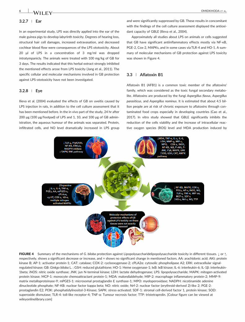
Approximately all studies about LPS on animals or cells suggested
that GB have significant antiinflammatory effects mostly via NF‐κB,
PGE‐2, Cox‐2, MAPKs, and in some cases viaTLR‐4 and HO‐1. A sum-
mary of molecular mechanisms of GB protection against LPS toxicity
was shown in Figure 4.
3.3 | Aflatoxin B1
Aflatoxin B1 (AFB1) is a common toxic member of the aflatoxins'
family, which was considered as the toxic fungal secondary metabo-
lite. Aflatoxins are produced by the fungi Aspergillus flavus, Aspergillus
parasiticus, and Aspergillus nominus. It is estimated that about 4.5 bil-
lion people are at risk of chronic exposure to aflatoxins through con-
taminated food crops especially in developing countries (Cao et al.,
2017). In vitro study showed that GBLE significantly inhibits the
reduction of the cells viability and the increase of intracellular reac-
tive oxygen species (ROS) level and MDA production induced by
popolysaccharidelipopolysaccharide toxicity in different tissues. ↓ or ↑,ificant change in mentioned factors. AA: arachidonic acid; AKt: protein‐2; cPLA2α: cytosolic phospholipase A2; ERK: extracellular signal‐me oxygenase‐1; IκB: IκB kinase; IL‐6: interleukin‐6; IL‐1β: interleukin‐te dehydrogenase; LPS: lipopolysaccharide; MAPK: mitogen‐activateddialdehyde; MIP‐2: macrophage inflammatory protein‐2; MMP‐9:e‐1; MPO: myeloperoxidase; NADPH: nicotinamide adeninee; Nrf‐2: nuclear factor (erythroid‐derived 2)‐like 2; PGE‐2:ted; SDF‐1: stromal cell‐derived factor 1, protein kinase; SOD:factor; TTP: tristetraprolin. [Colour figure can be viewed at
OMIDKHODA ET AL. 7
AFB1 in human hepatocytes (HepG2 cells) through antioxidant
effects (Hao et al., 2008). Another study was carried out by the
same authors on Wistar rats. They investigated the effects of
EGb761 on liver cancer induced by intra‐abdominal injection of
100‐200 μg/kg AFB1. The tests including MDA, GSH levels, and his-
topathological evaluation showed that after 42th and 55th weeks,
GB significantly reduced the size of the cancerous area and oxidative
stress (Hao et al., 2009). It seems that the inhibition of cancer can-
not only be due to the oxidative stress suppression; therefore, the
other more specific mechanisms should be considered.
3.4 | Lysophosphatidylcholine
Lysophosphatidylcholine (LPC) is an important lipid molecule in mam-
malian tissues and possesses different proinflammatory and athero-
genic effects (Matsumoto, Kobayashi, & Kamata, 2007). In vitro
study on H9c2 cell line showed that protective effect of GBLE against
LPC‐induced myocyte damage is partly attributed to the induction of
HO (an antioxidant enzyme which can mediate the adaptive cellular
response to oxidative stress and is responsible for regulating the intra-
cellular level of heme). The induction of HO was not observed by GB
terpenoids such as ginkgolide B and bilobalide. The induction of gene
expression and activity of HO by GBLE may be due to the alteration of
intracellular glutathione level (Chen, Zeng, Chen, Su, & Lai, 2001).
3.5 | Toxins derived from plants
3.5.1 | Lantadene
Latadenes (pentacyclic triterpenoides) are major toxic compounds
found in the leaves of the red flower variety of Lantana camara Linn.,
which belongs to Verbenaceae Family. Camara damages liver and kid-
neys and also induces photosensitization in ruminants. Among nonru-
minant animals, guinea pigs show the most typical symptoms
comparable to those observed in ruminants. It has been documented
that the methanolic leaf extract of GB was able to protect against
lantadene‐induced hepatic damage in guinea pigs. So that the elevated
levels of serum ALT, AST, and ALP induced by lantadenes were signif-
icantly decreased by methanolic GBLE dose‐dependently. The
increased levels of lipoperoxids and decreased levels of SOD, GSH
and catalase were markedly restored to that of control by methanolic
GBLE; moreover, the induction of apoptosis induced by lantadene was
suppressed in GB treated animals. The hepatoprotective effect of GB
was comparable to silymarin (Parimoo et al., 2014).
3.5.2 | Cassava
Cassava (Manihot esculenta Crantz) is an edible tuber due to its high
carbohydrate content. Cassava contains cyanogenic glycosides such
as linamarin and lotaustralin; so, long term consumption of cassava,
an improper processing, and a low protein diet have been associated
with neurodegenerative and neurological diseases. A study showed
the protective effect of GBLE (160 mg/kg/day, for 28 days, orally)
on disturbed locomotor activity and neuronal damage in hippocampus
induced by cassava juice (linamarin, 0.30 mg/kg) in rats. According to
the results of the study, in cassava‐treated rats, crossing and rearing
in the open field test and number of damaged neurons in the hippo-
campus were increased as compared with the vehicle group from
day 14 of treatment. Moreover, an uncoordinated swim characterized
by the lateral swim in the swim test was observed. GBLE attenuated
both behavioral and neuronal damages induced by cassava juice
administration possibly due to the high flavonoid content of GBLE
(Rivadeneyra‐Dominguez, Vazquez‐Luna, Rodriguez‐Landa, & Diaz‐
Sobac, 2013), because it is reported that flavonoids have the potential
of neuroprotection via multiple mechanisms, such as PI3K/Akt and
MAPK pathway regulation (Spencer, 2009).
3.5.3 | Gossypol
Pigment‐producing glands of the cotton seed contain a polyphenolic
compound known as gossypol, which can cause toxicity. Ingestion of
gossypol in high concentrations or for a long time decreases fertility
rate in ruminants and nonruminants probably because of increased
production of prooxidants and decreased antioxidant concentrations
(Santana et al., 2015). The protective effect of GBLE against
gossypol‐induced apoptosis in human lymphocytes was investigated.
Results showed that the level of apoptosis was decreased to 17.5%
and 20% following pretreatment of lymphocytes with 10 μg/ml EGb
761 for 30 min or 1 hr. Moreover, EGb 761 treatment (25‐150
μg/ml) reduced the percentage of apoptosis between 8 and 10% of
the control levels (Ergun, Yurtcu, & Ergun, 2005).
4 | CHEMICAL TOXICITIES
4.1 | Metals
4.1.1 | Aluminium
As aluminium (Al) is broadly distributed in the environment, exposure
to it is very common during daily life. Al and its salts are daily used
in drinking water, in our diets, food additives, and some beverages
(Lione, 1983). It is a well‐documented neurotoxicant due to its easy
access to the central nervous system and high accumulation in the
brain (Lakshmi, Sudhakar, & Prakash, 2015).
Several studies reported the beneficial effect of GBLE in Al‐
induced memory and learning impairments and oxidative stress in
the brain (Abd‐Elhady, Elsheikh, & Khalifa, 2013; Gong et al., 2006;
Gong, Wu, Huang, Sun, & Shi, 2005; Mohammadzadeh et al., 2017).
Learning and memory deficit were induced by both intragastric admin-
istration and drinking of AlCl3 solution for 3 (Gong et al., 2006) or 5
months (Gong et al., 2005). GBLE administration (50, 100, 200
mg/kg/day) for 2 months after AlCl3 intake ameliorated learning and
memory deficit through inhibition of the AChE expression in hippo-
campus (Gong et al., 2006) or by reduction of the levels of amyloid
precursor protein and caspase‐3 in hippocampus of Al‐treated rats
8 OMIDKHODA ET AL.
dose‐dependently (Gong et al., 2005). The improvement of memory
and learning by GB in the animals was confirmed by Morris water
maze test in both studies, so that GB caused significantly lower escape
latency and searching distance in comparison with AlCl3‐treated rats
(Gong et al., 2005; Gong et al., 2006). In another study, oral adminis-
tration of AlCl3 (10 mg/kg) for 3 months significantly increased TBARS
and decreased GSH, CAT, and SOD in the brain. Furthermore, AlCl3
reduced the levels of some neurotransmitters in brain tissue such as
noradrenaline, dopamine, and serotonin. Decrease in the amounts of
serum Zn, Cu, and Mg along with a significant increase in serum Fe
was observed in rats administered AlCl3. ALP and acid phosphatase
were increased by AlCl3. AlCl3 induced some degenerative changes
in rat brain tissues. These biochemical and histological changes were
improved by GB (200 mg/kg, orally for 3 months), which may be due
to its antioxidant effects (Mohammadzadeh et al., 2017). In the study
of Abd‐Elhady et al. (2013), rats were treated with 10 mg/kg of alu-
minium lactate intraperitoneally for 28 days in order to induce the alu-
minium toxicity. In this group, Al level was significantly raised in
serum, hippocampus, cortex, and whole brain; memory function in
the passive avoidance task test was disrupted; and AChE activity
was diminished. Twenty‐eight‐day oral administration of 200 mg/kg
of hydro‐alcoholic (50:50) GBLE caused a higher level of AChE activity
and memory function without any changes in Al levels in serum or
brain of the animals. GBLE also increased the reduced form of gluta-
thione and decreased MDA level (Abd‐Elhady et al., 2013).
4.1.2 | Lead
Lead, which is chemically named Pb, is one of the most important toxic
heavy metals that have been concentrated in the earth via human
industrial activities. This element can cause infertility, psychotic and
degenerative disorders, high blood pressure, renal dysfunction, and
interrupted zinc‐related functions in the body (Maret, 2017).
In an investigation, 50 and 100 mg/kg GBLE were used orally to
evaluate its possible effects on oxidative stress induced by 500 ppm
of lead acetate in drinking water (rats were treated with lead acetate
for 4 weeks). In this study, frontal cortex, cerebellum, hippocampus,
and brain stem of sacrificed animals were prepared and the content
of factors, including free radicals, MDA, and antioxidant enzymes were
measured. The findings demonstrated that GB dose‐dependently
protected the brain tissue from high levels of oxidative stress, which
is caused by lead acetate intoxication (Yallapragada & Velaga, 2015).
4.1.3 | Cadmium
This metal is a toxic element, which enters into the body through inha-
lation and ingestion. The smoke of cigarette reported as a major
source of exposure to cadmium in humans (Bernhoft, 2013).
In the original study of Predes et al. (2010), the single dose injec-
tion of 3 μmol/kg cadmium chloride, after 56 days, resulted in
decreased volume of rat leydig cells, their cytoplasm, and nucleus;
however, it did not induce any significant changes in the blood testos-
terone level, the size of each testicular parenchyma component, and
the number of leydig cells. Fifty‐six‐day treatment of animals with oral
administration of GB extract (a commercial product containing 19.2
mg/ml ginkgo flavonoids and 4.8 mg/ml terpenelactone) with the dose
of 100 mg/kg prevented the cellular histomorphological alterations.
The authors declared that the relation of these protective effects of
GB to its antioxidant properties needs to be confirmed with more
investigations (de Souza Predes, Monteiro, Matta, Garcia, & Dolder,
2011).
4.1.4 | Mercury
There is mercury (Hg) in the three forms of elemental, organic, and
salts (mono or divalent). Pharmacokinetic characteristics, toxicity mag-
nitude, and target organs of mercury depend on the form to which
humans are exposed (Bernhoft, 2012).
The effect of antioxidant activity of GB on mercury intoxication
was evaluated in an animal study. To develop the mercury toxicity,
rats were administered 5 mg/kg of HgCl2 as a single dose. Mercury
injection significantly elevated the blood level of BUN, Cr, ALT, AST,
TNF‐α, and LDH activity associated with increased MPO activity, free
radical overgeneration, and decreased GSH amount in kidney, liver,
brain, and lung tissues. Intraperitoneally injection of 150 mg/kg of
the GBLE for 5 days was considerably capable of restoring all these
changes (Çavuşolu, Yapar, & Yalçin, 2009).
4.1.5 | Heavy metal‐contained wastewater
This kind of water is often found nearby to industrial centres. Waters,
which consist of the high amount of heavy metals, can endanger the
life of its surrounding animals and plants. Cavusoglu et al. (2008) ana-
lyzed the wastewater, which is discharged into the Melet River in the
terms of heavy metals and evaluated its effects on germination and
growth of the Vicia faba L. seeds with and without exposure to GBLE.
The levels of MDA and micronuclei (MN) were also measured in the
plant roots. The analysis of the polluted water showed that the heavy
metals from the most to the least concentration, respectively, were
Pb, Al, Ni, Cr, Fe, Cu, Zn, and Cd. The number of germinated seeds,
which were fed with wastewater, was much less than those were
fed with the tap water. In the presence of the GBLE (10, 20, and 30
μM in the river water) germination percentage was significantly raised
in a dose‐dependent manner. The effects of polluted water and GB on
the root lengths and weights were similar to their effects on germina-
tion percentage. GB also had significant alleviative influence on
increased MDA and MN level induced by waste water feeding
(Cavusoglu et al., 2010).
4.1.6 | Triethyltin
This neurotoxic agent is the organic form of tin atom, which is
attached to three ethyl groups. It is demonstrated that GBLE could
inhibit the deleterious effects of triethyltin (TET) in rats, so that there
was no change in electrolyte and water balance of the brain in the ani-
mals, which were dosed GBLE (10 ml/kg of drinking water consists of
OMIDKHODA ET AL. 9
100 mg/kg GB) accompanying withTET (0.002% in drinking water) for
14 days, and TET administration was associated with significant
destructive cerebral oedema and histological damages in comparison
with normal animals (Otani, Chatterjee, Gabard, & Kreutzberg, 1986).
The other study focused on the possible molecular mechanism of GB
related to these protective effects. The results of this study clarified
that GBLE was able to restore the activity of the membrane‐bound
form of the PDE, which was reduced by TET administration, but it
did not affect on the soluble form. The performers showed that this
modulating effect of GBLE is not because of the formation of complex
with TET and it possibly had a direct or indirect activating effect. PDE
activation prevents the cyclic‐AMP accumulation in the cells and its
toxic effects on cellular metabolisms (Macovschi, Prigent, Nemoz, &
Pacheco, 1987).
In view of the effects of GBLE against neurotoxicity induced by
some metals, such as Al and Pb or the other toxic agents, it is interest-
ing to clarify the active ingredients of this herbal extract which is
effectively able to cross the brain‐blood barrier.
4.2 | Ethanol
Ethanol is one of the alcoholic compounds that is widely used and
abused in all over the world. Exposure to long term or high concentra-
tions of ethanol can lead to chronic or acute toxicity. The liver is the
major organ that affected by ethanol, because this substance is metab-
olized mostly in the liver, although a lot of tissues are susceptible to its
adverse effects (Rusyn & Bataller, 2013). The results of Chan and
Hsuuw research (2007) revealed that the ethanol in a concentration
range of 50‐400 mM for 36 hr significantly decreased Hep G2 cell via-
bility and increased the cellular apoptosis and ROS generation. When
ginkgolide B was added up to 25 mM to the cells, which were treated
with 100 mM of ethanol, cell viability, apoptosis percentage, and ROS
production were dose‐dependently improved; however, the higher
concentrations (50 and 100 mM) of ginkgolide B with the concomitant
of 100 mM of ethanol induced more apoptosis and ROS generation.
The effect of the concurrent use of ginkgolide B with ethanol on
caspase‐3 level and JNK/AP‐1 activity in hepatic cells was the same
as the results obtained from cell viability assay (Chan & Hsuuw,
2007); thus, it indicates that the hepatocellular protective effects of
ginkgolide B can be associated with suppression of caspase‐3, oxida-
tive stress, and regulation of JNK/AP‐1 pathway.
Yao et al. (2007, 2009) in two different studies exhibited the role
of HO‐1 in mediating antioxidant effects of GB on liver oxidative inju-
ries induced by ethanol. In the first study, animals were treated with
2.4 mg/kg of oral ethanol solution for 90 days in order to induce liver
toxicity, and in two separated groups, animals pretreated with 48 and
96 mg/kg of GBLE 1 hr before ethanol administration. In the next step,
after the sample preparation, serum ALT and AST level, MDA, GSH,
SOD, glutathione peroxidase (GPX), CAT level, and HO‐1 gene expres-
sion in the liver tissue were measured. The ethanol administration
resulted in much higher level of ALT, AST, and MDA than the control
group and significant reduction in tissue antioxidant enzymes and HO‐
1 expression. GB reversed all consequences of ethanol ingestion to
the normal level in a dose‐dependent manner (Yao et al., 2007). In
the second study, the effect of quercetin, one of the active ingredients
of GB and also a lot of other herbal extracts, on ethanol‐associated
hepatocyte toxicity via the HO‐1 pathway was assessed. It was dem-
onstrated that carbon monoxide (CO), an outcome of the heme
metabolism with HO‐1 activity rather than other metabolites, is effec-
tive in the protective pathways of quercetin. It was clarified that CO
inhibited the ethanol‐induced CYP2E1 up‐regulation, an enzyme
involved in oxidative stress. Therefore, quercetin can attenuate the
oxidative damages of ethanol through the up‐regulation of HO‐1
and then its metabolite, CO (Yao et al., 2009).
In another research, Qiu et al. (2015) investigated the effects of a
triple mixed herbal formulation named SGR on fatty liver developed
by chronic ethanol consumption. This formulation consists of three
herbs: Semen Hoveniae extract: 80%, GBLE: 10%, and Rosa roxburghii
Tratt extract: 10%. After mixing prepared Semen Hoveniae extract
with other two purchased extracts, the content analysis was carried
out by chromatography, and it illustrated the presence of
dihydromyricetin, dihydroquercetin, quercetin, and flavonoids. Find-
ings of this study represented that ethanol elevated the triglyceride
level in serum and liver tissue of mice through the decreased expres-
sion level of adiponectin, peroxisome proliferator‐activated receptor
(PPAR‐α) and AMPK‐β, and increased expression level of TNF‐α and
sterol regulatory element binding protein 1c (SREBP‐1c). SGR adminis-
tration for 30 days had the potential of regulating the expression of
adiponectin, PPAR‐α, TNF‐α, SREBP‐1c, and the level of phosphory-
lated AMPK‐β, instead of the total expression of AMPK‐β (Qiu et al.,
2015). In the studies, which are conducted on a combination of several
herbs, there is an uncertainty in making conclusion about the effects
of every herbal component, unless some treated groups with the iso-
lated or purified herbal extracts are added.
4.3 | Carbon tetrachloride (CCl4)
This compound is chemically a member of the haloalkanes that nowa-
days their uses have been minimized or forbidden because of their
toxic effects. The liver is the most important target organ of CCl4
(Weber, Boll, & Stampfl, 2003). CCl4 interaction with CYP2E1 leads
to CCl3̇ radical, which reacts with oxygen and generates the CCl3O
free radical. The formation of this radical, possibly, is the onset of
the consequent injuries like lipid peroxidation, cell membrane dysfunc-
tion, calcium homeostasis disturbances, cellular enzymatic leakage,
DNA damage and genotoxicity, hepatic fibrosis, cellular proliferative
changes, and finally liver cancer (Manibusan, Odin, & Eastmond, 2007).
There are four similar studies about the effects of GB on CCl4 hep-
atotoxicity in rats with the differences in dosing administration of CCl4
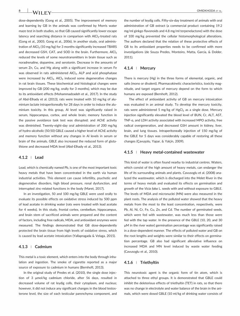
and GBLE. These studies were summarized in Table 1. In the study of
Shenoy et al. (2001), CCl4 was intraperitoneally injected at the dose of
0.5 mg/kg for 7 days, and in another group 50 mg/kg/day of GBLE
(with 0.24 mg of ginkgo flavonglycosides per gram of dry extract)
was simultaneously administered (IP) with CCl4. Oral silymarin (200
mg/kg) was given as a positive control. After treatments, the level of
ALT, AST, ALP, total protein (TP), and albumin (Alb) in the serum and
TABLE 1 Protective effects of G. biloba leaf extract against carbon tetrachloride
CCl4 and GBLE dosing
Evaluated
tissue G. biloba effects References
CCl4: 0.5 ml/kg/day, 7 days, IP
GBLE: 50 mg/kg/day, 7 days
liver ↓ALT, ↓AST, ↓ALP, ↑TP, ↑Alb in the blood
↓MDA, ↑GSH in the liver
Histopathological improvement
Ashok et al. (2001)
CCl4: 1 ml/kg/day of 50% (v/v) CCl4 solution in olive
oil, 7 days, IP
GBLE: 25 and 50 mg/kg/day, 10 days (3 days before
the start of CCl4 injection), IP
liver ↓ALT, ↓AST, ↓ALP, ↑TP, ↑Alb in the blood
↓MDA, ↑GSH, ↑SOD, ↑CAT, ↑GPX in the liver
Histopathological improvement
Naik and Panda (2007)
CCl4: 0.25 ml/kg of 50% CCl4 suspension in corn oil,
single dose, PO
GBLE: 4 mg/kg/day, 5 days before CCl4 administration,
PO
liver ↓ALT, ↓AST, ↑Alb in the blood
↓MDA, ↑TNF‐α and ↑IL‐6 mRNA level in the liver
Histopathological improvement
Chavez‐Morales et al. (2011)
CCl4: 5 ml/kg of 40% (v/v) CCl4 solution in olive oil for
the first time, then 3 ml/kg every 4 days, SC
GBLE: 10, 40 and 160 mg/kg/day, 6 weeks
liver ↓ALT, ↓AST, ↓ALP, ↑TP, ↑Alb, ↓hyaluronic acid,
↓laminin, ↓triglyceride, ↓total cholesterol in the
blood
↓MDA, ↑GSH, ↑SOD in the liver
Histopathological improvement
Yang et al. (2011)
CCl4: 0.25 ml of 50% CCl4 suspension in corn oil,
single dose, PO
GBLE: 4 mg/kg/day, 5 days before CCl4 administration
Kidney ↓TP and ↓glucose in the urine
↓MDA in the kidney
Histopathological improvement
Chavez‐Morales et al. (2017)
Note. ↓ or ↑, respectively, shows a significant decrease or increase in comparison with CCl4‐treated group.
10 OMIDKHODA ET AL.
the amount of MDA and GSH in the liver tissue were measured.
Moreover, the histopathological assessment of the livers was also per-
formed (Ashok et al., 2001). Other three researches were conducted
according to the protocols mentioned in the table below (Table 1).
Based on all four studies, we can totally conclude that carbon tetra-
chloride elevated the level of MDA and therefore caused the cellular
membrane damage that the rise in the blood level of hepatic enzymes
is the evidence of that. Drop in the level of albumin was an indicator
of the liver dysfunction due to the CCl4 administration, and the histo-
pathological assessment showed the tissue necrosis and fatty infiltra-
tion to some extent. Treatment of animals with GBLE preserved the
liver from being harmed by CCl4 intake (Ashok et al., 2001; Naik &
Panda, 2007; Chavez‐Morales et al., 2011; Yang, Wang, Ye, & Li,
2011). It should be noted that isolated and purified polyphenols of
GB leaves have been used in the study of Yang et al., 2011; therefore,
the polyphenols in addition to flavone glycosides and terpenoids of
GB leaves are effective at least on CCl4‐developed hepatic toxicity.
Moreover, the study of Naik and Panda (2007) has been the only
study in which the phytosomes of G. biloba extract (Ginkgoselect)
were used. These phytosomes also consist of flavone glycosides, ter-
penoids, tannins, and proteins.
One of the effects of CCl4 on the liver tissue in the study of Chavez‐
Morales et al. (2010) is the elimination of theTNF‐α expression in com-
parison with control (Chavez‐Morales et al., 2011). This effect is in the
vise versus line of some studies (Weber et al., 2003). The authors
believe that this result is due to the inhibition of protein expression by
CCl4 in the liver and the restoring effect of GBLE in this case was
expressed as a modulating influence (Chavez‐Morales et al., 2011).
Another experiment of Chavez‐Morales et al. (2017) demonstrated that
neither of renal functional factors, including urine flux, clearance of
inulin, and p‐aminohippurate was influenced by CCl4, but it significantly
increased proteinuria, MDA level, and induced tubular necrosis more in
the inner cortex of the kidneys. GBLE administration significantly allevi-
ated these injuries (Chavez‐Morales et al., 2017).
Although the mechanisms of CCl4 toxicity have partly been deter-
mined, in all mentioned studies, only the general oxidation or
inflammation‐related parameters were evaluated in GB protective
effect on CCl4. It would be more valuable that the effects of GB on
specific pathways involved in CCl4 toxicity, including CYP2E1 expres-
sion or DNA damage, were also assessed.
4.4 | Pesticides
4.4.1 | Insecticides
Methamidophos
Finkler, Silveira, Munaro, and Zanrosso (2012) investigated the possi-
ble effect of EGb 761 on the ototoxicity induced by methamidophos
(0.3 and 3 mg/kg/day for 7 days) as an organophosphorus pesticide.
The structure of the main part of cochlea including hair cells, named
organ of corti, in guinea pigs was analyzed by means of the electron
microscopy and the results indicated that GBLE (100 mg/kg/day, 90
min before methamidophos administration) prevented the organ of
corti structural deformation (Finkler et al., 2012).
Diazinon
Diazinon is the other insecticide, which is applied for agricultural pur-
poses. Several investigations demonstrated the diazinon genotoxicity
or its toxic effects on the animal vascular system, heart, and liver.
These toxicities were limited by herbal active ingredients (Lari et al.,
OMIDKHODA ET AL. 11
2015; Razavi, Hosseinzadeh, Abnous, & Imenshahidi, 2014; Razavi,
Hosseinzadeh, Abnous, Khoei, & Imenshahidi, 2016; Razavi,
Hosseinzadeh, Movassaghi, Imenshahidi, & Abnous, 2013; Zeinali,
Meybodi, Rezaee, Rafatpanah, & Hosseinzadeh, 2018). Considering
that this substance and the other similar compounds may enter the
waters and threat the marine animal lives, a study has been conducted
about its toxic effects on the immune system of Rainbow trout fishes,
one of the most cultured fish species. The immunomodulatory and
immunoprotective effects of GB were assessed on this toxicity. The
findings exhibited that 1 and 2 g of dried hydro‐alcoholic GB extract
per 1 kg of fish diet were able to restore the serum total immunoglob-
ulin level, peroxidise, and lysosome activity, which had been sup-
pressed by diazinon (0.287 mg/l). These two doses of GB also
inhibited the rise of renal transforming growth factor beta (TGF‐β1)
and IL‐1β level due to the diazinon exposure. The immunomodulatory
effects of GB did not appear in doses of 0.5 and 4 g/kg diet. The
authors have declared that GB in higher doses probably has
immunotoxic effects in this model, because it was observed that 4
g/kg of GB lessened total immunoglobulins, complement activity,
peroxidise, and lysosome activity with and without diazinon in com-
parison with control (Hajirezaee, Rafieepour, Shafiei, & Rahimi, 2019).
4.4.2 | Rodenticides
Bromethalin is a potent rodenticide, and it is used in the resistant
cases to anticoagulant poisons (DeClementi & Sobczak, 2018). The
mechanism of its toxic effects in the brain as a major target organ is
by reducing cellular ATP level following the uncoupling of mitochon-
drial oxidative phosphorylation. A decreasing in the ATP production
leads to dysfunction of the Na/K ATPase pump and finally the accu-
mulation of the Na and water (Peterson, 2013). Bromethalin was
administered with the dose of 1 mg/kg. After 24 hr, MDA, Na and
water level in the brain were determined. In the other group, rats
were treated with bromethalin with concurrent administration of
100 mg/kg of GBLE. This herbal extract could hamper the accumula-
tion of the excessive amounts of Na and water and the over‐
generation of MDA in the brain tissue of bromethalin‐treated rats.
Furthermore, GBLE significantly attenuated the neurotoxic signs,
which were consequences of the cerebral oedema and injuries
(Dorman, Cote, & Buck, 1992).
4.4.3 | Herbicides
Glyphosate
GBLE was also used to be checked that whether it has any effect on
glyphosate toxicity. To reach this, Cavusoglu, Yapar, Oruc, and Yalcin
(2011) orally administered 50 and 150 mg/kg of GBLE with intraperi-
toneal single injection of glyphosate (50 mg/kg) in mice. In the animals,
which received only glyphosate, ALT, AST, BUN, Cr, and MDA levels
in the liver and kidney significantly elevated in comparison with the
control group. In addition, genotoxicity was markedly developed in
this group. It was evaluated through the measurement of the related
factors, including MN frequency in red blood cells, mitotic index, and
chromosome aberrations in bone marrow cells. GB possessed the pro-
tective effect on all these abnormalities likely via its antioxidant prop-
erties (Cavusoglu et al., 2011).
Paraquat
Mostly, an accidental exposure to high doses of paraquat can lead to
toxicity and even death. The major target organ of this herbicide is
lung (Kurisaki, 1989); however, it may have neural toxicity.
The original study of Kang, Chen, Xu, Li, and Wang (2007) demon-
strated that GBLE (10, 20, and 40 μg/ml) dose‐dependently reduced
the injuries in PC12 cells treated with paraquat. Following the cell
incubation with paraquat, cell viability and B‐cell lymphoma 2 (Bcl‐2)
level significantly decreased and apoptosis‐related parameters
increased (Kang et al., 2007). Nevertheless, one cell culture study is
not valid enough to verify the protective effects of GB against
paraquat‐induced toxicity.
4.4.4 | Fungicides
Topsin
In the investigation of Sakr, Mahran, and Abdel‐Maksoud (2011), GB
was considered for its antioxidative activity in the case of ovarian
toxicity via exposing to dimethyl 4, 4,‐(o‐phenylene) bis (3‐
thioallophanate), a chemical fungicide, which is commercially named
topsin. It is often used as a plant seed preservative. The findings of this
study suggested that topsin (oral dose of 0.1 LD50 for 8 weeks)
caused pathologic alterations in the ovary components, including epi-
thelial cells, blood vessels, the structure, and the number of follicles. In
addition to histological changes, follicle‐stimulating hormone and
luteinizing hormone level were significantly diminished and serum
estradiol level obviously increased. The elevation of serum MDA level
and the significant reduction in serum SOD and CAT enzymes indi-
cated that topsin toxicity can be mediated through oxidative stress
and overgeneration of ROS. Remarkable efficacy of GBLE (40
mg/kg/day for 4 weeks after topsin administration), as a powerful
antioxidant, in the improvement of topsin toxicity confirmed this
hypothesis (Sakr et al., 2011).
4.5 | Cigarette smoke
In two separated assays of Wang et al. (2010 and 2011), GBLE was
utilized alone and with cobalt porphyrin in cigarette filter to minimize
the toxicity of its smoke. After preparing the GB‐contained cigarettes,
a system was applied to produce and trap the cigarette smoke in order
to test its free radical contents, in vivo acute and chronic toxicity in
the animals. The condensed form of cigarette smoke was also tested
for mutagenicity potential. Both modified versions of cigarettes had
less toxicity and mutagenicity than non‐modified cigarette and the
animals, which were exposed to GB‐contained cigarette experienced
milder toxicity‐related complications, so it means that GB lowered
the toxic effects of cigarette smoke. On the other hand, there were
much lower amounts of solid and gas‐phase free radicals related to
modified version in comparison with non‐modified one (Wang et al.,
12 OMIDKHODA ET AL.
2010; Wang et al., 2011). Therefore, the protective effects of GB
might originate from its radical scavenging and antioxidant activity.
4.6 | Monosodium glutamate
This compound is a food additive, which is widely applied in the proc-
essed food industry. The adverse effects of monosodium glutamate
(MSG) intoxication appear mostly in CNS, liver, and reproductive sys-
tem (Husarova & Ostatnikova, 2013).
An investigation represented that oral 1.5 mg/kg of MSG, twice a
week for a month, was attributed to dysfunction of the liver and kid-
ney in rats. About 80 mg/kg of GBLE, along with MGS administration,
markedly attenuated the liver enzymes, BUN, Cr, and MDA levels.
These findings demonstrated that the oxidative stress plays a crucial
role in the MSG toxicity (Elatrash & Abd El‐Haleim, 2015).
4.7 | Naphthalene
Naphthalene is broadly used in the industry as a precursor to produce
chemical compounds. Acute exposure to toxic amounts of this sub-
stance can result in hemolytic anaemia. Toxic effects of naphthalene
are mostly because of increased oxidative stress. A study revealed that
30‐day administration of 100 mg/kg of naphthalene induced overpro-
duction of free radicals, serum TNF‐α and IL‐β, and significantly sup-
pressed antioxidative capacity of lung, liver, and kidney of mice.
Furthermore, the histopathological evaluation of the animal lung, kid-
ney, and liver clarified that naphthalene harmed the structure of these
tissues. Most importantly, the oral treatment of naphthalene‐injected
animals with 150 mg/kg of GB prevented all of the mentioned injuries
(Tozan et al., 2007).
4.8 | Fluoride
Fluoride is one of the important trace elements for the human body,
but in the optimum level. In fact, consumption of the larger amounts
of this ion can lead to fluorosis and neural damage.
In an animal study, it is observed that the level of MDA and GSH
and the activity of SOD and CAT in fluoride‐treated mice (50 mg/L
in drinking water for 30 days) were significantly higher than both nor-
mal mice (with 0.04 ppm fluoride in drinking water) and fluoride‐
treated mice, which concomitantly received 20 mg/kg/day of EGb
761 for 30 days. Moreover, tissue staining of hippocampus for the his-
topathological assessment showed considerable neural degeneration
in the intoxicated animals, in comparison with control and EGb 761
plus fluoride‐treated group (Atmaca, Aksu, Yıldırım, & Atmaca, 2014).
Totally, it is concluded that EGb 761 is a protective agent against oxi-
dative stress induced by high concentration of fluoride.
4.9 | Chemotherapy medications
The effects of GB leaf extract have been investigated on different
toxicities developed by anticancer drugs including cisplatin,
doxorubicin, bleomycin, 5‐fluorouracil (5‐FU), and oxaliplatin. All
studies, which have been performed in this field up to now, were
summarized in Table 2. Overall, this valuable herbal extract illus-
trated its antioxidant, antiapoptotic, and antiinflammatory effects;
however, it also possessed the other special influences. This extract
even alleviated the structural and functional changes in the evalu-
ated tissues, and in some cases, it influenced even at the protein
expression level. In most of these studies the measurement of oxida-
tive stress‐associated parameters was in common (Abd‐Ellah &
Mariee, 2007; Amin et al., 2012; Astolfi, Simoni, Ciorba, & Martini,
2008; Daba et al., 2002; Erdogan et al., 2006; Esen et al., 2018;
Hauns, Haring, Kohler, Mross, & Unger, 2001; Huang, Whitworth,
& Rybak, 2007; Khafaga & Bayad, 2016; Liu et al., 2008; Marshall
et al., 2004; Naidu, Vijay, Krishna, Sundaram, & Singh, 2002;
Yeh et al., 2009).
All of the studies in the table below (Table 2) were carried out
on the cells or animals with the exception of two studies conducted
on humans by Hauns et al. (2001) and Marshall et al. (2004).
The results of Hauns et al. clinical trial in which 44 patients
with colorectal cancer were involved demonstrated that GBLE could
even enhance the efficacy of chemotherapy with 5‐FU, in addition
to the reduction of the drug toxic effects and the increment of
patient tolerability (Hauns et al., 2001). The limited number of cases
in this study is a weakness point of it. Another clinical study also
suggested the positive effect of GBLE on treatment efficacy and
its alleviative influence on oxaliplatin‐induced neurotoxicity (Marshall
et al., 2004).
About 50‐200 mg/kg of GBLE or EGb 761 were given to the ani-
mals in most studies; however, a very lower dose of this extract (5
mg/kg/dose) was used to assess its antidotal effects on doxorubicin
cardiotoxicity and reproductive toxicity and it was significantly effec-
tive (Liu et al., 2008; Yeh et al., 2009). Because a similar study on
doxorubicin‐induced cardiotoxicity was done using 100 mg/kg of GB
(Abd‐Ellah & Mariee, 2007), the only conclusion is that the lower
doses of EGb 761 than the usual doses have also enough potency to
influence at least on doxorubicin toxicities.
5 | RADIATION
Based on previous studies, which have been mentioned below, GB has
some protective effects on damages induced by different kinds of
radiation to which humans are exposed in their lifetime, such as waves
produced by cell phones and radioactive elements or scattered during
radiotherapy, radiography, or even sunshine.
5.1 | Cell phone
Gevrek, Aydin, Ozsoy, Aygun, and Bicer (2017) demonstrated that
intraperitoneal injection of 100 mg/kg/day of EGb761 reversed high
testicular apoptotic index and the testis histomorphological changes
and disturbances related to animal fertility, such as decreased sperm
numbers and testosterone hormone level induced by cell phone
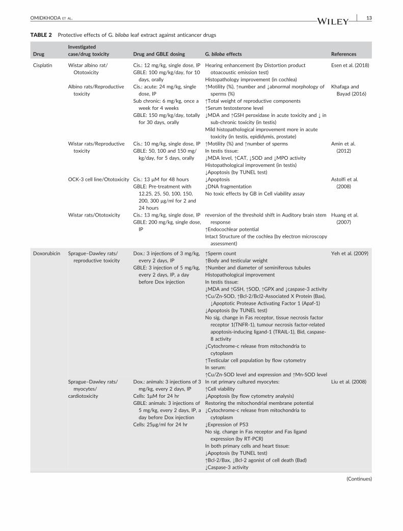
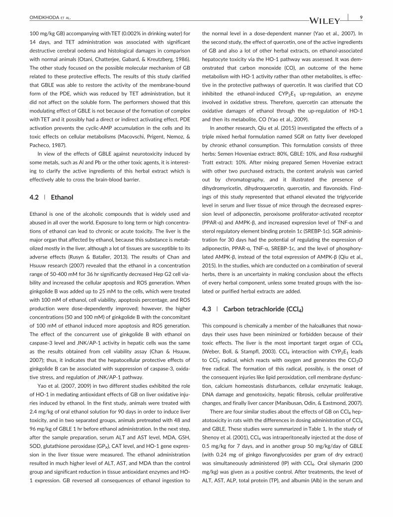
TABLE 2 Protective effects of G. biloba leaf extract against anticancer drugs
Drug
Investigated
case/drug toxicity Drug and GBLE dosing G. biloba effects References
Cisplatin Wistar albino rat/
Ototoxicity
Cis.: 12 mg/kg, single dose, IP
GBLE: 100 mg/kg/day, for 10
days, orally
Hearing enhancement (by Distortion product
otoacoustic emission test)
Histopathology improvement (in cochlea)
Esen et al. (2018)
Albino rats/Reproductive
toxicity
Cis.: acute: 24 mg/kg, single
dose, IP
Sub chronic: 6 mg/kg, once a
week for 4 weeks
GBLE: 150 mg/kg/day, totally
for 30 days, orally
↑Motility (%), ↑number and ↓abnormal morphology of
sperms (%)
↑Total weight of reproductive components
↑Serum testosterone level
↓MDA and ↑GSH peroxidase in acute toxicity and ↓ in
sub‐chronic toxicity (in testis)
Mild histopathological improvement more in acute
toxicity (in testis, epididymis, prostate)
Khafaga and
Bayad (2016)
Wistar rats/Reproductive
toxicity
Cis.: 10 mg/kg, single dose, IP
GBLE: 50, 100 and 150 mg/
kg/day, for 5 days, orally
↑Motility (%) and ↑number of sperms
In testis tissue:
↓MDA level, ↑CAT, ↓SOD and ↓MPO activity
Histopathological improvement (in testis)
↓Apoptosis (by TUNEL test)
Amin et al.
(2012)
OCK‐3 cell line/Ototoxicity Cis.: 13 μM for 48 hours
GBLE: Pre‐treatment with
12.25, 25, 50, 100, 150,
200, 300 μg/ml for 2 and
24 hours
↓Apoptosis↓DNA fragmentation
No toxic effects by GB in Cell viability assay
Astolfi et al.
(2008)
Wistar rats/Ototoxicity Cis.: 13 mg/kg, single dose, IP
GBLE: 200 mg/kg, single dose,
IP
reversion of the threshold shift in Auditory brain stem
response
↑Endocochlear potentialIntact Structure of the cochlea (by electron microscopy
assessment)
Huang et al.
(2007)
Doxorubicin Sprague–Dawley rats/
reproductive toxicity
Dox.: 3 injections of 3 mg/kg,
every 2 days, IP
GBLE: 3 injection of 5 mg/kg,
every 2 days, IP, a day
before Dox injection
↑Sperm count
↑Body and testicular weight
↑Number and diameter of seminiferous tubules
Histopathological improvement
In testis tissue:
↓MDA and ↑GSH, ↑SOD, ↑GPX and ↓caspase‐3 activity
↑Cu/Zn‐SOD, ↑Bcl‐2/Bcl2‐Associated X Protein (Bax),
↓Apoptotic Protease Activating Factor 1 (Apaf‐1)↓Apoptosis (by TUNEL test)
No sig. change in Fas receptor, tissue necrosis factor
receptor 1(TNFR‐1), tumour necrosis factor‐relatedapoptosis‐inducing ligand‐1 (TRAIL‐1), Bid, caspase‐8 activity
↓Cytochrome‐c release from mitochondria to
cytoplasm
↑Testicular cell population by flow cytometry
In serum:
↑Cu/Zn‐SOD level and expression and ↑Mn‐SOD level
Yeh et al. (2009)
Sprague–Dawley rats/
myocytes/
cardiotoxicity
Dox.: animals: 3 injections of 3
mg/kg, every 2 days, IP
Cells: 1μM for 24 hr
GBLE: animals: 3 injections of
5 mg/kg, every 2 days, IP, a
day before Dox injection
Cells: 25μg/ml for 24 hr
In rat primary cultured myocytes:
↑Cell viability↓Apoptosis (by flow cytometry analysis)
Restoring the mitochondrial membrane potential
↓Cytochrome‐c release from mitochondria to
cytoplasm
↓Expression of P53
No sig. change in Fas receptor and Fas ligand
expression (by RT‐PCR)In both primary cells and heart tissue:
↓Apoptosis (by TUNEL test)
↑Bcl‐2/Bax, ↓Bcl‐2 agonist of cell death (Bad)
↓Caspase‐3 activity
Liu et al. (2008)
(Continues)
OMIDKHODA ET AL. 13
TABLE 2 (Continued)
DrugInvestigatedcase/drug toxicity Drug and GBLE dosing G. biloba effects References
Sprague–Dawley rats/
hyperlipidemic
nephrotoxicity
Dox.: 5 mg/kg, single dose, IV
GBLE: 100 mg/kg/day, for 35
days, orally
In urine:
↓Total protein, ↓N‐acetyl‐β‐D‐glucosaminidase, ↓nitrite,↑creatinin clearance
In serum:
Lipid profile improvement, ↓urea, ↑total proteinIn kidney:
↓MDA and ↑GSH level, ↑SOD and CAT activity,↓ nitrite
Abd‐Ellah and
Mariee (2007)
Swiss Albino mice/
cardiotoxicity
Dox.: 4 mg/kg, once a week
for 4 weeks, IP
GBLX:100 mg/kg/day, for 4
weeks, orally
Enhancement of cardiac function (by
Electrocardiography)
Morphological improvement of myocytes (by electron
microscopy assessement)
↓MDA level, ↑SOD and ↑CAT activity (in heart)
↑Total antioxidant activity (in heart and plasma)
Naidu et al.
(2002)
Bleomycin Sprague–Dawley rats/
plasma oxidative injuries
Ble.: 2.5 U/kg, single dose,
intratracheal injection
GBLE:100 mg/kg/day, for 14
days, orally
In serum:
↓MDA and ↓nitric oxide level
↑SOD, ↑GPx and ↓XO activity
Erdogan et al.
(2006)
Wistar albino rats/lung
fibrosis
Ble.: 15 mg/kg, 3 times a week
for 4 weeks, IP
GBLE: 100 mg/kg, 3 times a
week for 4 weeks, orally, a
day after Bleomycin
administration
↓tension of pulmonary arterial rings in response to
serotonin
↓TNF‐α in serum
↓MDA and collagen level in lung tissue
Daba et al.
(2002)
5‐Fluorouracil Humans with advanced
progressive colorectal
cancer
5‐FU: 500 mg/m2/day, on day
2‐6 in every course which
was every 3 weeks, IV
infusion for 30 min
GBLE: 350 mg/day, on day 1‐6 in every course which was
every 3 weeks, IV infusion
for 30 min before 5‐FUinjection
↑Treatment efficacy
↑Treatment tolerability
↑overall quality of life (EORTC‐QLQ‐C30questionnaire)
Hauns et al.
(2001)
Oxaliplatin Humans with metastatic
colorectal cancer/
neuropathy
OX.: FOLFOX or CAPEOX
chemotherapy regimens
GBLE: 120 mg/kg, 2 times a
day, orally
↓Severity and duration of acute neuropathy
(Neuropathy monitoring by Ox‐specific neurotoxicitygrading scale)
Marshall et al.
(2004)
Note. ↓ or ↑, respectively, shows a significant decrease or increase in comparison with CCl4‐treated group. Cis.: cisplatin; Dox: doxorubicine; Bleo.:
bleomycin; 5‐FU: 5‐Fluorouracil; Ox.: oxaliplatin.
14 OMIDKHODA ET AL.
electromagnetic radiation (0.96 W/kg for six weeks and 4 hr/day;
Gevrek et al., 2017). Another study showed that long‐term exposure
of rats' brain to the cell phone radiofrequency increased cell apoptotic
markers in the hippocampus region and GB decreased cell mortality
(Gevrek, 2018). EGb761 has successfully been used as a preventive
agent against morphological and structural destruction of genome,
cytoplasm, and membrane of human lymphocytes exposed to simu-
lated radiofrequency waves like cell phone radiation (1.8 GHz;
Esmekaya et al., 2011).
5.2 | Radiotherapy/Radiography
Radiotherapy is the other source of radiation that may have some
side effects or complications. In a study, radiotherapy was conducted
with the dose of 36 Gy by means of cobalt‐60 induced dermatitis in
Wistar rats. Intraperitoneal administration of 100 mg/kg/day of
EGb761 for 5 days significantly increased GSH and decreased MDA
and NO levels in comparison with the control group. In addition, the
percentage of animals with dermatitis‐ diminished from 100% in radi-
ated group to 13% in EGb761‐treated group. Thus, these
changes supposed to be developed through inhibiting oxidative stress
and free radicals by antioxidant effects of GBLE (Yirmibesoglu et al.,
2012). Two similar studies displayed the antioxidant effect of oral
GBLE (40 mg/kg/day) in the lens of rats exposed to cobalt gamma
radiation of 5 Gy (Ertekin et al., 2004; Okumus et al., 2011). Ismail
and El‐Sonbaty (2016) showed that the fermentation of GB leaves
can improve its bioactivity. Their fermentation method was based
on indicated previous study of Zhang et al. (2012) by A. niger. The
methanolic extraction of fermented and nonfermented GB leaves
was analyzed by gas chromatography–mass spectrometry.
OMIDKHODA ET AL. 15
Comparison between contents of these two types of leaves demon-
strated that all GB constituents were raised or even some compounds
were produced after fermentation process, except isokaempferide,
which was disappeared. In this research, rats were treated with GB
leaves and its fermented form (50 mg/kg/day, intragastric), from 15
days before a single dose of gamma radiation (6 Gy). MDA, SOD,
GPx, and GSH as antioxidant agents; catecholamines as the stress‐
related hormones; IL‐1β and TNF‐α gene expression as inflammatory
signals; and DNA Fragmentation analysis were measured. The results
exhibited that fermented form had more antiinflammatory,
antiapoptotic, and antioxidative effects than intact form against radi-
ation injuries; however, both types were significantly effective (Ismail
& El‐Sonbaty, 2016).
Technetium‐99m as another gamma irradiating element has been
used to induce the model of lens injury in rats. The oral administration
of GBLE (0.11 mg/kg for 7 days before radiation) could compensate
the lens‐related oxidative injuries induced by intravenous injection
of 1 mCi of 99mTc‐sestamibi (Khedr, Shafaa, Abdel‐Ghaffar, & Saleh,
2018). Raafat et al. (2012) conducted another research with the sim-
ilar method, but on the rat liver. The findings demonstrated that
GBLE has the potential of protecting hepatocytes against oxidative
stress, histopathological damages, and programmed cell death due
to the Technetium‐99m injection (Raafat, Saleh, Shafaa, Khedr, &
Ghafaar, 2013).
Furthermore, the effect of GBLE on patients who treated with
radioactive iodine‐131 to ablate their thyroid tumour was assessed.
It was mentioned that the iodine‐131 can lead to genotoxicity asso-
ciated with the generation of free radicals and ROS, which is identi-
fied by clastogenic factors and MN in peripheral blood lymphocytes.
The increment of these factors was inhibited in a group of patients
received GBLE (120 mg/day of Tanakan for 3 days before, up to
one month after thyroid ablation) in concomitant with the main
treatment (with the dose of 3.7 GBq or 100 mCi), and there was
no detection of any side effects and changes in clinical outcome.
Although, the authors declared as their study weakness point, that
the dose of iodine therapy was twofold or higher than the standard
amount (Dardano et al., 2012).
5.3 | Radioactive elements
The injection of 5 mg/kg of uranium developed toxicity in the liver and
kidney of mice. It has been indicated that this toxicity is due to the oxi-
dative stress because it increases MDA content and decreases GSH
level. Two evaluated doses of GBLE (50 and 150 mg/kg/day for 5
days) had dose‐dependent alleviative effects on hepatic and renal
markers such as, AST, ALT, BUN, Cr, and reduced oxidative damages
(decreased MDA and increased GSH; Yapar, Cavusoglu, Oruc, &
Yalcin, 2010).
In Cavusoglu et al. (2009) investigation, an oral GBLE pretreatment
(50 and 150 mg/kg/day) had antidotal effects against DNA damages
and oxidative stress induced by uranium injection in mice (5 mg/kg;
Çavuşolu et al., 2009).
5.4 | Ultraviolet
There is some evidence of protective effects of GBLE on topical inju-
ries caused by ultraviolet (UV) radiation on skin.
In an in vivo experiment, 5‐day oral administration of GBLE (100
mg/kg/day) was able to increase SOD activity after its decline in the
dorsal animal skin by a single dose of 0.24 J/cm2 of UV‐B, which
was irradiated to the back of mice for 8 min. It also increases Zink
level, but it was not significant (Aricioglu et al., 2001). Dal Belo,
Gaspar, and Maia Campos (2011) formulated a topical combination
of glycolic extract of G. biloba, and green tea to evaluate its efficacy
against subsequent outcomes of UV‐A/UV‐B radiation generated by
a solar simulator in the animal skin, such as sunburn, skin thickness,
erythema, transepidermal water loss (TEWL), and also
dermohistological changes. These findings show that this combination
possesses significant positive effects on all above‐mentioned factors,
and in some aspects like TEWL and erythema index, GB was more
active than green tea and even the combination of both herbs. It
was suggested that these two herbs did not have absorption of UV
A/B and the effects are due to the antioxidant activity (Dal Belo
et al., 2011). In addition, there is another research about GB topical
formulation in combination with vitamins and red algae extract in
order to assess their photoprotective capacity. This formulation had
notable antiapoptotic and antiinflammatory influences in the skin of
mice exposed to UV‐A/B. Nevertheless, we cannot infer that how
much of the favourable effects are caused by GB (Mercurio et al.,
2015). In overall, based on reviewed articles, GB is a promising candi-
date for enhancing or preventing the injuries developed by radiation
exposure mostly via reinforcement of tissue antioxidant capacity and
prevention of DNA damage and apoptosis.
6 | CONCLUSION
Taken together, the effectiveness of GBLE was established against
various types of intoxications, which were developed through expo-
sure to some natural toxins, such as LPS, LPC, scorpion venom,
lantadenes, cassava, gossypol, aflatoxin B1, and chemical toxic agents,
such as metals, ethanol, pesticides, CCl4, cigarette smoke, naphtha-
lene, and monosodium glutamate and also the deleterious outcomes
of different kinds of radiations. It is worthwhile to indicate that the
fermentation of GB leaves increased its bioactivity in two studies.
In brief, GBLE attenuated the LPS toxicity in lung, CNS, cardiovas-
cular system, kidney, intestine, ear, and eye via pathways of NF‐κB,
PGE‐2, Cox‐2, MAPKs, TLR‐4, and HO‐1. Refer to the most studies,
the beneficial effects of this extract against almost all of the toxins
are possibly exerted by its antioxidant capacity (decreasing of lipid
peroxidation and restoring of GSH, GPx, SOD, and CAT) and its
antiinflammatory effects. The increment of HO, as a new antioxidant
pathway, by GBLE was suggested in LPS, LPC, and ethanol toxicities.
GBLE also has an AChE inhibiting effect in the cognitive disorder
induced by Al in addition to its antioxidative influences. GBLE through
regulation of JNK/AP‐1 pathway and apoptosis also reduces the
16 OMIDKHODA ET AL.
toxicities of some agents. PDE is another target of GBLE to improve
TET‐induced cerebral oedema.
It is worthwhile to indicate that the bi‐phasic pharmacological
effect is a typical phenomenon in antioxidative substances, such as
vitamin C, vitamin E, green tea polyphenols, and GBLE. The diverse
effects might also be attributed to different concentrations, different
cell types tested, different experimental conditions, etc. (Hou et al.,
2008). By the way, antioxidant substances such as GB are often pro-
oxidant in high doses (Lin et al., 2014).
Considering that the assessment of the possible injuries may be
developed by substances under investigation is critical in addition to
their beneficial effects; it is important to be mentioned that the Inter-
national Agency for Research on Cancer has recently been reported
that GBLE is in the Group 2B classification, which is referred to possi-
ble human carcinogens (Mei et al., 2017). Moreover, there are studies
that were performed with the purpose of toxicological evaluation of
GBLE in the animals, mice, and rats, in different GB doses, exposure
duration, and animal sex. The results of the study of Rider et al.
(2014) showed that the injuries in the liver, thyroid, and nose of ani-
mals were dose‐, time‐, and sex‐dependent so that the serious
destructive changes including tissue necrosis and atrophy developed
in higher doses than 250 mg/kg in 3‐month assessments. However,
these kinds of severe injuries appeared in lower doses in 2‐year
assessments (Rider et al., 2014). Based on studies that focused on pro-
tective effects of GB, almost all protective doses of GBLE used in ani-
mals were lower than 250 mg/kg for several days or weeks. It
demonstrates that in order to exploit the GB benefits, we should
choose the optimum dose and duration of treatment.
However, a few clinical studies have been conducted about the
present subject; the promising effects of this valuable herbal extract
will practically remain useless without carrying out more clinical stud-
ies and also proving the predomination of its protective effects on its
possible toxic effects in human beings.
CONFLICT OF INTEREST
The authors declare that there are no conflicts of interest.
ORCID
Seyedeh Farzaneh Omidkhoda https://orcid.org/0000-0003-0049-
906X
BiBi Marjan Razavi https://orcid.org/0000-0002-7450-9286
Hossein Hosseinzadeh https://orcid.org/0000-0002-3483-851X
REFERENCES
Abd‐Elhady, R. M., Elsheikh, A. M., & Khalifa, A. E. (2013). Anti‐amnestic
properties of Ginkgo biloba extract on impaired memory function
induced by aluminum in rats. International journal of developmental neu-
roscience, 31(7), 598–607. https://doi.org/10.1016/j.ijdevneu.
2013.07.006
Abd‐Ellah, M. F., & Mariee, A. D. (2007). Ginkgo biloba leaf extract (EGb
761) diminishes adriamycin‐induced hyperlipidaemic nephrotoxicity in
rats: Association with nitric oxide production. Biotechnology and Applied
Biochemistry, 46(1), 35–40.
Amin, A., Abraham, C., Hamza, A. A., Abdalla, Z. A., Al‐Shamsi, S. B.,
Harethi, S. S., & Daoud, S. (2012). A standardized extract of Ginkgo
biloba neutralizes cisplatin‐mediated reproductive toxicity in rats. Jour-
nal of biomedicine & biotechnology, 2012, 362049.
Amitai, Y. (1998). Clinical manifestations and management of scorpion
envenomation. Public health reviews, 26(3), 257–263.
Aricioglu, A., Bozkurt, M., Balabanli, B., Kilinc, M., Nazaroglu, N. K., &
Turkozkan, N. (2001). Changes in zinc levels and superoxide dismutase
activities in the skin of acute, ultraviolet‐B‐irradiated mice after treat-
ment with Ginkgo biloba extract. Biological trace element research,
80(2), 175–179. https://doi.org/10.1385/BTER:80:2:175
Ashok, S. K., Somayaji, S. N., & Bairy, K. L. (2001). Hepatoprotective effects
of Ginkgo biloba against carbon tetrachloride induced hepatic injury in
rats. Indian Journal of Pharmacology, 33(4), 260–266.
Astolfi, L., Simoni, E., Ciorba, A., & Martini, A. (2008). In vitro protective
effects of Ginkgo biloba against cisplatin toxicity in mouse cell line
OCk3. Audiological Medicine, 6(4), 251–258. https://doi.org/10.1080/16513860802527930
Atmaca, N., Aksu, D., Yıldırım, E., & Atmaca, H. T. (2014). Ginkgo biloba
extract (EGb 761) protects the mice brain against fluoride‐induced oxi-
dative stress. Fresenius Environmental Bulletin, 23, 1790–1794.
Bernatoniene, J., Majiene, D., Peciura, R., Laukeviciene, A., Bernatoniene,
R., Mekas, T., … Kopustinskiene, D. (2011). The effect of Ginkgo biloba
extract on mitochondrial oxidative phosphorylation in the normal and
ischemic rat heart. Phytotherapy research, 25(7), 1054–1060. https://doi.org/10.1002/ptr.3399
Bernhoft, R. A. (2012). Mercury toxicity and treatment: A review of the lit-
erature. Journal of environmental and public health, 2012, 460508.
Bernhoft, R. A. (2013). Cadmium toxicity and treatment. Scientific World
Journal, 2013, 394652.
Cao Z., Shao B., Xu F., Liu Y., Li Y., Zhu Y. (2017). Protective effect of sele-
nium on aflatoxin B1‐induced testicular toxicity in mice. Biological trace
element research, 180(2): 233‐238, Protective Effect of Selenium on
Aflatoxin B1‐Induced Testicular Toxicity in Mice, DOI: https://doi.
org/10.1007/s12011‐017‐0997‐z.
Cavusoglu, K., Yapar, K., Kinalioglu, K., Turkmen, Z., Cavusoglu, K., & Yalcin,
E. (2010). Protective role of Ginkgo biloba on petroleum wastewater‐induced toxicity in Vicia faba L. (Fabaceae) root tip cells. Journal of envi-
ronmental biology, 31(3), 319–324.
Cavusoglu, K., Yapar, K., Oruc, E., & Yalcin, E. (2011). Protective effect of
Ginkgo biloba L. leaf extract against glyphosate toxicity in Swiss albino
mice. Journal of medicinal food, 14(10), 1263–1272. https://doi.org/10.1089/jmf.2010.0202
Çavuşolu, K., Yapar, K., & Yalçin, E. (2009). Antioxidant potential of Ginkgo
biloba leaf extract against uranium‐induced genotoxicity and oxidative
stress in albino mice. Fresenius Environmental Bulletin, 18(9),
1551–1558.
Chan, W. H., & Hsuuw, Y. D. (2007). Dosage effects of ginkgolide B on
ethanol‐induced cell death in human hepatoma G2 cells. Annals of the
New York Academy of Sciences, 1095, 388–398. https://doi.org/
10.1196/annals.1397.042
Chavez‐Morales, R. M., Jaramillo‐Juarez, F., Posadas del Rio, F. A., Reyes‐Romero, M. A., Rodriguez‐Vazquez, M. L., & Martinez‐Saldana, M. C.
(2011). Protective effect of Ginkgo biloba extract on liver damage by
a single dose of CCl(4) in male rats. Human & experimental toxicology,
30(3), 209–216. https://doi.org/10.1177/0960327110371698
Chavez‐Morales, R. M., Jaramillo‐Juarez, F., Rodriguez‐Vazquez, M. L.,
Martinez‐Saldana, M. C., Del Rio, F. A. P., & Garfias‐Lopez, J. A.
(2017). The Ginkgo biloba extract (GbE) protects the kidney from dam-
age produced by a single and low dose of carbon tetrachloride in adult
OMIDKHODA ET AL. 17
male rats. Experimental and toxicologic pathology, 69(7), 430–434.https://doi.org/10.1016/j.etp.2017.04.003
Chen, J. X., Zeng, H., Chen, X., Su, C. Y., & Lai, C. C. (2001). Induction of
heme oxygenase‐1 by Ginkgo biloba extract but not its terpenoids par-
tially mediated its protective effect against lysophosphatidylcholine‐induced damage. Pharmacological research, 43(1), 63–69. https://doi.org/10.1006/phrs.2000.0753
Coskun, O., Armutcu, F., Kanter, M., & Kuzey, G. M. (2005). Protection of
endotoxin‐induced oxidative renal tissue damage of rats by vitamin E
or/and EGb 761 treatment. Journal of Applied Toxicology, 25(1), 8–12.https://doi.org/10.1002/jat.1002
Daba, M. H., Abdel‐Aziz, A. A. H., Moustafa, A. M., Al‐Majed, A. A., Al‐Shabanah, O. A., & El‐Kashef, H. A. (2002). Effects of L‐carnitine and
Ginkgo biloba extract (EGb 761) in experimental bleomycin‐inducedlung fibrosis. Pharmacological Research, 45(6), 461–467. https://doi.
org/10.1006/phrs.2002.0985
Dal Belo, S. E., Gaspar, L. R., & Maia Campos, P. M. (2011). Photoprotective
effects of topical formulations containing a combination of Ginkgo
biloba and green tea extracts. Phytotherapy research, 25(12),
1854–1860. https://doi.org/10.1002/ptr.3507
Dardano, A., Ballardin, M., Caraccio, N., Boni, G., Traino, C., Mariani, G., …Monzani, F. (2012). The effect of Ginkgo biloba extract on genotoxic
damage in patients with differentiated thyroid carcinoma receiving thy-
roid remnant ablation with iodine‐131. Thyroid, 22(3), 318–324.https://doi.org/10.1089/thy.2010.0398
de Souza Predes, F., Monteiro, J. C., Matta, S. L., Garcia, M. C., & Dolder, H.
(2011). Testicular histomorphometry and ultrastructure of rats treated
with cadmium and Ginkgo biloba. Biological trace element research,
140(3), 330–341. https://doi.org/10.1007/s12011‐010‐8702‐5
DeClementi, C., & Sobczak, B. R. (2018). Common rodenticide toxicoses in
small animals. The Veterinary clinics of North America. Small animal
practice, 48(6), 1027–1038. https://doi.org/10.1016/j.cvsm.2018.
06.006
Dorman, D. C., Cote, L. M., & Buck, W. B. (1992). Effects of an extract of
Gingko biloba on bromethalin‐induced cerebral lipid peroxidation and
edema in rats. American journal of veterinary research, 53(1), 138–142.
Dorri, M., Hashemitabar, S., & Hosseinzadeh, H. (2018). Cinnamon
(Cinnamomum zeylanicum) as an antidote or a protective agent against
natural or chemical toxicities: a review. Drug and chemical toxicology,
41(3), 338–351. https://doi.org/10.1080/01480545.2017.1417995
Elatrash, A. M., & Abd El‐Haleim, S. Z. (2015). Protective role of Ginkgo
biloba on monosodium glutamate: Induced liver and kidney toxicity in
rats. Research Journal of Pharmaceutical, Biological and Chemical Sci-
ences, 6(1), 1433–1441.
Erdogan, H., Fadillioglu, E., Kotuk, M., Iraz, M., Tasdemir, S., Oztas, Y., &
Yildirim, Z. (2006). Effects of Ginkgo biloba on plasma oxidant injury
induced by bleomycin in rats. Toxicology and industrial health, 22(1),
47–52. https://doi.org/10.1191/0748233706th245oa
Ergun, U., Yurtcu, E., & Ergun, M. A. (2005). Protective effect of ginkgo
biloba against gossypol‐induced apoptosis in human lymphocytes. Cell
biology international, 29(8), 717–720. https://doi.org/10.1016/j.
cellbi.2005.04.005
Ertekin, M. V., Kocer, I., Karslioglu, I., Taysi, S., Gepdiremen, A., Sezen, O., …Bakan, N. (2004). Effects of oral Ginkgo biloba supplementation on cat-
aract formation and oxidative stress occurring in lenses of rats exposed
to total cranium radiotherapy. Japanese journal of ophthalmology, 48(5),
499–502. https://doi.org/10.1007/s10384‐004‐0101‐z
Esen, E., Ozdogan, F., Gurgen, S. G., Ozel, H. E., Baser, S., Genc, S., &
Selçuk, A. (2018). Ginkgo biloba and lycopene are effective on cisplatin
induced ototoxicity? The journal of international advanced otology, 14(1),
22–26. https://doi.org/10.5152/iao.2017.3137
Esmekaya, M. A., Aytekin, E., Ozgur, E., Guler, G., Ergun, M. A., Omeroglu,
S., & Seyhan, N. (2011). Mutagenic and morphologic impacts of 1.8GHz
radiofrequency radiation on human peripheral blood lymphocytes
(hPBLs) and possible protective role of pre‐treatment with Ginkgo
biloba (EGb 761). The Science of the total environment, 410‐411,59–64. https://doi.org/10.1016/j.scitotenv.2011.09.036
Fanoudi, S., Alavi, M. S., Karimi, G., & Hosseinzadeh, H. (2018). Milk thistle
(Silybum marianum) as an antidote or a protective agent against natural
or chemical toxicities: a review. Drug and chemical toxicology, 1–15.https://doi.org/10.1080/01480545.2018.1485687
Fatani, A. J., Al‐Zuhair, H. A., Yaquob, H. I., Abdel‐Fattah, A. A., El‐Sayed, M.
I., & El‐Sayed, F. A. (2006). Protective effects of the antioxidant Ginkgo
biloba extract and the protease inhibitor aprotinin against Leiurus
quinquestriatus venom‐induced tissue damage in rats. Journal of Venom-
ous Animals and Toxins including Tropical Diseases, 12, 255–275.
Finkler, A. D., Silveira, A. F., Munaro, G., & Zanrosso, C. D. (2012).
Otoprotection in guinea pigs exposed to pesticides and Ginkgo biloba.
Brazilian journal of otorhinolaryngology, 78(3), 122–128. https://doi.
org/10.1590/S1808‐86942012000300020
Gargouri, B., Carstensen, J., Bhatia, H. S., Huell, M., Dietz, G. P. H., & Fiebich,
B. L. (2018). Anti‐neuroinflammatory effects of Ginkgo biloba extract
EGb761 in LPS‐activated primary microglial cells. Phytomedicine, 44,
45–55. https://doi.org/10.1016/j.phymed.2018.04.009
Gevrek, F. (2018). Histopathological, immunohistochemical, and stereolog-
ical analysis of the effect of Ginkgo biloba (Egb761) on the
hippocampus of rats exposed to long‐term cellphone radiation. Histol-
ogy and histopathology, 33(5), 463–473.
Gevrek, F., Aydin, D., Ozsoy, S., Aygun, H., & Bicer, C. (2017). Inhibition by
Egb761 of the effect of cellphone radiation on the male reproductive
system. Bratislavske lekarske listy, 118(11), 676–683. https://doi.org/10.4149/BLL_2017_128
Gong, Q. H., Wu, Q., Huang, X. N., Sun, A. S., Nie, J., & Shi, J. S. (2006). Pro-
tective effect of Ginkgo biloba leaf extract on learning and memory
deficit induced by aluminum in model rats. Chinese journal of integrative
medicine, 12(1), 37–41.
Gong, Q. H., Wu, Q., Huang, X. N., Sun, A. S., & Shi, J. S. (2005). Protective
effects of Ginkgo biloba leaf extract on aluminum‐induced brain dys-
function in rats. Life sciences, 77(2), 140–148. https://doi.org/
10.1016/j.lfs.2004.10.067
Hajirezaee, S., Rafieepour, A., Shafiei, S., & Rahimi, R. (2019).
Immunostimulating effects of Ginkgo biloba extract against toxicity
induced by organophosphate pesticide, diazinon in rainbow trout,
Oncorhynchus mykiss: innate immunity components and immune‐related genes. Environmental science and pollution research interna-
tional., 26, 8798–8807. https://doi.org/10.1007/s11356‐019‐04327‐7
Hao, Y. R., Yang, F., Cao, J., ou, C., Zhang, J. J., Duan, X. X., et al. (2008).
Protective effect of Ginkgo biloba extract on cytotoxicity induced by
aflatoxin B1 in HepG2 cells. 15, 1796–1799.
Hao, Y. R., Yang, F., Cao, J., Ou, C., Zhang, J. J., Yang, C., … Su, J. J. (2009).
Ginkgo biloba extracts (EGb761) inhibits aflatoxin B1‐inducedhepatocarcinogenesis in Wistar rats. Journal of Chinese medicinal mate-
rials, 32(1), 92–96.
Hauns, B., Haring, B., Kohler, S., Mross, K., & Unger, C. (2001). Phase II
study of combined 5‐fluorouracil/Ginkgo biloba extract (GBE 761
ONC) therapy in 5‐fluorouracil pretreated patients with advanced colo-
rectal cancer. Phytotherapy Research, 15(1), 34–38. https://doi.org/
10.1002/1099‐1573(200102)15:1<34::AID‐PTR755>3.0.CO;2‐2
Hosseini, A., & Hosseinzadeh, H. (2018). Antidotal or protective effects of
Curcuma longa (turmeric) and its active ingredient, curcumin, against
natural and chemical toxicities: A review. Biomedicine & pharmacother-
apy, 99, 411–421. https://doi.org/10.1016/j.biopha.2018.01.072
18 OMIDKHODA ET AL.
Hou, R. R., Chen, J. Z., Chen, H., Kang, X. G., Li, M. G., & Wang, B. R. (2008).
Neuroprotective effects of (−)‐epigallocatechin‐3‐gallate (EGCG) on
paraquat‐induced apoptosis in PC12 cells. Cell Biology International,
32, 22–30. https://doi.org/10.1016/j.cellbi.2007.08.007
Huang, C. H., Yang, M. L., Tsai, C. H., Li, Y. C., Lin, Y. J., & Kuan, Y. H.
(2013). Ginkgo biloba leaves extract (EGb 761) attenuates
lipopolysaccharide‐induced acute lung injury via inhibition of oxidative
stress and NF‐kappaB‐dependent matrix metalloproteinase‐9 pathway.
Phytomedicine, 20(3‐4), 303–309. https://doi.org/10.1016/j.
phymed.2012.11.004
Huang, X., Whitworth, C. A., & Rybak, L. P. (2007). Ginkgo biloba extract
(EGb 761) protects against cisplatin‐induced ototoxicity in rats. Otology
& neurotology, 28(6), 828–833. https://doi.org/10.1097/MAO.
0b013e3180430163
Husarova, V., & Ostatnikova, D. (2013). Monosodium Glutamate Toxic
Effects and Their Implications for Human Intake: A Review. JMED
Research, 2013(2013), 1–12.
Ihl, R., Bachinskaya, N., Korczyn, A. D., Vakhapova, V., Tribanek, M., Hoerr,
R., … Napryeyenko O, GOTADAY Study Group (2011). Efficacy and
safety of a once‐daily formulation of Ginkgo biloba extract EGb 761
in dementia with neuropsychiatric features: A randomized controlled
trial. International journal of geriatric psychiatry, 26(11), 1186–1194.
Ilieva, I., Ohgami, K., Shiratori, K., Koyama, Y., Yoshida, K., Kase, S., … Ohno,
S. (2004). The effects of Ginkgo biloba extract on lipopolysaccharide‐induced inflammation in vitro and in vivo. Experimental eye research,
79(2), 181–187. https://doi.org/10.1016/j.exer.2004.03.009
Ismail, A. F., & El‐Sonbaty, S. M. (2016). Fermentation enhances Ginkgo
biloba protective role on gamma‐irradiation induced
neuroinflammatory gene expression and stress hormones in rat brain.
Journal of photochemistry and photobiology. B, Biology, 158, 154–163.
Jang, C. H., Cho, Y. B., Kim, J. S., Cho, S. W., Yang, H. C., Jung, K. H., …Kang, S. I. (2011). Effect of Ginkgo biloba extract on endotoxin‐induced labyrinthitis. International journal of pediatric otorhinolaryngol-
ogy, 75(7), 905–909. https://doi.org/10.1016/j.ijporl.2011.04.003
Jang, S. H., Lee, E. K., Lim, M. J., Hong, N. J., Oh, I. S., Jin, Y. W., … Jang, Y.
S. (2012). Suppression of lipopolysaccharide‐induced expression of
inflammatory indicators in RAW 264.7 macrophage cells by extract
prepared from Ginkgo biloba cambial meristematic cells. Pharmaceutical
biology, 50(4), 420–428. https://doi.org/10.3109/13880209.
2011.610805
Kang, X., Chen, J., Xu, Z., Li, H., & Wang, B. (2007). Protective effects of
Ginkgo biloba extract on paraquat‐induced apoptosis of PC12 cells.
Toxicology in vitro, 21(6), 1003–1009. https://doi.org/10.1016/j.
tiv.2007.02.004
Keheyan, G., Dunn, L. A., & Hall, W. L. (2011). Acute effects of Ginkgo
biloba extract on vascular function and blood pressure. Plant foods for
human nutrition, 66(3), 209–211. https://doi.org/10.1007/s11130‐011‐0234‐4
Khafaga, A. F., & Bayad, A. E. (2016). Impact of Ginkgo biloba extract on
reproductive toxicity induced by single or repeated injection of cis-
platin in adult male rats. International Journal of Pharmacology, 12(4),
340–350. https://doi.org/10.3923/ijp.2016.340.350
Khedr, M. H., Shafaa, M. W., Abdel‐Ghaffar, A., & Saleh, A. (2018). Radio-
protective efficacy of Ginkgo biloba and Angelica archangelica extract
against technetium‐99m‐sestamibi induced oxidative stress and lens
injury in rats. International journal of radiation biology, 94(1), 37–44.https://doi.org/10.1080/09553002.2018.1407463
Kurisaki, E. (1989). Lung toxicity of Paraquat. Eisei kagaku, 35(4), 261–272.https://doi.org/10.1248/jhs1956.35.261
Lakshmi, B. V., Sudhakar, M., & Prakash, K. S. (2015). Protective effect of
selenium against aluminum chloride‐induced Alzheimer's disease:
Behavioral and biochemical alterations in rats. Biological trace element
research, 165(1), 67–74. https://doi.org/10.1007/s12011‐015‐0229‐3
Lari, P., Abnous, K., Imenshahidi, M., Rashedinia, M., Razavi, M., &
Hosseinzadeh, H. (2015). Evaluation of diazinon‐induced hepatotoxic-
ity and protective effects of crocin. Toxicology and industrial health,
31(4), 367–376. https://doi.org/10.1177/0748233713475519
Lee, C. Y., Yang, J. J., Lee, S. S., Chen, C. J., Huang, Y. C., Huang, K. H., &
Kuan, Y. H. (2014). Protective effect of Ginkgo biloba leaves extract,
EGb761, on endotoxin‐induced acute lung injury via a JNK‐ and Akt‐dependent NFkappaB pathway. Journal of agricultural and food chemis-
try, 62(27), 6337–6344. https://doi.org/10.1021/jf501913b
Lee, Y. W., Lin, J. A., Chang, C. C., Chen, Y. H., Liu, P. L., Lee, A. W., … Lin, F.
Y. (2011). Ginkgo biloba extract suppresses endotoxin‐mediated mono-
cyte activation by inhibiting nitric oxide‐ and tristetraprolin‐mediated
toll‐like receptor 4 expression. The Journal of nutritional biochemistry,
22(4), 351–359. https://doi.org/10.1016/j.jnutbio.2010.03.002
Li Y., Wu Y., Yao X., Hao F., Yu C., Bao Y., Wu Y., Song Z., Sun Y., Zheng L.,
Wang G., Huang Y., Sun L., Li Y. (2017). Ginkgolide A ameliorates LPS‐induced inflammatory responses in vitro and in vivo. International Jour-
nal of Molecular Sciences, 18(4): 794, Ginkgolide A Ameliorates LPS‐Induced Inflammatory Responses In Vitro and In Vivo, DOI: https://
doi.org/10.3390/ijms18040794.
Liao, H. J., Zheng, Y. F., Li, H. Y., & Peng, G. P. (2011). Two new ginkgolides
from the leaves of Ginkgo biloba. Planta medica, 77(16), 1818–1821.https://doi.org/10.1055/s‐0030‐1271153
Lin, F. Y., Chen, Y. H., Chen, Y. L., Wu, T. C., Li, C. Y., Chen, J. W., & Lin, S. J.
(2007). Ginkgo biloba extract inhibits endotoxin‐induced human aortic
smooth muscle cell proliferation via suppression of toll‐like receptor 4
expression and NADPH oxidase activation. Journal of agricultural and
food chemistry, 55(5), 1977–1984. https://doi.org/10.1021/jf062945r
Lin, H., Guo, X., Zhang, S., Dial, S. L., Guo, L., Manjanatha, M. G., … Mei, N.
(2014). Mechanistic evaluation of Ginkgo biloba leaf extract‐inducedgenotoxicity in L5178Y cells. Toxicological Sciences, 139(2), 338–349.https://doi.org/10.1093/toxsci/kfu037
Lione, A. (1983). The prophylactic reduction of aluminium intake. Food and
chemical toxicology, 21(1), 103–109. https://doi.org/10.1016/0278‐6915(83)90277‐6
Liu, T. J., Yeh, Y. C., Ting, C. T., Lee, W. L., Wang, L. C., Lee, H. W., … Lai, H.
C. (2008). Ginkgo biloba extract 761 reduces doxorubicin‐induced apo-
ptotic damage in rat hearts and neonatal cardiomyocytes.
Cardiovascular Research, 80(2), 227–235. https://doi.org/10.1093/
cvr/cvn192
Macovschi, O., Prigent, A. F., Nemoz, G., & Pacheco, H. (1987). Effects of
an extract of Ginkgo biloba on the 3',5'‐cyclic AMP phosphodiesterase
activity of the brain of normal and triethyltin‐intoxicated rats. Journal of
neurochemistry, 49(1), 107–114. https://doi.org/10.1111/j.1471‐4159.1987.tb03401.x
Manibusan, M. K., Odin, M., & Eastmond, D. A. (2007). Postulated carbon
tetrachloride mode of action: A review. Journal of environmental science
and health. Part C, Environmental carcinogenesis & ecotoxicology reviews,
25(3), 185–209. https://doi.org/10.1080/10590500701569398
Maret, W. (2017). The bioinorganic chemistry of lead in the context of its
toxicity. Metal ions in life sciences, 17.
Marshall J., Zakari A., Hwang J.J., Papadopoulos V., Rosenberg A., Silver C.
(2004). Ginkgo Biloba (GB) extract as a neuroprotective agent in
oxaliplatin (Ox)‐induced neuropathy. Journal of Clinical Oncology,
22(14_suppl): 3670‐3670.
Matsumoto, T., Kobayashi, T., & Kamata, K. (2007). Role of
lysophosphatidylcholine (LPC) in atherosclerosis. Current medicinal
chemistry, 14(30), 3209–3220. https://doi.org/10.2174/
092986707782793899
OMIDKHODA ET AL. 19
Mei, N., Guo, X., Ren, Z., Kobayashi, D., Wada, K., & Guo, L. (2017). Review
of Ginkgo biloba‐induced toxicity, from experimental studies to human
case reports. J Environ Sci Health C Environ Carcinog Ecotoxicol Rev,
35(1), 1–28. https://doi.org/10.1080/10590501.2016.1278298
Mercurio, D. G., Wagemaker, T. A., Alves, V. M., Benevenuto, C. G., Gaspar,
L. R., & Maia Campos, P. M. (2015). In vivo photoprotective effects of
cosmetic formulations containing UV filters, vitamins, Ginkgo biloba
and red algae extracts. Journal of photochemistry and photobiology. B,
Biology, 153, 121–126. https://doi.org/10.1016/j.jphotobiol.2015.
09.016
Mohamed, N. E., & Abd El‐Moneim, A. E. (2017). Ginkgo biloba extract alle-
viates oxidative stress and some neurotransmitters changes induced by
aluminum chloride in rats. Nutrition, 35, 93–99. https://doi.org/
10.1016/j.nut.2016.10.012
Mohammadzadeh, N., Mehri, S., & Hosseinzadeh, H. (2017). Berberis
vulgaris and its constituent berberine as antidotes and protective
agents against natural or chemical toxicities. Iranian journal of basic
medical sciences, 20(5), 538–551. https://doi.org/10.22038/IJBMS.
2017.8678
Mohanta, T. K., Tamboli, Y., & Zubaidha, P. K. (2014). Phytochemical and
medicinal importance of Ginkgo biloba L. Natural product research,
28(10), 746–752. https://doi.org/10.1080/14786419.2013.879303
van Beek T.A., Montoro P. (2009). Chemical analysis and quality control of
Ginkgo biloba leaves, extracts, and phytopharmaceuticals. Journal of
chromatography. A, 1216(11): 2002‐2032.
Naidu, M. U. R., Vijay, K. K., Krishna, M. I., Sundaram, C., & Singh, S. (2002).
Protective effect of Gingko biloba extract against doxorubicin‐inducedcardiotoxicity in mice. Indian Journal of Experimental Biology, 40(8),
894–900.
Naik, S. R., & Panda, V. S. (2007). Antioxidant and hepatoprotective effects
of Ginkgo biloba phytosomes in carbon tetrachloride‐induced liver
injury in rodents. Liver international, 27(3), 393–399. https://doi.org/10.1111/j.1478‐3231.2007.01463.x
Okumus, S., Taysi, S., Orkmez, M., Saricicek, E., Demir, E., Adli, M., & al, B.
(2011). The effects of oral Ginkgo biloba supplementation on radiation‐induced oxidative injury in the lens of rat. Pharmacognosy magazine,
7(26), 141–145. https://doi.org/10.4103/0973‐1296.80673
Otani, M., Chatterjee, S. S., Gabard, B., & Kreutzberg, G. W. (1986). Effect
of an extract of Ginkgo biloba on triethyltin‐induced cerebral edema.
Acta neuropathologica, 69(1‐2), 54–65. https://doi.org/10.1007/
BF00687039
Parimoo, H. A., Sharma, R., Patil, R. D., Sharma, O. P., Kumar, P., & Kumar,
N. (2014). Hepatoprotective effect of Ginkgo biloba leaf extract on
lantadenes‐induced hepatotoxicity in guinea pigs. Toxicon, 81, 1–12.https://doi.org/10.1016/j.toxicon.2014.01.013
Park, Y. M., Won, J. H., Yun, K. J., Ryu, J. H., Han, Y. N., Choi, S. K., & Lee,
K. T. (2006). Preventive effect of Ginkgo biloba extract (GBB) on the
lipopolysaccharide‐induced expressions of inducible nitric oxide syn-
thase and cyclooxygenase‐2 via suppression of nuclear factor‐kappaBin RAW 264.7 cells. Biological & pharmaceutical bulletin, 29(5),
985–990. https://doi.org/10.1248/bpb.29.985
Peterson, M. E. (2013). Bromethalin. Topics in companion animal medicine,
28(1), 21–23. https://doi.org/10.1053/j.tcam.2013.03.005
Qiu, P., Li, X., Kong, D. S., Li, H. Z., Niu, C. C., & Pan, S. H. (2015). Herbal
SGR formula prevents acute ethanol‐induced liver steatosis via inhibi-
tion of lipogenesis and enhancement fatty acid oxidation in mice.
Evidence‐based Complementary and Alternative Medicine, 2015.
Raafat, B. M., Saleh, A., Shafaa, M. W., Khedr, M., & Ghafaar, A. A. (2013).
Ginkgo biloba and Angelica archangelica bring back an impartial hepatic
apoptotic to anti‐apoptotic protein ratio after exposure to technetium
99mTc. Toxicology and industrial health, 29(1), 14–22. https://doi.org/10.1177/0748233711433938
Rameshrad, M., Razavi, B. M., & Hosseinzadeh, H. (2017). Protective
effects of green tea and its main constituents against natural and
chemical toxins: A comprehensive review. Food and chemical toxicology,
100, 115–137. https://doi.org/10.1016/j.fct.2016.11.035
Razavi, B. M., & Hosseinzadeh, H. (2015). Saffron as an antidote or a pro-
tective agent against natural or chemical toxicities. Daru, 23, 31.
https://doi.org/10.1186/s40199‐015‐0112‐y
Razavi, B. M., Hosseinzadeh, H., Abnous, K., & Imenshahidi, M. (2014).
Protective effect of crocin on diazinon induced vascular toxicity
in subchronic exposure in rat aorta ex‐vivo. Drug and chemical
toxicology, 37(4), 378–383. https://doi.org/10.3109/01480545.2013.866139
Razavi, B. M., Hosseinzadeh, H., Abnous, K., Khoei, A., & Imenshahidi, M.
(2016). Protective effect of crocin against apoptosis induced by sub-
chronic exposure of the rat vascular system to diazinon. Toxicology
and industrial health, 32(7), 1237–1245. https://doi.org/10.1177/
0748233714554941
Razavi, B. M., Hosseinzadeh, H., Movassaghi, A. R., Imenshahidi, M., &
Abnous, K. (2013). Protective effect of crocin on diazinon induced
cardiotoxicity in rats in subchronic exposure. Chemico‐biological interac-tions, 203(3), 547–555. https://doi.org/10.1016/j.cbi.2013.03.010
Rider, C. V., Nyska, A., Cora, M. C., Kissling, G. E., Smith, C., Travlos, G. S., …Chan, P. C. (2014). Toxicity and carcinogenicity studies of Ginkgo
biloba extract in rat and mouse: Liver, thyroid, and nose are targets.
Toxicologic pathology, 42(5), 830–843. https://doi.org/10.1177/
0192623313501235
Rivadeneyra‐Dominguez, E., Vazquez‐Luna, A., Rodriguez‐Landa, J. F., &Diaz‐Sobac, R. (2013). Neurotoxic effect of linamarin in rats associ-
ated with cassava (Manihot esculenta Crantz) consumption. Food and
chemical toxicology, 59, 230–235. https://doi.org/10.1016/j.fct.
2013.06.004
Rusyn, I., & Bataller, R. (2013). Alcohol and toxicity. Journal of hepatology,
59(2), 387–388. https://doi.org/10.1016/j.jhep.2013.01.035
Ryu, E. Y., Park, A. J., Park, S. Y., Park, S. H., Eom, H. W., Kim, Y. H., … Lee,
S. J. (2012). Inhibitory effects of Ginkgo biloba extract on inflammatory
mediator production by Porphyromonas gingivalis lipopolysaccharide in
murine macrophages via Nrf‐2 mediated heme oxygenase‐1 signaling
pathways. Inflammation, 35(4), 1477–1486. https://doi.org/10.1007/s10753‐012‐9461‐6
Sakr, S. A., Mahran, H. A., & Abdel‐Maksoud, A. M. (2011). Suppressive
effect of ginkgo biloba extract (egb 761) on topsin induced ovarian tox-
icity and oxidative stress in albino rats. Journal of Applied
Pharmaceutical Science, 1(4), 46–54.
Saleem, S., Zhuang, H., Biswal, S., Christen, Y., & Dore, S. (2008). Ginkgo
biloba extract neuroprotective action is dependent on heme oxygenase
1 in ischemic reperfusion brain injury. Stroke, 39(12), 3389–3396.https://doi.org/10.1161/STROKEAHA.108.523480
Santana, A. T., Guelfi, M., Medeiros, H. C. D., Tavares, M. A., Bizerra, P. F.
V., & Mingatto, F. E. (2015). Mechanisms involved in reproductive dam-
age caused by gossypol in rats and protective effects of vitamin E.
Biological research, 48(1), 43–43. https://doi.org/10.1186/s40659‐015‐0026‐7
Singh, S. K., Barreto, G. E., Aliev, G., & Echeverria, V. (2017). Ginkgo biloba
as an alternative medicine in the treatment of anxiety in dementia and
other psychiatric disorders. Current drug metabolism, 18(2), 112–119.https://doi.org/10.2174/1389200217666161201112206
Spencer, J. P. (2009). Flavonoids and brain health: Multiple effects
underpinned by common mechanisms. Genes & nutrition, 4(4),
243–250. https://doi.org/10.1007/s12263‐009‐0136‐3
20 OMIDKHODA ET AL.
Stackman, R. W., Eckenstein, F., Frei, B., Kulhanek, D., Nowlin, J., & Quinn,
J. F. (2003). Prevention of age‐related spatial memory deficits in a
transgenic mouse model of Alzheimer's disease by chronic Ginkgo
biloba treatment. Experimental neurology, 184(1), 510–520. https://
doi.org/10.1016/S0014‐4886(03)00399‐6
Sun, R., Zhang, H., Si, Q., & Wang, S. (2002). Protective effect of Ginkgo
biloba extract on acute lung injury induced by lipopolysaccharide in
D‐galactose aging rats. Chinese journal of tuberculosis and respiratory
diseases, 25(6), 352–355.
Tabeshpour, J., Mehri, S., Shaebani, B. F., & Hosseinzadeh, H. (2018). Pro-
tective effects of Vitis vinifera (grapes) and one of its biologically active
constituents, resveratrol, against natural and chemical toxicities: A
comprehensive review. Phytotherapy research, 32(11), 2164–2190.https://doi.org/10.1002/ptr.6168
Tang, Y., Zhou, G., Yao, L., Xue, P., Yu, D., Xu, R., … Duan, J. A. (2017). Pro-
tective effect of Ginkgo biloba leaves extract, EGb761, on myocardium
injury in ischemia reperfusion rats via regulation of TLR‐4/NF‐κB sig-
naling pathway. Oncotarget, 8(49), 86671–86680. https://doi.org/
10.18632/oncotarget.21372
Tavakkoli, A., Ahmadi, A., Razavi, B. M., & Hosseinzadeh, H. (2017). Black
seed (Nigella Sativa) and its constituent thymoquinone as an antidote
or a protective agent against natural or chemical toxicities. Iranian jour-
nal of pharmaceutical research, 16(Suppl), 2–23.
Tozan, A., Sehirli, O., Omurtag, G. Z., Cetinel, S., Gedik, N., & Sener, G.
(2007). Ginkgo biloba extract reduces naphthalene‐induced oxidative
damage in mice. Phytotherapy research, 21(1), 72–77. https://doi.org/10.1002/ptr.2027
Wadsworth, T. L., & Koop, D. R. (2001). Effects of Ginkgo biloba extract
(EGb 761) and quercetin on lipopolysaccharide‐induced release of
nitric oxide. Chemico‐biological interactions, 137(1), 43–58. https://doi.org/10.1016/S0009‐2797(01)00208‐3
Wadsworth, T. L., McDonald, T. L., & Koop, D. R. (2001). Effects of Ginkgo
biloba extract (EGb 761) and quercetin on lipopolysaccharide‐inducedsignaling pathways involved in the release of tumor necrosis factor‐alpha. Biochemical pharmacology, 62(7), 963–974. https://doi.org/
10.1016/S0006‐2952(01)00734‐1
Wang, C., Dai, Y., Feng, G., Yang, W., He, R., Zhou, X., … Tan, L. (2011).
Gingko biloba extract and cobalt porphyrin additive to remove harmful
components from cigarette smoke and reduce its toxicity. Toxicological
and Environmental Chemistry, 93(10), 2111–2122. https://doi.org/
10.1080/02772248.2011.626419
Wang, C. G., Dai, Y., Li, D. L., & Ma, K. Y. (2010). Ginkgo biloba leaf extract
action in scavenging free radicals and reducing mutagenicity and toxic-
ity of cigarette smoke in vivo. Journal of Environmental Science and
Health ‐ Part A Toxic/Hazardous Substances and Environmental Engineer-
ing, 45(4), 498–505.
Weber, L. W., Boll, M., & Stampfl, A. (2003). Hepatotoxicity and mecha-
nism of action of haloalkanes: Carbon tetrachloride as a toxicological
model. Critical reviews in toxicology, 33(2), 105–136. https://doi.org/10.1080/713611034
Wu, F., Shi, W., Zhou, G., Yao, H., Xu, C., Xiao, W., … Wu, X. (2016).
Ginkgolide B functions as a determinant constituent of Ginkgolides
in alleviating lipopolysaccharide‐induced lung injury. Biomedicine &
pharmacotherapy, 81, 71–78. https://doi.org/10.1016/j.biopha.
2016.03.048
Yallapragada P.R., Velaga M.K. (2015). Effect of Ginkgo biloba extract on
lead‐induced oxidative stress in different regions of rat brain. Journal
of environmental pathology, toxicology and oncology, 34(2): 161‐173,Effect of Ginkgo biloba Extract on Lead‐Induced Oxidative Stress in
Different Regions of Rat Brain, DOI: https://doi.org/10.1615/
JEnvironPatholToxicolOncol.2015013095.
Yang, L., Wang, C. Z., Ye, J. Z., & Li, H. T. (2011). Hepatoprotective effects
of polyprenols from Ginkgo biloba L. leaves on CCl4‐induced hepato-
toxicity in rats. Fitoterapia, 82(6), 834–840. https://doi.org/10.1016/j.fitote.2011.04.009
Yao, P., Hao, L., Nussler, N., Lehmann, A., Song, F., Zhao, J., … Nussler, A.
(2009). The protective role of HO‐1 and its generated products (CO,
bilirubin, and Fe) in ethanol‐induced human hepatocyte damage. Amer-
ican journal of physiology. Gastrointestinal and liver physiology, 296(6),
G1318–G1323. https://doi.org/10.1152/ajpgi.00555.2007
Yao, P., Li, K., Song, F., Zhou, S., Sun, X., Zhang, X., … Liu, L. (2007). Heme
oxygenase‐1 upregulated by Ginkgo biloba extract: Potential protection
against ethanol‐induced oxidative liver damage. Food and Chemical Tox-
icology, 45(8), 1333–1342. https://doi.org/10.1016/j.fct.2007.01.016
Yao, X., Chen, N., Ma, C. H., Tao, J., Bao, J. A., Zong‐Qi, C., … Li‐Yan, M. I.
(2015). Ginkgo biloba extracts attenuate lipopolysaccharide‐inducedinflammatory responses in acute lung injury by inhibiting the COX‐2and NF‐kappaB pathways. Chinese journal of natural medicines, 13(1),
52–58. https://doi.org/10.1016/S1875‐5364(15)60006‐1
Yapar, K., Cavusoglu, K., Oruc, E., & Yalcin, E. (2010). Protective role of
Ginkgo biloba against hepatotoxicity and nephrotoxicity in uranium‐treated mice. Journal of medicinal food, 13(1), 179–188. https://doi.org/10.1089/jmf.2009.0028
Yeh, K. Y., Shou, S. S., Lin, Y. X., Chen, C. C., Chiang, C. Y., & Yeh, C. Y.
(2015). Effect of Ginkgo biloba extract on lipopolysaccharide‐inducedanhedonic depressive‐like behavior in male rats. Phytotherapy research,
29(2), 260–266. https://doi.org/10.1002/ptr.5247
Yeh, Y. C., Liu, T. J., Wang, L. C., Lee, H. W., Ting, C. T., Lee, W. L., … Lai, H.
C. (2009). A standardized extract of Ginkgo biloba suppresses
doxorubicin‐induced oxidative stress and p53‐mediated mitochondrial
apoptosis in rat testes. British Journal of Pharmacology, 156(1), 48–61.https://doi.org/10.1111/j.1476‐5381.2008.00042.x
Yirmibesoglu, E., Karahacioglu, E., Kilic, D., Lortlar, N., Akbulut, G., &
Omeroglu, S. (2012). The protective effects of Ginkgo biloba extract
(EGb‐761) on radiation‐induced dermatitis: An experimental study.
Clinical and experimental dermatology, 37(4), 387–394. https://doi.org/10.1111/j.1365‐2230.2011.04253.x
Zeinali, M., Meybodi, N. T., Rezaee, S. A., Rafatpanah, H., & Hosseinzadeh,
H. (2018). Protective effects of chrysin on sub‐acute diazinon‐inducedbiochemical, hematological, histopathological alterations, and
genotoxicity indices in male BALB/c mice. Drug and chemical
toxicology, 41(3), 270–280. https://doi.org/10.1080/01480545.2017.1384834
Zhang, X., Zhao, L., Cao, F., Ahmad, H., Wang, G., & Wang, T. (2013).
Effects of feeding fermented Ginkgo biloba leaves on small intestinal
morphology, absorption, and immunomodulation of early
lipopolysaccharide‐challenged chicks. Poultry science, 92(1), 119–130.https://doi.org/10.3382/ps.2012‐02645
Zhaocheng, J., Jinfeng, L., Luchang, Y., Yequan, S., Feng, L., & Kai, W.
(2016). Ginkgolide A inhibits lipopolysaccharide‐induced inflammatory
response in human coronary artery endothelial cells via downregulation
of TLR4‐NF‐kappaB signaling through PI3K/Akt pathway. Die
Pharmazie, 71(10), 588–591.
How to cite this article: Omidkhoda SF, Razavi BBM,
Hosseinzadeh H. Protective effects of Ginkgo biloba L. against
natural toxins, chemical toxicities, and radiation: A comprehen-
sive review. Phytotherapy Research. 2019;1–20. https://doi.
org/10.1002/ptr.6469


![Shoot Growth and Heterophylly in Ginkgo Biloba · 2013-03-28 · 1970] CRITCHFIELD—GINKGO BILOBA 151 andhis co-workers (GunckelandWetmore1946a, 1946b; Gunckel and Thimann 1949;](https://img.pdfslide.us/doc/110x75/5f07ecc47e708231d41f7209/shoot-growth-and-heterophylly-in-ginkgo-biloba-2013-03-28-1970-critchfieldaginkgo.jpg)