Embed Size (px)
Citation preview
The Egyptian Journal of Hospital Medicine (July 2012) Vol., 48: 483– 495
483
Effect of Ginkgo biloba Leaves Aqueous Extract on Carbon
Tetrachloride Induced Acute hepatotoxicity in rats
Hala A.H. Khattab
Nutrition and Food Science Dept., Faculty of Home Economics, Helwan University
Abstract
Bachground: Oxidative stress plays a pivotal role in the pathogenesis and progression of various liver
diseases. Ginkgo biloba leaves extract (GbE) have been proved to be an effective antioxidant, thereby can
contribute to the prevention and treatment of diseases associated with oxidative stress. The present study
aimed to investigate the hepatoprotective effect of GbE on acute liver injury induced using carbon
tetrachloride (CCl4) in rats.
Material and Methods: Hepatotoxicity was induced in male rats by intraperitoneal (i.p) injection of CCl4
1mL/ kg body weight (b.w.) for every 72 h for 14 days, GbE was administered orally at a dose of 150 mg/kg
b.w., daily started two weeks prior to CCl4 injection and continued until the end of the experiment.
Results: CCl4 caused acute liver damage in rats, as evidenced by significant increase serum enzymes
activities of aspartate and alanine aminotransferase (ALT & AST) and alkaline phosphatase (ALP), and
hepatic malondialdehyde (MDA), as well as significant decrease in weight gain percent, serum total protein
(TP), high-density lipoprotein cholesterol (HDL-C), and hepatic reduced glutathione (GSH). Pretreatment
with GbE prior to CCl4 injection elicited hepatoprotetcive activity by significant decreased the activities of
liver enzymes and hepatic MDA, and significant increased the levels of TP, and hepatic GSH, as well as
induced significant ameliorated in weight gain percent and lipid profile parameters as compared with CCl4
group. Histopathological examination of the liver tissues of CCl4 group represented the presence of hepatic
necrosis associated with cells infiltration and vacuolar degeneration of hepatocytes, while the pretreatment
with GbE overcome these changes, the majority of the cells tend to be normal.
Conclusion: The present findings indicated that the hepatoprotective effect of GbE against CCl4-induced
oxidative damage may be due to its potent antioxidant activity. Therefore, GbE could be of potential help as
a medicament or food supplement for alleviation of liver toxicity.
Key words: Ginkgo biloba – aqueous extract, male rats, carbon tetrachloride, liver enzymes, lipid
parameters, malondialdehyde, reduced glutathione, hepatoprotective.
Introduction:
The liver, due to its metabolic enzymes,
plays a vital role in maintaining the homeostasis
of the body via the metabolism of endogenous and
exogenous molecules and it eases their
detoxification and elimination. Liver function can
be impaired and hepatocytes damaged upon
exposure to drugs, alcohol, infections, or
malnutrition (Mroueh et al., 2004). It has been
demonstrated that oxygen-derived free radicals
and lipid peroxidation play a critical role in the
pathogenesis of various liver diseases (Loguercio
and Federico, 2003 and Das et al., 2005).
Thereby, it has become the key to prevent and
cure hepatic damage by eliminating free radicals
and preventing lipid peroxidation (Han et al.,
2004 and Gedik et al., 2005), and are applied in
clinical medicine (He et al., 2004).
Ginkgo biloba L. (Family: Ginkgoaceae), is
an important herb medicine, achieving
unprecedented popularity over the past decade,
and the recognition of the important therapeutic
effects shown by this plant (Guo et al., 2011).
Chemically, the active constituents of Ginkgo
biloba leaf are mainly (kaempferol, quercitin and
isorhamnetin), diterpene lactones namely
Ginkgolides A, B, C, M and J and bilobalide,
biflavones (ginkgetin, isoginkgetin, bilobetin) and
organic acids such as 4-hydroxybenzoic acid, that
Effect of Ginkgo biloba leaves….
484
have presented various pharmacological activities
(Ahlemeyer and Krieglstein, 2003 and Boonkaew
and Camper, 2004). The extract of G .biloba
leaves have been proved to be an effective
antioxidant and found to possess cardioprotective,
antiasthmatic, antidiabetic, and potent central
nervous system activities, including enhancement
of memory, concentration, mental alertness and
decrease in mental fatigue (Naik et al., 2006 and
Naik and Panda, 2007). Also, it is used in the
management of cerebral insufficiency that occurs
during normal aging and treatment of neurological
diseases like Alzheimer’s, dementia, and other
cognitive dysfunctions (Kwon et al., 2004).
This extract has been shown several in vivo
effects, including augmentation of blood flow and
inhibition of platelet activating factor, it protects
the cell membrane against damage induced by free
radicals and presents protective effects against
myocardial and brain ischemia/reperfusion injury
(Zhang et al., 2000 and Ahlemeyer and
Krieglstein, 2003). The herb in addition posses
other important pharmacological actions. Ginkgo
biloba extract decreased gastric injury caused by
ethanol (Wang et al., 2000), protected against
chemically induced oxidative injury and fibrosis
(Ding et al., 2005). Therefore, the purpose of this
study was designed to investigate the protective
effect of GbE on CCl4-induced acute liver injury
in rats.
Material and Methods:
Drugs and chemicals:
Carbon tetrachloride and liquid paraffin
were purchased from Sigma Chemical Co. (St.
Louis, MO, USA). Carboxymethl cellulose,
Thiobarbituric acid, 1,1,3,3-tetramethoxy-
propane, trichloroacetic acid, and diethyl ether
were obtained from Sigma-Aldrich (USA).
Chemical Kits were obtained from Biodiagnostic
Co. Egypt. All chemicals used were analytical
grade of the highest laboratory purity. Casein was
obtained from Misr Scientific Co. Dokki, Giza,
Egypt. Cellulose and L-cystine were purchased
from Morgan Co. Cairo, Egypt. Starch and corn
oil were obtained from local market. Vitamins and
minerals constituent and sucrose were obtained
from El-Gomhoriya Pharm. and Chem. Ind. Co.
Cairo, Egypt.
Plant material:
Ginkgo biloba L. family (Ginkgoaceae),
leaves were obtained from International Garden,
Abbas El-Akkad St., Nasr City, Cairo, Egypt. It
was identified and collected by Agri. Eng.
Mohammed Abdul Latif Jadallah, Director
General of the International Garden. The plant
leaf of Ginkgo biloba was confirmed by Prof. Dr.
Al-Nowaihi, A. S. M., Prof. of Taxonomy,
Botany Department, Faculty of Science, Ain
Shams University.
Preparation of aqueous extract of G. biloba:
Ginkgo biloba leaves (100 gm dried
powder) were soaked in 1 liter boiling distilled
water. After 2 h it homogenized in the same
distilled water, stirred by using magnetic stirrer at
40° C for 1 h, then filtered through a two-layer of
cheese cloth. The residue was re-extracted with
fresh boiling distilled water by the same way. The
later aqueous extract was added to the first one.
This combined aqueous extract was condensed in
rotary evaporator under vacuum then lyophilized
and stored at 4 ºC until further use according to
Guo et al. (2011). Lyophilization was conducted
at Mycotoxins Central Lab & Food Safety,
National Research Center, Dokki, Cairo by using
Freeze-Dryer Lyophilizer Heidolph (Dura-Top-
Digital Programmer Bulk Tray Dryer FTS-
Systems, Dura-Dry MP, Egyptian Canadian Co.
Laborota, 4000 efficient, 90 rpm). 100 gm of G.
biloba L. leaves yielded 17.561 gm extract.
Pretreatment with GbE:
Ginkgo biloba extract was dissolved in
carboxymethyl cellulose (CMC), and a dose of
150 mg/kg b.w. was administered by gavage in (1
mL of 1%, w/v, CMC) according to Yapar et al.
(2010).
Induction of hepatotoxicity by CCl4:
Animals were injected intraperitoneally
(i.p) with CCl4 (1 mL/kg b.w., 1:1 v/v mixture of
CCl4 and liquid paraffin) every 72 h for 14 days
according to Karthikeyan and Deepa (2010).
Experimental animals:
Forty-two adult male albino rats, Sprague
Dawley strain, weighing (170 ± 10) g were
purchased from the animal house of the National
Research Center, Dokki, Egypt. Animals were
housed in plastic cages, fed on standard casein
diet according to Reeves et al. (1993) and given
Hala A.H. Khattab
485
tap water ad libitum. All rats were handled in
accordance with the standard guide for the care
and use of laboratory animals.
Experimental design:
After the period of adaptation (one week),
animals were divided into four groups (each of 6
rats) as following: Control group: Rats were
orally administered a single daily dose of (1 mL
of 1% , w/v, CMC), after two weeks injected i.p.
with liquid paraffin at a dose of 1 mL/kg b. w.
every 72 h for 14 days. CCl4 group: Rats were
injected i.p. with CCl4 in liquid paraffin (1:1) at a
dose of 1 mL/kg b.w. every 72 h for 14 days. GbE
group: Rats were administered orally by gavage
GbE at a dose of 150 mg/kg b.w. dissolved in (1
mL of 1%, w/v, CMC) for 28 days. Pretreated
GbE group: Rats were administered orally with
GbE at the same dose in GbE group, started two
weeks prior to CCl4 injection and continued until
the end of the experiment. During the
experimental period, food intake was recorded
daily, and all animals were weighed at the
beginning and biweekly intervals to monitor
changes and to adjust the dose of GbE and CCl4
accordingly.
Blood collection and serum separation:
Blood samples were withdrawn from the
retro orbital plexu of each animal, 48 h after the
last dose of the drug under anesthesia with diethyl
ether according to the method of Cocchetto and
Bjornsson (1983). Blood was allowed to clot, and
then centrifuged at 3000 rpm for 15 min to
separate serum, which kept at -20 ºC till
biochemical analysis. Immediately after blood
sampling, animals were sacrificed and the liver of
each animal was dissected out, a part of liver was
fixed in 10% formalin for histopathological
studies and the other part was washed with ice-
cold saline to remove as much blood as possible
and stored at -20 ºC until assayed.
Determination of liver enzymes activities and
total protein:
Separated serum samples were used for
determination of alanine and aspartate
aminotransferase activities (ALT&AST) (Reitman
and Frankel, 1957) and alkaline phosphatase
(ALP) (Belfied and Goldberg,1971). Furthermore,
serum samples were used for determination of
total protein (TP) (Henry, 1964).
Determination of lipid profile parameters:
Serum samples were used for determination
of triacylglycerol (TG) (Fossati and Prencipe,
1982), total cholesterol (TC) (Allain et al., 1974)
and high-density lipoprotein cholesterol (HDL-C)
(Demacker et al., 1980). While, low-density
lipoprotein cholesterol (LDL-C) and very low-
density lipoprotein cholesterol (VLDL-C) were
calculated according to the equation of
(Friedewald et al., 1972).
Determination of hepatic malondialdehyde and
reduced glutathione:
Liver homogenized (10%) was prepared in
ice cold saline (0.9%), and the homogenized
tissues were centrifuged at 3000 rpm at 4 ºC for
30 min. The obtained supernatants were used for
determination of malondialdehyde (MDA) as a
measure of lipid peroxidation (Yoshioka et al.,
1979), and reduced glutathione (GSH) (Beutler et
al., 1963).
Histopathological examination:
Specimens from liver were fixed
immediately in 10% neutral buffered formalin,
dehydrated in different grades of alcohol, cleared
in xylol, embedded in paraffin wax, sectioned at
4-6 u thick and stained with Haematoxylin and
Eosin (Bancroft et al., 1996) and examined
microscopically.
Statistical analysis:
Results were expressed as a (mean ± SE).
Data were analyzed statistically by analysis of
variance, for statistical significance using L.S.D.
test, one way ANOVA, post hoc multiple
comparisons according to Snedecor and Cochron
(1989). An IBM computer with a software system
SPSS version 20 was used for these calculations.
Results:
Biological evaluation:
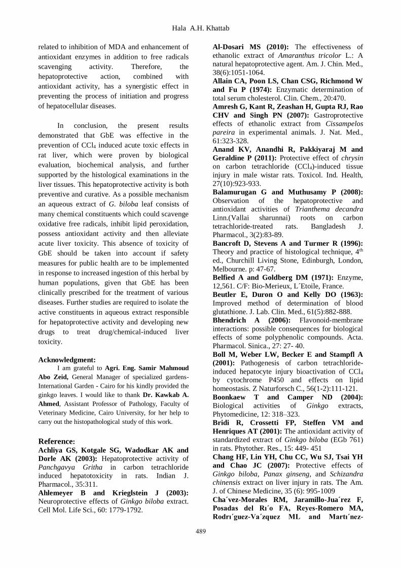
The result in Table (1) shows the effect of
GbE on biological evaluation (weight gain
percent, food intake and food efficiency ratio) in
CCl4 intoxicated rats. It is observed that there was
significant decrease in weight gain percent (p<
0.001), food intake (p< 0.001) and FER (p < 0.01) in
CCl4 intoxicated group as compared to control group.
Concerning the effect of GbE on rats, the ingestion
showed slightly increase in weight gain percent, food
intake and FER, there were no significant difference
Effect of Ginkgo biloba leaves….
486
as compared with control group. Pretreatment of rats
with GbE showed a significant increase in weight
gain percent (p< 0.001), food intake (p< 0.001) and
FER (p<0.01) as compared with CCl4 intoxicated
group indicating the sign of amelioration.
Biochemical results:
The effect of GbE on serum liver enzyme
activities (AST, ALT & ALP), and TP levels in
CCl4 intoxicated rats is illustrated in Table (2).
CCl4 intoxication caused a sharp significant
increase in serum AST, ALT and ALP activities
(p<0.001), and significant reduction in serum TP
level compared to control group (p<0.001).
Administration of GbE to rats showed non-
significant changes in serum liver enzyme
activities, and TP level as compared to control
group. Pretreatment of rats with GbE caused a
marked protection evidenced by significant
reduction (p<0.001) in serum AST, ALT and ALP
enzyme activities, and significant increase
(p<0.001) in TP level compared to CCl4 group.
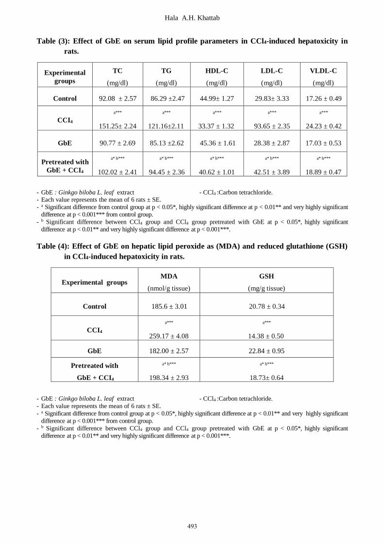
Table (3) shows the effect of GbE on lipid
profile parameters in CCl4 intoxicated rats. Serum
TC, TG, LDL-C and VLDL-C levels were
significantly increased (p<0.001) along with a
significant decrease in serum HDL-C levels
(p<0.001) in CCl4 intoxicated rats, as compared to
control rats. Administration of GbE to rats
revealed non-significant changes in all tested lipid
profile parameters compared to control group.
GbE pretreatment showed a significant
improvement in the levels of lipid parameters,
there was significant decrease in serum TC, TG,
LDL-C and VLDL-C levels (p<0.001) along with
a significant increase in serum HDL-C levels
(p<0.001) in rats group pretreated with GbE as
compared to CCl4 group.
Effect of GbE on hepatic malondialdehyde
(MDA) and reduced glutathione (GSH) in CCl4
intoxicated rats is presented in Table (4). Results
showed that, the level of MDA in the rats' liver
tissue, significantly elevated (p<0.001) in CCl4
intoxicated group compared to control group. On
the other hand pretreatment of rats with GbE
revealed amelioration in hepatic MDA content,
since the value of MDA showed significantly
reduced (p<0.001) as compared to CCl4 group,
while the results of rats receiving GbE tended to
match control value. Regarding, hepatic GSH, the
results revealed significant reduction (p<0.001) in
rats intoxicated with CCl4 as compared to control
group. Pretreatment of rats with GbE markedly
preserved hepatic GSH, the value of GSH near to
normal levels comparing with CCl4 group, at the
same time there was significant difference (p<
0.001) as compared with CCl4 group.
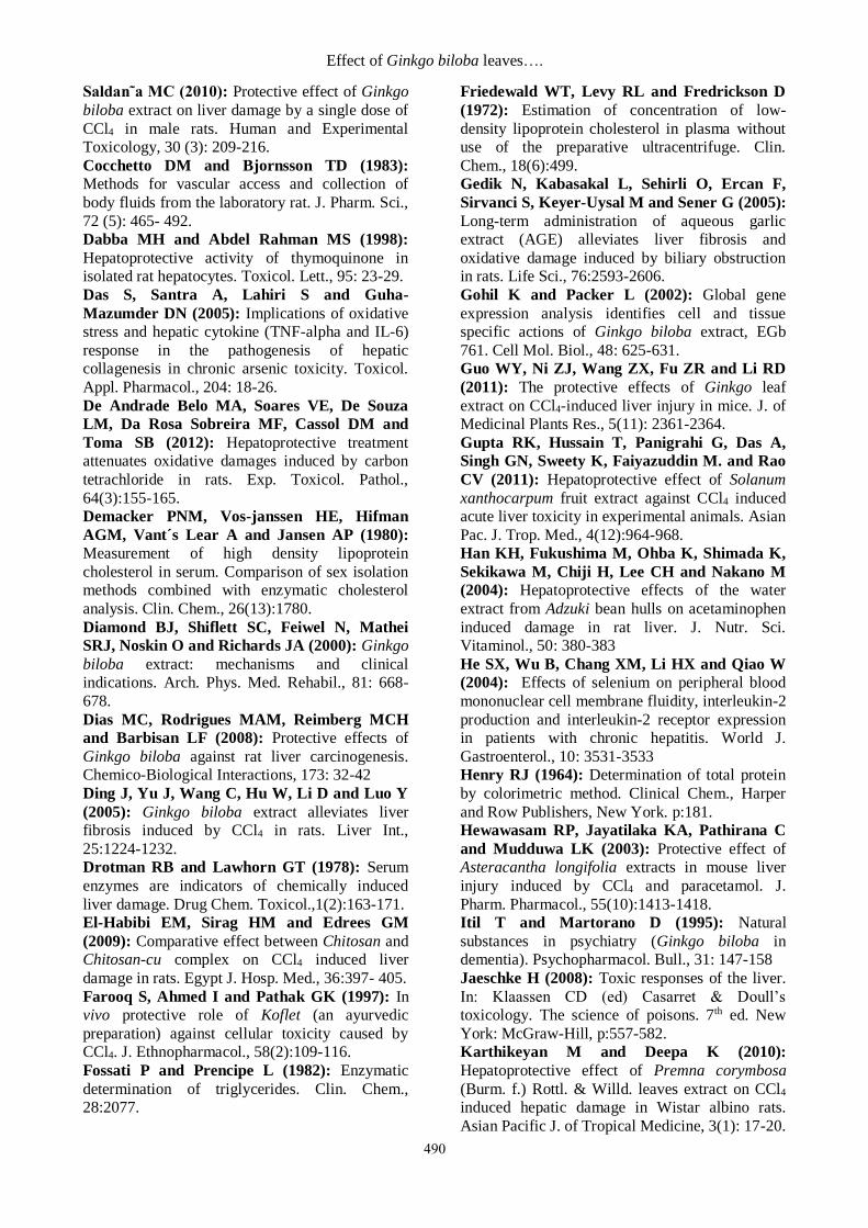
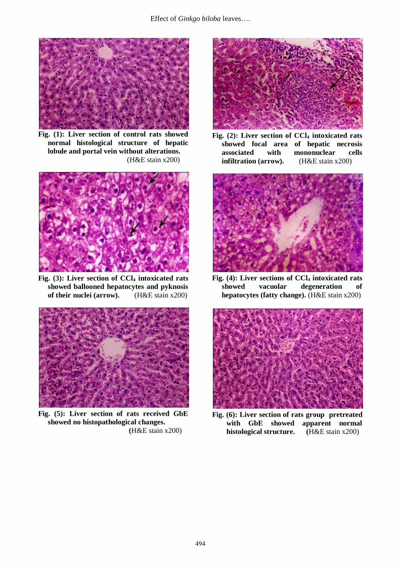
Histopathological results:
Microscopically, liver from control rat
group showed the normal histological structure of
hepatic lobule and portal vein without alterations
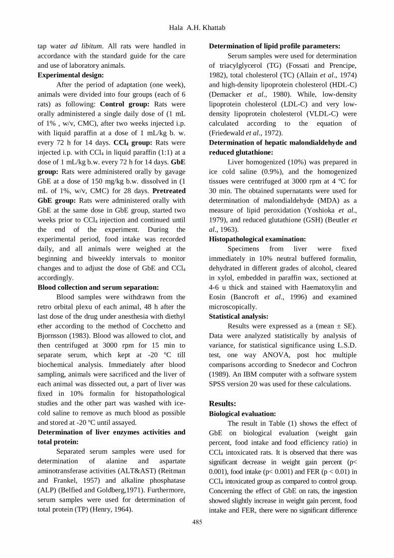
Fig. (1). Liver tissues in CCl4 intoxicated rats
showed focal area of hepatic necrosis associated
with mononuclear cells infiltration Fig. (2) and
ballooned hepatocytes and pyknosis of their nuclei
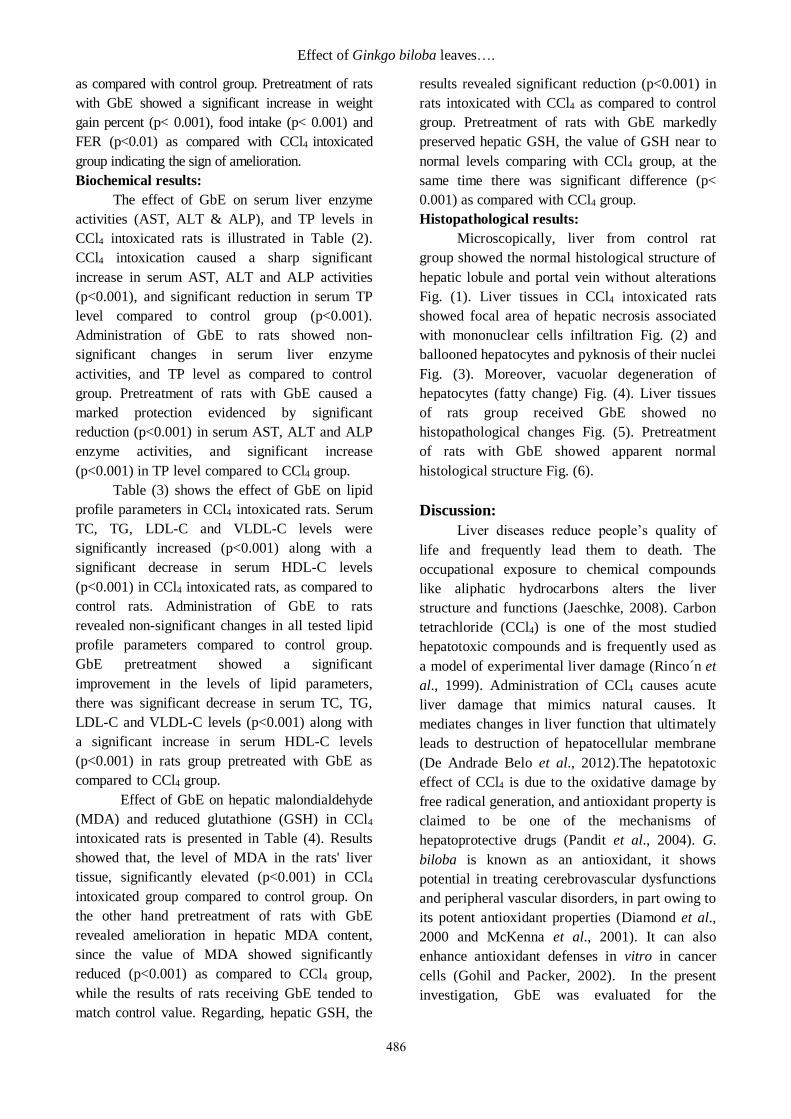
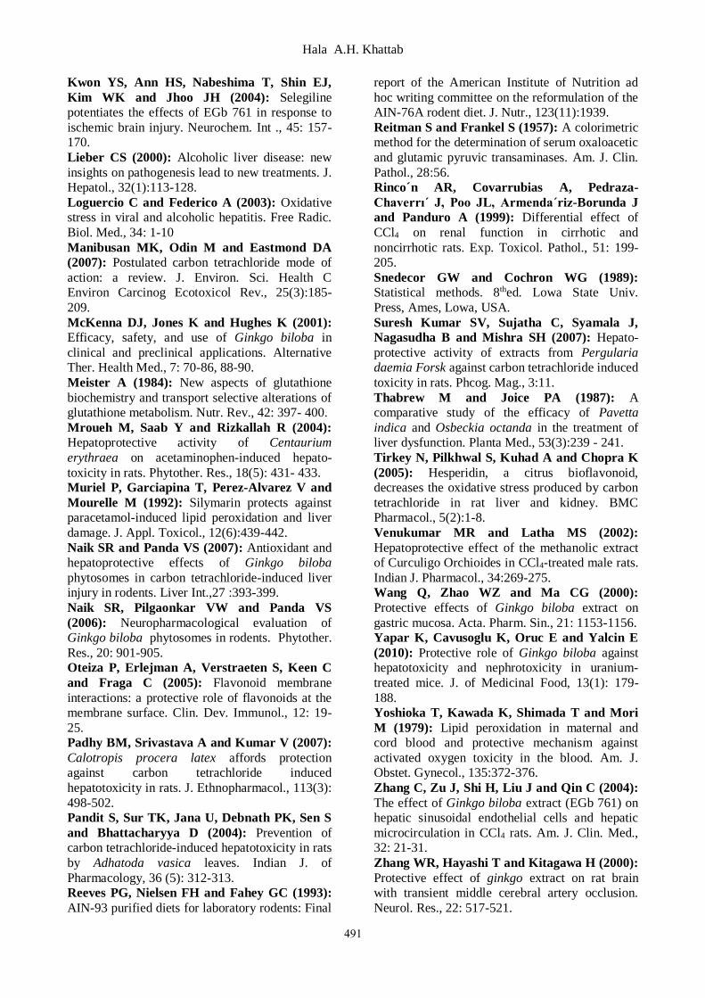
Fig. (3). Moreover, vacuolar degeneration of
hepatocytes (fatty change) Fig. (4). Liver tissues
of rats group received GbE showed no
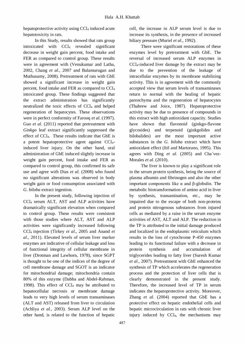
histopathological changes Fig. (5). Pretreatment
of rats with GbE showed apparent normal
histological structure Fig. (6).
Discussion:
Liver diseases reduce people’s quality of
life and frequently lead them to death. The
occupational exposure to chemical compounds
like aliphatic hydrocarbons alters the liver
structure and functions (Jaeschke, 2008). Carbon
tetrachloride (CCl4) is one of the most studied
hepatotoxic compounds and is frequently used as
a model of experimental liver damage (Rinco´n et
al., 1999). Administration of CCl4 causes acute
liver damage that mimics natural causes. It
mediates changes in liver function that ultimately
leads to destruction of hepatocellular membrane
(De Andrade Belo et al., 2012).The hepatotoxic
effect of CCl4 is due to the oxidative damage by
free radical generation, and antioxidant property is
claimed to be one of the mechanisms of
hepatoprotective drugs (Pandit et al., 2004). G.
biloba is known as an antioxidant, it shows
potential in treating cerebrovascular dysfunctions
and peripheral vascular disorders, in part owing to
its potent antioxidant properties (Diamond et al.,
2000 and McKenna et al., 2001). It can also
enhance antioxidant defenses in vitro in cancer
cells (Gohil and Packer, 2002). In the present
investigation, GbE was evaluated for the
Hala A.H. Khattab
487
hepatoprotective activity using CCl4 induced acute
hepatotoxicity in rats.
In this Study, results showed that rats group
intoxicated with CCl4 revealed significant
decrease in weight gain percent, food intake and
FER as compared to control group. These results
were in agreement with (Venukumar and Latha,
2002, Chang et al., 2007 and Balamurugan and
Muthusamy, 2008). Pretreatment of rats with GbE
showed a significant increase in weight gain
percent, food intake and FER as compared to CCl4
intoxicated group. These findings suggested that
the extract administration has significantly
neutralized the toxic effects of CCl4 and helped
regeneration of hepatocytes. These observations
were in perfect conformity of Farooq et al. (1997).
Guo et al. (2011) reported that pretreatment with
Ginkgo leaf extract significantly suppressed the
effect of CCl4. These results indicate that GbE is
a potent hepatoprotective agent against CCl4-
induced liver injury. On the other hand, oral
administration of GbE induced slightly increase in
weight gain percent, food intake and FER as
compared to control group, this confirmed its safe
use and agree with Dias et al. (2008) who found
no significant alterations was observed in body
weight gain or food consumption associated with
G. biloba extract ingestion.
In the present study, following injection of
CCl4 serum ALT, AST and ALP activities have
dramatically significant elevation when compared
to control group. These results were consistent
with those studies where ALT, AST and ALP
activities were significantly increased following
CCl4 injection (Tirkey et al., 2005 and Anand et
al., 2011). Elevated levels of serum liver marker
enzymes are indicative of cellular leakage and loss
of functional integrity of cellular membrane in
liver (Drotman and Lawhorn, 1978), since SGPT
is thought to be one of the indices of the degree of
cell membrane damage and SGOT is an indicator
for mitochondrial damage; mitochondria contain
80% of this enzyme (Dabba and Abdel-Rahman,
1998). This effect of CCl4 may be attributed to
hepatocellular necrosis or membrane damage
leads to very high levels of serum transaminases
(ALT and AST) released from liver to circulation
(Achliya et al., 2003). Serum ALP level on the
other hand, is related to the function of hepatic
cell, the increase in ALP serum level is due to
increase its synthesis, in the presence of increased
biliary pressure (Muriel et al., 1992).
There were significant restorations of these
enzymes level by pretreatment with GbE. The
reversal of increased serum ALP enzymes in
CCl4-induced liver damage by the extract may be
due to the prevention of the leakage of
intracellular enzymes by its membrane stabilizing
activity. This is in agreement with the commonly
accepted view that serum levels of transaminases
return to normal with the healing of hepatic
parenchyma and the regeneration of hepatocytes
(Thabrew and Joice, 1987). Hepatoprotective
activity may be due to presence of compounds in
this extract with high antioxidant capacity. Studies
have shown that flavonoid (ginkgo-flavone
glycosides) and terpenoid (ginkgolides and
bilobalides) are the most important active
substances in the G. biloba extract which have
antioxidant effect (Itil and Martorano, 1995). This
agrees with Ding et al. (2005) and Cha´vez-
Morales et al. (2010).
The liver is known to play a significant role
in the serum protein synthesis, being the source of
plasma albumin and fibrinogen and also the other
important components like α and β-globulin. The
metabolic biotransformation of amino acid in liver
by synthesis, transamination, etc., may be
impaired due to the escape of both non-proteins
and protein nitrogenous substances from injured
cells as mediated by a raise in the serum enzyme
activities of AST, ALT and ALP. The reduction in
the TP is attributed to the initial damage produced
and localized in the endoplasmic reticulum which
results in the loss of cytochrome P-450 enzymes
leading to its functional failure with a decrease in
protein synthesis and accumulation of
triglycerides leading to fatty liver (Suresh Kumar
et al., 2007). Pretreatment with GbE enhanced the
synthesis of TP which accelerates the regeneration
process and the protection of liver cells that is
clearly demonstrated in the present study.
Therefore, the increased level of TP in serum
indicates the hepatoprotective activity. Moreover,
Zhang et al. (2004) reported that GbE has a
protective effect on hepatic endothelial cells and
hepatic microcirculation in rats with chronic liver
injury induced by CCl4, the mechanisms may
Effect of Ginkgo biloba leaves….
488
involve its inhibition on platelet-activating factor
and lipid peroxidation.
The present results revealed significant
elevation in serum TC, TG, LDL-C and VLDL-C
levels along with significant reduction in serum
HDL-C levels in CCl4 intoxicated rats, as
compared to control rats. These results were in
agreement with the previous results of El-Habibi
et al. (2009) and Al-Dosari (2010). CCl4
intoxication increases the synthesis of fatty acids
and triglycerides from acetate, this could be
attributed to CCl4 positively affects the transport
of acetate into the liver cell, resulting in increased
acetate availability, CCl4 intoxication also results
in inhibition of synthesis of the bile acids from
cholesterol which is synthesized in liver or
derived from plasma lipids leading to increase
cholesterol level (Boll et al., 2001). On the other
hand, CCl4 lowers β- oxidation of fatty acids and
hydrolysis of triglycerides, this increases the
availability of fatty acids to esterification (Lieber,
2000). The current study showed that pretreatment
of rats with GbE resulted in significant
improvement in the tested lipid profile parameters,
it could be attributed to the active components of
GbE, mainly flavonoid fraction, which have many
beneficial effects, and antioxidant properties
(Oteiza et al., 2005 and Bhendrich, 2006).
Lipid peroxidation is one of the principal
causes of CCl4-induced liver injury and is
mediated by the free-radical derivatives of CCl4
(Manibusan et al., 2007). The level of lipid
peroxide is a measure of membrane damage and
alterations in structure and function of cellular
membranes. In the present study, elevation hepatic
MDA of rats intoxicated with CCl4 was observed.
The increase in hepatic MDA levels leading to
tissue damage and failure of antioxidant defense
mechanisms to prevent the formation of excessive
free radicals (Amresh et al., 2007). Pretreatment
of rats with GbE significantly reduced the
elevated levels of MDA through scavenging
oxygen radicals as the extract possesses a potent
antioxidant activity. Ginkgoʼs antioxidant activity
is attributed to its ability to increase levels of free
radical-scavenging enzymes and to neutralize
ferryl ion-induced peroxidation ((Bridi et al., 2001
and Naik and Panda, 2007).
Glutathione is one of the most abundant
tripeptide non-enzymatic biological antioxidant,
its functions include removal of free radicals such
as H2O2 and superoxide anions, maintenance of
membrane protein thiols and acting as a substrate
for glutathione peroxidase and glutathione
reductase (Meister, 1984). In the present study,
significant decrease in hepatic GSH level was
observed in CCl4 intoxicated group as compared
to control group. The depletion of hepatic GSH
has been shown to be associated with an enhanced
toxicity to CCl4 (Hewawasam et al., 2003). The
increase in hepatic GSH level in the rats
pretreated with GbE may be due GSH
regeneration. This effect of GbE may be due to an
initial reduction in hepatic peroxidative activities,
thereby leading to restoration of the GSH content
(Naik and Panda, 2007). These effects would also
contribute to partially explain the hepatoprotective
effect of GbE by inhibiting the biotransformation
of CCl4 and the consequent production of free
radicals (Cha´vez-Morales et al., 2010).
Histological results were in agreement with
the measured activities of serum liver enzymes
and provided supportive evidence for the
biochemical analysis, in the current study,
histopathological examination of the liver tissues
showed congestion in portal vein with infiltration
of mononuclear inflammatory cells, necrosis,
vacuolar degeneration and kupffer cells activation
in CCl4 group. Accordance to these findings,
Padhy et al. (2007) observed leukocytic
infiltration, centrilobular necrosis and vacuolation
in CCl4 treated rats. Gupta et al. (2011) observed
also fatty change, congestion in portal vein,
necrosis, ballooning degeneration and loss of
cellular boundaries. These finding relates with
high activities of serum liver enzyme activities
found in the CCl4 group in the present results.
Liver sections of rats pretreated with GbE showed
regeneration of hepatocytes near normal liver
architecture. This may be explained by the
constituents of GbE are scavengers of free
radicals, and inhibit lipid peroxidation, thus help
to maintain the integrity and permeability of cell
membranes and protects cells and tissues against
oxidative stress induced by free radicals (Naik and
Panda, 2007). Protective effect of GbE against
CCl4 induced hepatotoxicity in rats appears to be
Hala A.H. Khattab
489
related to inhibition of MDA and enhancement of
antioxidant enzymes in addition to free radicals
scavenging activity. Therefore, the
hepatoprotective action, combined with
antioxidant activity, has a synergistic effect in
preventing the process of initiation and progress
of hepatocellular diseases.
In conclusion, the present results
demonstrated that GbE was effective in the
prevention of CCl4 induced acute toxic effects in
rat liver, which were proven by biological
evaluation, biochemical analysis, and further
supported by the histological examinations in the
liver tissues. This hepatoprotective activity is both
preventive and curative. As a possible mechanism
an aqueous extract of G. biloba leaf consists of
many chemical constituents which could scavenge
oxidative free radicals, inhibit lipid peroxidation,
possess antioxidant activity and then alleviate
acute liver toxicity. This absence of toxicity of
GbE should be taken into account if safety
measures for public health are to be implemented
in response to increased ingestion of this herbal by
human populations, given that GbE has been
clinically prescribed for the treatment of various
diseases. Further studies are required to isolate the
active constituents in aqueous extract responsible
for hepatoprotective activity and developing new
drugs to treat drug/chemical-induced liver
toxicity.
Acknowledgment: I am grateful to Agri. Eng. Samir Mahmoud
Abo Zeid, General Manager of specialized gardens-
International Garden - Cairo for his kindly provided the
ginkgo leaves. I would like to thank Dr. Kawkab A.
Ahmed, Assistant Professor of Pathology, Faculty of
Veterinary Medicine, Cairo University, for her help to
carry out the histopathological study of this work.
Reference:
Achliya GS, Kotgale SG, Wadodkar AK and
Dorle AK (2003): Hepatoprotective activity of
Panchgavya Gritha in carbon tetrachloride
induced hepatotoxicity in rats. Indian J.
Pharmacol., 35:311.
Ahlemeyer B and Krieglstein J (2003): Neuroprotective effects of Ginkgo biloba extract.
Cell Mol. Life Sci., 60: 1779-1792.
Al-Dosari MS (2010): The effectiveness of
ethanolic extract of Amaranthus tricolor L.: A
natural hepatoprotective agent. Am. J. Chin. Med.,
38(6):1051-1064.
Allain CA, Poon LS, Chan CSG, Richmond W
and Fu P (1974): Enzymatic determination of
total serum cholesterol. Clin. Chem., 20:470.
Amresh G, Kant R, Zeashan H, Gupta RJ, Rao
CHV and Singh PN (2007): Gastroprotective
effects of ethanolic extract from Cissampelos
pareira in experimental animals. J. Nat. Med.,
61:323-328.
Anand KV, Anandhi R, Pakkiyaraj M and
Geraldine P (2011): Protective effect of chrysin on carbon tetrachloride (CCl4)-induced tissue
injury in male wistar rats. Toxicol. Ind. Health,
27(10):923-933.
Balamurugan G and Muthusamy P (2008):
Observation of the hepatoprotective and
antioxidant activities of Trianthema decandra
Linn.(Vallai sharunnai) roots on carbon
tetrachloride-treated rats. Bangladesh J.
Pharmacol., 3(2):83-89.
Bancroft D, Stevens A and Turmer R (1996):
Theory and practice of histological technique, 4th
ed., Churchill Living Stone, Edinburgh, London,
Melbourne. p: 47-67.
Belfied A and Goldberg DM (1971): Enzyme,
12,561. C/F: Bio-Merieux, L´Etoile, France.
Beutler E, Duron O and Kelly DO (1963):
Improved method of determination of blood
glutathione. J. Lab. Clin. Med., 61(5):882-888.
Bhendrich A (2006): Flavonoid-membrane
interactions: possible consequences for biological
effects of some polyphenolic compounds. Acta.
Pharmacol. Sinica., 27: 27- 40.
Boll M, Weber LW, Becker E and Stampfl A
(2001): Pathogenesis of carbon tetrachloride-
induced hepatocyte injury bioactivation of CCl4
by cytochrome P450 and effects on lipid
homeostasis. Z Naturforsch C., 56(1-2):111-121.
Boonkaew T and Camper ND (2004):
Biological activities of Ginkgo extracts,
Phytomedicine, 12: 318–323.
Bridi R, Crossetti FP, Steffen VM and
Henriques AT (2001): The antioxidant activity of
standardized extract of Ginkgo biloba (EGb 761)
in rats. Phytother. Res., 15: 449- 451
Chang HF, Lin YH, Chu CC, Wu SJ, Tsai YH
and Chao JC (2007): Protective effects of
Ginkgo biloba, Panax ginseng, and Schizandra chinensis extract on liver injury in rats. The Am.
J. of Chinese Medicine, 35 (6): 995-1009
Cha´vez-Morales RM, Jaramillo-Jua´rez F,
Posadas del Rı´o FA, Reyes-Romero MA,
Rodrı´guez-Va´zquez ML and Martı´nez-
Effect of Ginkgo biloba leaves….
490
Saldan˜a MC (2010): Protective effect of Ginkgo
biloba extract on liver damage by a single dose of
CCl4 in male rats. Human and Experimental
Toxicology, 30 (3): 209-216.
Cocchetto DM and Bjornsson TD (1983):
Methods for vascular access and collection of
body fluids from the laboratory rat. J. Pharm. Sci.,
72 (5): 465- 492.
Dabba MH and Abdel Rahman MS (1998):
Hepatoprotective activity of thymoquinone in
isolated rat hepatocytes. Toxicol. Lett., 95: 23-29.
Das S, Santra A, Lahiri S and Guha-
Mazumder DN (2005): Implications of oxidative
stress and hepatic cytokine (TNF-alpha and IL-6)
response in the pathogenesis of hepatic
collagenesis in chronic arsenic toxicity. Toxicol.
Appl. Pharmacol., 204: 18-26.
De Andrade Belo MA, Soares VE, De Souza
LM, Da Rosa Sobreira MF, Cassol DM and
Toma SB (2012): Hepatoprotective treatment
attenuates oxidative damages induced by carbon
tetrachloride in rats. Exp. Toxicol. Pathol.,
64(3):155-165.
Demacker PNM, Vos-janssen HE, Hifman
AGM, Vant´s Lear A and Jansen AP (1980):
Measurement of high density lipoprotein
cholesterol in serum. Comparison of sex isolation
methods combined with enzymatic cholesterol
analysis. Clin. Chem., 26(13):1780.
Diamond BJ, Shiflett SC, Feiwel N, Mathei
SRJ, Noskin O and Richards JA (2000): Ginkgo
biloba extract: mechanisms and clinical
indications. Arch. Phys. Med. Rehabil., 81: 668-
678.
Dias MC, Rodrigues MAM, Reimberg MCH
and Barbisan LF (2008): Protective effects of
Ginkgo biloba against rat liver carcinogenesis.
Chemico-Biological Interactions, 173: 32-42
Ding J, Yu J, Wang C, Hu W, Li D and Luo Y
(2005): Ginkgo biloba extract alleviates liver
fibrosis induced by CCl4 in rats. Liver Int.,
25:1224-1232.
Drotman RB and Lawhorn GT (1978): Serum
enzymes are indicators of chemically induced
liver damage. Drug Chem. Toxicol.,1(2):163-171. El-Habibi EM, Sirag HM and Edrees GM
(2009): Comparative effect between Chitosan and
Chitosan-cu complex on CCl4 induced liver
damage in rats. Egypt J. Hosp. Med., 36:397- 405.
Farooq S, Ahmed I and Pathak GK (1997): In
vivo protective role of Koflet (an ayurvedic
preparation) against cellular toxicity caused by
CCl4. J. Ethnopharmacol., 58(2):109-116.
Fossati P and Prencipe L (1982): Enzymatic
determination of triglycerides. Clin. Chem.,
28:2077.
Friedewald WT, Levy RL and Fredrickson D
(1972): Estimation of concentration of low-
density lipoprotein cholesterol in plasma without
use of the preparative ultracentrifuge. Clin.
Chem., 18(6):499.
Gedik N, Kabasakal L, Sehirli O, Ercan F,
Sirvanci S, Keyer-Uysal M and Sener G (2005):
Long-term administration of aqueous garlic
extract (AGE) alleviates liver fibrosis and
oxidative damage induced by biliary obstruction
in rats. Life Sci., 76:2593-2606.
Gohil K and Packer L (2002): Global gene
expression analysis identifies cell and tissue
specific actions of Ginkgo biloba extract, EGb
761. Cell Mol. Biol., 48: 625-631.
Guo WY, Ni ZJ, Wang ZX, Fu ZR and Li RD
(2011): The protective effects of Ginkgo leaf
extract on CCl4-induced liver injury in mice. J. of
Medicinal Plants Res., 5(11): 2361-2364.
Gupta RK, Hussain T, Panigrahi G, Das A,
Singh GN, Sweety K, Faiyazuddin M. and Rao
CV (2011): Hepatoprotective effect of Solanum
xanthocarpum fruit extract against CCl4 induced
acute liver toxicity in experimental animals. Asian
Pac. J. Trop. Med., 4(12):964-968.
Han KH, Fukushima M, Ohba K, Shimada K,
Sekikawa M, Chiji H, Lee CH and Nakano M
(2004): Hepatoprotective effects of the water
extract from Adzuki bean hulls on acetaminophen
induced damage in rat liver. J. Nutr. Sci.
Vitaminol., 50: 380-383
He SX, Wu B, Chang XM, Li HX and Qiao W
(2004): Effects of selenium on peripheral blood
mononuclear cell membrane fluidity, interleukin-2
production and interleukin-2 receptor expression
in patients with chronic hepatitis. World J.
Gastroenterol., 10: 3531-3533
Henry RJ (1964): Determination of total protein
by colorimetric method. Clinical Chem., Harper
and Row Publishers, New York. p:181.
Hewawasam RP, Jayatilaka KA, Pathirana C
and Mudduwa LK (2003): Protective effect of
Asteracantha longifolia extracts in mouse liver
injury induced by CCl4 and paracetamol. J.
Pharm. Pharmacol., 55(10):1413-1418. Itil T and Martorano D (1995): Natural
substances in psychiatry (Ginkgo biloba in
dementia). Psychopharmacol. Bull., 31: 147-158
Jaeschke H (2008): Toxic responses of the liver.
In: Klaassen CD (ed) Casarret & Doull’s
toxicology. The science of poisons. 7th ed. New
York: McGraw-Hill, p:557-582.
Karthikeyan M and Deepa K (2010):
Hepatoprotective effect of Premna corymbosa
(Burm. f.) Rottl. & Willd. leaves extract on CCl4
induced hepatic damage in Wistar albino rats.
Asian Pacific J. of Tropical Medicine, 3(1): 17-20.
Hala A.H. Khattab
491
Kwon YS, Ann HS, Nabeshima T, Shin EJ,
Kim WK and Jhoo JH (2004): Selegiline
potentiates the effects of EGb 761 in response to
ischemic brain injury. Neurochem. Int ., 45: 157-
170.
Lieber CS (2000): Alcoholic liver disease: new
insights on pathogenesis lead to new treatments. J.
Hepatol., 32(1):113-128.
Loguercio C and Federico A (2003): Oxidative
stress in viral and alcoholic hepatitis. Free Radic.
Biol. Med., 34: 1-10
Manibusan MK, Odin M and Eastmond DA
(2007): Postulated carbon tetrachloride mode of
action: a review. J. Environ. Sci. Health C
Environ Carcinog Ecotoxicol Rev., 25(3):185-
209.
McKenna DJ, Jones K and Hughes K (2001):
Efficacy, safety, and use of Ginkgo biloba in
clinical and preclinical applications. Alternative
Ther. Health Med., 7: 70-86, 88-90.
Meister A (1984): New aspects of glutathione
biochemistry and transport selective alterations of
glutathione metabolism. Nutr. Rev., 42: 397- 400.
Mroueh M, Saab Y and Rizkallah R (2004):
Hepatoprotective activity of Centaurium
erythraea on acetaminophen-induced hepato-
toxicity in rats. Phytother. Res., 18(5): 431- 433.
Muriel P, Garciapina T, Perez-Alvarez V and
Mourelle M (1992): Silymarin protects against
paracetamol-induced lipid peroxidation and liver
damage. J. Appl. Toxicol., 12(6):439-442.
Naik SR and Panda VS (2007): Antioxidant and
hepatoprotective effects of Ginkgo biloba
phytosomes in carbon tetrachloride-induced liver
injury in rodents. Liver Int.,27 :393-399.
Naik SR, Pilgaonkar VW and Panda VS
(2006): Neuropharmacological evaluation of
Ginkgo biloba phytosomes in rodents. Phytother.
Res., 20: 901-905.
Oteiza P, Erlejman A, Verstraeten S, Keen C
and Fraga C (2005): Flavonoid membrane
interactions: a protective role of flavonoids at the
membrane surface. Clin. Dev. Immunol., 12: 19-
25.
Padhy BM, Srivastava A and Kumar V (2007):
Calotropis procera latex affords protection
against carbon tetrachloride induced
hepatotoxicity in rats. J. Ethnopharmacol., 113(3):
498-502.
Pandit S, Sur TK, Jana U, Debnath PK, Sen S
and Bhattacharyya D (2004): Prevention of
carbon tetrachloride-induced hepatotoxicity in rats
by Adhatoda vasica leaves. Indian J. of
Pharmacology, 36 (5): 312-313.
Reeves PG, Nielsen FH and Fahey GC (1993):
AIN-93 purified diets for laboratory rodents: Final
report of the American Institute of Nutrition ad
hoc writing committee on the reformulation of the
AIN-76A rodent diet. J. Nutr., 123(11):1939.
Reitman S and Frankel S (1957): A colorimetric
method for the determination of serum oxaloacetic
and glutamic pyruvic transaminases. Am. J. Clin.
Pathol., 28:56.
Rinco´n AR, Covarrubias A, Pedraza-
Chaverrı´ J, Poo JL, Armenda´riz-Borunda J
and Panduro A (1999): Differential effect of
CCl4 on renal function in cirrhotic and
noncirrhotic rats. Exp. Toxicol. Pathol., 51: 199-
205.
Snedecor GW and Cochron WG (1989):
Statistical methods. 8thed. Lowa State Univ.
Press, Ames, Lowa, USA.
Suresh Kumar SV, Sujatha C, Syamala J,
Nagasudha B and Mishra SH (2007): Hepato-
protective activity of extracts from Pergularia daemia Forsk against carbon tetrachloride induced
toxicity in rats. Phcog. Mag., 3:11.
Thabrew M and Joice PA (1987): A
comparative study of the efficacy of Pavetta
indica and Osbeckia octanda in the treatment of
liver dysfunction. Planta Med., 53(3):239 - 241.
Tirkey N, Pilkhwal S, Kuhad A and Chopra K
(2005): Hesperidin, a citrus bioflavonoid,
decreases the oxidative stress produced by carbon
tetrachloride in rat liver and kidney. BMC
Pharmacol., 5(2):1-8.
Venukumar MR and Latha MS (2002):
Hepatoprotective effect of the methanolic extract
of Curculigo Orchioides in CCl4-treated male rats.
Indian J. Pharmacol., 34:269-275.
Wang Q, Zhao WZ and Ma CG (2000):
Protective effects of Ginkgo biloba extract on
gastric mucosa. Acta. Pharm. Sin., 21: 1153-1156.
Yapar K, Cavusoglu K, Oruc E and Yalcin E
(2010): Protective role of Ginkgo biloba against
hepatotoxicity and nephrotoxicity in uranium-
treated mice. J. of Medicinal Food, 13(1): 179-
188.
Yoshioka T, Kawada K, Shimada T and Mori
M (1979): Lipid peroxidation in maternal and
cord blood and protective mechanism against
activated oxygen toxicity in the blood. Am. J.
Obstet. Gynecol., 135:372-376.
Zhang C, Zu J, Shi H, Liu J and Qin C (2004):
The effect of Ginkgo biloba extract (EGb 761) on
hepatic sinusoidal endothelial cells and hepatic
microcirculation in CCl4 rats. Am. J. Clin. Med.,
32: 21-31.
Zhang WR, Hayashi T and Kitagawa H (2000):
Protective effect of ginkgo extract on rat brain
with transient middle cerebral artery occlusion.
Neurol. Res., 22: 517-521.
Effect of Ginkgo biloba leaves….
492
Table (1): Effect of GbE on weight gain percent, food intake and food efficiency ratio (FER) in CCl4-
induced hepatoxicity in rats.
Experimental groups Weight gain percent Food Intake
(g/ rat/day) FER
Control 27.71 ± 0.72 19.16 ± 0.74 0.09 ± 0.002
CCl4 a***
12.28 ± 0.88
a***
11.62 ± 0.55
a**
0.068± 0.006
GbE 26.54 ± 1.14 19.47 ± 0.58 0.086± 0.006
Pretreated with
GbE + CCl4
a * b ***
24.16 ± 0.83
a * b ***
17.31 ± 0.57
b **
0.087± 0.001
- GbE : Ginkgo biloba L. leaf extract - CCl4 :Carbon tetrachloride.
- Each value represents the mean of 6 rats ± SE.
- a Significant difference from control group at p < 0.05*, highly significant difference at p < 0.01** and very highly significant
difference at p < 0.001*** from control group.
- b Significant difference between CCl4 group and CCl4 group pretreated with GbE at p < 0.05*, highly significant
difference at p < 0.01** and very highly significant difference at p < 0.001***.
Table (2): Effect of GbE on serum aminotransferase (ALT, AST) and alkaline phophatase (ALP)
enzyme activities, as well as total protein (TP) levels in CCl4-induced hepatoxicity in rats.
Experimental
groups
ALT
(U/L)
AST
(U/L)
ALP
(U/L)
T P
(g/dl)
Control 39.1 ± 1.73 99.99 ± 3.99 70.68 ± 1.44 6.498± 0. 14
CCl4
a***
106.17 ± 1.54
a***
215.86 ± 3.12
a***
143.59 ± 1.93
a***
4.54± 0.15
GbE 38.92 ± 1.16 97.98 ± 2.02 69.14 ± 1.24 6.71± 0.12
Pretreated with
GbE + CCl4
a * b ***
46.01 ± 2.28
a * b ***
111.6 ± 1.88
a * b ***
77.66 ± 2.23
a * b ***
5.92± 0.16
- GbE : Ginkgo biloba L. leaf extract - CCl4 :Carbon tetrachloride.
- Each value represents the mean of 6 rats ± SE.
- a Significant difference from control group at p < 0.05*, highly significant difference at p < 0.01** and very highly significant
difference at p < 0.001*** from control group.
- b Significant difference between CCl4 group and CCl4 group pretreated with GbE at p < 0.05*, highly significant
difference at p < 0.01** and very highly significant difference at p < 0.001***.
Hala A.H. Khattab
493
Table (3): Effect of GbE on serum lipid profile parameters in CCl4-induced hepatoxicity in
rats.
Experimental
groups
TC
(mg/dl)
TG
(mg/dl)
HDL-C
(mg/dl)
LDL-C
(mg/dl)
VLDL-C
(mg/dl)
Control 92.08 ± 2.57 86.29 ±2.47 44.99± 1.27 29.83± 3.33 17.26 ± 0.49
CCI4
a***
151.25± 2.24
a***
121.16±2.11
a***
33.37 ± 1.32
a***
93.65 ± 2.35
a***
± 0.4224.23
GbE 90.77 ± 2.69 85.13 ±2.62 45.36 ± 1.61 28.38 ± 2.87 17.03 ± 0.53
Pretreated with
GbE + CCI4
a* b***
102.02 ± 2.41
a* b***
94.45 ± 2.36
a* b***
40.62 ± 1.01
a* b***
42.51 ± 3.89
a* b***
18.89 ± 0.47
- GbE : Ginkgo biloba L. leaf extract - CCl4 :Carbon tetrachloride. - Each value represents the mean of 6 rats ± SE.
- a Significant difference from control group at p < 0.05*, highly significant difference at p < 0.01** and very highly significant
difference at p < 0.001*** from control group.
- b Significant difference between CCl4 group and CCl4 group pretreated with GbE at p < 0.05*, highly significant
difference at p < 0.01** and very highly significant difference at p < 0.001***.
Table (4): Effect of GbE on hepatic lipid peroxide as (MDA) and reduced glutathione (GSH)
in CCl4-induced hepatoxicity in rats.
Experimental groups MDA
(nmol/g tissue)
GSH
(mg/g tissue)
Control 185.6 ± 3.01 20.78 ± 0.34
CCI4
a***
259.17 ± 4.08
a***
14.38 ± 0.50
GbE 182.00 ± 2.57 22.84 ± 0.95
Pretreated with
GbE + CCI4
a* b***
198.34 ± 2.93
a* b***
18.73± 0.64
- GbE : Ginkgo biloba L. leaf extract - CCl4 :Carbon tetrachloride.
- Each value represents the mean of 6 rats ± SE.
- a Significant difference from control group at p < 0.05*, highly significant difference at p < 0.01** and very highly significant
difference at p < 0.001*** from control group.
- b Significant difference between CCl4 group and CCl4 group pretreated with GbE at p < 0.05*, highly significant
difference at p < 0.01** and very highly significant difference at p < 0.001***.
Effect of Ginkgo biloba leaves….
494
Fig. (2): Liver section of CCl4 intoxicated rats
showed focal area of hepatic necrosis
associated with mononuclear cells
infiltration (arrow). (H&E stain x200)
Fig. (4): Liver sections of CCl4 intoxicated rats
showed vacuolar degeneration of
hepatocytes (fatty change). (H&E stain x200)
Fig. (6): Liver section of rats group pretreated
with GbE showed apparent normal
histological structure. (H&E stain x200)
Fig. (1): Liver section of control rats showed
normal histological structure of hepatic
lobule and portal vein without alterations.
(H&E stain x200)
Fig. (3): Liver section of CCl4 intoxicated rats
showed ballooned hepatocytes and pyknosis
of their nuclei (arrow). (H&E stain x200)
Fig. (5): Liver section of rats received GbE
showed no histopathological changes.
(H&E stain x200)
Hala A.H. Khattab
495
نبات الجنكوبيلوبا علي التسمم الكبدي الحادلأوراق تأثير المستخلص المائي
فئران الرابع كلوريد الكربون في ب ثالمُحد
هالة عبد الرحمن حسن خطاب
جامعة حلوان -كلية الإقتصاد المنزلي -قسم التغذية وعلوم الأطعمة
الملخص العربي
نبات لأوراق المستخلص المائي ثبتأ مختلف امراض الكبد. وقدوتقدم في احداث بالغ الاهمية دور الضغط التأكسدي له
تهدف هذه دور في الوقاية والعلاج للامراض المرتبطة بالضغط التأكسدي.له بذلك فإن ،مضادة للاكسدةكمادة ة تالجنكوبيلوبا فعالي
رابع كلوريد ب ثحدالم علي التسمم الكبدي الحادبيلوبا وراق نبات الجنكو لأللكبد للمستخلص المائي يقاتأثير الوالالدراسة الي تقييم
بجرعة ابع كلوريد الكربون رالفئران وذلك بالحقن داخل الغشاء البريتوني بذكور تم إحداث التسمم الكبدي في فئران.الفي الكربون
عن يوميا جنكوال نبات وراقالمائي لأمستخلص التم إعطاء بينما ، يوما 14ساعة لمدة 72مل/كجم من وزن الجسم كل 1مقدارها
رابع كلوريد الكربون واستمرحتى نهاية ن الجسم لمدة أسبوعين قبل الحقن بمجم/كجم من وز 150طريق الفم بجرعة مقدارها
.التجربة
في حصائيةإذو دلالة رتفاع إأحدث تلف حاد في الكبد, حيث وجد قد رابع كلوريد الكربون الحقن ب النتائج أنأظهرت
ومحتوي الكبد من ، في مصل الدم ، وإنزيم الفوسفاتيز القلويلانيننزيم ناقلة أمين الأسبارتات، إنزيم ناقلة أمين الآإمستويات
البروتين الكلي، الكوليستيرول ،المكتسبوزن للالنسبة المئوية في حصائيةإذو دلالة اانخفاضوجد وكذلك المالونداي ألدهيد،
المعالجة المسبقة أظهرت وقد ومحتوي الكبد من الجلوتاثيون المختزل.في مصل الدم ليبوبروتينات ذات الكثافة العالية بالالمرتبط
ذو دلالة نخفاض ٳحيث وجد ،حماية الكبد فعالية فيقبل الحقن برابع كلوريد الكربون الجنكو نبات وراق المائي لأمستخلص الب
في البروتين ذو دلالة إحصائيةرتفارع ٳ متزامنا مع حدوث، ومحتواه من المالونداي ألدهيد نشاط انزيمات الكبد فيحصائية إ
في النسبة المئوية للوزن المكتسب معنويا ادوث تحسن، أيضا أظهرت النتائج ح ون المختزلحتوي الكبد من الجلوتاثيمو الكلي،
وقد اظهر الفحص الهستوباثولوجي لانسجة . تم حقنها برابع كلوريد الكربونمقارنة بالمجموعة التي الدهون عند ال مقاييسوكذلك
مع وجود ارتشاح الخلايا الالتهابية موت موضعي،تغييرات نسيجية ودوث الحقن برابع كلوريد الكربون قد تسبب في ح الكبد أن
التغلب علي هذة الي بالمستخلص المائي لأوراق نبات الجنكو المعالجة المسبقة في الخلايا الكبدية، بينما أدت التنكس الفجويو
.أغلبية الخلايا الكبدية أصبحت أقرب الي الحالة الطبيعيةحدثة، حيث أن م التغييرات ال
ضد التلف التأكسدي لمستخلص المائي لاوارق نبات الجنكولللكبد التأثير الواقي تشير نتائج هذه الدراسة إلى أن
يمكن لذلك قد التأكسدي. ولضغط اتثبيط دورة في مضاد للأكسدة و كمادة فعاليتةقد يرجع إلى ث برابع كلوريد الكربوندحالم
لتقليل من السمية لفائدتة في ا في المستحضرات الدوائية ومكملات الاغذية المائي لاوراق نبات الجنكو أستعمال هذا المستخلص
الكبدية.
المالونداي ،الدهون مقاييس الكبد،أنزيمات رابع كلوريد الكربون، ذكور الفئران، نبات الجنكو بيلوبا، المستخلص المائي، :الكلمات المفتاحية
.يألدهيد، الجلوتاثيون المختزل، التأثير الواق
![Shoot Growth and Heterophylly in Ginkgo Biloba · 2013-03-28 · 1970] CRITCHFIELD—GINKGO BILOBA 151 andhis co-workers (GunckelandWetmore1946a, 1946b; Gunckel and Thimann 1949;](https://img.pdfslide.us/doc/110x75/5f07ecc47e708231d41f7209/shoot-growth-and-heterophylly-in-ginkgo-biloba-2013-03-28-1970-critchfieldaginkgo.jpg)