Embed Size (px)
Citation preview
http://poi.sagepub.com/Prosthetics and Orthotics International
http://poi.sagepub.com/content/35/4/342The online version of this article can be found at:
DOI: 10.1177/0309364611420201
2011 35: 342 originally published online 26 September 2011Prosthet Orthot IntPedro V Munuera and Rocio Mazoteras-Pardo
Benefits of custom-made foot orthoses in treating patellofemoral pain
Published by:
http://www.sagepublications.com
On behalf of:
International Society for Prosthetics and Orthotics
can be found at:Prosthetics and Orthotics InternationalAdditional services and information for
http://poi.sagepub.com/cgi/alertsEmail Alerts:
http://poi.sagepub.com/subscriptionsSubscriptions:
http://www.sagepub.com/journalsReprints.navReprints:
http://www.sagepub.com/journalsPermissions.navPermissions:
What is This?
- Sep 26, 2011Proof
- Nov 28, 2011Version of Record >>
at Universidad de Sevilla. Biblioteca on December 7, 2011poi.sagepub.comDownloaded from
INTERNATIONALSOCIETY FOR PROSTHETICSAND ORTHOTICS
Original Research Report
Benefits of custom-made foot orthoses intreating patellofemoral pain
Pedro V Munuera and Rocio Mazoteras-Pardo
AbstractBackground: Patellofemoral pain is one of the most common disorders affecting the knee. Forefoot varus and excessivesubtalar pronation can be associated with patellofemoral pain. Foot orthotics may produce an improvement insymptoms.Objectives: The aim of this study was to test whether patellofemoral pain is improved after four weeks of using custom-made foot orthoses.Study Design: Clinical trial without control group.Methods: Twenty-one subjects with patellofemoral pain were given custom-made foot orthoses (2-mm thick polypro-pylene and 4-mm thick polyethylene foam liner of 45 shore A hardness). Patellofemoral pain was evaluated with a visualanalogue scale before applying the treatment, and at two weeks and four weeks follow-up. At the two-week check-up, aforefoot varus posting was added to the orthoses.Results: Improvements in patellofemoral pain was significant in all comparisons: initial pain with pain at the two-weekcheck-up (P<0.001), initial pain with pain at four weeks (P<0.001), and pain at two weeks with pain at four weeks(P<0.001). The effect size was large in all comparisons.Conclusion: For the participants in this study, the custom-made foot orthoses were found to be an effective conservativetreatment to reduce the symptoms of patellofemoral pain.
Clinical relevanceAs foot orthoses are a conservative, simple and low-cost treatment, the improvement in symptoms represents majorbenefits for the patients with patellofemoral pain themselves, and for the public health system since it could reduce thecosts deriving from other more complex treatments, such as surgery or prolonged periods of rehabilitation.
KeywordsFoot orthoses, forefoot varus, patellofemoral pain, treatment
Date received: 4 July 2011; accepted: 21 July 2011
Background
Patellofemoral pain is one of the most common disor-ders affecting the knee and may affect up to 25% of thepopulation.1 It usually represents a major problemespecially in active adolescents or young adults, or sub-jects with intense sports activity. It is a disorder with awide range of symptoms, as persistent retropatellarpain, accentuated pain climbing or descending stairs,after a period of prolonged sitting, squatting down,crepitation of the joint or feeling of instability.2
Despite its high incidence, there is no clear consen-sus on what are the causative mechanisms leading to
this common problem. Explanations have been asdiverse as changes in the Q angle, imbalances in theperiarticular soft tissues of the knee, quadriceps weak-ness, vastus muscle imbalance, or bone abnormalities.2–4
University of Seville, Calle Avicena, s/n, Seville, Spain.
Corresponding author:
Pedro V Munuera, University of Seville, Calle Avicena, s/n, Seville, 41009,
Spain
Email: [email protected]
Prosthetics and Orthotics International
35(4) 342–349
� The International Society for
Prosthetics and Orthotics 2011
Reprints and permissions:
sagepub.co.uk/journalsPermissions.nav
DOI: 10.1177/0309364611420201
poi.sagepub.com
at Universidad de Sevilla. Biblioteca on December 7, 2011poi.sagepub.comDownloaded from
Misalignment of the patellofemoral system is present inmost of these theories either as a primary problem oras a result of some other alteration in the lower limb.This misalignment may not be caused solely by localmechanical problems of the joint, but may also resultfrom excessive subtalar pronation which may alternormal tibial rotation in the stance phase of gait, lead-ing to biomechanical defects in the patellofemoralmechanism.5–7 Tiberio stated that compensatory femo-ral internal rotation due to increased subtalar prona-tion may cause compression of the lateral aspect ofthe patella on the lateral femoral condyle.8 Fourteenof the 16 individuals with patellofemoral pain whoparticipated in the Johnston and Gross study hadforefoot varus,2 an alteration which often leads tocompensatory subtalar pronation.9,10 Late excessiverearfoot eversion during the stance phase of gaitincreased the internal rotation of the lower limb ina study carried out by Souza et al.,7 and this couldhave been related to patellofemoral pain as an overuselesion.
Foot orthoses are often recommended as a treatmentto control excessive pronation of the subtalar and mid-tarsal joint during the stance phase of gait. Althoughthe mechanism by which they act on the mechanics ofthe knee is not clearly understood, numerous studieshave shown a marked improvement in patellofemoralpain with their use.2,5,11–15 Various types of foot ortho-ses have been studied to check their effectiveness inknee pain. These consist of simple lateral or medialwedges added to footwear,16–18 or standard19 orcustom-made foot orthoses,2 all yielding a diversity ofresults.
The overall objective of the present study was to testthe effectiveness of one type of custom-made footorthosis in reducing patellofemoral pain. Secondaryobjectives were: to check what type of forefoot align-ment existed in the patellofemoral pain patients whoparticipated in the study, and to check whether theyexperienced a greater improvement when a forefootposting was introduced into the foot orthoses. Thenull hypotheses tested in the study were: (i) the footorthoses led to no reduction in patellofemoral pain;(ii) patients with patellofemoral pain have no tendencyto present forefoot varus deformity; and (iii) the fore-foot varus posting did not enhance the relief of thepatellofemoral pain.
Methods
Subjects
Twenty-one subjects with patellofemoral pain, sixwomen and 15 men, mean age 26.57�11.05 years,participated in the study. They had presented at the
Podiatric Clinical Area of the University of Sevilleseeking professional consultation in the periodJanuary 2009 to May 2011. The subjects were consid-ered suitable candidates for orthotic intervention if theymet the following criteria: retropatellar or peripatellarpain caused by at least two activities from among run-ning, walking, hopping, squatting, stair climbing,kneeling or prolonged sitting; duration of symptomslonger than six weeks; pain of insidious onset; painunrelated to trauma; worst pain in the previous weekof at least 30 mm on a 100-mm visual analoguescale (VAS); and pain elicited by patellar palpation,patellofemoral joint compression, or resisted isometricquadriceps contraction with knee slightly flexed.15
Exclusion criteria were: musculoskeletal disorders ofthe knee different than patellofemoral pain; previoussurgery on the knee; neurological lower limb disorders,knee pain treated with drugs, foot orthoses, orrehabilitation.
This work was in accordance with the protocol andfollowed the ethical and humane principles of research.Written informed consent for participation and publi-cation, including publication of photographs of partic-ipants, has been obtained. The study was approved bythe Research Ethics Committee of the University ofSeville.
Procedure
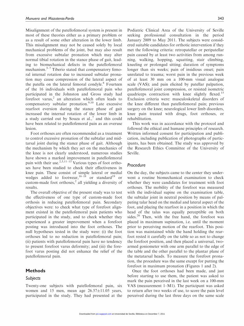
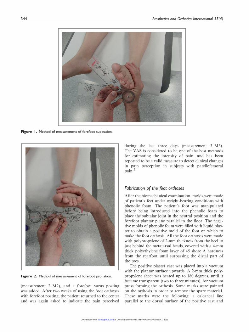
On the day, the subjects came to the center they under-went a routine biomechanical examination to checkwhether they were candidates for treatment with footorthoses. The mobility of the forefoot was measuredwith the individual supine on the examination table,the subtalar joint in neutral position by means of pal-pating talar head on the medial and lateral aspect of thefoot, and placing the rearfoot in a position in which thehead of the talus was equally perceptible on bothsides.20 Then, with the free hand, the forefoot wasplaced in maximum supination, i.e. until the momentprior to perceiving motion of the rearfoot. This posi-tion was maintained while the hand holding the rear-foot rested it carefully on the table so as not to changethe forefoot position, and then placed a universal, two-armed goniometer with one arm parallel to the edge ofthe table and the other parallel to the plantar plane ofthe metatarsal heads. To measure the forefoot prona-tion, the procedure was the same except for putting theforefoot in maximum pronation (Figures 1 and 2).
Once the foot orthoses had been made, and justbefore starting to use them, the patient was asked tomark the pain perceived in the last week on a 100-mmVAS (measurement 1–M1). The participant was askedto return after two weeks of use, to score the pain levelperceived during the last three days on the same scale
Munuera and Mazoteras-Pardo 343
at Universidad de Sevilla. Biblioteca on December 7, 2011poi.sagepub.comDownloaded from
(measurement 2–M2), and a forefoot varus postingwas added. After two weeks of using the foot orthoseswith forefoot posting, the patient returned to the centerand was again asked to indicate the pain perceived
during the last three days (measurement 3–M3).The VAS is considered to be one of the best methodsfor estimating the intensity of pain, and has beenreported to be a valid measure to detect clinical changesin pain perception in subjects with patellofemoralpain.21
Fabrication of the foot orthoses
After the biomechanical examination, molds were madeof patient’s feet under weight-bearing conditions withphenolic foam. The patient’s foot was manipulatedbefore being introduced into the phenolic foam toplace the subtalar joint in the neutral position and theforefoot plantar plane parallel to the floor. The nega-tive molds of phenolic foam were filled with liquid plas-ter to obtain a positive mold of the foot on which tomake the foot orthosis. All the foot orthoses were madewith polypropylene of 2-mm thickness from the heel tojust behind the metatarsal heads, covered with a 4-mmthick polyethylene foam layer of 45 shore A hardnessfrom the rearfoot until surpassing the distal part ofthe toes.
The positive plaster cast was placed into a vacuumwith the plantar surface upwards. A 2-mm thick poly-propylene sheet was heated up to 180 degrees, until itbecame transparent (two to three minutes), for vacuumpress forming the orthosis. Some marks were paintedon the orthosis in order to remove the spare material.These marks were the following: a calcaneal lineparallel to the dorsal surface of the positive cast and
Figure 1. Method of measurement of forefoot supination.
Figure 2. Method of measurement of forefoot pronation.
344 Prosthetics and Orthotics International 35(4)
at Universidad de Sevilla. Biblioteca on December 7, 2011poi.sagepub.comDownloaded from
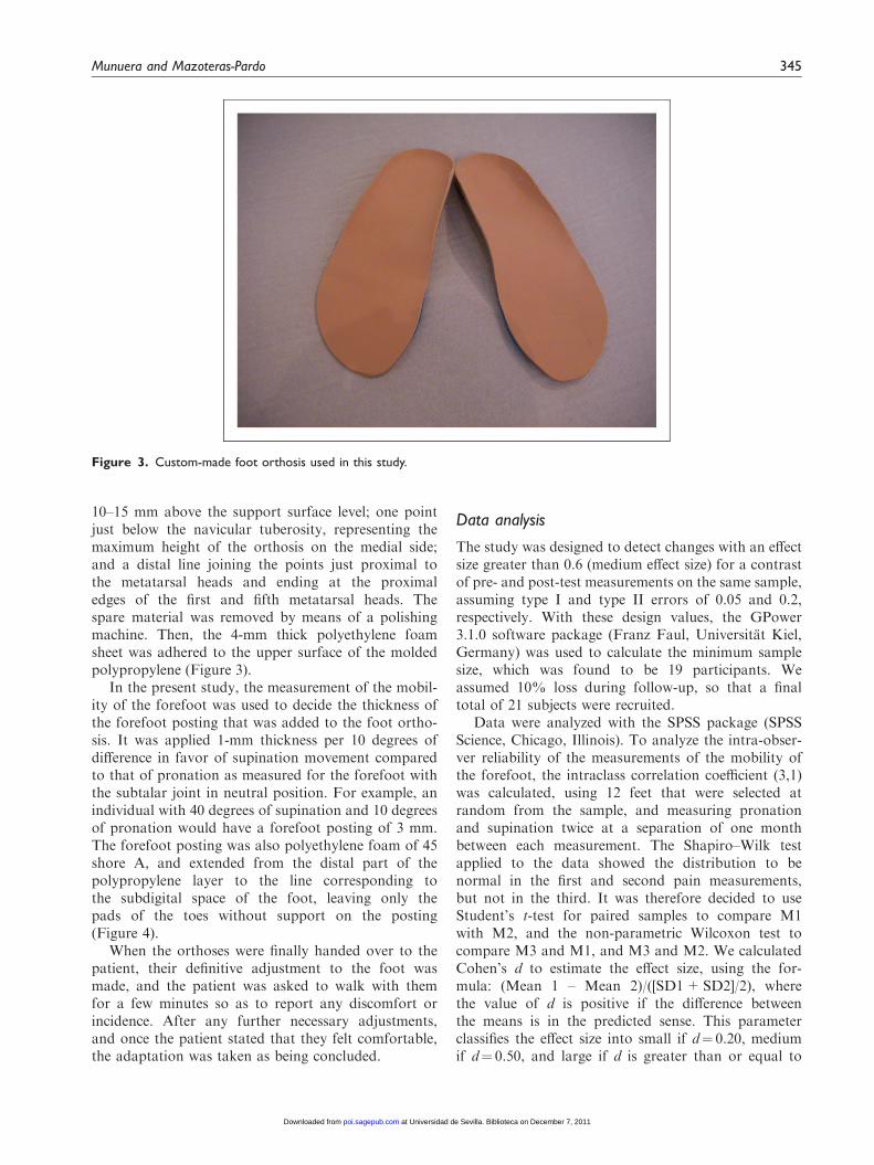
10–15 mm above the support surface level; one pointjust below the navicular tuberosity, representing themaximum height of the orthosis on the medial side;and a distal line joining the points just proximal tothe metatarsal heads and ending at the proximaledges of the first and fifth metatarsal heads. Thespare material was removed by means of a polishingmachine. Then, the 4-mm thick polyethylene foamsheet was adhered to the upper surface of the moldedpolypropylene (Figure 3).
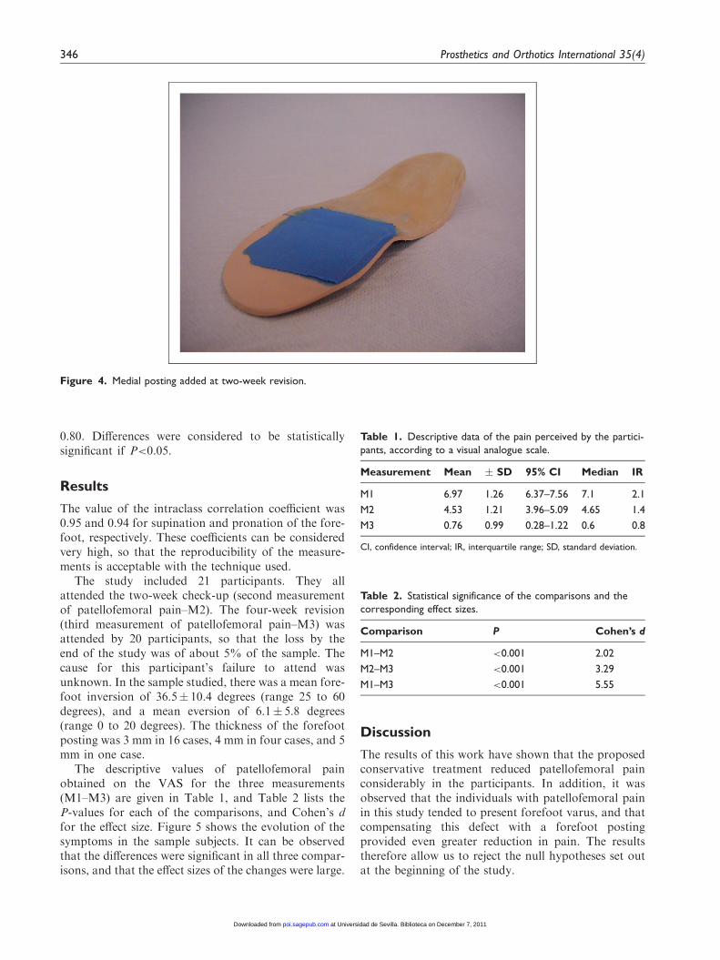
In the present study, the measurement of the mobil-ity of the forefoot was used to decide the thickness ofthe forefoot posting that was added to the foot ortho-sis. It was applied 1-mm thickness per 10 degrees ofdifference in favor of supination movement comparedto that of pronation as measured for the forefoot withthe subtalar joint in neutral position. For example, anindividual with 40 degrees of supination and 10 degreesof pronation would have a forefoot posting of 3 mm.The forefoot posting was also polyethylene foam of 45shore A, and extended from the distal part of thepolypropylene layer to the line corresponding tothe subdigital space of the foot, leaving only thepads of the toes without support on the posting(Figure 4).
When the orthoses were finally handed over to thepatient, their definitive adjustment to the foot wasmade, and the patient was asked to walk with themfor a few minutes so as to report any discomfort orincidence. After any further necessary adjustments,and once the patient stated that they felt comfortable,the adaptation was taken as being concluded.
Data analysis
The study was designed to detect changes with an effectsize greater than 0.6 (medium effect size) for a contrastof pre- and post-test measurements on the same sample,assuming type I and type II errors of 0.05 and 0.2,respectively. With these design values, the GPower3.1.0 software package (Franz Faul, Universitat Kiel,Germany) was used to calculate the minimum samplesize, which was found to be 19 participants. Weassumed 10% loss during follow-up, so that a finaltotal of 21 subjects were recruited.
Data were analyzed with the SPSS package (SPSSScience, Chicago, Illinois). To analyze the intra-obser-ver reliability of the measurements of the mobility ofthe forefoot, the intraclass correlation coefficient (3,1)was calculated, using 12 feet that were selected atrandom from the sample, and measuring pronationand supination twice at a separation of one monthbetween each measurement. The Shapiro–Wilk testapplied to the data showed the distribution to benormal in the first and second pain measurements,but not in the third. It was therefore decided to useStudent’s t-test for paired samples to compare M1with M2, and the non-parametric Wilcoxon test tocompare M3 and M1, and M3 and M2. We calculatedCohen’s d to estimate the effect size, using the for-mula: (Mean 1 – Mean 2)/([SD1+SD2]/2), wherethe value of d is positive if the difference betweenthe means is in the predicted sense. This parameterclassifies the effect size into small if d¼ 0.20, mediumif d¼ 0.50, and large if d is greater than or equal to
Figure 3. Custom-made foot orthosis used in this study.
Munuera and Mazoteras-Pardo 345
at Universidad de Sevilla. Biblioteca on December 7, 2011poi.sagepub.comDownloaded from
0.80. Differences were considered to be statisticallysignificant if P<0.05.
Results
The value of the intraclass correlation coefficient was0.95 and 0.94 for supination and pronation of the fore-foot, respectively. These coefficients can be consideredvery high, so that the reproducibility of the measure-ments is acceptable with the technique used.
The study included 21 participants. They allattended the two-week check-up (second measurementof patellofemoral pain–M2). The four-week revision(third measurement of patellofemoral pain–M3) wasattended by 20 participants, so that the loss by theend of the study was of about 5% of the sample. Thecause for this participant’s failure to attend wasunknown. In the sample studied, there was a mean fore-foot inversion of 36.5� 10.4 degrees (range 25 to 60degrees), and a mean eversion of 6.1� 5.8 degrees(range 0 to 20 degrees). The thickness of the forefootposting was 3 mm in 16 cases, 4 mm in four cases, and 5mm in one case.
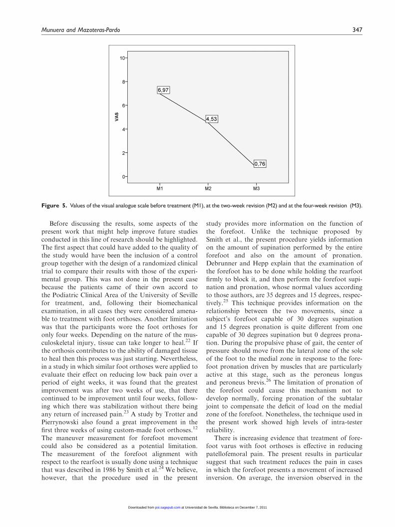
The descriptive values of patellofemoral painobtained on the VAS for the three measurements(M1–M3) are given in Table 1, and Table 2 lists theP-values for each of the comparisons, and Cohen’s dfor the effect size. Figure 5 shows the evolution of thesymptoms in the sample subjects. It can be observedthat the differences were significant in all three compar-isons, and that the effect sizes of the changes were large.
Discussion
The results of this work have shown that the proposedconservative treatment reduced patellofemoral painconsiderably in the participants. In addition, it wasobserved that the individuals with patellofemoral painin this study tended to present forefoot varus, and thatcompensating this defect with a forefoot postingprovided even greater reduction in pain. The resultstherefore allow us to reject the null hypotheses set outat the beginning of the study.
Figure 4. Medial posting added at two-week revision.
Table 1. Descriptive data of the pain perceived by the partici-pants, according to a visual analogue scale.
Measurement Mean � SD 95% CI Median IR
M1 6.97 1.26 6.37–7.56 7.1 2.1
M2 4.53 1.21 3.96–5.09 4.65 1.4
M3 0.76 0.99 0.28–1.22 0.6 0.8
CI, confidence interval; IR, interquartile range; SD, standard deviation.
Table 2. Statistical significance of the comparisons and thecorresponding effect sizes.
Comparison P Cohen’s d
M1–M2 <0.001 2.02
M2–M3 <0.001 3.29
M1–M3 <0.001 5.55
346 Prosthetics and Orthotics International 35(4)
at Universidad de Sevilla. Biblioteca on December 7, 2011poi.sagepub.comDownloaded from
Before discussing the results, some aspects of thepresent work that might help improve future studiesconducted in this line of research should be highlighted.The first aspect that could have added to the quality ofthe study would have been the inclusion of a controlgroup together with the design of a randomized clinicaltrial to compare their results with those of the experi-mental group. This was not done in the present casebecause the patients came of their own accord tothe Podiatric Clinical Area of the University of Sevillefor treatment, and, following their biomechanicalexamination, in all cases they were considered amena-ble to treatment with foot orthoses. Another limitationwas that the participants wore the foot orthoses foronly four weeks. Depending on the nature of the mus-culoskeletal injury, tissue can take longer to heal.22 Ifthe orthosis contributes to the ability of damaged tissueto heal then this process was just starting. Nevertheless,in a study in which similar foot orthoses were applied toevaluate their effect on reducing low back pain over aperiod of eight weeks, it was found that the greatestimprovement was after two weeks of use, that therecontinued to be improvement until four weeks, follow-ing which there was stabilization without there beingany return of increased pain.23 A study by Trotter andPierrynowski also found a great improvement in thefirst three weeks of using custom-made foot orthoses.12
The maneuver measurement for forefoot movementcould also be considered as a potential limitation.The measurement of the forefoot alignment withrespect to the rearfoot is usually done using a techniquethat was described in 1986 by Smith et al.24 We believe,however, that the procedure used in the present
study provides more information on the function ofthe forefoot. Unlike the technique proposed bySmith et al., the present procedure yields informationon the amount of supination performed by the entireforefoot and also on the amount of pronation.Debrunner and Hepp explain that the examination ofthe forefoot has to be done while holding the rearfootfirmly to block it, and then perform the forefoot supi-nation and pronation, whose normal values accordingto those authors, are 35 degrees and 15 degrees, respec-tively.25 This technique provides information on therelationship between the two movements, since asubject’s forefoot capable of 30 degrees supinationand 15 degrees pronation is quite different from onecapable of 30 degrees supination but 0 degrees prona-tion. During the propulsive phase of gait, the center ofpressure should move from the lateral zone of the soleof the foot to the medial zone in response to the fore-foot pronation driven by muscles that are particularlyactive at this stage, such as the peroneus longusand peroneus brevis.26 The limitation of pronation ofthe forefoot could cause this mechanism not todevelop normally, forcing pronation of the subtalarjoint to compensate the deficit of load on the medialzone of the forefoot. Nonetheless, the technique used inthe present work showed high levels of intra-testerreliability.
There is increasing evidence that treatment of fore-foot varus with foot orthoses is effective in reducingpatellofemoral pain. The present results in particularsuggest that such treatment reduces the pain in casesin which the forefoot presents a movement of increasedinversion. On average, the inversion observed in the
Figure 5. Values of the visual analogue scale before treatment (M1), at the two-week revision (M2) and at the four-week revision (M3).
Munuera and Mazoteras-Pardo 347
at Universidad de Sevilla. Biblioteca on December 7, 2011poi.sagepub.comDownloaded from
participants was six times greater than the eversion, ahigher ratio than that proposed by some authors asnormal.25 We would argue that this alteration of theforefoot causes it to function as a forefoot varusdeformity. Similar results have been reported by otherworkers. Johnston and Gross examined the effect offoot orthoses on the quality of life of patients withpatellofemoral pain, and added forefoot varus postingto 80% of their sample.2 They showed improvement inpain, stiffness and physical activity after using footorthoses for two weeks and in the follow-up afterthree months of use.2 Eng and Pierrynowski foundthat the subjects who formed the group with patellofe-moral pain in their study had a mean (�SD) of12.4 (�3.7) degrees of forefoot varus.5 They observedthat the use of soft foot orthoses for eight weeks,together with a program of isometric exercises forthe quadriceps muscle, reduced patellofemoral painmore than the exercise program alone. Saxena andHaddad examined 102 subjects of whom nearly 90%were diagnosed with patellofemoral pain, retropatellardysplasia, or chondromalacia patellae, and needed toadd forefoot varus posting in 91.2% of the cases, whichled them to suggest that there might be a correlationbetween forefoot varus deformity and patellofemoralpain.11 At other times, rather than forefoot varus,it has been excessive subtalar pronation which hasbeen associated with patellofemoral pain.13,15,27 Itshould be noted that one of the compensation mecha-nisms of forefoot varus in gait is increased subtalarpronation.9,14,28
All these studies point to the existence of a strongfunctional relationship between the foot and knee.Pronation of the subtalar joint and flexion of theknee cause internal tibial rotation in a weight-bearingposition. This internal tibial rotation occurs because theknee begins the stance phase of gait in an almost fullyextended position, and then flexes to cushion theimpact together with subtalar pronation.6 Undernormal conditions, these movements change to the mid-stance phase of gait, in which the knee begins to extendand the subtalar joint begins to supine. However, whenthe subtalar pronation is excessive or prolonged in time,for example because it is needed to compensate highforefoot inversion,28 the external rotation of the tibiamay be delayed. Tiberio calls this situation a ‘biome-chanical dilemma’ for the knee joint, and it may be thesource of patellofemoral problems since the movementsof the patella are influenced by tibial rotation.8 In thefirst degrees of flexion, the patella is looser, less pressedagainst the femur, thus allowing more movement,29 andchanges may be produced in the normal position of thepatella on the femur. These changes may cause patello-femoral problems that can be controlled with the use offoot orthoses.
There might be several reasons why foot orthoses areeffective in decreasing patellofemoral pain. One is thatfoot orthoses to control subtalar pronation woulddiminish internal tibial rotation in the late midstanceand propulsive phases of gait, thus mitigating the ‘bio-mechanical dilemma’ referred to by Tiberio.8 If the sub-talar joint pronates during heel-off and push-off, themechanics of the knee may be affected by changes intibial rotation. When the knee is extended during gait,the tibia has to undergo external rotation. Subtalar pro-nation, however, forces internal tibial rotation. Footorthoses would reduce this conflict. A second reasonis that the Q angle would acquire a more physiologicalposition with the use of foot orthoses, since increasedor decreased Q angles would both result in areas ofexcessive pressure of contact of the patella on thefemur.30 A third reason would be that foot orthosescontribute to a more centralized position of the patellawith respect to the femoral condyles,31 possibly becauseof their effect on the rotations in the frontal and trans-verse planes of the tibia on the femur. Therefore, thereaction forces of the patellofemoral joint would bemore evenly distributed between the two condyles.5
Conclusions
The results of the present study suggest that custom-made foot orthoses, fabricated with 2-mm thick poly-propylene and a 4-mm thick polyethylene foam liner of45 shore A, reduced the patellofemoral pain in the par-ticipants. In most cases the improvement was increasedby adding forefoot varus posting to the orthoses, sinceit was observed that the participants presentedincreased movement of inversion of the forefoot rela-tive to that of eversion.
Funding
This research received no specific grant from any funding
agency in the public, commercial, or not-for-profit sectors.
Conflict of interest
The authors declare that there is no conflict of interest.
References
1. Callaghan MJ and Oldham JA. The role of quadriceps
exercise in the treatment of patellofemoral pain syndrome.
Sports Med 1996; 21: 384–391.2. Johnston LB and Gross MT. Effects of foot orthoses
on quality of life for individuals with patellofemoral
pain syndrome. J Orthop Sports Phys Ther 2004; 34:
440–448.
348 Prosthetics and Orthotics International 35(4)
at Universidad de Sevilla. Biblioteca on December 7, 2011poi.sagepub.comDownloaded from
3. Pitmann D and Jack D. A clinical investigation to deter-mine the effectiveness of biomechanical foot orthoses asinitial treatment for patellofemoral pain syndrome.
J Prosthet Orthot 2000; 12: 110–116.4. Thijs Y, Van Tiggelen D, Roosen P, De Clercq D and
Witwrouw E. A prospective study on gait-related intrinsicrisk factors for patellofemoral pain. Clin J Sport Med
2007; 17: 437–445.5. Eng JJ and Pierrynowski MR. Evaluation of soft foot
orthotics in the treatment of patellofemoral pain syn-
drome. Phys Ther 1993; 73: 62–70.6. Nawoczenski DA, Cook TM and Saltzman CL. The
effect of foot orthotics on three-dimensional kinematics
of the leg and rearfoot during running. J Orthop SportPhys Ther 1995; 21: 317–327.
7. Souza TR, Pinto RZ, Trede RG, Kirkwood RN, Pertence
AE and Fonseca ST. Late rearfoot eversion and lower-limb internal rotation caused by changes in the interac-tion between forefoot and support surface. J Am PodiatrMed Assoc 2009; 99: 503–511.
8. Tiberio D. The effect of excessive subtalar joint pronationon patellofemoral mechanics: a theoretical model.J Orthop Sports Phys Ther 1987; 9: 160–165.
9. Root ML, Orien WP and Weed JH. Normal and abnormalfunction of the foot, Vol 2. Los Angeles: ClinicalBiomechanics Corp, 1977.
10. Michaud TC. Foot orthoses and others forms of conserva-tive foot care. Boston, MA: Williams and Wilkins, 1996.
11. Saxena A and Haddad J. The effect of foot orthoses onpatellofemoral pain syndrome. J Am Podiatr Med Assoc
2003; 93: 264–271.12. Trotter LC and Pierrynowski MR. The short-term effec-
tiveness of full-contact custom-made foot orthoses and
prefabricated shoe inserts on lower-extremity musculo-skeletal pain: a randomized clinical trial. J Am PodiatrMed Assoc 2008; 98: 357–363.
13. Barton CJ, Bonanno D, Levinger P and Menz HB. Footand ankle characteristics in patellofemoral pain syn-drome: a case control and reliability study. J Orthop
Sports Phys Ther 2010; 40: 286–296.14. Johanson MA, Greenfeld L, Hung C, Walters R and
Watson C. The relationship between forefoot and rear-foot static alignment in pain-free individuals with above-
average forefoot varus angles. Foot Ankle Spec 2010; 3:112–116.
15. Barton CJ, Menz HB and Crossley KM. The immediate
effects of foot orthoses on functional performance in indi-viduals with patellofemoral pain syndrome. Br J SportsMed 2011; 45: 193–197.
16. Baker K, Goggins J, Xie H, et al. A randomized cross-over trial of a wedged insole for treatment of knee oste-oarthritis. Arthritis Rheum 2007; 56: 1198–1203.
17. Hinman RS, Payne C, Metcalf BR, Wrigley TV andBennell KL. Lateral wedges in knee osteoarthritis:What are their immediate clinical and biomechanical
effects and can these predict a three-month clinical out-come? Arthritis Rheum 2008; 59: 408–415.
18. Rodrigues PT, Ferreira AF, Pereira RMR, Bonfa E,Borba EF and Fuller R. Effectiveness of medial-wedge
insole treatment for valgus knee osteoarthritis. ArthritisRheum 2008; 59: 603–608.
19. Barton CJ, Menz HB and Crossley KM. Effects of pre-
fabricated foot orthoses on pain and function in individ-uals with patellofemoral pain syndrome: a cohort study.Phys Ther Sport 2011; 12: 70–75.
20. Elveru RA, Rothstein JM, Lamb RL and Riddle DL.Methods for taking subtalar joint measurements. A clin-ical report. Phys Ther 1988; 68: 678–682.
21. Chesworth BM, Culham E, Tata GE and Peat M.Validation of outcome measures in patients with patello-femoral syndrome. J Orthop Sports Phys Ther 1989; 10:302–308.
22. De Marchi A, Robba T, Ferrarese E and Faletti C.Imaging in musculoskeletal injuries: state of the art.Radiol Med 2005; 110: 115–131.
23. Castro A and Munuera PV. Efecto del tratamiento orto-podologico en el dolor lumbar. Rev Esp Podol 2010; 21:94–99.
24. Smith LS, Clarke TE, Hamill CL and Santopietro F. Theeffects of soft and semi-rigid orthoses upon rearfootmovement in running. J Am Podiatr Med Assoc 1986;76: 227–233.
25. Debrunner HU and Hepp WR. Diagnostico en ortopedia.Barcelona: Grass-Iatros Ediciones, 1996.
26. Perry J and Burnfield JM. Gait analysis: Normal and
pathological function. Thorofare: Slack, Inc, 2010.27. Barton JC, Levinger P, Webster KE and Menz HB.
Walking kinematics in individuals with patellofemoral
pain syndrome: a case-control study. Gait Posture 2011;33: 286–291.
28. Johanson MA, Donatelli R, Wooden MJ, Andrew PD
and Cummings GS. Effects of three different postingmethods on controlling abnormal subtalar pronation.Phys Ther 1994; 74: 149–158.
29. van Kampen A and Huiskes R. The three-dimensional
tracking pattern of the human patella. J Orthop Res1990; 8: 372–382.
30. Huberti HH and Hayes WC. Patellofemoral contact pres-
sures. The influence of q-angle and tendofemoral contact.J Bone Joint Surg Am 1984; 66: 715–724.
31. Klingman RE, Liaos SM and Hardin KM. The effect of
subtalar joint posting on patellar glide position in sub-jects with excessive rearfoot pronation. J Orthop SportsPhys Ther 1997; 25: 185–191.
Munuera and Mazoteras-Pardo 349
at Universidad de Sevilla. Biblioteca on December 7, 2011poi.sagepub.comDownloaded from