Embed Size (px)
Citation preview
Research Report
Evaluation of Soft Foot Orthotics in the Treatment of Patellofemoral Pain Syndrome
Background and Purpose, The efictiveness of soft foot orthotics in the treat- ment of patients who have patellofemoral pain syndrome was investigated. Sub- jects. Subjects were 20 adolescent female patients, aged 13 to 17 years @=14.8, SD = 1.2), who were diagnosed with patellofemoral pain syndrome and who ex- hibited excessive forefoot varus or calcaneal valgus. Metbods. Subjects were ran- domly mgned to one of two groups: a control group (n=10), which took part in an exercise program, or a treatment group (n=IO), which used soft foot orthotics in addition to participating in the exercise program. The exercise program con- sisted of quadriceps fernoris and hamstring muscle strengthening and stretching exerckes. A visual analogue scale was used to assess the level of pain of the sub- jects over an 8-week penen&. Results. Both the treatment and control groups demonstrated a signijicant decrease in the level of pain, but the improvement of the treatment group was significantly greater than that of the control group. Con- clusion and Dlscuswbn. The results suggest that in addition to an exercise program, the use of so3 foot orthotics is an e$ective means of treatment for the patient with patellofemoral pain syndrome. [Eng JJ, Piemynowski MR. Evaluation of soj2 foot orthotics in the treatment of patellofmral pain syndrome. Phys Ther. 1993; 7362-70.1
Patellofemoral pain syndrome (PFPS) is the leading cause of chronic knee pain in adolescents.1 The diagnosed incidence is on the rise, most likely as a result of greater emphasis on fitness in our society and an increased aware- ness of the condition by medical prac- titioners. Retropatellar pain experi- enced with PFPS can become a severe problem for adolescents, denying them full participation in sports and leisure activities. It is a significant psy-
chological blow to those adolescents who are so restricted by their pain that they must abandon sports and related activities during their teen years. Much of the persistence of daily activity into adulthood depends on the perceptions of physical activity formed during childhood and adolescence.*
Although the exact etiology of PFPS is unknown, investigatorss5 propose that abnormal patellofemoral mechan-
JJ Eng, PT, is a doctoral candidate, Department of Kinesiology, University of Waterloo, Waterloo, Ontario, Canada N2L 3G1. She was a student in the master's degree program, Institute of Biomedi- cal Engineering and Department of Community Health, Faculty of Medicine, University of Toronto, when the study was completed in partial fulfillment of her degree requirements. Address all corre- spondence to Ms Eng.
MR Pierrynowski, PhD, is Associate Professor, School of Occupational Therapy and Physiotherapy, McMaster University, Hamilton, Ontario, Canada L8N 325. He was Associate Professor, School of Physical and Health Education, Faculty of Medicine, and Institute of Biomedical Engineering, Uni- versity of Toronto, when this study was completed.
This study was approved by the Hospital for Sick Children Human Subjects Review Board.
This study was supported in pan by the University of Toronto (Open Fellowship to Ms Eng)
This article was submitted October I, 1991, and was accepted September 14, 1992.
Janice J Eng Michael R
Pier rynowskl
ics are the primary cause of PFPS. A disturbance of the normal patellofem- oral relationship results in an uneven distribution of shearing and compres- sive forces acting on the patellofemo- ral joint during normal a~tivity.~
Malalignment of the patellofemoral mechanism is not only caused by local patellofemoral mechanics, but reflects anatomical variations through- out the entire lower extremities; in- deed, PFPS is highly correlated with excessive pr0nation.~-7 Excessive subtalar pronation during the stance phase can alter the normal rotation of the tibia in the frontal and transverse planes as a result of the anatomical congruency of the talus within the ankle mortise.10 In turn, aberrant tibia1 rotation can disrupt the normal patellofemoral relationship.7J*J* To alter aspects of lower-extremity me- chanics, one can use a foot orthotic, a device inserted between the foot and shoe, to modify foot positioning and
8/62 Physical Therapy/Volume 73, Number 2February 1993
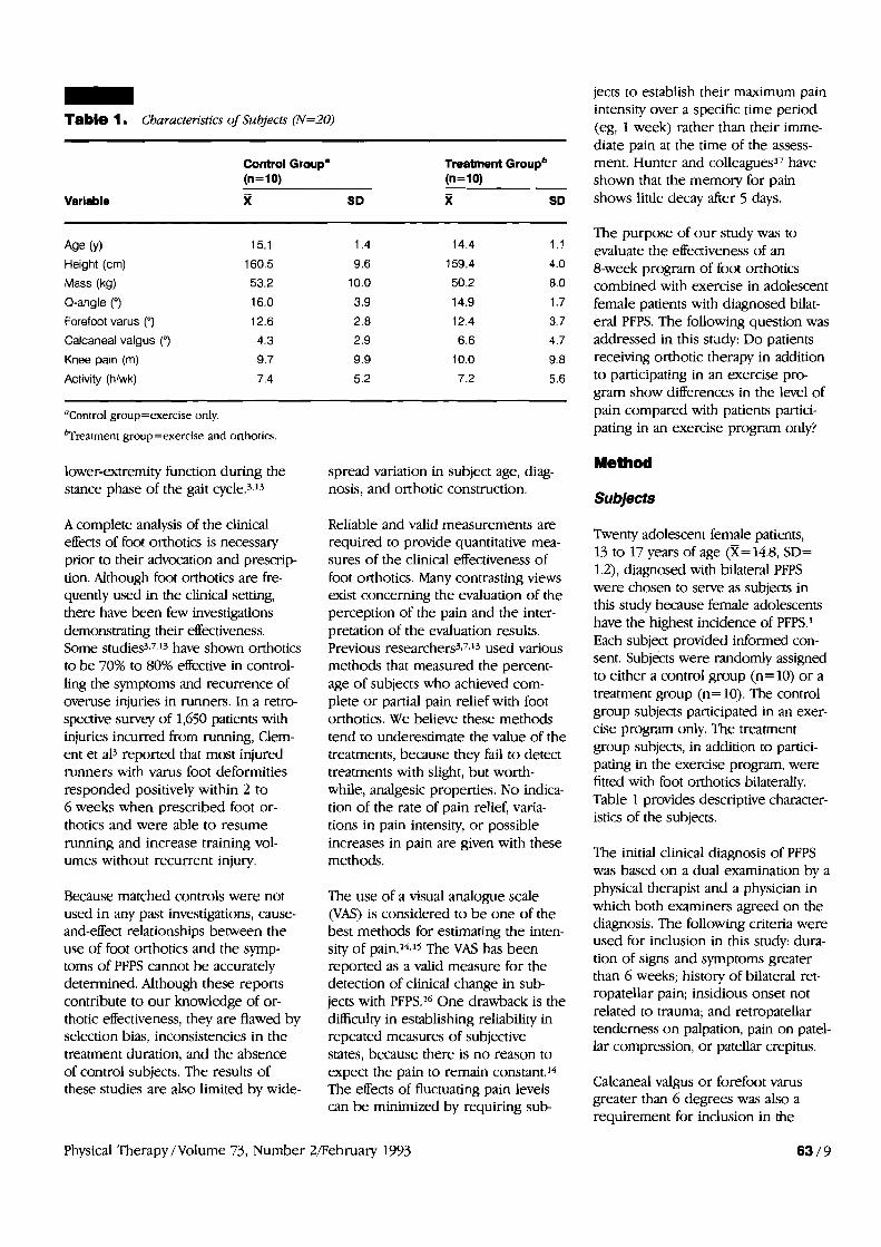
- Table 1. Characteristics of Subjects (N=20)
Control Groupa Treatment Groupb (n=lO) (n=10)
X SD x SD
Age (Y) Height (cm)
Mass (kg)
Q-angle (")
Forefoot varus (q
Calcaneal valgus (q
Knee pain (m)
Activity (hlwk)
"Control group=exercise only.
b~reatrnent group=exercise and onhotics.
lower-extremity function during the stance phase of the gait cycle.3J3
A complete analysis of the clinical effects of foot orthotics is necessary prior to their advocation and prescrip- tion. Although foot orthotics are fre- quently used in the clinical setting, there have been few investigations demonsvating their effectiveness. Some studies3.7J3 have shown orthotics to be 70% to 80% effective in control- ling the symptoms and recurrence of overuse injuries in runners. In a retro- spective survey of 1,650 patients with injuries incurred from running, Clem- ent et a13 reported that most injured runners with varus foot deformities responded positively within 2 to 6 weeks when prescribed foot or- thotics and were able to resume running and increase training vol- umes without recurrent injury.
Because matched controls were not used in any past investigations, cause- and-effect relationships between the use of foot orthotics and the symp- toms of PFPS cannot be accurately determined. Although these reports contribute to our knowledge of or- thotic effectiveness, they are flawed by selection bias, inconsistencies in the treatment duration, and the absence of control subjects. The results of these studies are also limited by wide-
spread variation in subject age, diag- nosis, and orthotic construction.
Reliable and valid measurements are required to provide quantitative mea- sures of the clinical effectiveness of foot orthotics. Many contrasting views exist concerning the evaluation of the perception of the pain and the inter- pretation of the evaluation results. Previous researchers3'7J3 used various methods that measured the percent- age of subjects who achieved com- plete or partial pain relief with foot orthotics. We believe these methods tend to underestimate the value of the treatments, because they fail to detect treatments with slight, but worth- while, analgesic properties. No indica- tion of the rate of pain relief, varia- tions in pain intensity, or possible increases in pain are given with these methods.
The use of a visual analogue scale WAS) is considered to be one of the best methods for estimating the inten- sity of pain.14315 The VAS has been reported as a valid measure for the detection of clinical change in sub- jects with PFPS.I6 One drawback is the difficulty in establishing reliability in repeated measures of subjective states, because there is no reason to expect the pain to remain constant.14 The effects of fluctuating pain levels can be minimized by requiring sub-
jects to establish their maximum pain intensity over a specific time period (eg, 1 week) rather than their imme- diate pain at the time of the assess- ment. Hunter and colleagues17 have shown that the memoly for pain shows little decay after 5 days.
The purpose of our study was to evaluate the effectiveness of an 8-week program of foot orthotics combined with exercise in adolescent female patients with diagnosed bilat- eral PFPS. The following question was addressed in this sti~dy: Do patients receiving orthotic therapy in addition to participating in an exercise pro- gram show differences in the level of pain compared with patients partici- pating in an exercise program only?
Method
Subjects
Twenty adolescent female patients, 13 to 17 years of age @=14.8, SD= 1.2), diagnosed with bilateral PFPS were chosen to sewe as subjects in this study because female adolescents have the highest incidence of PFPS.I Each subject provided informed con- sent. Subjects were randomly assigned to either a control group (n= 10) or a treatment group (n=10). The control group subjects participated in an arer- cise program only. The treatment group subjects, in addition to partici- pating in the exercise program, were fitted with foot orthotics bilaterally. Table 1 provides descriptive character- istics of the subjects.
The initial clinical diagnosis of PFPS was based on a dual examination by a physical therapist and a physician in which both examiners agreed on the diagnosis. The following criteria were used for inclusion in this study: dura- tion of signs and symptoms greater than 6 weeks; history of bilateral ret- ropatellar pain; insidious onset not related to trauma; and retropatellar tenderness on palpation, pain on patel- lar compression, or patellar crepitus.
Calcaneal valgus or forefoot varus greater than 6 degrees was also a requirement for inclusion in the
Physical Therapy /Volume 73, Number 2/February 1993
enced over the last week for each of the activities.
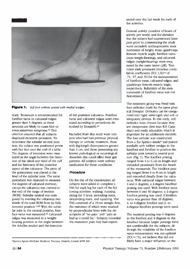
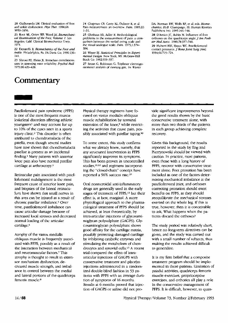
Figure 1. Soft foot orthotic posted with
study. Treatment is recommended for forefoot varus or calcaneal valgus greater than 5 degrees, as these amounts are likely to cause foot or lowerextremity symptoms.18 This criterion ensured that all subjects displayed excessive pronation. To determine the subtalar neutral posi- tion, the subject was positioned prone with her feet over the end of a table. The degrees of inversion were mea- sured as the angle between the bisec- tion of the distal one third of the calf and the bisection of the posterior aspect of the calcaneus. The pivot of the goniometer was placed at the level of the subtalar joint. The same procedure was repeated to measure the degrees of calcaneal eversion, except the calcaneus was everted to the end of the range of motion (ROM). Subtalar neutral was com- puted by everting the calcaneus two thirds of the total ROM from its fully inverted position.19.20 With the subta- lar joint in the neutral position, fore- foot varus was measured.21 Calcaneal valgus was measured in a weight- bearing position as the angle between the Achilles tendon and the bisection
medial wedges.
of the posterior calcaneus. Forefoot varus and calcaneal valgus were mea- sured according to procedures de- scribed by Donatelli.22
Excluded from this study were sub- jects who had had previous physical therapy or orthotic treatment, those with leg-length discrepancies greater than 1 cm, and those possessing any known pathological or neurological disorders that could affect their gait patterns. All subjects were without medication for these conditions.
Procedure
On the day of the examination, all subjects were asked to complete a VAS for each leg for each of the fol- lowing activities: walking, running, sitting for 1 hour, ascending stairs, descending stairs, and squatting. The VAS consisted of a 10-cm straight line, the extremes of which were marked by perpendicular lines with the de- scriptors of "no pain" and "pain as bad as it could be." Subjects recorded the maximum pain they had experi-
'Spenco Sports Medicine Products, Toronto, Ontario, Canada M4W 3L9
General activity (number of hours of activity per week) and the duration that the subject had experienced knee pain prior to commencing the study were recorded. Anthropometric mea- surements of height, mass, quadriceps femoris muscle angle, forefoot varus (non-weight-bearing), and calcaneal valgus (weight-bearing) were mea- sured by the same tester (JJE). Test- retest trials produced intraclass corre. lation coefficients (ICC[1,1])23 of .71, .97, and .94 for the measurements of forefoot varus, calcaneal valgus, and quadriceps femoris muscle angle, respectively. Reliability of the ma- surements of forefoot varus was not determined.
The treatment group was fitted with - - foot orthotics made by the same phys- ical therapist. Orthotics can be catego- rized into rigid, semi-rigid, and soft or temporary devices. In this study, soft orthotics were chosen because they are inexpensive (less than $15 Cana- dian) and easily adjustable, which is important for an adolescent clientele. The foot orthotic was constructed from a flat Spenco insole* and posted medially with rubber wedges in the hindfoot and forefoot to position the subtalar joint toward a neutral posi- tion (Fig. 1). The forefoot posting ranged from 4 to 6 cm in length and extended proximally from the heads of the metatarsals. The hindfoot post- ing ranged from 6 to 8 cm in length and extended distally from the calca- neus. With calcaneal valgus between 4 and 6 degrees, a 2degree hindfoot posting was used. With forefoot varus between 6 and 10 degrees, a 2-degree forefoot posting was used. If forefoot varus was greater than 10 degrees, 4- to 6degree forefoot and 2- to 4-degree hindfoot postings were used.
The maximal posting was 6 degrees in the forefoot and 4 degrees in the hindfoot because larger postings were not comfortable for the subjects. Al- though the reliability of the forefoot varus measurement was not optimal (ICC = .71), we believe this did not likely have a major influence on the
Physical Therapy/Volume 73, Number 2fiebruary 1993
prescription of the postings, because the majority of subjects exhibited such large magnitudes of calcaneal valgus and forefoot varus (Tab. 1) that the maximal amount of posting was prescribed. The control group sub- jects were fitted with flat Spenco insoles, which were inserted into their shoes without any postings to decrease the bias between the two groups. The orthotic insole was worn whenever the subject was wearing shoes and could be transferred into different shoes (eg, running shoes, school shoes), depending on the subject's needs.
Subjects were monitored for 8 weeks. During this time, they visited the clinic twice each week. Every 2 weeks, the subjects completed six VASs to measure their pain response to the activities of walking, running, stairs ascent, stairs descent, sitting for 1 hour, and squatting. As all subjects were students, a regular 1-hour school period was the criterion that they used to estimate their pain for the activity of sitting for 1 hour. Sub- jects also recorded the number of hours they had participated in physi- cal activities.
Exerclse Program
On the first visit, all subjects were instructed in an exercise program of isometric quadriceps femoris muscle contractions and straight leg raising in a supine position. On the second visit, subjects were instructed in quadriceps femoris and hamstring muscle stretch- ing exercises. To stretch the quadri- ceps femoris muscles, the subject stood on one leg and grasped the contralateral ankle. In the contralat- era1 limb, the knee joint was flexed while the subject maintained a neutral or extended hip joint position. While one knee was flexed to 45 degrees in a long-sitting position, the subject stretched the hamstring muscle by lowering the chest toward the ex- tended knee. Resisted straight leg raising and hamstring muscle strengthening were initiated on suc- cessive visits. Resistance was provided by small weights or by using an elas- tic material looped around the ankles.
One set of 10 repetitions of all the exercises was to be performed twice a day at home. Three random phone calls were made to each subject to determine whether large discrepan- cies occurred between the control and treatment groups in their compli- ance with the exercise program. A positive response was given if the subject had performed the exercises the previous day. No significant differ- ence was noted between the two groups regarding exercise compliance using a sign test (P<.05).
Data Analysls
Means and standard deviations were calculated for the descriptive charac- teristics. Independent t tests were used to compare these variables be- tween the two groups. Significance was accepted at the .05 level.
As all subjects experienced bilateral knee pain, analysis was performed for the knee that was considered the most painful on the initial assessment. The VAS data were found to be nor- mal in distribution using the Shapiro- Wilk W statistic of normality. In addi- tion, the variance within the control and treatment groups was homoge- neous. Parametric methods of analysis have been recommended for VAS data if the distribution of the variances is found to be homogeneou~.~4~25 Inde- pendent t tests were performed to compare each activity of the VAS com- pleted on the first visit to determine whether the control and treatment groups commenced the study from a similar baseline. The main statistical procedure was a three-factor (group versus activity versus week) analysis of variance (ANOVA) for repeated mea- sures followed by a Newrnan-Keuls post hoc analysis when a significant F-ratio test result was observed.26 The level of significance was accepted at .05.
Resutts
No significant differences were ob- served between the groups for the initial pain scales o r for any of the descriptive variables (anthropomeuic measurements, duration of pain prior
to the study, general activity during the study), suggesting that the groups were well matched.
Figure 2 demonstrates the differences between the control and treatment groups. The results of the repeated- measures ANOVA are presented in Table 2. Overall, subjects in both groups showed a significant reduction in the pain response. A significant difference of the pain response among the six activities was also observed.
Although both groups demonstrated a significant reduction in the reported pain, the treatment group demon- strated a significantly greater reduc- tion than the control group. Post hoc analyses were performed to compare the treatment and control groups (1) for weeks 2, 4, 6, and 8 and (2) for the activities of walking, running, stairs ascent, stairs descent, sitting for 1 hour, and squatting. The post hoc analysis revealed significant differ- ences between the treatment and control groups for weeks 4, 6, and 8, with the treatment group demon- strating a greater reduction of pain over the control group. The treat- ment and control groups demon- strated significant differences in the activities of running, ascent of stairs, descent of stairs, and squatting.
It was expected that the activities would demonstrate significantly differ- ent pain responses, as the activities varied in the amount of stress placed on the patellofemoral joint. Both groups reported a significant reduc- tion in the pain response. This im- provement might be attributed to the exercise program, which was de- signed to encourage contraction of the vastus medialis oblique muscle for stabilization of the patella within the femoral groove and to stretch muscles that may contribute to in- creased patellar forces.
The treatment group reported a sig- nificant decrease in reported pain when compared with the control group at weeks 4, 6, and 8. It is diB-
Physical Therapy /Volume 73, Number
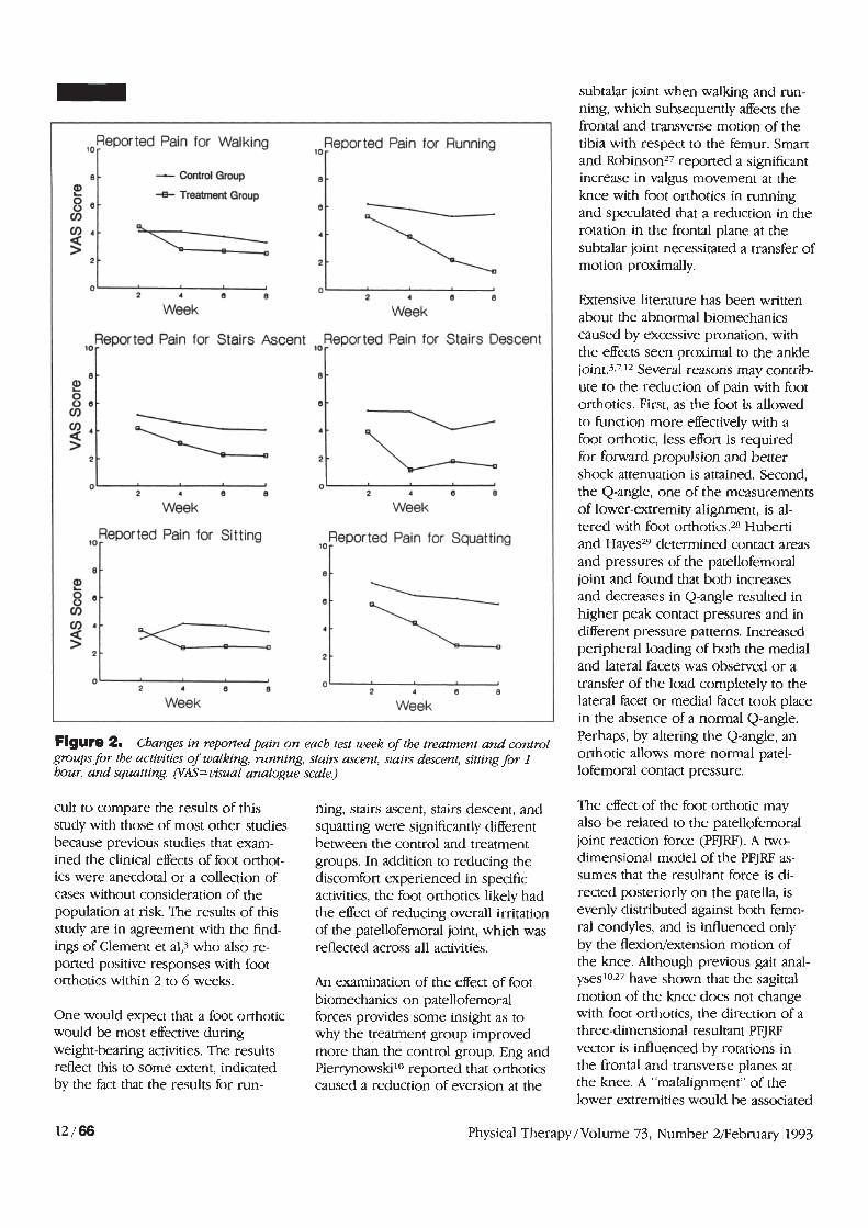
Figure 2. Changes in reportedpain on each test week of the treatment and control groups for the activities of walking, running, stairs ascent, stairs descent, sitting for I hour, and squattfng. pM=uisual analogue scale.)
lor Reported Pain for Walking
10 - Reported Pain for Running
8 - - control Group 8 - E? -a- Treatment Group 8 8 - 0
2 4 - > sp; 2 -
J ; 7 0 2 4 8 8 2 4 6 8
Week Week
10- Reported Pain for Stairs Ascent loReported Pain for Stairs Descent
8 - 8 - 2 8 e - e - V)
-
I cl 0 2 4 6 8 2 4 8 8
Week Week
lo Reported Pain for Sitting 10 Reported Pain for Squatting
cult to compare the results of this study with those of most orher studies because previous studies that exam- ined the clinical effects of foot orthot- ics were anecdotal or a collection of cases without consideration of the population at risk. The results of this study are in agreement with the find- ings of Clement et al? who also re- ported positive responses with foot orthotics within 2 to 6 weeks.
8
E? 8 e - cn V) 4 -
s 2 -
One would expect that a foot orthotic would be most effective during weight-bearing activities. The results reflect this to some extent, indicated by the fact that the results for run-
ning, stairs ascent, stairs descent, and squatting were significantly different between the control and treatment groups. In addition to reducing the discomfort experienced in specific activities, the foot orthotics likely had the effect of reducing overall irritation of the patellofemoral joint, which was reflected across all activities.
0 2 4 8 6 0
2 4 6 8
Week Week
- 8
An examination of the effect of foot biomechanics on patellofemoral forces provides some insight as to why the treatment group improved more than the control group. Eng and Pierrynowski10 reported that orthotics caused a reduction of eversion at the
-
subtalar joint when walking and run- ning, which subsequently affects the frontal and transverse motion of the tibia with respect to the femur. Smart and Robinson2' reported a significant increase in valgus movement at the knee with foot orthotics in running and speculated that a reduction in [he rotation in the frontal plane at the subtalar joint necessitated a transfer of motion proximally.
P
Extensive literature has been written about the abnormal biomechanics caused by excessive pronation, with the effects seen proximal to the ankle joint.3r7J2 Several reasons may contrib- ute to the reduction of pain with foot orthotics. First, as the foot is allowed to function more effectively with a foot orthotic, less effort is required for forward propulsion and better shock attenuation is attained. Second, the Q-angle, one of the measurements of lower-extremity alignment, is al- tered with foot orthotics.28 Huberti and Hayes29 determined contact areas and pressures of the patellofemoral joint and found that both increases and decreases in Q-angle resulted in higher peak contact pressures and in different pressure patterns. Increased peripheral loading of both the medial and lateral facets was observed or a transfer of the load completely to the lateral facet or medial facet took place in the absence of a normal Q-angle. Perhaps, by altering the Q-angle, an orthotic allows more normal patel- lofemoral contact pressure.
The effect of the foot orthotic may also be related to the patellofemoral joint reaction force (PFJRF). A two- dimensional model of the PFJRF as- sumes that the resultant force is di- rected posteriorly on the patella, is evenly distributed against both femo- ral condyles, and is influenced only by the flexion/extension motion of the knee. Although previous gait anal- ysesloaZ7 have shown that the sagittal motion of the knee does not change with foot orthotics, the direction of a three-dimensional resultant PFJRF vector is influenced by rotations in the frontal and transverse planes at the knee. A "malalignment" of the lower extremities would be associared
Physical Therapy/Volume 73, Number 2February 1993
cine Clinic of the Hospital for Sick Children for their kind assistance.
Table 2. Analysis of Variance Sumnary for the Pain Response
Source df SS MS F P
Group 1 831.37 831.37 7.92 ,012
Error 18 1889.40 104.97
Week 3 263.97 87.99 25.72 .0001
Weekxgroup 3 39.32 13.10 3.83 ,015
Error 54 184.72 3.42
Activity 5 277.29 55.46 4.82 ,0006
Activity xgroup 5 83.70 16.40 1.45 .21
Error 90 1035.48 11.50
Weekxactivity 15 17.34 1.16 1.13 .33
Weekxgroupxactivity 15 19.12 1.27 1.24 .24
Error 270 277.04 1.03
with unequal transmission of the resultant PFJRF to the medial and lateral femoral condyles. If forces are applied to only one femoral condyle, one would expect a subsequent in- crease in load to the overlying patel- lar facets. It may be postulated that the foot orthotic has some influence on the location of the PFJRF. Perhaps one of the reasons for the effect of the orthotic is that by affecting the transverse and frontal rotations of the tibia on the femur, the location of the PFJRF is more evenly distributed be- tween both condyles.
Clinical lmpllcations
One might argue that a more appro- priate study would compare the use of foot orthotics with a control with- out foot orthotics (no exercise pro- gram included). Although the merits of such a study are recognized, the realistic: conservative management of PFPS is an eclectic one with the appli- cation of various treatment regimens. This study has examined only one of the possible treatments that may be beneficial to the patient with PFPS; in the actual clinical setting, several approaches are often applied at the same time, depending on the needs of the patient.
Soft foot orthotics is a very inexpen- sive and simple treatment for patients with PFPS who display excessive fore-
foot varus o r calcaneal valgus. If clini- cians select foot orthotics as a treat- ment for PFPS, we believe they should consider at least a 4-week trial period for their patients, as significant differences were not found at the 2-week period in this study. Patients who have success with orthotic treat- ment may then progress to a more permanent type of foot orthotic, be- cause the soft orthotic will tend to break down with time and repeated usage.
Summary and ConclusCons
In this clinical study, foot orthotics and an exercise program were found to reduce pain more significantly in female patients with PFPS than just an exercise program alone. Only a short- term follow-up was performed in this study, and recommendations beyond the 8-week period cannot be ad- dressed. Hypotheses to explain the reduction of pain included a relation- ship between the motion of the ti- biofemoral joint and (1) the distribu- tion of forces between the medial and lateral femoral condyles and (2) the contact pressure and pattern between the patella and femoral condyles.
Acknowledgments
We would like to thank Dr Iris Mar- shall and the staff of the Sports Medi-
References
1 Baxter MP. Knee pain in the paediatric ath- lete. Paediafric Medicine. 1986; 1:211-218. 2 Shephard RJ. Physical activity and "wellness" of the child. In: Boileau RA, ed. Adt~ances in Pediatric Sport Sciences: Biological Issues. Champaign, Ill: Human Kinetics Publishers Inc; I984;l:l-27. 3 Clement DB, Taunton JE, Smart GW, McNi- col KL. A survey of overuse running injuries. The Physician and Sportsmedicine. 1981;9(5): 47-58. 4 Hvid I, Anderson LI, Schmidt H. Chondro- malacia patellae: the relationship to abnormal patellofemoral joint mechanics. Acla Orrhop Scand 1981;52:661466. 5 Insall JN, Aglietti P, Tria AJ. Patellar pain and incongruence, 2: clinical application. Clin Or- thop. 1983;176:225232. 6 Sikorski JM, Peters J, Watt T. Importance of femoral rotation in chondromalacia patellae as shown by serial radiography. JBone Joint Surg [Br]. 1979;61:435442. 7 James SL, Bates BT, Osternig LR. Injuries to runners. Am J Sports Med 1978;6:40-j0. 8 Jernick S, Heifitz NM. An investigation into the relationship of foot pronation to chondro- malacia patellae. In: Rinaldi RR, Sabia ML, eds. Sports Medicine '79. Mt Kisco, NY: Futura Pub- lishing Co Inc; 1979:l-31. 9 McConnell JC. An investigation of certain biomechanical variables predisposing an ado- lescent male to retropatellar pain. Presented at the Second Australasian Physiotherapy Con- gress; 1984; Perth, Western Australia, Australia. 10 Eng JJ, Pierrynowski MR. Effect of foot or- thotics on the kinematics of the knee joint. In: Proceedings of the 121h International Con- gress of Biomechanics; June 26-30, 1969; Los Angeles, California. Abstract. 11 Muller W. The Knee Joint. New York, NY: Springer-Verlag New York Inc; 1983. 12 Tiberio D. Effect of excessive subtalar joint pronation on patellofemoral mechanics: a the- oretical model. Journal of Orrhopaedic and Sports Physical Therapy. 1987;9:160-165. 13 Eggold JF. Onhotics in the prevention of runners' overuse injuries. The Physician and Sportsmedicine. 1981;9(3):125-13 1. 14 Huskisson EC. Measurement of pain. Lan- cet. 1974;2:1127-1131. 15 Scott J, Huskisson EC. Graphic representa- tion of pain. Pain. 1976;2:175184. 16 Chesworth BM, Culham EG, Tata GE, Peat M. Validation of outcome measures in patients with patellofemoral syndrome. Journal of Or- thopaedic and Sports Physical Therapy. 1989; 10:302-308. 1 7 Hunter M, Phillips C, Rachman S. Memory for pain. Pain. 1979;6:3546. 18 Sgarlato TE. Compendium of Podiatric Bio- mechanics. San Francisco, Calif: California Col- lege of Podiatric Medicine; 1971. 19 Elveru R, Rothstein JM, Lamb RL, Riddle DL. Methods for taking subtalar joint measure- ments. Phys Ther. 1988;68:678482.
Physical Therapy/Volume 73, Number 2IFebruary 1993
20 Giallonardo LM. Clinical evaluation of foot and ankle dysfunction. Phys Ther. 1988;68: 1850-1856. 21 Root ML, Orien WP, Weed JH. Biomechani- cal Examination of the Foot, Volume I . Los Angeles, Calif: clinical Biomechanics Corp; 1971. 22 Donatelli R. Biomechanics of the Foot and Ankle. Philadelphia, Pa: FA Davis Co; 1990:136 141. 23 Shrout PE, Fleiss JL. Intraclass correlations: uses in assessing rater reliability. Psycho1 Bull. 1979;86:42&428.
Commentarv
Patellofemoral pain syndrome (PFPS) is one of the most frequent muscu- loskeletal disorders affecting athletic youngsters1 and may account for up to 10% of the cases seen in a sports injury clinic.2 This disorder is often attributed to chondromalacia of the patella, even though several studies have now shown that chondromalacia patellae is present as an incidental finding.3 Many patients with anterior knee pain also have normal patellar cartilage at arthroscopy.*
Retinacular pain associated with patel- lofemoral malalignment is the most frequent cause of anterior knee pain, and biopsies of the lateral retinacu- lum have shown that small nerves in this area can be injured as a result of chronic patellar imbalance.5 Over time, patellofemoral imbalance can cause articular damage because of increased local stresses and decreased normal loading of the articular ~art i lage.~
Atrophy of the vastus medialis obliquus muscle is frequently associ- ated with PFPS, possibly as a result of the interaction between mechanical and neuromuscular factors.' This atrophy is thought to result in exten- sor mechanism dysfunction, de- creased muscle strength, and imbal- ance in control between the medial and lateral portions of the quadriceps femoris m u s ~ l e . ~
24 Chapman CR, Casey KL, Dubner R, et al. Pain measurement: an overview. Pain. 1985;22: 1-3 1. 25 Ohnhaus EE, Adler R. Methodological problems in the measurement of pain: a com- parison between the verbal rating scale and the visual analogue scale. Pain. 1975;1:374- 384. 26 Winer BJ. Statistical Principles in Experi- mental Design. New York, NY: McGraw-Hill Book CO; 1962:319-337. 27 Smart G , Robinson G. Triplanar electrogo- niometer analysis of running gait. In: Winter
DA, Norman RW, Wells RP, et al, eds. Biome- chanics, 1X-B. Champaign, Ill: Human Kinetics Publishers Inc; 1985:144-148. 28 D'Amico JC, Rubin M. Influence of foot orthoses on the quadriceps angle. JAm Podi- atr Med Auoc. 1986;78:337-340. 29 Huberti HH, Hayes WC. Patellofemoral contact pressures. J Bone Joint Surg [Am]. 1984;66:715-724.
Physical therapy regimens have fo- cused on vastus medialis obliquus muscle rehabilitation by terminal extension of the knee? while restrict- ing the activities that cause pain, pos- sibly associated with patellar taping.lO
To some extent, this study confirms what we already knew, namely, that any structured intervention in PFPS significantly improves its symptoms. This has been proven in uncontrolled studies,llJ2 and regimens incorporat- ing the "closed-chain" concept have reported a 96% success rate.1°
Oral nonsteroidal anti-inflammatory drugs are generally used in the early stages of treatment of PFPS,l3 but their effect is, at best, marginal. A more physiological approach to the pharma- cological treatment of PFPS should be achieved, at least theoretically, by intra-articular injections of glycosami- noglycan polysulphate (GAGPS). Gly- cosaminoglycan polysulphate shows good affinity for the cartilage matrix, possibly protecting damaged cartilage by inhibiting catabolic enzymes and stimulating the metabolism of chon- drocytes and synovial cells.13 A recent trial compared the effect of intra- articular injections of GAGPS with conservative treatment and placebo injections administered in a random- ized double-blind fashion in 53 pa- tients with PFPS with an average dura- tion of symptoms of 16 months. Results at 6 months proved that injec- tion of GAGPS or saline did not pro-
vide significant improvements beyond the good results shown by the basic conservative treatment alone, with more than two thirds of the patients in each group achieving complete recovery.
Given this background, the results reported in the study by Eng and Pierrynowski should be viewed with caution. In practice, most patients, even those with a long history of PFPS, recover with conservative treat- ment alone. Foot pronation has been included as one of the factors deter- mining mechanical imbalance at the patellofemoral joint, and orthoses correcting pronation should exert benefits on PFPS, as they should reequilibrate the mechanical stresses exerted on the whole leg. If this is true, however, then it is conceivable to ask, What happens when the pa- tients discard the orthoses?
The study period was relatively short, hence no long-term directives can be given, and the study was carried out with a small number of subjects, thus making the results achieved difficult to generalize.
It is my firm belief that a composite treatment program should be imple- mented in these patients. Limitation of painful activities, quadriceps femoris muscle exercises, proprioceptive exercises, and orthotics all play a role in the conservative management of PFPS. It is difficult, however, to quan-
Physical Therapy/Volume 73, Number 2/February 1993