Embed Size (px)
Citation preview

Prolactin Regulates Cyclin D1 Promoter Activity via Serine-threonine Kinase PAK1 and
Adapter Protein Nck Maria Diakonova, Peter Oladimeji, Leah Rider
Department of Biological Sciences University of Toledo, USA
1 Introduction
1.1 Prolactin in Human Breast Cancer
Prolactin (PRL), a hormone of the growth hormone/cytokine family, exerts both endocrine and autocrine/ paracrine effects and functions in both reproduction and as a cytokine (Bernichtein et al., 2010; Ben-Jonathan et al., 2008). Accumulating evidence from a variety of sources demonstrate a link between PRL and breast cancer (Tworoger and Hankinson, 2006; Clevenger, 2003). It has been shown that high circu-lating levels of PRL increase the risk of breast cancer in women (Tworoger and Hankinson, 2006; Hankinson et al., 1999), PRL receptor (PRLR) is overexpressed in 95% of breast cancer cases and PRLR protein can be stabilized by oncogenic pathways (Swaminathan et al., 2008; Meng et al., 2004; Touraine et al., 1998; Clevenger et al., 1995; Ginsburg and Vonderhaar, 1995). The examination of transgenic mouse models provided experimental evidence that a sustained increase in the levels of circulating lacto-genic hormones, in particular PRL, causes mammary cancer (Wennbo et al., 1997; Tornell et al., 1991). Moreover, human breast cancer cells are able to upregulate the local synthesis of PRL, suggesting that this hormone can act in an autocrine manner to promote the proliferation of neoplastic cells (Clevenger et al., 1995; Ginsburg and Vonderhaar, 1995).
Initiation of PRL signaling involves PRL binding to PRLR and activation of the tyrosine kinase JAK2 which, in turn, phosphorylates the PRLR. Phosphorylated tyrosines (Tyr) within the receptor and JAK2 recruit an array of effector and/or signaling proteins. The best identified target of JAK2 is a family of transcription factors termed Signal Transducers and Activators of Transcription (STATs). STATs exist within the cytoplasm in a latent or inactive state; they are recruited by cytokine receptor complexes through an interaction involving a phosphotyrosine (on the cytokine receptor and/or the associated JAK) and the SH2 domain of the STAT protein (Reich, 2007; Schindler et al., 2007; Lim and Cao, 2006). Three members of the STAT family participate in PRL signaling: STAT1, STAT3 and STAT5 (both A and B isoforms) (Schaber et al., 1998; DaSilva et al., 1996; Ball et al., 1988). Additionally, phosphoryla-tion of STAT6 during pregnancy has also been recently demonstrated (Khaled et al., 2007). STAT5 was originally identified as mammary gland factor (Wakao et al., 1994) and is the major STAT activated by PRL. JAK2/STAT5 pathway mediates most PRL action in lobuloalveolar development and lactation. However, the role of this pathway in the development and progression of breast cancer is more complex. Current data support the concept of dual roles of STAT5 as promoter of mammary tumorigenesis, and as suppressors of the progression of established breast cancer (Tan and Nevalainen, 2008; Wagner and Rui, 2008). JAK2 phopshorylation of STATs leads to their dimerization and translocation into the nucleus where they bind to specific response elements (GAS sequence) in the promoter of target genes, including promoter for cyclin D1 gene. The human cyclin D1 promoter contains two consensus GAS sites at -457 and -224. PRL induces STAT5 binding to the more distal GAS site (GAS1) to enhance cyclin D1 pro-moter activity (Brockman et al., 2002). PRL also induces cyclin D1 promoter activity by removing a ubiquitous transcriptional factor Oct-1 from the GAS2 site in the cyclin D1 promoter (Brockman and Schuler, 2005). In addition, using mammary cells from JAK2 knockout mice, JAK2 has been shown to control expression of the cyclin D1 mRNA and regulate the accumulation of cyclin D1 protein in the nu-cleus by inhibiting signal transducers that mediate the phosphorylation and nuclear export of cyclin D1 (Sakamoto et al., 2007).

1.2 Cyclin D1 As a Breast Cancer Oncogene
Cyclins regulate progression through the cell cycle and dysregulated expression of cyclins and/or cyclin-dependent kinases can lead to aberrant cellular growth, proliferation and tumorigenesis. D-type cyclins (i.e., cyclin D1, D2, and D3) are regulators of the cyclin-dependent kinases 4 and 6 (CDK4 and CDK6) and mediate the growth factor-induced progression through the G1 phase of the cell cycle (Sherr, 1995; Diehl, 2002). Activation of these kinases by D cyclins results in phosphorylation of retinoblastoma pro-tein, leading to increased transcription of E2F-responsive genes, and subsequent mitosis. In addition, cy-clin D1 regulates multiple other processes relevant to oncogenesis, including other actions in cell cycle progression, adhesion and migration, responses to DNA damage, protein synthesis, metabolism, and dif-ferentiation, in many cases, independently of CDK4/6 or its kinase activity (Arnold and Papanikolaou, 2005; Fu et al., 2004; Coqueret, 2002) (Musgrove et al., 2011). Cyclin D1 is the most extensively studied member of the D-type cyclins due to its suggested pivotal role as a protooncogene in a number of human malignancies including breast cancer ((Knudsen et al., 2006; Lee and Sicinski, 2006; Sutherland and Musgrove, 2004; Suzuki et al., 1999; Dickson et al., 1995).
Among regulators of the cell cycle, cyclin D1 is a strong candidate target of PRL signaling since females deficient in cyclin D1 exhibit impaired mammary gland development similar to STAT5 knockout mice (Fantl et al., 1995; Sicinski et al., 1995). PRL is thought to influence cell proliferation and growth by altering the expression of cyclins D1 and B1 (Brockman and Schuler, 2005; Brockman et al., 2002; Schroeder et al., 2002). In addition to cyclins D1 and B1, a significant increase in cyclins A and E ex-pression has been also detected in many breast cancers (Megha et al., 1999; Keyomarsi and Pardee, 1993). The cyclin D1 gene is amplified or overexpressed in up to 50% of human breast cancers (Dickson et al., 1995; McIntosh et al., 1995), the overexpression of cyclin D1 in the mammary epithelium leads to the formation of tumors in transgenic mice after a latency of more than 1 year (Wang et al., 1994), and inter-ference of its nuclear export and proteolytic degradation has been shown to accelerate mammary carcino-genesis (Lin et al., 2008). Moreover, the targeted ablation of cyclin D1 or the inhibition of its correct functional association with Cdk4/6 was suggested to completely prevent the onset of ErbB2-associated mammary cancer (Jeselsohn et al., 2010; Landis et al., 2006; Yu et al., 2001). Interestingly, two inde-pendent mouse models have been recently established to demonstrate that lack of cycline D1 was associ-ated with a compensatory upregulation of cyclin D3 indicating that cyclin D1 is an important but not es-sential mediator of PRL-induced mammary proliferation although is critical for differentiation and lacta-tion (Asher et al., 2012; Zhang et al., 2011). How we mentioned earlier, not all actions of cyclin D– CDK4/CDK6 depend on substrate phosphorylation. Indeed, in addition to promotion of cell proliferation, cyclin D1 has been shown to regulate multiple other processes relevant to oncogenesis independently of CDK4/6 or its kinase activity (Musgrove et al., 2011). One major non-catalytic function of the D-cyclins is transcriptional regulation. Cyclin D1 is tethered to the promoters of many genes during normal devel-opment, probably through interactions with various transcription factors. Thus, Sicinski and colleagues examining cyclin D1–associated proteins in mouse embryos determined that about one third of the identi-fied proteins were transcription factors (Bienvenu et al., 2010). It is clear that understanding the media-tors of PRL/cyclin D1 action in carcinogenesis will reveal potential sites for preventative and therapeutic interventions.

1.3 Serine-Threonine kinase PAK1 is involved in Breast Cancer Progression
We have recently linked PRL signaling to the serine-threonine kinase PAK1 and shown that JAK2 direct-ly phosphorylates PAK1 (Rider et al., 2007) (Figure 1).
Figure 1: Schematic diagram depicting PAK1. The N-terminus of PAK1 contains five PXXP motifs (yellow bars) of which the first two bind SH2 domain of Nck and Grb2, respectively. PBD (p21-binding domain) domain is responsible for PAK1 activation by Rac1-3, Cdc42, Chp, TC-10 and Wrch-1 (dark-green box, amino acids 67-113). The autoinhibitory domain (AID, blue box, amino acids 87-149) overlaps with PBD, it associates in trans with the kinase domain of PAK1 (orange box, amino acids 255-529). Non-classical proline-rich domain (light-green box, amino acids 182-203) associates with PAX/GIT proteins. Forteen tyrosines of PAK1 and their sequences are shown below. Tyrosines 153, 201 and 285 (shown in red) are sites of JAK2 phopshorylation.
PAK1 is a member of a conserved family of p21-activated serine-threonine kinases, and is im-portant for a variety of cellular functions, including cell morphogenesis, motility, survival, mitosis and malignant transformation (for review Kumar et al., 2006; Zhao and Manser, 2005; Bokoch, 2003). The emerging roles of PAK1 in the regulation of multiple fundamental cellular processes have directed signif-icant attention towards understanding how PAK1 activity is controlled. Autoinhibition of the PAK1 C-terminal catalytic domain by the N-terminal domain is a key mechanism of PAK1 regulation. Several layers of inhibition, involving dimerization and occupation of the catalytic cleft by contact between the N- and C-terminal domains, keep PAK1 kinase activity in check (Lei et al., 2000). Autoinhibition of PAK1 occurs in trans, meaning that the inhibitory domain of one PAK1 molecule interacts with the ki-nase domain of another PAK1 molecule (Parrini et al., 2002). Association of GTP-bound forms of Cdc42 and Rac1 with the PAK1 PBD/CRIB domain induces conformational changes in the N-terminal domain that no longer support its autoinhibitory function. In addition to Cdc42 and Rac1, PAK1 is activated by the binding of small GTPases, Rac2 and Rac3, as well as TC10, CHP and Wrich-1 proteins (Tao et al., 2001; Mira et al., 2000; Aronheim et al., 1998; Knaus and Bokoch, 1998; Neudauer et al., 1998; Manser et al., 1994). PAK1 is a predominantly cytoplasmic protein, but is activated upon recruitment to the cell membrane. PAK1 membrane localization occurs through interaction with adaptor proteins Nck, Grb2 and PIX, all of which are activated by ligation of growth-factor receptors (Zhao et al., 2000b; Daniels et al.,

1998; Lu et al., 1997; Bokoch et al., 1996). Membrane recruitment of PAK1 via adapter proteins and subsequent PAK1 activation may involve phosphorylation at Thr 423 (a site that is also autophosphory-lated when PAK1 is activated by Rac1 and Cdc42) by PDK1 (King et al., 2000) or interaction with lipids, such as sphingosine, that can activate PAK1 in a GTPase-independent manner (Bokoch et al., 1998). In addition to PDK, several other protein kinases regulate PAK1. Thus, Akt1 phosphorylates PAK1 at Ser 21, decreasing Nck binding to the PAK1 N-terminus and stimulating PAK1 activity (Tang et al., 2000; Zhao et al., 2000a). The p35-bound form of Cdk5, a neuron-specific protein kinase, associates with and phosphorylates PAK1 at Thr 212 and inhibits PAK1 kinase activity (Rashid et al., 2001; Nikolic et al., 1998). The cyclin B-bound form of Cdc2 also phosphorylates PAK1 at Thr 212 (Banerjee et al., 2002; Thiel et al., 2002), affecting PAK1 protein-protein interaction but not PAK1 activation (Thiel et al., 2002).
Accumulating evidence suggests that some PAK1 functions can be kinase-independent. Thus, PAK1 can regulate the actin cytoskeleton in both kinase-dependent and -independent ways (Vadlamudi et al., 2002; Manser et al., 1997). It has been shown that the kinase inhibitory domain of PAK1 (KID) in-duces cell cycle arrest independently of PAK1 kinase activity (Thullberg et al., 2007). We have previous-ly proposed that tyrosyl phosphorylation of PAK1 by JAK2 creates high-affinity docking sites for binding to SH2-domain-containing proteins and alters the ability of PAK1 to find, bind, and/or phosphorylate intracellular targets, thereby amplifying the effect of PAK1 on cell functions (Rider et al., 2007).
PAK1 is involved in breast cancer progression (for review Gururaj et al., 2005; Kumar and Vadlamudi, 2002). PAK1 is overexpressed (Bekri et al., 1997) or up-regulated (Balasenthil et al., 2004; Salh et al., 2002; Vadlamudi et al., 2000) in some breast cancers. Overexpression of PAK1 was observed in 34 of 60 breast tumor specimens (Balasenthil et al., 2004) and expression of PAK1 in human breast tumors correlates with tumor grade, with higher expression observed in less differentiated ductal breast carcinomas (grade III) than in grade I and II tumors (Salh et al., 2002). Highly proliferating human breast cancer cell lines and tumor tissues express hyperactive PAK1 and its upstream regulator Rac3 (Mira et al., 2000). Activated PAK1 increased cell invasion of breast cancer cells and expression of a kinase-dead PAK1 mutant in the highly invasive breast cancer cell lines led to a reduction in invasiveness (Adam et al., 2000). Conversely, hyperactivation of the PAK1 pathway in the non-invasive breast cancer cell line
MCF-7 promotes cell migration and anchorage-independent growth (Vadlamudi et al., 2000). Recently PAK1 has been shown to phosphorylate dynein light chain 1 (DLC1) that plays a critical role in tumor-igenic phenotypes of DLC1 in breast cancer cells (Vadlamudi et al., 2004). Thus, PAK1 has become one of the focal points in the investigation into the mechanism and onset of human breast cancer.
PAK1 has also been implicated in regulation of cyclin D1 gene expression. Overexpression of cat-alytically active PAK1 T423E in MCF7 cells leads to cyclin D1 expression while overexpression of PAK1 lacking the nuclear localization signals does not (Holm et al., 2006; Rayala et al., 2006; Bal-asenthil et al., 2004). Reducing PAK1 expression by PAK1-siRNA is accompanied by a significant re-duction of cyclins D1 and B1 expression (Balasenthil et al., 2004). PAK1 has a well-established role in the nucleus, where it associates with chromatin, phosphorylates histone H3 and several transcription fac-tors and transcriptional coregulators (Park et al., 2007; Singh et al., 2005; Li et al., 2002, for review Rayala and Kumar, 2007).
Understanding the mechanism by which PRL stimulates mitogenesis and how it interacts with oth-er factors important in breast cancer may lead to improved diagnostic assays and therapeutic approaches. In this study we have linked PRL and PAK1 as the JAK2 substrate to the stimulation of cyclin D1 pro-moter activity. We have proposed two mechanisms by which PRL regulates cyclin D1 promoter activity.

The first is a positive effect that depends on the PRL-dependent phosphorylation of three tyrosines on PAK1 and the presence of PAK1 nuclear localization signals. The second is a counter-regulatory mecha-nism that involves the interaction between PAK1 and adapter protein Nck, which keeps the Nck-PAK1 complex in the cytoplasm.
2 Results
2.1 Prolactin-activated Tyrosyl Phosphorylated PAK1 Stimulates Cyclin D1 Promoter Activity
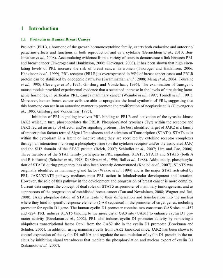
Both PAK1 and prolactin have previously been implicated in the regulation of cyclin D1 promoter activi-ty (Balasenthil et al., 2004; Brockman et al., 2002). Since we have recently shown that PRL causes tyro-syl phosphorylation of PAK1 by JAK2 kinase (Rider et al., 2007), we decided to investigate whether ty-rosyl phosphorylation of PAK1 is important for cyclin D1 regulation in response to PRL. First, we meas-ured the induction of cyclin D1 promoter activity in T47D cells treated with or without PRL. As shown in Figure 2A, T47D cells transfected with a human cyclin D1 promoter-luciferase construct increased lucif-erase expression in response to PRL as expected. Second, co-transfection of T47D cells with luciferase construct and PAK1 WT results in a 4.6-fold increase in luciferase expression in the absence of PRL that corresponds to previously published data (white bars in Figure 2B) (Balasenthil et al., 2004). Interesting-ly, treatment of the PAK1 WT-expressing cells with PRL causes a 14-fold increase in luciferase expres-sion as compared with the cells not expressing PAK1 WT and treated with PRL (black bars in Figure 2B). Overexpression of PAK1 lacking the three phosphorylated tyrosines (PAK1 Y3F) which are sites of JAK2 phosphorylation, reduced PAK1’s effect on cyclin D1 promoter activity by 55% compared to PAK1 WT in the presence of PRL. These data suggest that Tyr(s) 153, 201 and 285 of PAK1 are required for maximal cyclin D1 promoter activity in response to PRL.
2.2 PAK1 Shuttles between the Cytoplasm and Nucleus and PRL Promotes PAK1 Nuclear Ac-cumulation
Data from the literature suggest that PAK1 translocates into the nucleus in response to EGF (Singh et al., 2005) and we wished to investigate the potential significance of PAK1 nuclear localization for cyclin D1 regulation. We first studied whether PRL can stimulate nuclear translocation of PAK1. Figure 3 indicates that treatment of T47D cells with PRL for 24h caused nuclear accumulation of endogenous PAK1. Inter-estingly, extended incubation of T47D cells with PRL up to 48h led to re-distribution of PAK1 back to the cytoplasm. These immunofluorescence data were confirmed by fractionation assay demonstrating the presence of PAK1 in both cytoplasmic and nuclear fractions before PRL treatment, elevated levels of PAK1 in the nuclear fraction 24h after PRL addition and a decrease in nuclear PAK1 after 48h (not shown).
In order to investigate the role of the three sites of JAK2-dependent tyrosyl phosphorylation of the PAK1 molecule in nuclear translocation, we overexpressed either PAK1 WT or PAK1 Y3F in T47D cells, treated them with or without PRL to activate JAK2 and defined the amount of cells with nuclear PAK1 (Figure 4A and B). There were significantly more cells with intranuclear PAK1 WT after PRL treatment than without PRL, while there was no PRL-dependent difference in localization of PAK1 Y3F mutant, suggesting that these three tyrosines may play a role in PAK1 nuclear translocation. We have also seen significant PAK1 translocation into the nucleus when we overexpressed PAK1 WT with JAK2 in COS-7 and MCF-7 cells (Figure 5, left two bars in each plot).
(a) (b)
Figure 2: Prolactin stimulates cyclin D1 promoter activity through tyrosines 153, 201 and 285 of PAK1. T47D cells were transfected with cyclin D1-luciferase reporter (A) or cotransfected with cyclin D1-luciferase reporter with vector, PAK1 WT or PAK1 Y3F (B). The cells were treat-ed with (black bars) or without (white bars) 500 ng/ml of prolactin for an additional 24h, lysed, and luciferase activity was measured. Luciferase activity was normalized with !-galactosidase ac-tivity. Bars represent mean ±S.E., *, p <0.05, n=3.
Figure 3: Prolactin causes translocation of endogenous PAK1 into nucleus. T47D cells were deprived of serum for 24h and treated with or without 500 ng/ml PRL for 0, 24 or 48h. Endoge-nous PAK1 was subjected to confocal immunofluorescence with "PAK1 antibody. Scale bar, 50 µm.

(a) (b)
Figure 4: Tyrosyl phosphorylation of PAK1 is required for translocation of PAK1 into nu-cleus in response to PRL. (A) T47D cells were transfected with either PAK1 WT or PAK1 Y3F. The cells were serum deprived for 24h, treated with or without 500 ng/ml of PRL for an additional 24h and PAK1 was immunolocalized with "PAK1 antibody. Scale bar, 25 µm. (B) The percentage of cells with PAK1 nuclear localization was counted and plotted. 100 PAK1-expressing cells were assessed for PAK1 or PAK1 Y3F immunolocalization in each experiment for each type of treat-ment. Bars represent mean ±S.E., *, p<0.05, n=3.
Since the maximal amount of nuclear endogenous PAK1 was observed 24h but not 48h after PRL
treatment, we hypothesize that PAK1 may shuttle between the nucleus and the cytoplasm. To test this hypothesis, we treated T47D cells with Leptomycin B (LMB), a specific inhibitor of Crm1-dependent nuclear export. Indeed, LMB treatment lead to nuclear accumulation of overexpressed PAK1 WT in T47D, MCF-7 and COS-7 cells indicating that nucleo-cytoplasmic shuttling of PAK1 was happening and that this occurred in a cell-type independent manner (Figure 5).
2.3 Effect of Nuclear Localization Signals and Tyrosyl Phosphorylation of PAK1 on Cyclin D1 Promoter Activity
To further implicate a regulatory role of PAK1 tyrosyl phosphorylation in nuclear localization and the regulation of cyclin D1 transcription, we used a previously described PAK1 mutant in which three nucle-ar localization signals (NLS) have been mutated by replacing the three basic lysine residues with alanines (amino acids 48-51 for NLS1, 243-245 for nLS2 and 267-269 for NLS3) (Singh et al., 2005). Because overexpression of this PAK1 mutant lacking the three functional nuclear localization signals (PAK1 mutNLS) decreased but did not eliminate augmentation of cyclin D1 promoter activity compared to PAK1 WT (Holm et al., 2006), we hypothesize that mutation of Tyr(s) 153, 201 and 285 in PAK1 mut-NLS will further reduce PRL-induced activation of cyclin D1 promoter. To test this, we transiently ex-pressed PAK1 mutNLS or PAK1 mutNLS Y3F mutants in T47D cells, treated the cells with or without PRL and performed experiments as described above. As shown in Fig.6A, expression of PAK1 mutNLS decreased both PRL-dependent and PRL-independent cyclin D1 transcription activity by 46% i. e. to a similar level caused by expression of PAK1 Y3F mutant (47% inhibition in this experiment). Expression of PAK1 mutNLS Y3F significantly decreased the effect of PRL on cyclin D1 promoter activity by 68%,

Figure 5: PAK1 shuttles between cytoplasm and nucleus. PAK1 alone (T47D cells) or PAK1 and JAK2 (COS-7 and MCF-7 cells) were overexpressed in the indicated cells. The cells were in-cubated with Leptomycin B (LMB) for 8h and processed for immunolocalization of PAK1 with αPAK1 antibody. T47D cells were treated with 500 ng/ml of PRL for 48h before LMB was added. The percentage of cells with PAK1 nuclear localization was counted and plotted. 100 PAK1-positive (for T47D cells) and both PAK1- and JAK2-positive (for COS-7 and MCF-7 cells) cells were assessed for PAK1 immunolocalization in each experiment for each type of treatment. Scale bar, 25 µm. Bars represent mean ±S.E., *, p<0.05, n=3.

Figure 6: Effect of PAK1 nuclear localization signals and tyrosyl phosphorylation on cyclin D1 promoter activity. T47D cells were cotransfected with cyclin D1-luciferase reporter with ei-ther vector, PAK1 WT, PAK1 mutNLS (three nuclear localization signals mutated), PAK1 Y3F or PAK1 mutNLS Y3F. The cells were serum deprived for 24h , treated with (black bars) or without (white bars) 500 ng/ml of prolactin for an additional 24h, lysed, and luciferase activity was meas-ured. Luciferase activity was normalized with β-galactosidase activity. Bars represent mean ±S.E., *, p <0.05, n=3. The expression levels of PAK1 WT and PAK1 mutants are indicated (A). Total cell lysates, cytosolic (C) and nuclear (N) fractions of T47D cells transfected with PAK1 WT or PAK1 mutants and treated with or without PRL were separated by SDS-PAGE, transferred to ni-trocellulose and immunoblotted with αPAK1, αpaxillin as a cytosolic marker and αRARα as a nu-clear marker (B).

suggesting that both nuclear localization and tyrosyl phosphorylation of PAK1 are required for the maxi-mal effect of PRL on cyclin D1 promoter activity. Figure 6B indicates that treatment of T47D cells with PRL caused nuclear accumulation of overexpressed PAK1 WT but not PAK1 mutNLS, PAK1 Y3F or PAK1 mutNLS Y3F mutants.
2.4 Nck Regulates PAK1 Nuclear Localization and Inhibits PAK1-stimulated Cyclin D1 Promot-er Activity
In our search for additional PAK1-dependent mechanisms of cyclin D1 promoter activation, we investi-gated a role for the adapter protein Nck since Nck is a known binding partner of PAK1 (Bokoch et al., 1996; Galisteo et al., 1996), that is known to shuttle between the cytoplasm and nucleus (Kremer et al., 2007). We first investigated the effect of Nck expression on PAK1 nuclear relocation in response to PRL by overexpressing PAK1 WT alone, Nck alone or PAK1 with Nck together and treating T47D cells with or without PRL. The number of the cells with nuclear PAK1 and the number of cells with nuclear Nck were counted and plotted (Figure 7 B, C). As illustrated in Figure 7A-C, Nck retained PAK1 in the cyto-plasm (2 left white bars in Figure 7B) and inhibited PAK1 nuclear translocation in response to PRL (2 left black bars in Figure 7B). This effect was partially inhibited by expressing PAK1 Y3F instead of PAK1 WT, but only for PRL-untreated cells (PAK1 WT+Nck vs. PAK1 Y3F +Nck without PRL, white bars in Figure 7B). We did not see a significant difference between the cells expressing the same con-structs but treated with PRL (PAK1 WT+Nck vs. PAK1 Y3F-Nck with PRL, black bars in Figure 7B). These data suggest that the three tyrosines on PAK1 may play a role in localization of the PAK1-Nck complex, but this effect is not affected by PRL-dependent tyrosyl phosphorylation. Interestingly, the per-centage of cells in which Nck localized to both the cytoplasmic and nuclear compartments was decreased by up to 45% when it was co-expressed with PAK1 (Figure 7A and C). This effect was independent of PRL treatment (Figure 7A and C). These data suggest that Nck sequesters PAK1 in the cytoplasm and that it stays in the cytoplasm itself when complexed with PAK1. Tyrosyl phosphorylation of PAK1 on the three tyrosines assessed does not play a role in this process (Figure 7A and C, last two bars).
Data from the experiments with the luciferase-cyclin D1 promoter construct demonstrated that co-expression of Nck with PAK1 WT strongly inhibited (by 95%) the impact of PAK1 on cyclin D1 pro-moter activity both in the presence and absence of PRL (Figure 8A). This inhibition was much stronger than that caused by expression of PAK1 Y3F (by 60%).
To study further whether the inhibitory effect of Nck on PAK1-induced cyclin D1 promoter activi-ty was relieved by disruption of Nck-PAK1 binding, we used two mutants: Nck W143R mutant has a mutation in the second SH3 domain and fails to bind to PAK (Zhu et al., 2010), and PAK1 P13A mutant, a mutant that is unable to interact with Nck (Bokoch et al., 1996; Galisteo et al., 1996). Our co-immunoprecipitation experiments confirmed that only PAK1 WT and Nck WT bound to each other in vivo while PAK1 P13A and Nck W143R did not (Figure 9B). Data in Fig. 9A demonstrate that PAK1 P13A and PAK1 WT augment cyclin D1 promoter activity. Mutation of tryptophan 143 to arginine in Nck had no effect on cyclin D1 promoter activity as compared with Nck WT. Expression of both PAK WT and Nck WT strongly inhibited cyclin D1 promoter activity as described before. However, disruption of Nck-PAK1 binding by expression of mutated PAK1 and Nck partially but significantly relieved the repression of PRL-dependent stimulation, induced by Nck WT (Figure 9A). These data confirm that a functional Nck-PAK1 complex is required for optimal regulation of cyclin D1 promoter activity.

Figure 7: Nck retains PAK1 in the cytoplasm. T47D cells were either transfected with PAK1 WT or Nck, or co-transfected with Nck and either PAK1 WT or PAK1 Y3F, serum deprived for 24h, treated with or without 500 ng/ml of PRL for an additional 24h. PAK1 and Nck were immunolocal-ized with αPAK1 or αNck correspondingly. Scale bar, 25 µm. (A). The percentage of cells with PAK1 (B) or Nck (C) nuclear localization was counted and plotted. 100 PAK1-expressing or both PAK1- and Nck-expressing cells were assessed for PAK1 or Nck immunolocalization in each exper-iment for each type of treatment. Bars represent mean ±S.E., *, p<0.05, n=3.

Figure 8: Nck blocks the amplifying effect of PAK1 on cyclin D1 promoter activity. (A) T47D cells were cotransfected with cyclin D1-luciferase reporter, and either PAK1 WT, PAK1 and Nck, PAK1 Y3F or PAK1 Y3F and Nck. The cells were serum deprived for 24h, treated with (black bars) or without (white bars) 500 ng/ml of PRL for an additional 24h, lysed, and luciferase activity was measured. Lucif-erase activity was normalized with β-galactosidase activity. Bars represent mean ±S.E., *, p <0.05 com-pared with cells expressing PAK1 WT and untreated with PRL, n=3. (B) Whole-cell lysates of T47D cells transfected with PAK1 WT, PAK1 Y3F and Nck were subjected to αPAK1 and αNck Western blotting. The expression levels of PAK1 WT, PAK1 Y3F and Nck are indicated.

3 Discussion
The effect of PRL on regulation of the cell cycle progression increases our understanding of the mecha-nism by which PRL may stimulate growth during mammary development. Furthermore, in an abnormal genetic or environmental context, this action may contribute to mammary carcinogenesis and may point toward potential targets for pharmacological intervention in this process. Here we introduce the serine-threonine kinase PAK1 as a possible target in the PRL-dependent signaling pathway leading to cyclin D1 activation.
PAK1 has been suggested to serve as the key effector for Rac1 activation of cyclin D1 (Westwick et al., 1997). Later, PAK1 was shown to activate cyclin D1 in vivo and in vitro (Balasenthil et al., 2004). Overexpression of both wild type and catalytically active PAK1 T423E in different cell lines led to in-creased cyclin D1 promoter activity, and the overexpression of PAK1 T423E in MCF-7 cells also elevat-ed levels of cyclin D1 mRNA, protein and nuclear accumulation of cyclin D1. Reducing PAK1 expres-sion by PAK1-siRNA or overexpression of dominant negative PAK1 were accompanied by a significant reduction of cyclin D1 expression (Balasenthil et al., 2004). The same authors demonstrated that hyper-plastic mammary glands from PAK1 T423E transgenic mice exhibited increased expression of cyclin D1 as compared to the wild type mice. The authors proposed a model wherein PAK1 regulation of cyclin D1 expression involves an NFkB-dependent pathway (Balasenthil et al., 2004). Merlin, the NF2 tumor sup-pressor gene product, has been proposed as a negative regulator of PAK1-stmulated cyclin D1 promoter activity by inhibition of PAK1 activity (Xiao et al., 2005). Nheu et al. (2004) demonstrated that PAK activity is essential for rennin-angiotensin system-induced upregulation of cyclin D1 (Nheu et al., 2004). All of the aforementioned studies explained the effect of PAK1 on cyclin D1 activity by the serine-threonine kinase activity of PAK1.
However, many of the effects of PAK1 seem to be independent of its kinase activity but dependent on protein-protein interaction. Thus, the kinase inhibitory domain of PAK1 (KID) induced a cell cycle arrest and inhibition of cyclin D1 and D2 expression. More importantly, this arrest could not be rescued by the expression of activated PAK1 T423E demonstrating that KID-induced cell cycle arrest occurs in-dependently of PAK1 kinase activity (Thullberg et al., 2007).
Here we linked upstream PRL-triggered signaling via JAK2 tyrosine kinase to downstream PAK1 which is a JAK2 target. We have previously demonstrated that PAK1 is a novel binding partner and a substrate of JAK2 in response to PRL activation, and identified three tyrosines (Tyr(s) 153, 201 and 285) of PAK1 that are phosphorylated by JAK2 (Rider et al., 2007). Here we have shown that PAK1, in re-sponse to PRL, causes an increase of cyclin D1 promoter activity and mutation of three tyrosines (Tyrs 153, 201, 285) inhibit this amplifying effect by 55%. In an attempt to find a mechanism of this pTyr-PAK1 action, we noticed that PRL causes translocation of PAK1 into the nuclei. Nuclear translocation of PAK1 in response to EGF has been previously described (Singh et al., 2005; for review Rayala and Ku-mar, 2007). Thus, endogenous PAK1 localizes in the nucleus in 18-24% of the interphase MCF-7 cells and directly phosphorylates histone H3 (Li et al., 2002). PAK1 associates with the promoter of PFK-M gene and stimulates PFK-M expression, and also with a portion of the NFAT1 gene and represses expres-sion of this gene (Singh et al., 2005). In addition, increased levels of nuclear PAK1 were linked to intrin-sic tamoxifen resistance of breast cancer cells (Holm et al., 2006; Li et al., 2002). We extended these findings and demonstrated that PAK1 shuttles between the cytoplasm and the nucleus in different cell lines including T47D, COS-7 and MCF-7. Furthermore, we have shown that PRL-dependent PAK1 nu-clear translocation depends on Tyrs 153, 201 and 285 since the PAK1 Y3F mutant does not translocate

into the nucleus in response to PRL. Three nuclear localization signals have been mapped on PAK1, and PAK1 lacking these three functional NLS (PAK1 mutNLS) fails to translocate into the nucleus in re-sponse to EGF (Singh et al., 2005). We have shown here that PAK1 mutNLS exhibited 46% less impact on cyclin D promoter activity as compared to PAK1 WT. We hypothesized that by eliminating both PAK1 tyrosine phosphorylation and functional NLSs, we would completely inhibit PRL-dependent am-plification of cyclin D1 promoter activity. However, PAK1 mutNLS Y3F exhibited only 68% inhibition, suggesting that both the three nuclear localization signals and three tyrosines which are phosporylated by JAK2 in response to PRL contribute to but are not exclusively required for the PRL-dependent induction of cyclin D1 promoter activity. Why do PAK1 mutNLS and PAK1 mutNLS Y3F, which both retain in the cytoplasm, still have an amplifying effect on cyclin D1 activity? PAK1 may activate cyclin D1 pro-moter activity in response to PRL via multiple mechanisms. For example, PAK1 phosphorylates specific cytoplasmic proteins that can directly or indirectly regulate cyclin D1 promoter activity. In this context it is interesting to note that, although PRL signals via STAT5 to the distal GAS1 binding sites in the cyclin D1 promoter, co-expression of dominant negative STAT5A and PAK1 WT showed no effect on cyclin D1 promoter activity (Brockman and Schuler, 2005; Balasenthil et al., 2004; Brockman et al., 2002). We are currently investigating which regulatory elements of cyclin D1 promoter are affected by PRL-dependent PAK1 tyrosyl phosphorylation.
In attempt to find another mechanism that can regulate the action of PRL on the cyclin D1, we fo-cused on Nck for several reasons. First, adapter protein Nck is a binding partner of PAK1 (Bokoch et al., 1996; Galisteo et al., 1996). Nck W143R mutant with a mutation in the second SH3 domain, fails to bind to PAK (Zhu et al., 2010) and the PAK1 P13A mutant is unable to interact with Nck (Bokoch et al., 1996; Galisteo et al., 1996). Second, Nck is present in both the cytoplasm and the nucleus (Lawe et al., 1997). Nck rapidly accumulates in the nucleus after the introduction of DNA damage (Kremer et al., 2007). In agreement with Lawe et.al., who showed that nuclear localization of Nck does not depend on growth factor stimulation (Lawe et al., 1997), we have shown here that PRL also does not cause Nck nu-clear translocation and that around 65% of cells contain nuclear Nck regardless of PRL treatment. How-ever, when we co-expressed both Nck and PAK1, both molecules were mostly retained in the cytoplasm. This effect was especially dramatic for PAK1, since 3-fold fewer cells had nuclear PAK1 as compared with cells without Nck co-expression. More importantly, Nck abolished the ability of PRL to induce PAK1 nuclear translocation. These data suggest that Nck can sequester PAK1 in the cytoplasm. This se-questering has a physiological role since co-expression of both PAK1 and Nck inhibits the amplifying effect of PRL-induced PAK1 on cyclin D1 promoter activity (95% inhibition). This inhibition was par-tially abolished by disruption of the PAK1-Nck binding by using either PAK1 P13A mutant, Nck W143R mutant, or both. This partial inhibition implies the presence of Nck-PAK1-interaction-independent mech-anisms that affect cyclin D1 promoter activity. Nck is a common target for a variety of growth factor re-ceptors and becomes phosphorylated on serine, threonine and tyrosine residues after growth factor stimu-lation (Chou et al., 1992; Li et al., 1992; Park and Rhee, 1992). Nck is implicated in the regulation of different signal transduction pathways including c-Jun N-terminal kinase (JNK) and mixed lineage kinase 2 (MLK2) pathways (Miyamoto et al., 2004; Poitras et al., 2003; Murakami et al., 2002; Becker et al., 2000; Stein et al., 1998; Su et al., 1997). Furthermore, nuclear Nck is essential for activation of p53 in response to UV-induced DNA damage (Kremer et al., 2007). Transcriptional regulation of the cyclin D1 gene as a complex and many different transcription factors have been identified that regulate the cyclin D1 promoter (reviewed in Wang et al., 2004). The regulation of cyclin D1 by integrin signaling is well-

documented (reviewed in Musgrove, 2006) and Nck is an important member of focal adhesions and in-tegrin-dependent pathway (reviewed in Buday et al., 2002).
It is possible that there are mechanisms other than retention of Nck-pTyr-PAK1 complex in the cy-toplasm, to regulate the cyclin D1 promoter activity. Nck is a binding partner of PTP-PEST (a cytosolic protein tyrosine phosphatase) (Zhao et al., 2000a). PTP-PEST has been shown to dephosphorylate PRL-activated JAK2 in vitro (Horsch et al., 2001). We can speculate that Nck brings PTP-PEST to the PRL-induced JAK2-PAK1 complex that may lead to dephosphorylation and inactivation of JAK2. Inactive JAK2 cannot tyrosyl phosphorylate PAK1 which leads to inability of PAK1 to enhance cyclin D1 activa-tion. Another possible common target which binds to both Nck and JAK2 is the ubiquitin ligase c-Cbl, which is a negative regulator of various signaling pathways. C-Cbl becomes tyrosyl-phosphorylated after stimulation of a wide variety of receptors including the PRL receptor (Hunter et al., 1997). The negative regulation of PRL signaling by c-Cbl is confirmed by observations that c-Cbl repression leads to en-hanced JAK2/STAT activation while c-Cbl overexpression results in increased ubiquitination and proteo-somal degradation of STAT5 (Goh et al., 2002; Wang et al., 2002). Since c-Cbl binds to Nck (Wunder-lich et al., 1999; Rivero-Lezcano et al., 1994), we can speculate that Nck may bring c-Cbl to the PRL-induced JAK2-PAK1 complex, thus leading to proteosomal degradation of JAK2 followed by attenuation of pTyr-PAK1’s effect on cyclin D1 promoter activity. Nck also binds to SOCS-3 and recruits Nck to the plasma membrane (Sitko et al., 2004). SOCS-3 negatively regulates PRL signaling by interacting with phosphorylated PRLR leading to JAK2 supression and, probably, by directly interacting with JAK2 which has been shown for the erythropoietin receptor (Dif et al., 2001; Sasaki et al., 2000; Tomic et al., 1999). It would be attractive to speculate that Nck-SOCS-3 complex is recruited to the plasma membrane to bind to the PRLR leading to inactivation of JAK2 and decreased pTyr-PAK1’s activity toward cyclin D1 promoter. We should, however, note that we have not seen relocation of Nck to the plasma membrane in response to PRL. It is possible that the redistribution of Nck to the plasma membrane occurs shortly after ligand treatment (for example, in 30 min as it was described for PDGF treatment (Sitko et al., 2004)) while long-term treatment of PRL (24 h in the current research) has no effect on the predominant-ly intranuclear localization of Nck (Figure 7).
To summarize, Figure 10 shows an overall view of mechanisms of cyclin D1 promoter activity regulation by tyrosyl phosphorylated PAK1 in response to PRL. First, PRL binds to the PRL receptor and activates JAK2 which tyrosyl phosphorylates PAK1 on three tyrosines. Tyrosyl phosphorylated PAK1 translocates into the nucleus where it stimulates cyclin D1 promoter activity. Both tyrosyl phosphoryla-tion of PAK1 and the three intact NLSs are required for this maximal effect of PRL on cyclin D1 because PAK1 mutNLS Y3F mutant has 68% reduced ability to activate the cyclin D1 promoter. However, the more critical mechanism for PAK1-dependent regulation of cyclin D1 activation is the formation of the PAK1-Nck complex in the cytoplasm. This complex retains PAK1 in the cytoplasm which leads to the inhibition of the amplifying effect of PAK1 on cyclin D1 promoter activity in response to PRL. Which protein(s) can regulate the formation of the Nck-PAK1 complex and what kind of role the phosphorylated tyrosines on PAK1 play are currently under our investigation.
It is of note that both PRL and PAK1 are oncogenic. Considering that cyclin D1 promoter activity is positively regulated by tyrosyl phosphorylated PAK1 in a PRL-dependent manner, we may speculate that the PRL-activated JAK2/PAK1 axis plays a role in breast cancer promotion. Whether JAK2-dependent phosphorylation of PAK1 plays a role in normal cells and in other types of cancer requires future investigation.

Figure 9: PAK1-Nck binding is required for the effect of Nck on cyclin D1 promoter activity. (A) T47D cells were cotransfected with cyclin D1-luciferase reporter and cDNAs encoding the in-dicated proteins and treated as in Figure 7A. Luciferase activity was normalized with β-galactosidase activity. Bars represent mean ±S.E., *, p <0.05 compared with cells expressing PAK1 WT and untreated with PRL, n=3. (B) Nck WT is co-immunoprecipitated with PAK1 WT (lane 5) but not with PAK1 P13A (lane 6). Nck W148R is co-immunoprecipitated neither with PAK1 WT (lane 7) nor with PAK1 P13A (lane 8). HA-tagged Nck was immunoprecipitated with αHA from T47D cells overexpressing the indicated proteins and immunoblotted with the indicated antibodies. The light bands in lanes 1 and 3 in the αNck blot represent endogenous Nck.

Figure 10: PAK1 regulates prolactin-dependent cyclin D1 promoter activity by two distinct mechanisms. In response to cytokines such as prolactin, PAK1 is tyrosyl phosphorylated by JAK2 and translocates into the nucleus where it stimulates cyclin D1 promoter activity. The nuclear locali-zation signals (NLS) and the three tyrosines of PAK1 are sufficient but not required for this PAK1 function since deletion of these three phosphorylated tyrosines (PAK1 Y3F) and mutation of the three NLSs (PAK1 mutNLS) decreased PAK1 WT activity on cyclin D1 promoter activity by 55% and 46%, respectively. The double mutant of PAK1 (mutNLS Y3F) decreased PAK1 WT activity on cy-clin D1 promoter activity by 68%. Another mechanism regulating the PAK1 function is Nck-PAK1 binding, since co-expression of PAK1 and Nck inhibited PAK1-dependent stimulation of cyclin D1 promoter activity by 95%. We propose that the Nck-PAK1 complex sequesters both molecules in the cytoplasm, thereby abolishing the amplifying effect of PAK1 on the prolactin-induced activation of cyclin D1.
Acknowledgement
The article has been adapted from the journal “Molecular Endocrinology” ("PAK1-Nck Regulates Cyclin D1 Promoter Activity in Response to Prolactin", v. 25, 9, 2011, p. 1565-1578; doi:10.1210/me.2011-0062)

References
Adam, L., Vadlamudi, R., Mandal, M., Chernoff, J. and Kumar, R. (2000) Regulation of microfilament reorganization and invasiveness of breast cancer cells by kinase dead p21-activated kinase-1. J Biol Chem. 275: 12041-12050.
Arnold, A. and Papanikolaou, A. (2005) Cyclin D1 in breast cancer pathogenesis. J Clin Oncol. 23: 4215-4224.
Aronheim, A., Broder, Y.C., Cohen, A., Fritsch, A., Belisle, B. and Abo, A. (1998) Chp, a homologue of the GTPase Cdc42Hs, activates the JNK pathway and is implicated in reorganizing the actin cytoskeleton. Curr Biol. 8: 1125-1128.
Asher, J.M., O'Leary, K.A., Rugowski, D.E., Arendt, L.M. and Schuler, L.A. (2012) Prolactin promotes mammary patho-genesis independently from cyclin D1. Am J Pathol. 181: 294-302.
Balasenthil, S., Sahin, A.A., Barnes, C.J., Wang, R.A., Pestell, R.G., Vadlamudi, R.K. and Kumar, R. (2004) p21-activated kinase-1 signaling mediates cyclin D1 expression in mammary epithelial and cancer cells. J Biol Chem. 279: 1422-1428.
Ball, R.K., Friis, R.R., Schoenenberger, C.A., Doppler, W. and Groner, B. (1988) Prolactin regulation of beta-casein gene expression and of a cytosolic 120-kd protein in a cloned mouse mammary epithelial cell line. Embo J. 7: 2089-2095.
Banerjee, M., Worth, D., Prowse, D.M. and Nikolic, M. (2002) Pak1 phosphorylation on t212 affects microtubules in cells undergoing mitosis. Curr Biol. 12: 1233-1239.
Becker, E., Huynh-Do, U., Holland, S., Pawson, T., Daniel, T.O. and Skolnik, E.Y. (2000) Nck-interacting Ste20 kinase couples Eph receptors to c-Jun N-terminal kinase and integrin activation. Mol Cell Biol. 20: 1537-1545.
Bekri, S., Adelaide, J., Merscher, S., Grosgeorge, J., Caroli-Bosc, F., Perucca-Lostanlen, D., et al (1997) Detailed map of a region commonly amplified at 11q13-->q14 in human breast carcinoma. Cytogenet Cell Genet. 79: 125-131.
Ben-Jonathan, N., LaPensee, C.R. and LaPensee, E.W. (2008) What can we learn from rodents about prolactin in humans? Endocr Rev. 29: 1-41.
Bernichtein, S., Touraine, P. and Goffin, V. (2010) New concepts in prolactin biology. J Endocrinol. 206: 1-11.
Bienvenu, F., Jirawatnotai, S., Elias, J.E., Meyer, C.A., Mizeracka, K., Marson, A., et al (2010) Transcriptional role of cyclin D1 in development revealed by a genetic-proteomic screen. Nature. 463: 374-378.
Bokoch, G.M. (2003) Biology of the p21-activated kinases. Annu Rev Biochem. 72: 743-781.
Bokoch, G.M., Wang, Y., Bohl, B.P., Sells, M.A., Quilliam, L.A. and Knaus, U.G. (1996) Interaction of the Nck adapter protein with p21-activated kinase (PAK1). J Biol Chem. 271: 25746-25749.
Bokoch, G.M., Reilly, A.M., Daniels, R.H., King, C.C., Olivera, A., Spiegel, S. and Knaus, U.G. (1998) A GTPase-independent mechanism of p21-activated kinase activation. Regulation by sphingosine and other biologically active lipids. J Biol Chem. 273: 8137-8144.
Brockman, J.L. and Schuler, L.A. (2005) Prolactin signals via Stat5 and Oct-1 to the proximal cyclin D1 promoter. Mol Cell Endocrinol. 239: 45-53.
Brockman, J.L., Schroeder, M.D. and Schuler, L.A. (2002) PRL activates the cyclin D1 promoter via the Jak2/Stat pathway. Mol Endocrinol. 16: 774-784.
Buday, L., Wunderlich, L. and Tamas, P. (2002) The Nck family of adapter proteins: regulators of actin cytoskeleton. Cell Signal. 14: 723-731.
Chou, M.M., Fajardo, J.E. and Hanafusa, H. (1992) The SH2- and SH3-containing Nck protein transforms mammalian fibroblasts in the absence of elevated phosphotyrosine levels. Mol Cell Biol. 12: 5834-5842.
Clevenger, C.V. (2003) Role of prolactin/prolactin receptor signaling in human breast cancer. Breast Dis. 18: 75-86.

Clevenger, C.V., Chang, W.P., Ngo, W., Pasha, T.L., Montone, K.T. and Tomaszewski, J.E. (1995) Expression of prolactin and prolactin receptor in human breast carcinoma. Evidence for an autocrine/paracrine loop. Am J Pathol. 146: 695-705.
Coqueret, O. (2002) Linking cyclins to transcriptional control. Gene. 299: 35-55.
Daniels, R.H., Hall, P.S. and Bokoch, G.M. (1998) Membrane targeting of p21-activated kinase 1 (PAK1) induces neurite outgrowth from PC12 cells. Embo J. 17: 754-764.
DaSilva, L., Rui, H., Erwin, R.A., Howard, O.M., Kirken, R.A., Malabarba, M.G., et al (1996) Prolactin recruits STAT1, STAT3 and STAT5 independent of conserved receptor tyrosines TYR402, TYR479, TYR515 and TYR580. Mol Cell Endocrinol. 117: 131-140.
Dickson, C., Fantl, V., Gillett, C., Brookes, S., Bartek, J., Smith, R., et al (1995) Amplification of chromosome band 11q13 and a role for cyclin D1 in human breast cancer. Cancer Lett. 90: 43-50.
Diehl, J.A. (2002) Cycling to cancer with cyclin D1. Cancer Biol Ther. 1: 226-231.
Dif, F., Saunier, E., Demeneix, B., Kelly, P.A. and Edery, M. (2001) Cytokine-inducible SH2-containing protein suppresses PRL signaling by binding the PRL receptor. Endocrinology. 142: 5286-5293.
Fantl, V., Stamp, G., Andrews, A., Rosewell, I. and Dickson, C. (1995) Mice lacking cyclin D1 are small and show defects in eye and mammary gland development. Genes Dev. 9: 2364-2372.
Fu, M., Wang, C., Li, Z., Sakamaki, T. and Pestell, R.G. (2004) Minireview: Cyclin D1: normal and abnormal functions. Endocrinology. 145: 5439-5447.
Galisteo, M.L., Chernoff, J., Su, Y.C., Skolnik, E.Y. and Schlessinger, J. (1996) The adaptor protein Nck links receptor tyrosine kinases with the serine-threonine kinase Pak1. J Biol Chem. 271: 20997-21000.
Ginsburg, E. and Vonderhaar, B.K. (1995) Prolactin synthesis and secretion by human breast cancer cells. Cancer Res. 55: 2591-2595.
Goh, E.L., Zhu, T., Leong, W.Y. and Lobie, P.E. (2002) c-Cbl is a negative regulator of GH-stimulated STAT5-mediated transcription. Endocrinology. 143: 3590-3603.
Gururaj, A.E., Rayala, S.K. and Kumar, R. (2005) p21-activated kinase signaling in breast cancer. Breast Cancer Res. 7: 5-12.
Hankinson, S.E., Willett, W.C., Michaud, D.S., Manson, J.E., Colditz, G.A., Longcope, C., et al (1999) Plasma prolactin levels and subsequent risk of breast cancer in postmenopausal women. J Natl Cancer Inst. 91: 629-634.
Holm, C., Rayala, S., Jirstrom, K., Stal, O., Kumar, R. and Landberg, G. (2006) Association between Pak1 expression and subcellular localization and tamoxifen resistance in breast cancer patients. J Natl Cancer Inst. 98: 671-680.
Horsch, K., Schaller, M.D. and Hynes, N.E. (2001) The protein tyrosine phosphatase-PEST is implicated in the negative regulation of epidermal growth factor on PRL signaling in mammary epithelial cells. Mol Endocrinol. 15: 2182-2196.
Hunter, S., Koch, B.L. and Anderson, S.M. (1997) Phosphorylation of cbl after stimulation of Nb2 cells with prolactin and its association with phosphatidylinositol 3-kinase. Mol Endocrinol. 11: 1213-1222.
Jeselsohn, R., Brown, N.E., Arendt, L., Klebba, I., Hu, M.G., Kuperwasser, C. and Hinds, P.W. (2010) Cyclin D1 kinase activity is required for the self-renewal of mammary stem and progenitor cells that are targets of MMTV-ErbB2 tu-morigenesis. Cancer Cell. 17: 65-76.
Keyomarsi, K. and Pardee, A.B. (1993) Redundant cyclin overexpression and gene amplification in breast cancer cells. Proc Natl Acad Sci U S A. 90: 1112-1116.
Khaled, W.T., Read, E.K., Nicholson, S.E., Baxter, F.O., Brennan, A.J., Came, P.J., et al (2007) The IL-4/IL-13/Stat6 sig-nalling pathway promotes luminal mammary epithelial cell development. Development. 134: 2739-2750.

King, C.C., Gardiner, E.M., Zenke, F.T., Bohl, B.P., Newton, A.C., Hemmings, B.A. and Bokoch, G.M. (2000) p21-activated kinase (PAK1) is phosphorylated and activated by 3-phosphoinositide-dependent kinase-1 (PDK1). J Biol Chem. 275: 41201-41209.
Knaus, U.G. and Bokoch, G.M. (1998) The p21Rac/Cdc42-activated kinases (PAKs). Int J Biochem Cell Biol. 30: 857-862.
Knudsen, K.E., Diehl, J.A., Haiman, C.A. and Knudsen, E.S. (2006) Cyclin D1: polymorphism, aberrant splicing and can-cer risk. Oncogene. 25: 1620-1628.
Kremer, B.E., Adang, L.A. and Macara, I.G. (2007) Septins regulate actin organization and cell-cycle arrest through nu-clear accumulation of NCK mediated by SOCS7. Cell. 130: 837-850.
Kumar, R. and Vadlamudi, R.K. (2002) Emerging functions of p21-activated kinases in human cancer cells. J Cell Physiol. 193: 133-144.
Kumar, R., Gururaj, A.E. and Barnes, C.J. (2006) p21-activated kinases in cancer. Nat Rev Cancer. 6: 459-471.
Landis, M.W., Pawlyk, B.S., Li, T., Sicinski, P. and Hinds, P.W. (2006) Cyclin D1-dependent kinase activity in murine de-velopment and mammary tumorigenesis. Cancer Cell. 9: 13-22.
Lawe, D.C., Hahn, C. and Wong, A.J. (1997) The Nck SH2/SH3 adaptor protein is present in the nucleus and associates with the nuclear protein SAM68. Oncogene. 14: 223-231.
Lee, Y.M. and Sicinski, P. (2006) Targeting cyclins and cyclin-dependent kinases in cancer: lessons from mice, hopes for therapeutic applications in human. Cell Cycle. 5: 2110-2114.
Lei, M., Lu, W., Meng, W., Parrini, M.C., Eck, M.J., Mayer, B.J. and Harrison, S.C. (2000) Structure of PAK1 in an auto-inhibited conformation reveals a multistage activation switch. Cell. 102: 387-397.
Li, F., Adam, L., Vadlamudi, R.K., Zhou, H., Sen, S., Chernoff, J., et al (2002) p21-activated kinase 1 interacts with and phosphorylates histone H3 in breast cancer cells. EMBO Rep. 3: 767-773.
Li, W., Hu, P., Skolnik, E.Y., Ullrich, A. and Schlessinger, J. (1992) The SH2 and SH3 domain-containing Nck protein is oncogenic and a common target for phosphorylation by different surface receptors. Mol Cell Biol. 12: 5824-5833.
Lim, C.P. and Cao, X. (2006) Structure, function, and regulation of STAT proteins. Mol Biosyst. 2: 536-550.
Lin, D.I., Lessie, M.D., Gladden, A.B., Bassing, C.H., Wagner, K.U. and Diehl, J.A. (2008) Disruption of cyclin D1 nuclear export and proteolysis accelerates mammary carcinogenesis. Oncogene. 27: 1231-1242.
Lu, W., Katz, S., Gupta, R. and Mayer, B.J. (1997) Activation of Pak by membrane localization mediated by an SH3 do-main from the adaptor protein Nck. Curr Biol. 7: 85-94.
Manser, E., Leung, T., Salihuddin, H., Zhao, Z.S. and Lim, L. (1994) A brain serine/threonine protein kinase activated by Cdc42 and Rac1. Nature. 367: 40-46.
Manser, E., Huang, H.Y., Loo, T.H., Chen, X.Q., Dong, J.M., Leung, T. and Lim, L. (1997) Expression of constitutively active alpha-PAK reveals effects of the kinase on actin and focal complexes. Mol Cell Biol. 17: 1129-1143.
McIntosh, G.G., Anderson, J.J., Milton, I., Steward, M., Parr, A.H., Thomas, M.D., et al (1995) Determination of the prog-nostic value of cyclin D1 overexpression in breast cancer. Oncogene. 11: 885-891.
Megha, T., Lazzi, S., Ferrari, F., Vatti, R., Howard, C.M., Cevenini, G., et al (1999) Expression of the G2-M checkpoint regulators cyclin B1 and P34CDC2 in breast cancer: a correlation with cellular kinetics. Anticancer Res. 19: 163-169.
Meng, J., Tsai-Morris, C.H. and Dufau, M.L. (2004) Human prolactin receptor variants in breast cancer: low ratio of short forms to the long-form human prolactin receptor associated with mammary carcinoma. Cancer Res. 64: 5677-5682.
Mira, J.P., Benard, V., Groffen, J., Sanders, L.C. and Knaus, U.G. (2000) Endogenous, hyperactive Rac3 controls prolif-eration of breast cancer cells by a p21-activated kinase-dependent pathway. Proc Natl Acad Sci U S A. 97: 185-189.

Miyamoto, Y., Yamauchi, J., Mizuno, N. and Itoh, H. (2004) The adaptor protein Nck1 mediates endothelin A receptor-regulated cell migration through the Cdc42-dependent c-Jun N-terminal kinase pathway. J Biol Chem. 279: 34336-34342.
Murakami, H., Yamamura, Y., Shimono, Y., Kawai, K., Kurokawa, K. and Takahashi, M. (2002) Role of Dok1 in cell sig-naling mediated by RET tyrosine kinase. J Biol Chem. 277: 32781-32790.
Musgrove, E.A. (2006) Cyclins: roles in mitogenic signaling and oncogenic transformation. Growth Factors. 24: 13-19.
Musgrove, E.A., Caldon, C.E., Barraclough, J., Stone, A. and Sutherland, R.L. (2011) Cyclin D as a therapeutic target in cancer. Nat Rev Cancer. 11: 558-572.
Neudauer, C.L., Joberty, G., Tatsis, N. and Macara, I.G. (1998) Distinct cellular effects and interactions of the Rho-family GTPase TC10. Curr Biol. 8: 1151-1160.
Nheu, T., He, H., Hirokawa, Y., Walker, F., Wood, J. and Maruta, H. (2004) PAK is essential for RAS-induced upregula-tion of cyclin D1 during the G1 to S transition. Cell Cycle. 3: 71-74.
Nikolic, M., Chou, M.M., Lu, W., Mayer, B.J. and Tsai, L.H. (1998) The p35/Cdk5 kinase is a neuron-specific Rac effector that inhibits Pak1 activity. Nature. 395: 194-198.
Park, D. and Rhee, S.G. (1992) Phosphorylation of Nck in response to a variety of receptors, phorbol myristate acetate, and cyclic AMP. Mol Cell Biol. 12: 5816-5823.
Park, J.B., Kim, E.J., Yang, E.J., Seo, S.R. and Chung, K.C. (2007) JNK- and Rac1-dependent induction of immediate early gene pip92 suppresses neuronal differentiation. J Neurochem. 100: 555-566.
Parrini, M.C., Lei, M., Harrison, S.C. and Mayer, B.J. (2002) Pak1 kinase homodimers are autoinhibited in trans and dis-sociated upon activation by Cdc42 and Rac1. Mol Cell. 9: 73-83.
Poitras, L., Jean, S., Islam, N. and Moss, T. (2003) PAK interacts with NCK and MLK2 to regulate the activation of jun N-terminal kinase. FEBS Lett. 543: 129-135.
Rashid, T., Banerjee, M. and Nikolic, M. (2001) Phosphorylation of Pak1 by the p35/Cdk5 kinase affects neuronal mor-phology. J Biol Chem. 276: 49043-49052.
Rayala, S.K. and Kumar, R. (2007) Sliding p21-activated kinase 1 to nucleus impacts tamoxifen sensitivity. Biomed Phar-macother.
Rayala, S.K., Talukder, A.H., Balasenthil, S., Tharakan, R., Barnes, C.J., Wang, R.A., et al (2006) P21-activated kinase 1 regulation of estrogen receptor-alpha activation involves serine 305 activation linked with serine 118 phosphoryla-tion. Cancer Res. 66: 1694-1701.
Reich, N.C. (2007) STAT dynamics. Cytokine Growth Factor Rev. 18: 511-518.
Rider, L., Shatrova, A., Feener, E.P., Webb, L. and Diakonova, M. (2007) JAK2 tyrosine kinase phosphorylates PAK1 and regulates PAK1 activity and functions. J Biol Chem. 282: 30985-30996.
Rivero-Lezcano, O.M., Sameshima, J.H., Marcilla, A. and Robbins, K.C. (1994) Physical association between Src homolo-gy 3 elements and the protein product of the c-cbl proto-oncogene. J Biol Chem. 269: 17363-17366.
Sakamoto, K., Creamer, B.A., Triplett, A.A. and Wagner, K.U. (2007) The Janus kinase 2 is required for expression and nuclear accumulation of cyclin D1 in proliferating mammary epithelial cells. Mol Endocrinol. 21: 1877-1892.
Salh, B., Marotta, A., Wagey, R., Sayed, M. and Pelech, S. (2002) Dysregulation of phosphatidylinositol 3-kinase and downstream effectors in human breast cancer. Int J Cancer. 98: 148-154.
Sasaki, A., Yasukawa, H., Shouda, T., Kitamura, T., Dikic, I. and Yoshimura, A. (2000) CIS3/SOCS-3 suppresses erythro-poietin (EPO) signaling by binding the EPO receptor and JAK2. J Biol Chem. 275: 29338-29347.
Schaber, J.D., Fang, H., Xu, J., Grimley, P.M. and Rui, H. (1998) Prolactin activates Stat1 but does not antagonize Stat1 activation and growth inhibition by type I interferons in human breast cancer cells. Cancer Res. 58: 1914-1919.

Schindler, C., Levy, D.E. and Decker, T. (2007) JAK-STAT signaling: from interferons to cytokines. J Biol Chem. 282: 20059-20063.
Schroeder, M.D., Symowicz, J. and Schuler, L.A. (2002) PRL modulates cell cycle regulators in mammary tumor epithelial cells. Mol Endocrinol. 16: 45-57.
Sherr, C.J. (1995) D-type cyclins. Trends Biochem Sci. 20: 187-190.
Sicinski, P., Donaher, J.L., Parker, S.B., Li, T., Fazeli, A., Gardner, H., et al (1995) Cyclin D1 provides a link between development and oncogenesis in the retina and breast. Cell. 82: 621-630.
Singh, R.R., Song, C., Yang, Z. and Kumar, R. (2005) Nuclear localization and chromatin targets of p21-activated kinase 1. J Biol Chem. 280: 18130-18137.
Sitko, J.C., Guevara, C.I. and Cacalano, N.A. (2004) Tyrosine-phosphorylated SOCS3 interacts with the Nck and Crk-L adapter proteins and regulates Nck activation. J Biol Chem. 279: 37662-37669.
Stein, E., Huynh-Do, U., Lane, A.A., Cerretti, D.P. and Daniel, T.O. (1998) Nck recruitment to Eph receptor, EphB1/ELK, couples ligand activation to c-Jun kinase. J Biol Chem. 273: 1303-1308.
Su, Y.C., Han, J., Xu, S., Cobb, M. and Skolnik, E.Y. (1997) NIK is a new Ste20-related kinase that binds NCK and MEKK1 and activates the SAPK/JNK cascade via a conserved regulatory domain. Embo J. 16: 1279-1290.
Sutherland, R.L. and Musgrove, E.A. (2004) Cyclins and breast cancer. J Mammary Gland Biol Neoplasia. 9: 95-104.
Suzuki, R., Kuroda, H., Komatsu, H., Hosokawa, Y., Kagami, Y., Ogura, M., et al (1999) Selective usage of D-type cyclins in lymphoid malignancies. Leukemia. 13: 1335-1342.
Swaminathan, G., Varghese, B. and Fuchs, S.Y. (2008) Regulation of prolactin receptor levels and activity in breast cancer. J Mammary Gland Biol Neoplasia. 13: 81-91.
Tan, S.H. and Nevalainen, M.T. (2008) Signal transducer and activator of transcription 5A/B in prostate and breast can-cers. Endocr Relat Cancer. 15: 367-390.
Tang, Y., Zhou, H., Chen, A., Pittman, R.N. and Field, J. (2000) The Akt proto-oncogene links Ras to Pak and cell survival signals. J Biol Chem. 275: 9106-9109.
Tao, W., Pennica, D., Xu, L., Kalejta, R.F. and Levine, A.J. (2001) Wrch-1, a novel member of the Rho gene family that is regulated by Wnt-1. Genes Dev. 15: 1796-1807.
Thiel, D.A., Reeder, M.K., Pfaff, A., Coleman, T.R., Sells, M.A. and Chernoff, J. (2002) Cell cycle-regulated phosphoryla-tion of p21-activated kinase 1. Curr Biol. 12: 1227-1232.
Thullberg, M., Gad, A., Beeser, A., Chernoff, J. and Stromblad, S. (2007) The kinase-inhibitory domain of p21-activated kinase 1 (PAK1) inhibits cell cycle progression independent of PAK1 kinase activity. Oncogene. 26: 1820-1828.
Tomic, S., Chughtai, N. and Ali, S. (1999) SOCS-1, -2, -3: selective targets and functions downstream of the prolactin re-ceptor. Mol Cell Endocrinol. 158: 45-54.
Tornell, J., Rymo, L. and Isaksson, O.G. (1991) Induction of mammary adenocarcinomas in metallothionein promoter-human growth hormone transgenic mice. Int J Cancer. 49: 114-117.
Touraine, P., Martini, J.F., Zafrani, B., Durand, J.C., Labaille, F., Malet, C., et al (1998) Increased expression of prolactin receptor gene assessed by quantitative polymerase chain reaction in human breast tumors versus normal breast tis-sues. J Clin Endocrinol Metab. 83: 667-674.
Tworoger, S.S. and Hankinson, S.E. (2006) Prolactin and breast cancer risk. Cancer Lett. 243: 160-169.
Vadlamudi, R.K., Li, F., Adam, L., Nguyen, D., Ohta, Y., Stossel, T.P. and Kumar, R. (2002) Filamin is essential in actin cytoskeletal assembly mediated by p21-activated kinase 1. Nat Cell Biol. 4: 681-690.
Vadlamudi, R.K., Bagheri-Yarmand, R., Yang, Z., Balasenthil, S., Nguyen, D., Sahin, A.A., et al (2004) Dynein light chain 1, a p21-activated kinase 1-interacting substrate, promotes cancerous phenotypes. Cancer Cell. 5: 575-585.

Vadlamudi, R.K., Adam, L., Wang, R.A., Mandal, M., Nguyen, D., Sahin, A., et al (2000) Regulatable expression of p21-activated kinase-1 promotes anchorage-independent growth and abnormal organization of mitotic spindles in human epithelial breast cancer cells. J Biol Chem. 275: 36238-36244.
Wagner, K.U. and Rui, H. (2008) Jak2/Stat5 signaling in mammogenesis, breast cancer initiation and progression. J Mammary Gland Biol Neoplasia. 13: 93-103.
Wakao, H., Gouilleux, F. and Groner, B. (1994) Mammary gland factor (MGF) is a novel member of the cytokine regulat-ed transcription factor gene family and confers the prolactin response. Embo J. 13: 2182-2191.
Wang, C., Li, Z., Fu, M., Bouras, T. and Pestell, R.G. (2004) Signal transduction mediated by cyclin D1: from mitogens to cell proliferation: a molecular target with therapeutic potential. Cancer Treat Res. 119: 217-237.
Wang, L., Rudert, W.A., Loutaev, I., Roginskaya, V. and Corey, S.J. (2002) Repression of c-Cbl leads to enhanced G-CSF Jak-STAT signaling without increased cell proliferation. Oncogene. 21: 5346-5355.
Wang, T.C., Cardiff, R.D., Zukerberg, L., Lees, E., Arnold, A. and Schmidt, E.V. (1994) Mammary hyperplasia and carci-noma in MMTV-cyclin D1 transgenic mice. Nature. 369: 669-671.
Wennbo, H., Gebre-Medhin, M., Gritli-Linde, A., Ohlsson, C., Isaksson, O.G. and Tornell, J. (1997) Activation of the pro-lactin receptor but not the growth hormone receptor is important for induction of mammary tumors in transgenic mice. J Clin Invest. 100: 2744-2751.
Westwick, J.K., Lambert, Q.T., Clark, G.J., Symons, M., Van Aelst, L., Pestell, R.G. and Der, C.J. (1997) Rac regulation of transformation, gene expression, and actin organization by multiple, PAK-independent pathways. Mol Cell Biol. 17: 1324-1335.
Wunderlich, L., Goher, A., Farago, A., Downward, J. and Buday, L. (1999) Requirement of multiple SH3 domains of Nck for ligand binding. Cell Signal. 11: 253-262.
Xiao, G.H., Gallagher, R., Shetler, J., Skele, K., Altomare, D.A., Pestell, R.G., et al (2005) The NF2 tumor suppressor gene product, merlin, inhibits cell proliferation and cell cycle progression by repressing cyclin D1 expression. Mol Cell Biol. 25: 2384-2394.
Yu, Q., Geng, Y. and Sicinski, P. (2001) Specific protection against breast cancers by cyclin D1 ablation. Nature. 411: 1017-1021.
Zhang, Q., Sakamoto, K., Liu, C., Triplett, A.A., Lin, W.C., Rui, H. and Wagner, K.U. (2011) Cyclin D3 compensates for the loss of cyclin D1 during ErbB2-induced mammary tumor initiation and progression. Cancer Res. 71: 7513-7524.
Zhao, Z.S. and Manser, E. (2005) PAK and other Rho-associated kinases--effectors with surprisingly diverse mechanisms of regulation. Biochem J. 386: 201-214.
Zhao, Z.S., Manser, E. and Lim, L. (2000a) Interaction between PAK and nck: a template for Nck targets and role of PAK autophosphorylation. Mol Cell Biol. 20: 3906-3917.
Zhao, Z.S., Manser, E., Loo, T.H. and Lim, L. (2000b) Coupling of PAK-interacting exchange factor PIX to GIT1 promotes focal complex disassembly. Mol Cell Biol. 20: 6354-6363.
Zhu, J., Attias, O., Aoudjit, L., Jiang, R., Kawachi, H. and Takano, T. (2010) p21-activated kinases regulate actin remodel-ing in glomerular podocytes. Am J Physiol Renal Physiol. 298: F951-961.


![biblio.ugent.be · Web viewsignaling to the cyclin D1 pathway [153], regulate the immune response [154] and influence both cell proliferation and migration [155,156]. Some hyaladherins](https://img.pdfslide.us/doc/110x75/5e4dbc7ce6cf513c4646ba64/web-view-signaling-to-the-cyclin-d1-pathway-153-regulate-the-immune-response.jpg)


![Cyclin D1-CDK4 activity drives sensitivity to bortezomib ... · gets overexpressed as a result of this translocation [3]. As a binding partner and activator of the cyclin-dependent](https://img.pdfslide.us/doc/110x75/605a8db8d2c67b15837b18b1/cyclin-d1-cdk4-activity-drives-sensitivity-to-bortezomib-gets-overexpressed.jpg)