Embed Size (px)
Citation preview
1998, 18(11):6605. Mol. Cell. Biol.
Michael F. Reed and Mark E. EwenMohamed H. Ladha, Kwang Y. Lee, Todd M. Upton, and Cyclin D1-CDK4Regulation of Exit from Quiescence by p27
http://mcb.asm.org/content/18/11/6605Updated information and services can be found at:
These include:
REFERENCEShttp://mcb.asm.org/content/18/11/6605#ref-list-1at:
This article cites 97 articles, 54 of which can be accessed free
CONTENT ALERTS more»articles cite this article),
Receive: RSS Feeds, eTOCs, free email alerts (when new
http://journals.asm.org/site/misc/reprints.xhtmlInformation about commercial reprint orders: http://journals.asm.org/site/subscriptions/To subscribe to to another ASM Journal go to:
on February 23, 2013 by P
EN
N S
TA
TE
UN
IVhttp://m
cb.asm.org/
Dow
nloaded from
MOLECULAR AND CELLULAR BIOLOGY,0270-7306/98/$04.0010
Nov. 1998, p. 6605–6615 Vol. 18, No. 11
Copyright © 1998, American Society for Microbiology. All Rights Reserved.
Regulation of Exit from Quiescence by p27 andCyclin D1-CDK4
MOHAMED H. LADHA, KWANG Y. LEE, TODD M. UPTON,† MICHAEL F. REED,‡AND MARK E. EWEN*
The Dana-Farber Cancer Institute and the Harvard Medical School, Boston, Massachusetts 02115
Received 30 April 1998/Returned for modification 17 June 1998/Accepted 18 August 1998
The synthesis of cyclin D1 and its assembly with cyclin-dependent kinase 4 (CDK4) to form an activecomplex is a rate-limiting step in progression through the G1 phase of the cell cycle. Using an activated alleleof mitogen-activated protein kinase kinase 1 (MEK1), we show that this kinase plays a significant role inpositively regulating the expression of cyclin D1. This was found both in quiescent serum-starved cells and incells expressing dominant-negative Ras. Despite the observation that cyclin D1 is a target of MEK1, in cyclingcells, activated MEK1, but not cyclin D1, is capable of overcoming a G1 arrest induced by Ras inactivation.Either wild-type or catalytically inactive CDK4 cooperates with cyclin D1 in reversing the G1 arrest induced byinhibition of Ras activity. In quiescent NIH 3T3 cells expressing either ectopic cyclin D1 or activated MEK1,cyclin D1 is able to efficiently associate with CDK4; however, the complex is inactive. A significant percentageof the cyclin D1-CDK4 complexes are associated with p27 in serum-starved activated MEK1 or cyclin D1 celllines. Reduction of p27 levels by expression of antisense p27 allows for S-phase entry from quiescence in NIH3T3 cells expressing ectopic cyclin D1, but not in parental cells.
Both positive and negative extracellular growth factors exerttheir influence upon proliferation during the G1 phase of thecell cycle. At a point in late G1, termed the restriction point(R), cells become largely refractory to these factors and oncepast R are committed to completing the mitotic cycle (62, 87,97). The action of serum growth factors is mediated, in part,through their activation of receptor tyrosine kinases. Ras, aninner plasma membrane-bound GTPase, plays a significantrole in receiving and transducing extracellular signals by func-tioning as a downstream mediator of several membrane-boundreceptor and nonreceptor tyrosine kinases (5, 45, 46, 69). Rasinfluences, and is required for, both the G0/G1 transition andpassage through G1 to a point temporally coincident with therestriction point. In certain situations, Ras has been shown tobe sufficient for progression from G0 to S phase, as demon-strated by the ability of microinjected activated or wild-typeRas to induce DNA synthesis from quiescence in the absenceof serum factors (19, 84). Furthermore, microinjection of mu-rine fibroblasts with anti-Ras neutralizing antibodies preventsefficient serum-induced S-phase entry from a G0 state (16, 52),and expression of a dominant-negative Ras protein (RasN17)inhibits the proliferation of NIH 3T3 and 32D myeloid cells,resulting in a G1 arrest (8, 18, 59).
In terms of nuclear events influencing progression throughthe G1 phase of the cell cycle, a key role is played by G1 cyclinsand their catalytic subunits, the cyclin-dependent kinases(CDKs). The three D-type cyclins associate with and activateCDK4 and CDK6. Cyclin E is required for the activation ofCDK2. The synthesis of D-type cyclins, whose expression israte limiting for G1 progression, is regulated by various mito-gens (79, 80). The assembly of D cyclins with their catalyticpartners is also a mitogen-regulated process occurring in early
to mid-G1 (48, 49). Cyclin D-CDK4/CDK6 complexes promoteG1 progression, at least in part, by phosphorylating the retino-blastoma protein, Rb, thereby inactivating its ability to act as atranscriptional repressor in a complex with E2F (74, 80). Inturn, the expression of cyclin E, which is also rate limiting forG1 progression, is controlled in part by Rb (22, 26, 57). CyclinE-CDK2 complexes then participate in the phosphorylationand inactivation of Rb but are also thought to have othercritical substrates whose phosphorylation is required for DNAsynthesis (11, 34, 44, 58, 71, 78, 98). The phosphorylation of Rbroughly coincides with the passage of cells from serum depen-dence to serum independence and, like Ras, has been linked torestriction point control (26, 92).
The activity of G1 cyclin-CDK complexes is regulated, inpart, by the CDK inhibitors p21 and p27. These CDK inhibi-tors can directly inhibit the activity of both cyclin D-CDK4/CDK6 and cyclin E-CDK2 holoenzymes or prevent the activa-tion of these kinases by cyclin-dependent kinase-activatingkinase (CAK) (28, 32, 66, 82). Indeed, the accumulation of p27is required for cells to efficiently exit from the cell cycle andenter a quiescent state (12, 72). Conversely, p21 and p27 mayhave a positive role in G1 progression. p21, and to a lesserextent p27, can promote the assembly and nuclear localizationof cyclin D-CDK4 complexes (35). In many but not all cellsystems described elsewhere, p27 levels are high and p21 levelsare low during quiescence, with mitogenic stimulation leadingto a reciprocal reduction in p27 levels and elevation in p21levels (21, 24, 39, 55, 56). These observations indicate thephysiological importance of the connection between the influ-ence of mitogens on the expression of CDK inhibitors andtheir actions during G1.
The influence of Ras on G1 progression has recently beenshown to be mediated through direct effects on the cell cyclemachinery. Ras transduces extracellular signals through mul-tiple downstream pathways, and activation of more than oneeffector pathway is required for efficient mitogenesis (27). Inthis context, the Ras effector pathway involving the Raf1, mi-togen-activated protein kinase kinase 1 (MEK1), and mitogen-activated protein kinase (p42mapk and p44mapk [also known
* Corresponding author. Mailing address: Dana-Farber Cancer In-stitute, 44 Binney Street, Boston, MA 02115. Phone: (617) 632-2206.E-mail: [email protected].
† Present address: Cytomatrix, Inc., Cambridge, MA 02139.‡ Present address: Department of Surgery, Brigham and Women’s
Hospital and Harvard Medical School, Boston, MA 02115.
6605
on February 23, 2013 by P
EN
N S
TA
TE
UN
IVhttp://m
cb.asm.org/
Dow
nloaded from
as ERK1 and ERK2, respectively, and hereafter referred to asMAPK]) cascade is best characterized. Each element of thispathway has been shown to participate in G1 progression (7,14, 33, 61) and to positively regulate cyclin D1 expression (2,20, 25, 36, 40, 94). The importance of this link between Ras andcyclin D1 is highlighted by a number of observations: (i) theactivity of cyclin D1-CDK4 appears to be required only in cellsexpressing functional Rb (81); (ii) the CDK4/CDK6-specificinhibitor p16 inhibits Ras-induced mitogenesis (75); (iii) theexpression of dominant-negative Ras can inhibit the expressionof cyclin D1 (2, 65); and (iv) inactivation of Ras activity leadingto a G1 arrest is Rb dependent (38, 50, 65). In addition, Ras-mediated signaling has also been suggested to be involved inregulating the expression of CDK inhibitors. Both Ras-depen-dent and -independent pathways have been suggested to havea role in downregulating the expression of p27 during G1progression (1, 86, 93). With Raf-nuclear receptor fusions, theacute induction of Raf kinase activity has been shown to inducethe expression of p21 (76, 77, 96).
Here we have reassessed the relative contribution of cyclinD1 expression to Ras-induced G1 progression, with particularfocus on the Raf/MEK/MAPK pathway. This is motivated by anumber of observations. Previously, we have shown that ex-pression of cyclin D1 plus CDK4, achieved by transient trans-fection, can reverse the G1 cell cycle arrest induced by domi-nant-negative Ras (65). This result, together with theobservation that the Ras/Raf/MEK/MAPK pathway regulatesthe expression of cyclin D1, might be taken to suggest that thiscyclin is the sole critical target of Ras during mitogenic stim-ulation. However, recent findings question the physiologicalrelevance of such overexpression studies. For example, al-though p16 can inhibit Ras-induced mitogenesis, Ras plus Myccan drive cells from G0 to S phase in the absence of theactivation of cyclin D1-CDK4 and without inducing Rb phos-phorylation (38). In addition, microinjection of active cyclinD-CDK4 complexes, or overexpression of cyclin E, can alsopromote G1 progression in the absence of Rb phosphorylation(37, 43). We have thus also addressed the potential importanceof the coordinate regulation of cyclin D1 and p27. This ismotivated by the realization that both downregulation of cyclinD1 and elevated expression of p27, two events which can bebrought about through the inhibition of Ras activity, are char-acteristic of quiescent cells. Together with the finding that p27can inhibit the activity of, yet paradoxically possibly promotethe assembly of, cyclin D-CDK4, we have asked whether reg-ulation of the levels of cyclin D1 and p27 is required to main-tain a quiescent state.
MATERIALS AND METHODS
Plasmids and antibodies. The retroviral plasmid encoding activated MEK1(MEK-E217/E221) (14) was kindly provided by C. Marshall. pCMV5-asp27 (72)for expression of antisense p27 was kindly provided by N. Rivard and J. Pouys-segur. pMT-RasN17 (and its empty vector counterpart, pMT-DBam), pDCR-HRasV12, pCMV-Rb, pRc/CMV-cyclin D1, and pcDNA3-p16 plasmids encodingdominant-negative Ras, oncogenic Ras, Rb, murine cyclin D1, and p16, respec-tively, have been described previously (65). pBABE-puro (51) was kindly pro-vided by J. Morgenstern and H. Land. pCMV-CDK4 (91) encoding humanCDK4 was kindly provided by E. Harlow. pRc/CMV-CDK4K35M (30) encodinginactive murine CDK4 was kindly provided by C. Sherr. pcDNA3-GFP encodinggreen fluorescent protein (GFP) was kindly provided by P. Silver. Polyclonalantibody to cyclin D1 was from Upstate Biotechnology Inc. Polyclonal antibodyto CDK4 (C-22) was from Santa Cruz Biotechnology Inc. Monoclonal antibodiesto p27 (catalog no. K25020) and MEK1 (catalog no. M17020) were from Trans-duction Laboratories. Monoclonal antibodies to cyclin D2 (DCS-5.2) and cyclinD3 (DCS-22) were from NeoMarkers.
Cell lines. NIH 3T3 derivatives expressing activated MEK1 (MEK1-E217/E221, MEK-EE) were made with retroviral supernatants prepared from thepackaging cell line Bosc23 (64) transfected with pBABE-puro-MEK-EE orpBABE-puro (control). Pooled populations of infected NIH 3T3 cells were
generated by selection in puromycin (2 mg/ml; Sigma) for 4 to 6 days. Cyclin D1derivatives of NIH 3T3 cells and the parental line (70) were kindly provided byM. Roussel and C. Sherr. NIH 3T3 cells and their derivatives were maintained inDulbecco’s modified Eagle medium (DMEM) supplemented with 5% bovine calfserum (BCS; HyClone), glutamine, penicillin, and streptomycin. Cell lines wererendered quiescent by culture in DMEM–0.25% BCS for 96 h. Rb2/2 3T3 cellswere maintained as described previously (65).
Immunoprecipitations and Western blot analysis. Cells (2 3 105) were washedtwice with phosphate-buffered saline (PBS) and then lysed in DIP buffer (50 mMHEPES [pH 7.2], 150 mM NaCl, 1 mM EDTA, 2.5 mM EGTA, 10% glycerol,0.1% Tween 20 and 1% Triton X-100 containing 1 mM NaF, 0.5 mM phenyl-methylsulfonyl fluoride, 0.5 mM sodium orthovanadate, 10 mM b-glycerophos-phate, 1 mg of aprotinin per ml, 1 mg of leupeptin per ml) for 30 min on ice. Thesamples were centrifuged at 16,000 3 g, and supernatants were precipitated withappropriate antibodies as indicated in the figure legends. Immune complexeswere collected with protein A-Sepharose beads, then washed three times withDIP buffer containing inhibitors, and then suspended in sample buffer. Boiledsamples were electrophoresed through a sodium dodecyl sulfate–10 or 12%polyacrylamide gel electrophoresis gel. The gels were then transferred ontopolyvinylidene difluoride membranes. After the transfer, the filter was blockedwith 5% nonfat dry milk in Tris-buffered saline containing 0.05% Tween 20(TBST), and the filter was incubated at room temperature for 60 min withprimary antibodies in TBST. After three washes with TBST, 60 min of incubationwith horseradish peroxidase-conjugated secondary antibody (anti-mouse or anti-rabbit), and three more washes with TBST, detection was performed with en-hanced chemiluminescence (Amersham). Signal was detected with XAR5 film(Kodak).
CDK4 kinase assays. CDK4 kinase assays were performed as described else-where (48, 54). In time course assays, aliquots of lysates containing equivalentamounts of total protein, 100 mg, determined by the Bradford assay (Bio-Rad),were used for kinase assays. Magnetic sorting was performed by transfecting cellswith a plasmid encoding the CD20 surface marker alone with the other indicatedplasmids. Forty-two hours after transfection, cells were trypsinized and incubatedwith monoclonal antibody to CD20 as described elsewhere (65), followed byincubation with magnetic beads covalently attached to sheep anti-mouse immu-noglobulin G antibody (Dynabeads; Dynal, Inc.). The CD20-positive transfectedcells were then magnetically separated from the untransfected cell population.The sorted cells were then used to perform CDK4 kinase assays.
Cell cycle analysis. Cell cycle phase distribution analysis of transfected cellswas performed as described elsewhere (17, 65). When these assays are per-formed, titrations with both RasN17- and p16-encoding plasmids are carried out.The data shown are for the plasmid concentrations which result in a maximalincrease in the G1 population for a given cell line.
Microinjection. Microinjection experiments were performed as described pre-viously (65) with the following modifications. Cells to be microinjected wereplated on coverslips and made quiescent by serum starvation. The indicatedplasmids were injected into the nuclei of starved cells. Approximately 100 to 150cells were microinjected per experiment. To monitor injected cells, a plasmid,pcDNA3-GFP, encoding GFP was coinjected. At 24 h after injection, the cellswere incubated for 12 to 14 h with 5-bromodeoxyuridine (BrdU) at 10 mM. Cellswere fixed in 3% paraformaldehyde–2% glucose in PBS and then washed withPBS. A PBS solution containing 0.2% Triton X-100 was used to permeabilize thecells. After being washed, cells were blocked with 0.1% Triton X-100–5% fetalbovine serum in PBS with subsequent washing in 5 mM MgSO4 in PBS. Cellswere then incubated for 1 h in 5 mM MgSO4 in PBS containing 100 U of DNase(Boehringer Mannheim) per ml and anti-BrdU monoclonal antibody (unconju-gated; Becton Dickinson) at a 1/10 dilution in a humidified chamber at roomtemperature. Cells were then washed in 0.1% Triton X-100 in PBS and incubatedwith rhodamine-conjugated goat anti-mouse antibody (Jackson ImmunoRe-search Laboratories, Inc.) at a 1/200 dilution for 15 min at room temperature.After being washed with 0.1% Triton X-100 in PBS, nuclei were stained withDAPI (49,6-diamidino-2-phenylindole), washed, and mounted. Cells were exam-ined by fluorescence microscopy.
RESULTS
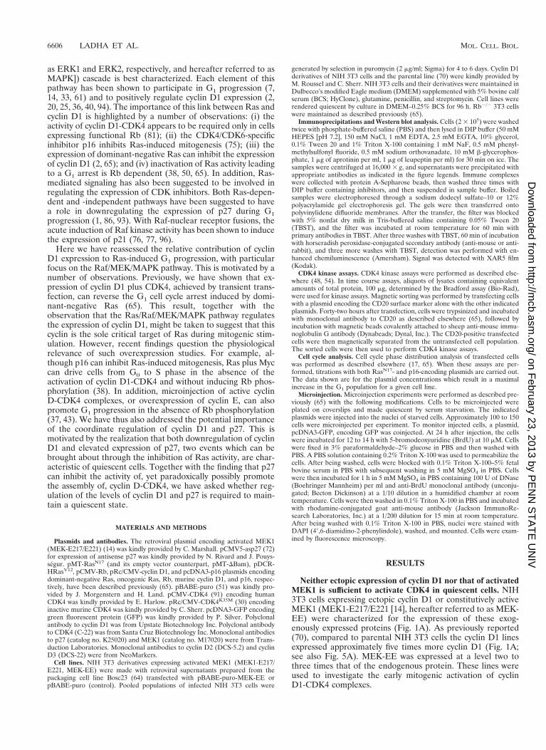
Neither ectopic expression of cyclin D1 nor that of activatedMEK1 is sufficient to activate CDK4 in quiescent cells. NIH3T3 cells expressing ectopic cyclin D1 or constitutively activeMEK1 (MEK1-E217/E221 [14], hereafter referred to as MEK-EE) were characterized for the expression of these exog-enously expressed proteins (Fig. 1A). As previously reported(70), compared to parental NIH 3T3 cells the cyclin D1 linesexpressed approximately five times more cyclin D1 (Fig. 1A;see also Fig. 5A). MEK-EE was expressed at a level two tothree times that of the endogenous protein. These lines wereused to investigate the early mitogenic activation of cyclinD1-CDK4 complexes.
6606 LADHA ET AL. MOL. CELL. BIOL.
on February 23, 2013 by P
EN
N S
TA
TE
UN
IVhttp://m
cb.asm.org/
Dow
nloaded from
The parental, cyclin D1, and MEK-EE lines were renderedquiescent by serum starvation for a period of 96 h. The in-crease in the G0/G1 population (50%), under these conditions,was approximately the same for the MEK-EE and parentallines (Fig. 1B). By comparison, the cyclin D1 line did not arrestquite as efficiently (40% increase in G0/G1) as the other linesunder these conditions. Cyclin D1 was undetectable in theparental line. By contrast, a significant amount of cyclin D1 wasdetected in the MEK-EE line, consistent with the role of theRas/Raf/MEK/MAPK pathway in the positive regulation ofcyclin D1 synthesis. The levels of cyclin D1 in the starvedMEK-EE lines were comparable to that found in an asynchro-nous culture of parental or MEK-EE cycling cells (Fig. 1A).The levels of CDK4 were similar in both starved and asynchro-nous cultures in each of the lines.
To begin to understand why both the cyclin D1 andMEK-EE lines could be arrested in G0/early G1 by serumwithdrawal, the ability of CDK4 to support kinase activityunder these conditions was determined. In serum-starved cy-clin D1 and MEK-EE lines, CDK4 immune complexes did notshow associated kinase activity against a recombinant Rb sub-strate (Fig. 1C). Thus, despite the continued expression ofcyclin D1, CDK4 was inactive. These results provide a possibleexplanation for why neither the ectopic expression of activatedMEK1 nor that of cyclin D1 is sufficient to efficiently drive NIH3T3 cells from a quiescent state into S phase.
Expression of activated MEK1 or cyclin D1 and CDK4, butnot cyclin D1 alone, can overcome G1 arrest induced by dom-inant-negative Ras. In cells stimulated out of a quiescent state,
either serum withdrawal or inhibition of Ras activity in thepresence of serum, prior to the restriction point, prevents S-phase entry. Likewise, in an asynchronous population Ras in-hibition leads to a G1 arrest. Given that neither the serum-starved cyclin D1 line nor the MEK-EE line showed detectableamounts of CDK4 activity, a characteristic of mid- to late G1cells, they have likely not progressed beyond the G0/G1 tran-sition under these conditions. This motivated us to next deter-mine if ectopic expression of cyclin D1 or MEK-EE has similareffects on the cell cycle in an asynchronous population ofcycling cells with respect to Ras-mediated signaling. Specifi-cally, we compared the effects of serum withdrawal (notedabove) and inhibition of Ras activity. We also reinvestigatedthe requirements of cyclin D1 and CDK4 with respect to theirparticipation in the override of a G1 arrest induced by theexpression of dominant-negative Ras.
We have previously demonstrated that expression of domi-nant-negative Ras, RasN17, results in an Rb-dependent G1arrest that is mediated, at least in part, by the downregulationof cyclin D1 expression (65). This mutant of Ras has a prefer-ential affinity for GDP and is thought to function by seques-tering guanine nucleotide exchange factors, thereby inhibitingendogenous Ras (8, 18, 83, 85), and blocking the activation ofMAPKs in response to receptor tyrosine kinases (15, 73, 89,95). Furthermore, we had shown that transient transfection ofboth cyclin D1 and CDK4 could override a G1 arrest caused byinhibition of Ras activity (65). Given that transient overexpres-sion studies can sometimes lead to aphysiological results, wedetermined whether stable expression of cyclin D1 could over-
FIG. 1. Expression of activated MEK1 (MEK-EE) and cyclin D1 in NIH 3T3 cells. (A) Parental NIH 3T3 cells and their cyclin D1 and MEK-EE derivatives wererendered quiescent by serum starvation or maintained in a cycling asynchronous state. Under these two conditions, the levels of endogenous and exogenous MEK1,cyclin D1, and CDK4 were determined by Western blot analysis of whole-cell lysates. (B) Same as panel A, except that the cell cycle distribution of the cells wasmonitored by fluorescence-activated cell sorting analysis. (C) Lysates were prepared from serum-starved and asynchronous cultures of parental NIH 3T3 cells and thecyclin D1 and MEK-EE derivatives. Immunoprecipitation (IP) with antibody to CDK4 were performed. Immune complexes were assayed for CDK4-associated kinaseactivity with recombinant glutathione S-transferase (GST)–Rb as substrate. NRS, normal rabbit serum.
VOL. 18, 1998 REGULATION OF EXIT FROM QUIESCENCE 6607
on February 23, 2013 by P
EN
N S
TA
TE
UN
IVhttp://m
cb.asm.org/
Dow
nloaded from
ride a G1 arrest induced by RasN17 and whether there was arequirement for the coexpression of CDK4. This would alsoallow us to more accurately compare the outcomes when an-alyzing the cyclin D1 and MEK-EE lines, which, as opposed totransient transfection of cyclin D1, express roughly the sameamount of cyclin D1.
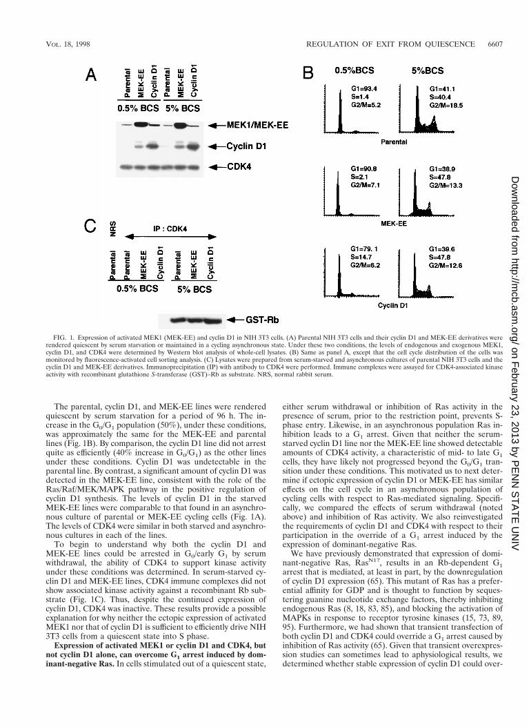
Either stable or transient expression of cyclin D1 alone onlypartially overrode a G1 arrest induced by RasN17 (Fig. 2A), bythe same experimental approach we had previously employed(65). Ectopic expression of CDK4 caused a significant reversalin the G1 arrest induced by dominant-negative Ras in the cyclinD1 lines but not in the parental line, consistent with our pre-vious results. Given that the expression of cyclin D1 and notthat of CDK4 is thought to be rate limiting for G1 progressionand that the expression of the former but not of the latter isinhibited by expression of RasN17, we also investigated therequirement for CDK4 activity in these override experiments.Expression of a catalytically inactive allele of CDK4,CDK4K35M (30), also allowed the cyclin D1 line, but not pa-rental NIH 3T3 cells, to override the RasN17-induced G1 arrest(Fig. 2A). Thus, expression of cyclin D1 alone is not sufficientto override a cell cycle arrest mediated by the inhibition of Rasactivity. That there is a requirement for either wild-type orcatalytically inactive CDK4 suggests the possibility that titra-tion of CDK inhibitors, an established function of cyclin D-CDK4 complexes (10, 66), participates in the override.
The findings noted above differ from an Rb-mediated G1arrest in Rb-deficient fibroblasts. Here ectopic expression of
cyclin D1 alone was sufficient to significantly reverse the cellcycle block induced by Rb (Fig. 2B). Together with our previ-ous results, these new data suggest that, though a G1 arrestcaused by Ras inactivation is Rb dependent, there are differentrequirements with respect to cyclin D1 and CDK4 in reversingan Rb- versus a RasN17-induced G1 arrest.
In contrast to the cyclin D1 lines, the MEK-EE lines effi-ciently reversed a G1 arrest induced by expression of RasN17.Similar results were obtained by transient transfection of plas-mids encoding MEK-EE and RasN17 (data not shown). Sincethe levels of cyclin D1 in both starved and asynchronousMEK-EE cells did not exceed those observed in the cyclin D1line, the result cannot be entirely attributable to MEK1-medi-ated regulation of cyclin D1, and additional events play asignificant role in the ability of MEK-EE to reverse the G1arrest induced by dominant-negative Ras. Together with theresults noted above, these data suggest that ectopic expressionof cyclin D1 and that of MEK-EE have different cellular re-sponses to serum withdrawal and Ras inactivation.
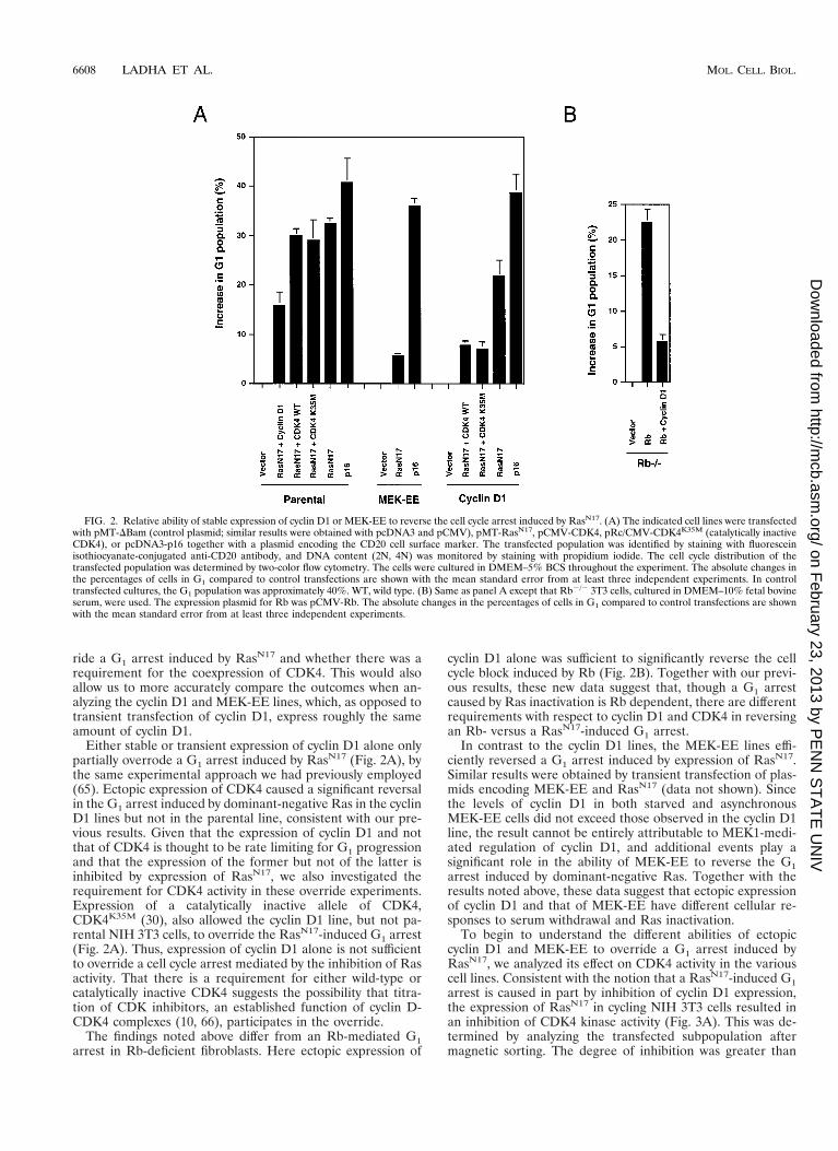
To begin to understand the different abilities of ectopiccyclin D1 and MEK-EE to override a G1 arrest induced byRasN17, we analyzed its effect on CDK4 activity in the variouscell lines. Consistent with the notion that a RasN17-induced G1arrest is caused in part by inhibition of cyclin D1 expression,the expression of RasN17 in cycling NIH 3T3 cells resulted inan inhibition of CDK4 kinase activity (Fig. 3A). This was de-termined by analyzing the transfected subpopulation aftermagnetic sorting. The degree of inhibition was greater than
FIG. 2. Relative ability of stable expression of cyclin D1 or MEK-EE to reverse the cell cycle arrest induced by RasN17. (A) The indicated cell lines were transfectedwith pMT-DBam (control plasmid; similar results were obtained with pcDNA3 and pCMV), pMT-RasN17, pCMV-CDK4, pRc/CMV-CDK4K35M (catalytically inactiveCDK4), or pcDNA3-p16 together with a plasmid encoding the CD20 cell surface marker. The transfected population was identified by staining with fluoresceinisothiocyanate-conjugated anti-CD20 antibody, and DNA content (2N, 4N) was monitored by staining with propidium iodide. The cell cycle distribution of thetransfected population was determined by two-color flow cytometry. The cells were cultured in DMEM–5% BCS throughout the experiment. The absolute changes inthe percentages of cells in G1 compared to control transfections are shown with the mean standard error from at least three independent experiments. In controltransfected cultures, the G1 population was approximately 40%. WT, wild type. (B) Same as panel A except that Rb2/2 3T3 cells, cultured in DMEM–10% fetal bovineserum, were used. The expression plasmid for Rb was pCMV-Rb. The absolute changes in the percentages of cells in G1 compared to control transfections are shownwith the mean standard error from at least three independent experiments.
6608 LADHA ET AL. MOL. CELL. BIOL.
on February 23, 2013 by P
EN
N S
TA
TE
UN
IVhttp://m
cb.asm.org/
Dow
nloaded from
50% compared to approximately 75% for p16-transfected cells.The difference may be in part to due to our observations thatthe RasN17 expression specifically inhibits the expression ofcyclin D1 but does not affect the synthesis of cyclin D2 andcyclin D3 (Fig. 3B). With the available antibodies, we have notbeen able to accurately assess whether the above-noted differ-ence in CDK4 activity is due to cyclin D2- and cyclin D3-associated kinase activity.
Expression of MEK-EE largely prevented the downregula-tion of cyclin D1 and the inhibition of CDK4 kinase activitycaused by expression of RasN17 (Fig. 4). Transient transfectionof plasmids encoding activated Ras, RasV12 (5), or MEK-EEwas also able to prevent the inhibition of CDK4 kinase activitycaused by expression of RasN17 (Fig. 3 and data not shown). Bycontrast, in the cyclin D1 line, CDK4 activity was inhibited toa similar degree as that found for the parental NIH 3T3 line(Fig. 4A). Thus, the relative ability of ectopic cyclin D1 andMEK-EE to override a RasN17-induced G1 arrest correlates, toa significant extent, with their ability to prevent the inhibitionof CDK4 activity following Ras inactivation.
Cyclin D1 and CDK4 complex formation in cell lines thatconstitutively express cyclin D1 during G1 progression. Theabove results suggest that cyclin D1 and activated MEK1 differin their abilities to overcome Ras inactivation in cycling cells,while both the cyclin D1 and activated MEK1 lines respondedsimilarly to serum deprivation and subsequently to quiescence;both lines could be arrested in G0/G1 with CDK4 inactive. Asa first step in determining why CDK4 might be inactive in thecyclin D1 and MEK-EE lines, we assayed the ability of cyclinD1 and CDK4 to form complexes in serum-starved cells. CyclinD1 immunoprecipitates were found to contain CDK4, andconversely, CDK4 immune complexes were found to containcyclin D1. The degree to which complexes between cyclin D1
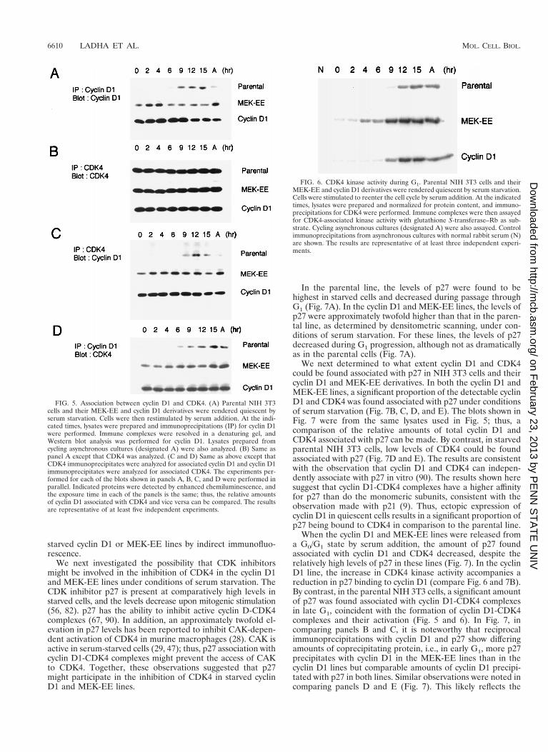
and CDK4 could be detected in serum-starved cells was com-parable to that observed in mid- to late G1 or in cycling cells(Fig. 5). The parental line, used as a control, did not showcomplex formation between cyclin D1 and CDK4 in starvedand early G1 cells, simply due to the lack of cyclin D1 expres-sion under these conditions. Cyclin D1 was detected at 6 to 9 hafter serum stimulation of quiescent parental NIH 3T3 cells.At this time in mid- to late G1, complexes could be detectedbetween cyclin D1 and CDK4 (Fig. 5) (48), commensurate withthe appearance of kinase activity (see below) (48). The resultssuggest that the inability to detect CDK4-associated kinaseactivity in starved cyclin D1 and MEK-EE lines was not due tothe inability of cyclin D1 to associate with CDK4.
We also measured CDK4-associated kinase activity duringG1 following serum stimulation. In parental NIH 3T3 cells,CDK4 activity was first detected at 9 h after stimulation at thetime when complexes between cyclin D1 could be detected(Fig. 5 and 6). The cyclin D1 and MEK-EE lines reproduciblydisplayed CDK4 activity at 9 and 4 h poststimulation, respec-tively (Fig. 6).
p27 is bound to inactive CDK4 in quiescent cyclin D1 andMEK-EE lines. We next sought to determine why CDK4 wasinactive in the serum-starved NIH 3T3 cells expressing cyclinD1 and MEK-EE. Three possibilities were considered. First,under conditions of serum starvation, CDK4 might be ren-dered inactive due to tyrosine phosphorylation at position 17(88). Second, cyclin D1 when expressed in serum-starved cellsmight fail to localize to the nucleus. In some cell systems, cyclinD1 has been reported to be localized to the cytoplasm instarved cells (4). Third, one or more CDK inhibitors might beresponsible for maintaining cyclin D1-CDK4 in an inactivestate. In terms of the first possibility, we were unable to detectthe presence of tyrosine phosphate in CDK4 either in theparental NIH 3T3 cells or in the cyclin D1 or MEK-EE deriv-atives, with an antiphosphotyrosine antibody. Similarly, we didnot detect significant amounts of cyclin D1 in the cytoplasm of
FIG. 3. Effect of RasN17 expression on CDK4 activity and D cyclin levels. (A)NIH 3T3 cells, cultured in DMEM–5% BCS, were transfected with plasmidsencoding the indicated proteins or vector alone together with a plasmid encodingthe surface marker CD20. Forty-two hours later, transfected cells were isolatedby magnetic sorting with Dynabeads (see Materials and Methods). Extracts wereprepared, normalized for protein content, and subjected to immunoprecipitationfor CDK4. CDK4-associated kinase was measured by using glutathione S-trans-ferase (GST)–Rb as a substrate. In lane 1, control normal rabbit serum was used.Lane 2 is the control (pMT-DBam, vector-alone transfection; the same resultswere obtained when pcDNA3 was used as the vector control) set to 100% kinaseactivity. The bar graph represents the relative kinase activity with the meanstandard error from at least three independent experiments. A representativeautoradiogram is shown. (B) Same as panel A except that cell extracts were usedfor Western blot analysis for cyclin D1, cyclin D2, cyclin D3, and CDK4. Indi-cated proteins were detected by enhanced chemiluminescence.
FIG. 4. The effect of activated MEK1 on CDK4 activity and cyclin D1 levelsin cells transiently expressing RasN17. (A) NIH 3T3 cells expressing ectopic cyclinD1 or MEK-EE or cells infected with control virus were transiently transfectedwith control plasmid (pMT-DBam), pMT-RasN17, or pcDNA3-p16. The trans-fected population, cultured in DMEM–5% BCS, was magnetically sorted, andCDK4 kinase assays were performed as described for Fig. 3B. Kinase activity forvector control transfections was set to 100% as indicated. The bar graph repre-sents the relative kinase activity with the mean standard error from at least threeindependent experiments. A representative autoradiogram is shown. GST, glu-tathione S-transferase. (B) Same as panel A except that cell lysates were used toassess the relative amounts of cyclin D1 protein present as a function of RasN17
expression by Western blot analysis.
VOL. 18, 1998 REGULATION OF EXIT FROM QUIESCENCE 6609
on February 23, 2013 by P
EN
N S
TA
TE
UN
IVhttp://m
cb.asm.org/
Dow
nloaded from
starved cyclin D1 or MEK-EE lines by indirect immunofluo-rescence.
We next investigated the possibility that CDK inhibitorsmight be involved in the inhibition of CDK4 in the cyclin D1and MEK-EE lines under conditions of serum starvation. TheCDK inhibitor p27 is present at comparatively high levels instarved cells, and the levels decrease upon mitogenic stimulation(56, 82). p27 has the ability to inhibit active cyclin D-CDK4complexes (67, 90). In addition, an approximately twofold el-evation in p27 levels has been reported to inhibit CAK-depen-dent activation of CDK4 in murine macrophages (28). CAK isactive in serum-starved cells (29, 47); thus, p27 association withcyclin D1-CDK4 complexes might prevent the access of CAKto CDK4. Together, these observations suggested that p27might participate in the inhibition of CDK4 in starved cyclinD1 and MEK-EE lines.
In the parental line, the levels of p27 were found to behighest in starved cells and decreased during passage throughG1 (Fig. 7A). In the cyclin D1 and MEK-EE lines, the levels ofp27 were approximately twofold higher than that in the paren-tal line, as determined by densitometric scanning, under con-ditions of serum starvation. For these lines, the levels of p27decreased during G1 progression, although not as dramaticallyas in the parental cells (Fig. 7A).
We next determined to what extent cyclin D1 and CDK4could be found associated with p27 in NIH 3T3 cells and theircyclin D1 and MEK-EE derivatives. In both the cyclin D1 andMEK-EE lines, a significant proportion of the detectable cyclinD1 and CDK4 was found associated with p27 under conditionsof serum starvation (Fig. 7B, C, D, and E). The blots shown inFig. 7 were from the same lysates used in Fig. 5; thus, acomparison of the relative amounts of total cyclin D1 andCDK4 associated with p27 can be made. By contrast, in starvedparental NIH 3T3 cells, low levels of CDK4 could be foundassociated with p27 (Fig. 7D and E). The results are consistentwith the observation that cyclin D1 and CDK4 can indepen-dently associate with p27 in vitro (90). The results shown heresuggest that cyclin D1-CDK4 complexes have a higher affinityfor p27 than do the monomeric subunits, consistent with theobservation made with p21 (9). Thus, ectopic expression ofcyclin D1 in quiescent cells results in a significant proportion ofp27 being bound to CDK4 in comparison to the parental line.
When the cyclin D1 and MEK-EE lines were released froma G0/G1 state by serum addition, the amount of p27 foundassociated with cyclin D1 and CDK4 decreased, despite therelatively high levels of p27 in these lines (Fig. 7). In the cyclinD1 line, the increase in CDK4 kinase activity accompanies areduction in p27 binding to cyclin D1 (compare Fig. 6 and 7B).By contrast, in the parental NIH 3T3 cells, a significant amountof p27 was found associated with cyclin D1-CDK4 complexesin late G1, coincident with the formation of cyclin D1-CDK4complexes and their activation (Fig. 5 and 6). In Fig. 7, incomparing panels B and C, it is noteworthy that reciprocalimmunoprecipitations with cyclin D1 and p27 show differingamounts of coprecipitating protein, i.e., in early G1, more p27precipitates with cyclin D1 in the MEK-EE lines than in thecyclin D1 lines but comparable amounts of cyclin D1 precipi-tated with p27 in both lines. Similar observations were noted incomparing panels D and E (Fig. 7). This likely reflects the
FIG. 5. Association between cyclin D1 and CDK4. (A) Parental NIH 3T3cells and their MEK-EE and cyclin D1 derivatives were rendered quiescent byserum starvation. Cells were then restimulated by serum addition. At the indi-cated times, lysates were prepared and immunoprecipitations (IP) for cyclin D1were performed. Immune complexes were resolved in a denaturing gel, andWestern blot analysis was performed for cyclin D1. Lysates prepared fromcycling asynchronous cultures (designated A) were also analyzed. (B) Same aspanel A except that CDK4 was analyzed. (C and D) Same as above except thatCDK4 immunoprecipitates were analyzed for associated cyclin D1 and cyclin D1immunoprecipitates were analyzed for associated CDK4. The experiments per-formed for each of the blots shown in panels A, B, C, and D were performed inparallel. Indicated proteins were detected by enhanced chemiluminescence, andthe exposure time in each of the panels is the same; thus, the relative amountsof cyclin D1 associated with CDK4 and vice versa can be compared. The resultsare representative of at least five independent experiments.
FIG. 6. CDK4 kinase activity during G1. Parental NIH 3T3 cells and theirMEK-EE and cyclin D1 derivatives were rendered quiescent by serum starvation.Cells were stimulated to reenter the cell cycle by serum addition. At the indicatedtimes, lysates were prepared and normalized for protein content, and immuno-precipitations for CDK4 were performed. Immune complexes were then assayedfor CDK4-associated kinase activity with glutathione S-transferase–Rb as sub-strate. Cycling asynchronous cultures (designated A) were also assayed. Controlimmunoprecipitations from asynchronous cultures with normal rabbit serum (N)are shown. The results are representative of at least three independent experi-ments.
6610 LADHA ET AL. MOL. CELL. BIOL.
on February 23, 2013 by P
EN
N S
TA
TE
UN
IVhttp://m
cb.asm.org/
Dow
nloaded from
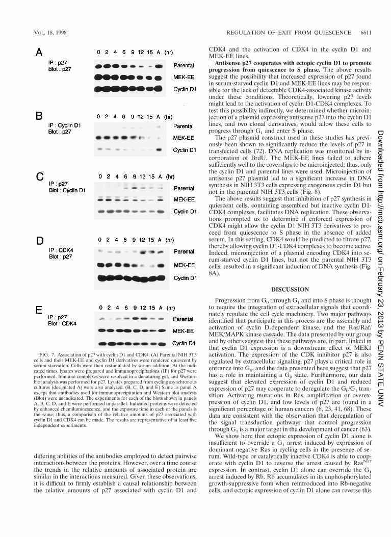
differing abilities of the antibodies employed to detect pairwiseinteractions between the proteins. However, over a time coursethe trends in the relative amounts of associated protein aresimilar in the interactions measured. Given these observations,it is difficult to firmly establish a causal relationship betweenthe relative amounts of p27 associated with cyclin D1 and
CDK4 and the activation of CDK4 in the cyclin D1 andMEK-EE lines.
Antisense p27 cooperates with ectopic cyclin D1 to promoteprogression from quiescence to S phase. The above resultssuggest the possibility that increased expression of p27 foundin serum-starved cyclin D1 and MEK-EE lines may be respon-sible for the lack of detectable CDK4-associated kinase activityunder these conditions. Theoretically, lowering p27 levelsmight lead to the activation of cyclin D1-CDK4 complexes. Totest this possibility indirectly, we determined whether microin-jection of a plasmid expressing antisense p27 into the cyclin D1lines, and two clonal derivatives, would allow these cells toprogress through G1 and enter S phase.
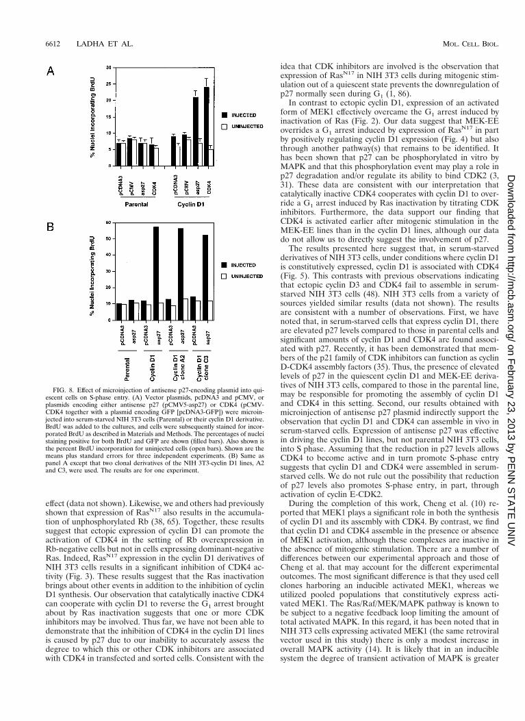
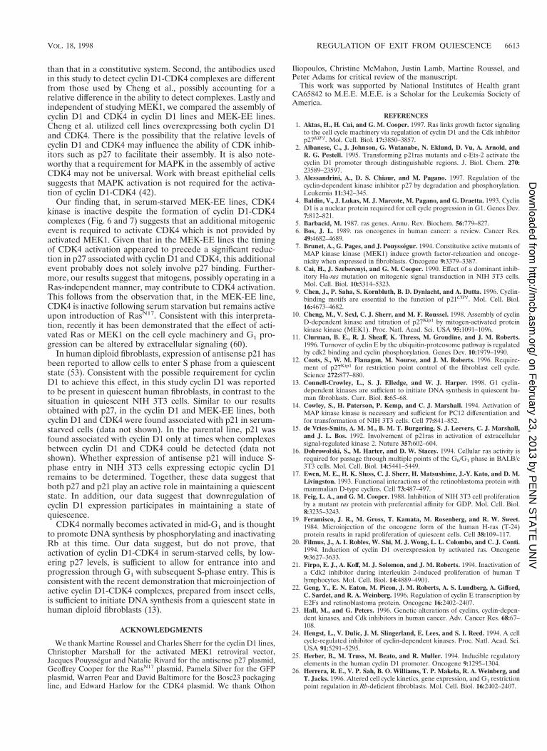
The p27 plasmid construct used in these studies has previ-ously been shown to significantly reduce the levels of p27 intransfected cells (72). DNA replication was monitored by in-corporation of BrdU. The MEK-EE lines failed to adheresufficiently well to the coverslips to be microinjected; thus, onlythe cyclin D1 and parental lines were used. Microinjection ofantisense p27 plasmid led to a significant increase in DNAsynthesis in NIH 3T3 cells expressing exogenous cyclin D1 butnot in the parental NIH 3T3 cells (Fig. 8).
The above results suggest that inhibition of p27 synthesis inquiescent cells, containing assembled but inactive cyclin D1-CDK4 complexes, facilitates DNA replication. These observa-tions prompted us to determine if enforced expression ofCDK4 might allow the cyclin D1 NIH 3T3 derivatives to pro-ceed from quiescence to S phase in the absence of addedserum. In this setting, CDK4 would be predicted to titrate p27,thereby allowing cyclin D1-CDK4 complexes to become active.Indeed, microinjection of a plasmid encoding CDK4 into se-rum-starved cyclin D1 lines, but not the parental NIH 3T3cells, resulted in a significant induction of DNA synthesis (Fig.8A).
DISCUSSION
Progression from G0 through G1 and into S phase is thoughtto require the integration of extracellular signals that coordi-nately regulate the cell cycle machinery. Two major pathwaysidentified that participate in this process are the assembly andactivation of cyclin D-dependent kinase, and the Ras/Raf/MEK/MAPK kinase cascade. The data presented by our groupand by others suggest that these pathways are, in part, linked inthat cyclin D1 expression is a downstream effect of MEK1activation. The expression of the CDK inhibitor p27 is alsoregulated by extracellular signaling. p27 plays a critical role inentrance into G0, and the data presented here suggest that p27has a role in maintaining a G0 state. Furthermore, our datasuggest that elevated expression of cyclin D1 and reducedexpression of p27 may cooperate to deregulate the G0/G1 tran-sition. Activating mutations in Ras, amplification or overex-pression of cyclin D1, and low levels of p27 are found in asignificant percentage of human cancers (6, 23, 41, 68). Thesedata are consistent with the observation that deregulation ofthe signal transduction pathways that control progressionthrough G1 is a major target in the development of cancer (63).
We show here that ectopic expression of cyclin D1 alone isinsufficient to override a G1 arrest induced by expression ofdominant-negative Ras in cycling cells in the presence of se-rum. Wild-type or catalytically inactive CDK4 is able to coop-erate with cyclin D1 to reverse the arrest caused by RasN17
expression. In contrast, cyclin D1 alone can override the G1arrest induced by Rb. Rb accumulates in its unphosphorylatedgrowth-suppressive form when reintroduced into Rb-negativecells, and ectopic expression of cyclin D1 alone can reverse this
FIG. 7. Association of p27 with cyclin D1 and CDK4. (A) Parental NIH 3T3cells and their MEK-EE and cyclin D1 derivatives were rendered quiescent byserum starvation. Cells were then restimulated by serum addition. At the indi-cated times, lysates were prepared and immunoprecipitations (IP) for p27 wereperformed. Immune complexes were resolved in a denaturing gel, and Westernblot analysis was performed for p27. Lysates prepared from cycling asynchronouscultures (designated A) were also analyzed. (B, C, D, and E) Same as panel Aexcept that antibodies used for immunoprecipitation and Western blot analysis(Blot) were as indicated. The experiments for each of the blots shown in panelsA, B, C, D, and E were performed in parallel. Indicated proteins were detectedby enhanced chemiluminescence, and the exposure time in each of the panels isthe same; thus, a comparison of the relative amounts of p27 associated withcyclin D1 and CDK4 can be made. The results are representative of at least fiveindependent experiments.
VOL. 18, 1998 REGULATION OF EXIT FROM QUIESCENCE 6611
on February 23, 2013 by P
EN
N S
TA
TE
UN
IVhttp://m
cb.asm.org/
Dow
nloaded from
effect (data not shown). Likewise, we and others had previouslyshown that expression of RasN17 also results in the accumula-tion of unphosphorylated Rb (38, 65). Together, these resultssuggest that ectopic expression of cyclin D1 can promote theactivation of CDK4 in the setting of Rb overexpression inRb-negative cells but not in cells expressing dominant-negativeRas. Indeed, RasN17 expression in the cyclin D1 derivatives ofNIH 3T3 cells results in a significant inhibition of CDK4 ac-tivity (Fig. 3). These results suggest that the Ras inactivationbrings about other events in addition to the inhibition of cyclinD1 synthesis. Our observation that catalytically inactive CDK4can cooperate with cyclin D1 to reverse the G1 arrest broughtabout by Ras inactivation suggests that one or more CDKinhibitors may be involved. Thus far, we have not been able todemonstrate that the inhibition of CDK4 in the cyclin D1 linesis caused by p27 due to our inability to accurately assess thedegree to which this or other CDK inhibitors are associatedwith CDK4 in transfected and sorted cells. Consistent with the
idea that CDK inhibitors are involved is the observation thatexpression of RasN17 in NIH 3T3 cells during mitogenic stim-ulation out of a quiescent state prevents the downregulation ofp27 normally seen during G1 (1, 86).
In contrast to ectopic cyclin D1, expression of an activatedform of MEK1 effectively overcame the G1 arrest induced byinactivation of Ras (Fig. 2). Our data suggest that MEK-EEoverrides a G1 arrest induced by expression of RasN17 in partby positively regulating cyclin D1 expression (Fig. 4) but alsothrough another pathway(s) that remains to be identified. Ithas been shown that p27 can be phosphorylated in vitro byMAPK and that this phosphorylation event may play a role inp27 degradation and/or regulate its ability to bind CDK2 (3,31). These data are consistent with our interpretation thatcatalytically inactive CDK4 cooperates with cyclin D1 to over-ride a G1 arrest induced by Ras inactivation by titrating CDKinhibitors. Furthermore, the data support our finding thatCDK4 is activated earlier after mitogenic stimulation in theMEK-EE lines than in the cyclin D1 lines, although our datado not allow us to directly suggest the involvement of p27.
The results presented here suggest that, in serum-starvedderivatives of NIH 3T3 cells, under conditions where cyclin D1is constitutively expressed, cyclin D1 is associated with CDK4(Fig. 5). This contrasts with previous observations indicatingthat ectopic cyclin D3 and CDK4 fail to assemble in serum-starved NIH 3T3 cells (48). NIH 3T3 cells from a variety ofsources yielded similar results (data not shown). The resultsare consistent with a number of observations. First, we havenoted that, in serum-starved cells that express cyclin D1, thereare elevated p27 levels compared to those in parental cells andsignificant amounts of cyclin D1 and CDK4 are found associ-ated with p27. Recently, it has been demonstrated that mem-bers of the p21 family of CDK inhibitors can function as cyclinD-CDK4 assembly factors (35). Thus, the presence of elevatedlevels of p27 in the quiescent cyclin D1 and MEK-EE deriva-tives of NIH 3T3 cells, compared to those in the parental line,may be responsible for promoting the assembly of cyclin D1and CDK4 in this setting. Second, our results obtained withmicroinjection of antisense p27 plasmid indirectly support theobservation that cyclin D1 and CDK4 can assemble in vivo inserum-starved cells. Expression of antisense p27 was effectivein driving the cyclin D1 lines, but not parental NIH 3T3 cells,into S phase. Assuming that the reduction in p27 levels allowsCDK4 to become active and in turn promote S-phase entrysuggests that cyclin D1 and CDK4 were assembled in serum-starved cells. We do not rule out the possibility that reductionof p27 levels also promotes S-phase entry, in part, throughactivation of cyclin E-CDK2.
During the completion of this work, Cheng et al. (10) re-ported that MEK1 plays a significant role in both the synthesisof cyclin D1 and its assembly with CDK4. By contrast, we findthat cyclin D1 and CDK4 assemble in the presence or absenceof MEK1 activation, although these complexes are inactive inthe absence of mitogenic stimulation. There are a number ofdifferences between our experimental approach and those ofCheng et al. that may account for the different experimentaloutcomes. The most significant difference is that they used cellclones harboring an inducible activated MEK1, whereas weutilized pooled populations that constitutively express acti-vated MEK1. The Ras/Raf/MEK/MAPK pathway is known tobe subject to a negative feedback loop limiting the amount oftotal activated MAPK. In this regard, it has been noted that inNIH 3T3 cells expressing activated MEK1 (the same retroviralvector used in this study) there is only a modest increase inoverall MAPK activity (14). It is likely that in an induciblesystem the degree of transient activation of MAPK is greater
FIG. 8. Effect of microinjection of antisense p27-encoding plasmid into qui-escent cells on S-phase entry. (A) Vector plasmids, pcDNA3 and pCMV, orplasmids encoding either antisense p27 (pCMV5-asp27) or CDK4 (pCMV-CDK4 together with a plasmid encoding GFP [pcDNA3-GFP]) were microin-jected into serum-starved NIH 3T3 cells (Parental) or their cyclin D1 derivative.BrdU was added to the cultures, and cells were subsequently stained for incor-porated BrdU as described in Materials and Methods. The percentages of nucleistaining positive for both BrdU and GFP are shown (filled bars). Also shown isthe percent BrdU incorporation for uninjected cells (open bars). Shown are themeans plus standard errors for three independent experiments. (B) Same aspanel A except that two clonal derivatives of the NIH 3T3-cyclin D1 lines, A2and C3, were used. The results are for one experiment.
6612 LADHA ET AL. MOL. CELL. BIOL.
on February 23, 2013 by P
EN
N S
TA
TE
UN
IVhttp://m
cb.asm.org/
Dow
nloaded from
than that in a constitutive system. Second, the antibodies usedin this study to detect cyclin D1-CDK4 complexes are differentfrom those used by Cheng et al., possibly accounting for arelative difference in the ability to detect complexes. Lastly andindependent of studying MEK1, we compared the assembly ofcyclin D1 and CDK4 in cyclin D1 lines and MEK-EE lines.Cheng et al. utilized cell lines overexpressing both cyclin D1and CDK4. There is the possibility that the relative levels ofcyclin D1 and CDK4 may influence the ability of CDK inhib-itors such as p27 to facilitate their assembly. It is also note-worthy that a requirement for MAPK in the assembly of activeCDK4 may not be universal. Work with breast epithelial cellssuggests that MAPK activation is not required for the activa-tion of cyclin D1-CDK4 (42).
Our finding that, in serum-starved MEK-EE lines, CDK4kinase is inactive despite the formation of cyclin D1-CDK4complexes (Fig. 6 and 7) suggests that an additional mitogenicevent is required to activate CDK4 which is not provided byactivated MEK1. Given that in the MEK-EE lines the timingof CDK4 activation appeared to precede a significant reduc-tion in p27 associated with cyclin D1 and CDK4, this additionalevent probably does not solely involve p27 binding. Further-more, our results suggest that mitogens, possibly operating in aRas-independent manner, may contribute to CDK4 activation.This follows from the observation that, in the MEK-EE line,CDK4 is inactive following serum starvation but remains activeupon introduction of RasN17. Consistent with this interpreta-tion, recently it has been demonstrated that the effect of acti-vated Ras or MEK1 on the cell cycle machinery and G1 pro-gression can be altered by extracellular signaling (60).
In human diploid fibroblasts, expression of antisense p21 hasbeen reported to allow cells to enter S phase from a quiescentstate (53). Consistent with the possible requirement for cyclinD1 to achieve this effect, in this study cyclin D1 was reportedto be present in quiescent human fibroblasts, in contrast to thesituation in quiescent NIH 3T3 cells. Similar to our resultsobtained with p27, in the cyclin D1 and MEK-EE lines, bothcyclin D1 and CDK4 were found associated with p21 in serum-starved cells (data not shown). In the parental line, p21 wasfound associated with cyclin D1 only at times when complexesbetween cyclin D1 and CDK4 could be detected (data notshown). Whether expression of antisense p21 will induce S-phase entry in NIH 3T3 cells expressing ectopic cyclin D1remains to be determined. Together, these data suggest thatboth p27 and p21 play an active role in maintaining a quiescentstate. In addition, our data suggest that downregulation ofcyclin D1 expression participates in maintaining a state ofquiescence.
CDK4 normally becomes activated in mid-G1 and is thoughtto promote DNA synthesis by phosphorylating and inactivatingRb at this time. Our data suggest, but do not prove, thatactivation of cyclin D1-CDK4 in serum-starved cells, by low-ering p27 levels, is sufficient to allow for entrance into andprogression through G1 with subsequent S-phase entry. This isconsistent with the recent demonstration that microinjection ofactive cyclin D1-CDK4 complexes, prepared from insect cells,is sufficient to initiate DNA synthesis from a quiescent state inhuman diploid fibroblasts (13).
ACKNOWLEDGMENTS
We thank Martine Roussel and Charles Sherr for the cyclin D1 lines,Christopher Marshall for the activated MEK1 retroviral vector,Jacques Pouyssegur and Natalie Rivard for the antisense p27 plasmid,Geoffrey Cooper for the RasN17 plasmid, Pamela Silver for the GFPplasmid, Warren Pear and David Baltimore for the Bosc23 packagingline, and Edward Harlow for the CDK4 plasmid. We thank Othon
Iliopoulos, Christine McMahon, Justin Lamb, Martine Roussel, andPeter Adams for critical review of the manuscript.
This work was supported by National Institutes of Health grantCA65842 to M.E.E. M.E.E. is a Scholar for the Leukemia Society ofAmerica.
REFERENCES1. Aktas, H., H. Cai, and G. M. Cooper. 1997. Ras links growth factor signaling
to the cell cycle machinery via regulation of cyclin D1 and the Cdk inhibitorp27KIP1. Mol. Cell. Biol. 17:3850–3857.
2. Albanese, C., J. Johnson, G. Watanabe, N. Eklund, D. Vu, A. Arnold, andR. G. Pestell. 1995. Transforming p21ras mutants and c-Ets-2 activate thecyclin D1 promoter through distinguishable regions. J. Biol. Chem. 270:23589–23597.
3. Alessandrini, A., D. S. Chiaur, and M. Pagano. 1997. Regulation of thecyclin-dependent kinase inhibitor p27 by degradation and phosphorylation.Leukemia 11:342–345.
4. Baldin, V., J. Lukas, M. J. Marcote, M. Pagano, and G. Draetta. 1993. CyclinD1 is a nuclear protein required for cell cycle progression in G1. Genes Dev.7:812–821.
5. Barbacid, M. 1987. ras genes. Annu. Rev. Biochem. 56:779–827.6. Bos, J. L. 1989. ras oncogenes in human cancer: a review. Cancer Res.
49:4682–4689.7. Brunet, A., G. Pages, and J. Pouyssegur. 1994. Constitutive active mutants of
MAP kinase kinase (MEK1) induce growth factor-relaxation and oncoge-nicity when expressed in fibroblasts. Oncogene 9:3379–3387.
8. Cai, H., J. Szeberenyi, and G. M. Cooper. 1990. Effect of a dominant inhib-itory Ha-ras mutation on mitogenic signal transduction in NIH 3T3 cells.Mol. Cell. Biol. 10:5314–5323.
9. Chen, J., P. Saha, S. Kornbluth, B. D. Dynlacht, and A. Dutta. 1996. Cyclin-binding motifs are essential to the function of p21CIP1. Mol. Cell. Biol.16:4673–4682.
10. Cheng, M., V. Sexl, C. J. Sherr, and M. F. Roussel. 1998. Assembly of cyclinD-dependent kinase and titration of p27Kip1 by mitogen-activated proteinkinase kinase (MEK1). Proc. Natl. Acad. Sci. USA 95:1091–1096.
11. Clurman, B. E., R. J. Sheaff, K. Thress, M. Groudine, and J. M. Roberts.1996. Turnover of cyclin E by the ubiquitin-proteosome pathway is regulatedby cdk2 binding and cyclin phosphorylation. Genes Dev. 10:1979–1990.
12. Coats, S., W. M. Flanagan, M. Nourse, and J. M. Roberts. 1996. Require-ment of p27Kip1 for restriction point control of the fibroblast cell cycle.Science 272:877–880.
13. Connell-Crowley, L., S. J. Elledge, and W. J. Harper. 1998. G1 cyclin-dependent kinases are sufficient to initiate DNA synthesis in quiescent hu-man fibroblasts. Curr. Biol. 8:65–68.
14. Cowley, S., H. Paterson, P. Kemp, and C. J. Marshall. 1994. Activation ofMAP kinase kinase is necessary and sufficient for PC12 differentiation andfor transformation of NIH 3T3 cells. Cell 77:841–852.
15. de Vries-Smits, A. M. M., B. M. T. Burgering, S. J. Leevers, C. J. Marshall,and J. L. Bos. 1992. Involvement of p21ras in activation of extracellularsignal-regulated kinase 2. Nature 357:602–604.
16. Dobrowolski, S., M. Harter, and D. W. Stacey. 1994. Cellular ras activity isrequired for passage through multiple points of the G0/G1 phase in BALB/c3T3 cells. Mol. Cell. Biol. 14:5441–5449.
17. Ewen, M. E., H. K. Sluss, C. J. Sherr, H. Matsushime, J.-Y. Kato, and D. M.Livingston. 1993. Functional interactions of the retinoblastoma protein withmammalian D-type cyclins. Cell 73:487–497.
18. Feig, L. A., and G. M. Cooper. 1988. Inhibition of NIH 3T3 cell proliferationby a mutant ras protein with preferential affinity for GDP. Mol. Cell. Biol.8:3235–3243.
19. Feramisco, J. R., M. Gross, T. Kamata, M. Rosenberg, and R. W. Sweet.1984. Microinjection of the oncogene form of the human H-ras (T-24)protein results in rapid proliferation of quiescent cells. Cell 38:109–117.
20. Filmus, J., A. I. Robles, W. Shi, M. J. Wong, L. L. Colombo, and C. J. Conti.1994. Induction of cyclin D1 overexpression by activated ras. Oncogene9:3627–3633.
21. Firpo, E. J., A. Koff, M. J. Solomon, and J. M. Roberts. 1994. Inactivation ofa Cdk2 inhibitor during interleukin 2-induced proliferation of human Tlymphocytes. Mol. Cell. Biol. 14:4889–4901.
22. Geng, Y., E. N. Eaton, M. Picon, J. M. Roberts, A. S. Lundberg, A. Gifford,C. Sardet, and R. A. Weinberg. 1996. Regulation of cyclin E transcription byE2Fs and retinoblastoma protein. Oncogene 16:2402–2407.
23. Hall, M., and G. Peters. 1996. Genetic alterations of cyclins, cyclin-depen-dent kinases, and Cdk inhibitors in human cancer. Adv. Cancer Res. 68:67–108.
24. Hengst, L., V. Dulic, J. M. Slingerland, E. Lees, and S. I. Reed. 1994. A cellcycle-regulated inhibitor of cyclin-dependent kinases. Proc. Natl. Acad. Sci.USA 91:5291–5295.
25. Herber, B., M. Truss, M. Beato, and R. Muller. 1994. Inducible regulatoryelements in the human cyclin D1 promoter. Oncogene 9:1295–1304.
26. Herrera, R. E., V. P. Sah, B. O. Williams, T. P. Makela, R. A. Weinberg, andT. Jacks. 1996. Altered cell cycle kinetics, gene expression, and G1 restrictionpoint regulation in Rb-deficient fibroblasts. Mol. Cell. Biol. 16:2402–2407.
VOL. 18, 1998 REGULATION OF EXIT FROM QUIESCENCE 6613
on February 23, 2013 by P
EN
N S
TA
TE
UN
IVhttp://m
cb.asm.org/
Dow
nloaded from
27. Joneson, T., M. A. White, M. H. Wigler, and D. Bar-Sagi. 1996. Stimulationof membrane ruffling and MAP kinase activation by distinct effectors ofRAS. Science 271:810–812.
28. Kato, J.-Y., M. Matsuoka, K. Polyak, J. Massague, and C. J. Sherr. 1994.Cyclic AMP-induced G1 phase arrest mediated by an inhibitor (p27Kip1) ofcyclin-dependent kinase 4 activation. Cell 79:487–496.
29. Kato, J.-Y., M. Matsuoka, D. K. Strom, and C. J. Sherr. 1994. Regulation ofcyclin D-dependent kinase 4 (cdk4) by cdk4-activating kinase. Mol. Cell.Biol. 14:2713–2721.
30. Kato, J.-Y., H. Matsushime, S. W. Hiebert, M. E. Ewen, and C. J. Sherr.1993. Direct binding of cyclin D to the retinoblastoma gene product (pRb)and pRb phosphorylation by the cyclin D-dependent kinase cdk4. GenesDev. 7:331–342.
31. Kawada, M., S. Yamagoe, Y. Murakami, K. Suzuki, S. Mizuno, and Y.Uehara. 1997. Induction of p27Kip1 degradation and anchorage indepen-dence through the MAP kinase signaling pathway. Oncogene 15:629–637.
32. Koff, A., M. Ohtsuki, K. Polyak, J. M. Roberts, and J. Massague. 1993.Negative regulation of G1 in mammalian cells: inhibition of cyclin E-depen-dent kinase by TGF-b. Science 260:536–539.
33. Kolch, W., G. Heidecker, P. Lloyd, and U. R. Rapp. 1991. Raf-1 proteinkinase is required for growth of induced NIH/3T3 cells. Nature 12:426–428.
34. Krude, T., M. Jackman, J. Pines, and R. A. Lasky. 1997. Cyclin/Cdk-depen-dent initiation of DNA replication in a human cell-free system. Cell 88:109–119.
35. LaBaer, J., M. D. Garrett, L. F. Stevenson, J. M. Slingerland, C. Sandhu,H. S. Chou, A. Fattaey, and E. Harlow. 1997. New functional activities for thep21 family of CDK inhibitors. Genes Dev. 11:847–862.
36. Lavoie, J. N., G. L’Allemain, A. Brunet, R. Muller, and J. Pouyssegur. 1996.Cyclin D1 expression is regulated positively by the p42/p44MAPK and nega-tively by the p38/HOGMAPK pathway. J. Biol. Chem. 271:20608–20616.
37. Leng, X., L. Connell-Crowley, D. Goodrich, and J. W. Harper. 1997. S-phaseentry upon ectopic expression of G1 cyclin-dependent kinases in the absenceof retinoblastoma protein phosphorylation. Curr. Biol. 7:709–712.
38. Leone, G., J. DeGregori, R. Sears, L. Jakoi, and J. R. Nevins. 1997. Myc andRas collaborate in inducing accumulation of active cyclin E/Cdk2 and E2F.Nature 387:422–426.
39. Li, Y., C. W. Jenkins, M. A. Nichols, and Y. Xiong. 1994. Cell cycle expres-sion and p53 regulation of the cyclin-dependent kinase inhibitor p21. Onco-gene 9:2261–2268.
40. Liu, J.-J., J.-R. Chao, M.-C. Jiang, S.-Y. Ng, J. J.-Y. Yen, and H. F. Yang-Yen.1995. Ras transformation results in an elevated level of cyclin D1 and ac-celeration of G1 progression in NIH 3T3 cells. Mol. Cell. Biol. 15:3654–3663.
41. Loda, M., B. Cukor, S. W. Tam, P. Lavin, M. Fiorentino, G. F. Draetta, J. M.Jessup, and M. Pagano. 1997. Increased proteosome-dependent degradationof the cyclin-dependent kinase inhibitor p27 in aggressive colorectal carci-nomas. Nat. Med. 3:231–234.
42. Lukas, J., J. Bartkova, and J. Bartek. 1996. Convergence of mitogenicsignalling cascades from diverse classes of receptors at the cyclin D–cyclin-dependent kinase–pRb-controlled G1 checkpoint. Mol. Cell. Biol. 16:6917–6925.
43. Lukas, J., T. Herzinger, K. Hansen, M. C. Moroni, D. Resnitzky, K. Helin,S. I. Reed, and J. Bartek. 1997. Cyclin E-induced S phase without activationof the pRb/E2F pathway. Genes Dev. 11:1479–1492.
44. Lundberg, A. S., and R. A. Weinberg. 1998. Functional inactivation of theretinoblastoma protein requires sequential modification by at least two dis-tinct cyclin-cdk complexes. Mol. Cell. Biol. 18:753–761.
45. Marshall, C. J. 1995. Specificity of receptor tyrosine kinase signaling: tran-sient versus sustained extracellular signal-regulated kinase activation. Cell80:179–185.
46. Marshall, C. J. 1997. Ras effectors. Curr. Opin. Cell Biol. 8:197–204.47. Matsuoka, M., J.-Y. Kato, R. P. Fisher, D. O. Morgan, and C. J. Sherr. 1994.
Activation of cyclin-dependent kinase 4 (cdk4) by mouse MO15-associatedkinase. Mol. Cell. Biol. 14:7265–7275.
48. Matsushime, H., D. E. Quelle, S. A. Shurtleff, M. Shibuya, C. J. Sherr, andJ.-Y. Kato. 1994. D-type cyclin-dependent kinase activity in mammalian cells.Mol. Cell. Biol. 14:2066–2076.
49. Meyerson, M., and E. Harlow. 1994. Identification of G1 kinase activity forcdk6, a novel cyclin D partner. Mol. Cell. Biol. 14:2077–2086.
50. Mittnacht, S., H. Paterson, M. F. Olson, and C. J. Marshall. 1997. Rassignalling is required for inactivation of tumour suppressor pRb cell-cyclecontrol protein. Curr. Biol. 7:219–221.
51. Morgenstern, J. P., and H. Land. 1990. Advanced mammalian gene transfer:high titre retroviral vectors with multiple drug selection markers and acomplementary helper-free packaging line. Nucleic Acids Res. 18:3587–3596.
52. Mulcahy, L. S., M. R. Smith, and D. W. Stacey. 1985. Requirement for rasproto-oncogene function during serum-stimulated growth of NIH3T3 cells.Nature 313:241–243.
53. Nakanashi, M., G. R. Adami, R. S. Robetorye, A. Noda, S. F. Venable, D.Dimitrov, O. M. Pereira-Smith, and J. R. Smith. 1995. Exit from G0 andentry into the cell cycle of cells expressing p21Sdi1 antisense RNA. Proc. Natl.Acad. Sci. USA 92:4352–4356.
54. Neuman, E., M. H. Ladha, N. Lin, T. M. Upton, S. J. Miller, J. DiRenzo,R. G. Pestell, P. W. Hinds, S. F. Dowdy, M. Brown, and M. E. Ewen. 1997.Cyclin D1 stimulation of estrogen receptor transcriptional activity indepen-dent of cdk4. Mol. Cell. Biol. 17:5338–5347.
55. Noda, A., Y. Ning, S. F. Venable, O. M. Pereira-Smith, and J. R. Smith. 1994.Cloning of senescent cell-derived inhibitors of DNA synthesis using an ex-pression screen. Exp. Cell Res. 211:90–98.
56. Nourse, J., E. Firpo, W. M. Flanagan, S. Coats, K. Polyak, M. H. Lee, J.Massague, G. R. Crabtree, and J. M. Roberts. 1994. Interleukin-2-mediatedelimination of the p27Kip1 cyclin-dependent kinase inhibitor prevented byrapamycin. Nature 372:570–573.
57. Ohtsubo, M., and J. M. Roberts. 1993. Cyclin-dependent regulation of G1 inmammalian fibroblasts. Science 259:1908–1912.
58. Ohtsubo, M., A. M. Theodoras, J. Schumacher, J. M. Roberts, and M.Pagano. 1995. Human cyclin E, a nuclear protein essential for the G1-to-Sphase transition. Mol. Cell. Biol. 15:2612–2624.
59. Okuda, K., T. J. Ernst, and J. D. Griffin. 1994. Inhibition of p21ras activationblocks proliferation but not differentiation of interleukin-3-dependent my-eloid cells. J. Biol. Chem. 269:24602–24607.
60. Olson, M. F., H. F. Paterson, and C. J. Marshall. 1998. Signals from Ras andRho GTPases interact to regulate expression of p21Waf1/Cip1. Nature 394:295–299.
61. Pages, G., P. Lenorman, G. L’Allemain, J.-C. Chambard, S. Meloche, and J.Pouyssegur. 1993. Mitogen-activated protein kinase p42mapk and p44mapkare required for fibroblast proliferation. Proc. Natl. Acad. Sci. USA 90:8319–8323.
62. Pardee, A. B. 1974. A restriction point for control of normal animal cellproliferation. Proc. Natl. Acad. Sci. USA 71:1286–1290.
63. Pardee, A. B. 1989. G1 events and regulation of cell proliferation. Science246:603–608.
64. Pear, W. S., G. P. Nolan, M. L. Scott, and D. Baltimore. 1993. Production ofhigh-titer helper-free retroviruses by transient transfection. Proc. Natl. Acad.Sci. USA 90:8392–8396.
65. Peeper, D. S., T. M. Upton, M. H. Ladha, E. Neuman, J. Zalvide, R. Ber-nards, J. A. DeCaprio, and M. E. Ewen. 1997. Ras signalling linked to thecell-cycle machinery by the retinoblastoma protein. Nature 386:177–181.
66. Polyak, K., J.-Y. Kato, M. J. Solomon, C. J. Sherr, J. Massague, J. M.Roberts, and A. Koff. 1994. p27Kip1, a cyclin-cdk inhibitor, links transforminggrowth factor-b and contact inhibition to cell cycle arrest. Genes Dev. 8:9–22.
67. Polyak, K., M.-H. Lee, H. Erdjument-Bromage, A. Koff, J. M. Roberts, P.Tempst, and J. Massague. 1994. Cloning of p27Kip1, a cyclin-dependentkinase inhibitor and a potential mediator of extracellular antimitogenic sig-nal. Cell 78:59–66.
68. Porter, P. L., K. E. Malone, P. J. Heagerty, G. M. Alexander, L. A. Gatti, E. J.Firpo, and J. R. Daling. 1997. Expression of cell-cycle regulators p27Kip1 andcyclin E, alone and in combination, correlates with survival in young breastcancer patients. Nat. Med. 3:222–225.
69. Pronk, G. J., and J. L. Bos. 1994. The role of p21ras in receptor tyrosinekinase signalling. Biochim. Biophys. Acta 1198:131–147.
70. Quelle, D. E., R. A. Ashmun, S. A. Shurtleff, J. Kato, D. Bar-Sagi, M. F.Roussel, and C. J. Sherr. 1993. Overexpression of mouse D-type cyclinsaccelerates G1 phase in rodent fibroblasts. Genes Dev. 7:1559–1571.
71. Resnitzky, D., and S. I. Reed. 1995. Different roles for cyclins D1 and E inregulation of the G1-to-S transition. Mol. Cell. Biol. 15:3463–3469.
72. Rivard, N., G. L’Allemain, J. Bartek, and J. Pouyssegur. 1996. Abrogation ofp27Kip1 by cDNA antisense suppresses quiescence (G0 state) in fibroblasts.J. Biol. Chem. 271:18337–18341.
73. Robbins, D. J., M. Cheng, E. Zhen, C. A. Vanderbilt, L. A. Feig, and M. H.Cobb. 1992. Evidence for a Ras-dependent extracellular signal-related pro-tein kinase (ERK) cascade. Proc. Natl. Acad. Sci. USA 89:6924–6928.
74. Sellers, W. R., and W. G. Kaelin. 1996. RB as a modulator of transcription.Biochim. Biophys. Acta. 1288:M1–M5.
75. Serrano, M., E. Gomez-Lohoz, R. A. DePinho, D. Beach, and D. Bar-Sagi.1995. Inhibition of Ras-induced proliferation and transformation by p16INK4.Science 267:249–252.
76. Serrano, M., A. W. Lin, M. E. McCurrach, D. Beach, and S. W. Lowe. 1997.Oncogenic ras provokes premature cell senescence associated with accumu-lation of p53 and p16INK4a. Cell 88:593–602.
77. Sewing, A., B. Wiseman, A. LLoyd, and H. Land. 1997. High-intensity Rafsignal causes cell cycle arrest mediated by p21Cip1. Mol. Cell. Biol. 17:5588–5597.
78. Sheaff, R. J., M. Groudine, M. Gordon, J. M. Roberts, and B. E. Clurman.1997. Cyclin E-cdk2 is a regulator of p27Kip1. Genes Dev. 11:1464–1478.
79. Sherr, C. J. 1993. Mammalian G1 cyclins. Cell 73:1059–1065.80. Sherr, C. J. 1994. G1 phase progression: cycling on cue. Cell 79:551–555.81. Sherr, C. J. 1996. Cancer cell cycles. Science 274:1672–1677.82. Sherr, C. J., and J. M. Roberts. 1995. Inhibitors of mammalian G1 cyclin-
dependent kinases. Genes Dev. 9:1149–1163.83. Stacey, D. W., L. A. Feig, and J. B. Gibbs. 1991. Dominant inhibitor Ras
mutants selectively inhibit the activity of either cellular or oncogenic Ras.Mol. Cell. Biol. 11:4053–4064.
6614 LADHA ET AL. MOL. CELL. BIOL.
on February 23, 2013 by P
EN
N S
TA
TE
UN
IVhttp://m
cb.asm.org/
Dow
nloaded from
84. Stacey, D. W., and H. F. Kung. 1984. Transformation of NIH 3T3 cells bymicroinjection of Ha-ras protein. Nature 310:508–511.
85. Szeberenyi, J., H. Cai, and G. M. Cooper. 1990. Effect of a dominant inhib-itory Ha-ras mutation on neuronal differentiation of PC12 cells. Mol. Cell.Biol. 10:5324–5332.
86. Takuwa, N., and Y. Takuwa. 1997. Ras activity late in G1 phase required forp27kip1 downregulation, passage through the restriction point, and entry intoS phase in growth factor-stimulated NIH 3T3 fibroblasts. Mol. Cell. Biol.17:5348–5358.
87. Temin, H. 1971. Stimulation by serum of multiplication of stationary chickencells. J. Cell. Physiol. 78:161–170.
88. Terada, Y., M. Tatsuka, S. Jinno, and H. Okayama. 1995. Requirement fortyrosine phosphorylation of Cdk4 in G1 arrest induced by ultraviolet radia-tion. Nature 376:358–362.
89. Thomas, S. M., M. DeMarco, G. D’Arcangelo, S. Helegoua, and J. S. Brugge.1992. Ras is essential for nerve growth factor- and phorbol ester-inducedtyrosine phosphorylation of MAP kinases. Cell 68:1031–1040.
90. Toyoshima, H., and T. Hunter. 1994. p27, a novel inhibitor of G1 cyclin-cdkprotein kinase activity, is related to p21. Cell 78:67–74.
91. van den Heuvel, S., and E. Harlow. 1993. Distinct roles for cyclin-dependentkinases in cell cycle control. Science 262:2050–2054.
92. Weinberg, R. A. 1995. The retinoblastoma protein and cell cycle control. Cell81:323–330.
93. Winston, J., F. Dong, and W. J. Pledger. 1996. Differential modulation of G1cyclins and the cdk inhibitor p27Kip1 by platelet-derived growth factor andplasma factors in density-arrested fibroblasts. J. Biol. Chem. 271:11253–11260.
94. Winston, J. T., S. R. Coats, Y.-Z. Wang, and W. J. Pledger. 1996. Regulationof the cell cycle machinery by oncogenic Ras. Oncogene 12:127–134.
95. Wood, K. W., C. Sarnecki, T. M. Roberts, and J. Blenis. 1992. ras mediatesnerve growth factor receptor modulation of three signal-transducing proteinkinases: MAP kinase, Raf-1, and RSK. Cell 68:1041–1050.
96. Woods, D., D. Parry, H. Cherwinski, E. Bosch, E. Lees, and M. McMahon.1997. Raf-induced proliferation or cell cycle arrest is determined by the levelof Raf activity with arrest mediated by p21Cip1. Mol. Cell. Biol. 17:5598–5611.
97. Zetterberg, A., O. Larsson, and K. G. Wiman. 1995. What is the restrictionpoint? Curr. Opin. Cell Biol. 7:835–842.
98. Zhao, J., B. Dynlacht, T. Imai, T.-A. Hori, and E. Harlow. 1998. Expressionof NPAT, a novel substrate of cyclin E-cdk2, promotes S-phase entry. GenesDev. 12:456–461.
VOL. 18, 1998 REGULATION OF EXIT FROM QUIESCENCE 6615
on February 23, 2013 by P
EN
N S
TA
TE
UN
IVhttp://m
cb.asm.org/
Dow
nloaded from