Embed Size (px)
Citation preview
Behavioural Neurology 25 (2012) 285–289 285DOI 10.3233/BEN-2012-110219IOS Press
Short Communication
Progressive crossed-apraxia of speech as afirst manifestation of a probable corticobasaldegeneration
Frederic Assala,∗, Marina Laganarob, Corinne Dubois Remundc and Claire Ragno PaquiercaNeurology, Department of Clinical Neurosciences, HUG and Faculty of Medicine, Geneva, SwitzerlandbFAPSE, University of Geneva, 40 bd du Pont-d’Arve, Geneva 4, SwitzerlandcGeriatric Psychiatry, Department of Psychiatry, HUG, Petit-Bel-Air 2, Chene-Bourg, Switzerland
Abstract. We present the longitudinal neurolinguistic, neuropsychological and neurologic follow-up of a 64 y.o. right-handedwoman, who developed progressive apraxia of speech (PAOS), followed by peripheral agraphia then a left corticobasal syn-drome (CBS). Neuroimaging (CT, MRI and FDG-PET) unequivocally showed progressive right hemispheric atrophy and hy-pometabolism. This particular evolution first confirms that PAOS is a phenotype of probable corticobasal degeneration (CBD).More importantly, this case underpins the neural organisation of motor planning processing in relation with speech, as well asgraphic and limb praxis impairments, and constitutes a rare example of crossed-PAOS.
Keywords: Crossed-apraxia of speech, corticobasal degeneration, peripheral agraphia
Apraxia of speech (AOS) is a complex motor speechdisorder which has been attributed to impaired plan-ning/programming of gestural scores for articulation [1,2]. Progressive AOS (PAOS) has been increasingly de-scribed in the last ten years in isolation or combinedwith progressive non fluent aphasia (PNFA), dysarthriaor orofacial apraxia, and is now considered as a mark-er of corticobasal degeneration (CBD) and progressivesupranuclear palsy (PSP) neuropathology [3].
A 64 yo right-handed housewife with thirteen yearsof education, presented with speech difficulties charac-terized by effortful and dysprosodic speech, substitu-tions, additions, omissions or inversions of sounds withspontaneous auto-corrections, impaired repetition withpolysyllabic words and phonetically complex soundssuch as consonant clusters that gradually evolved tofull AOS (see table), that were later accompanied by
∗Corresponding author: Frederic Assal, MD, Neurology, Dept ofClinical Neuroscience, HUG, Gabrielle Perret-Gentil 4, 1211 Gene-va, Switzerland. Tel.: +41 223728318; Fax: +41 223728332; E-mail: [email protected].
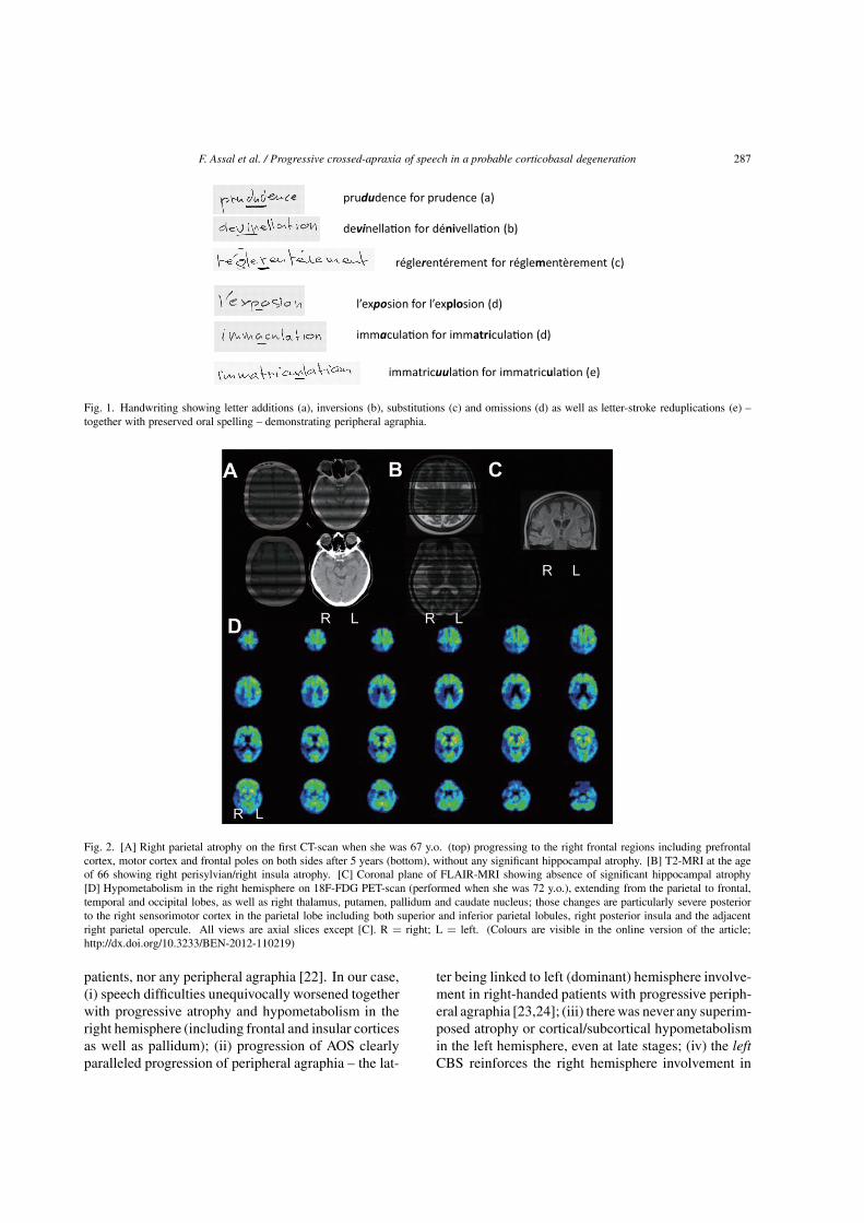
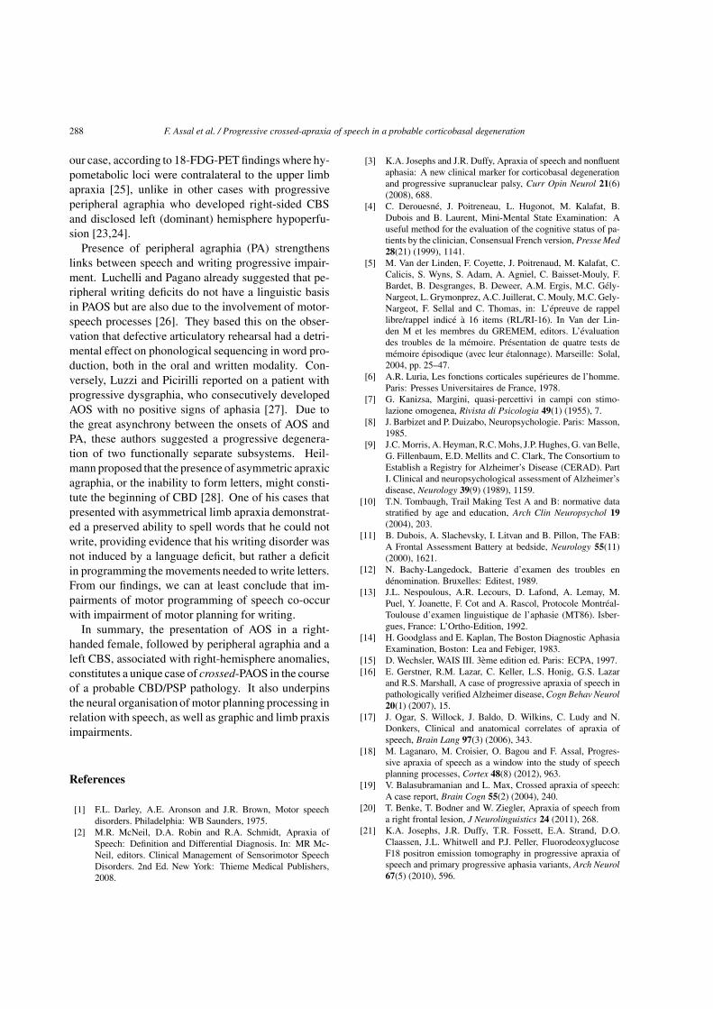
peripheral agraphia (preserved oral spelling and let-ter additions, inversions, substitutions and omissionsas well as letter-stroke reduplications in handwriting,see Fig. 1) then a corticobasal syndrome (CBS). Neu-ropsychological and extensive neurological examina-tion at the age of 72 years old indicated an unmistakableCBS of the left upper limb characterized by ideomotorapraxia, severe rigidity, bradykinesia, postural tremor,myoclonus, left-sided extinction on bilateral stimula-tion and alien hand sign (hand not recognized as herown hand). There was also a parkinsonian gait with lefthand levitation. Structural and functional neuroimag-ing disclosed atrophy and/or hypometabolism in theright hemisphere (see Fig. 2).
This longitudinal follow-up of almost 8 years ofa PAOS followed by peripheral agraphia then a left-sided corticobasal syndrome (CBS) constitutes, to ourknowledge, a rare case of crossed-PAOS. We first con-firm that PAOS can be isolated for several years, thenfollowed by a CBS and therefore constitute a mark-er of CBD/PSP spectrum [2]. PAOS associated withAlzheimer’s disease neuropathology has been reported
ISSN 0953-4180/12/$27.50 2012 – IOS Press and the authors. All rights reserved
286 F. Assal et al. / Progressive crossed-apraxia of speech in a probable corticobasal degeneration
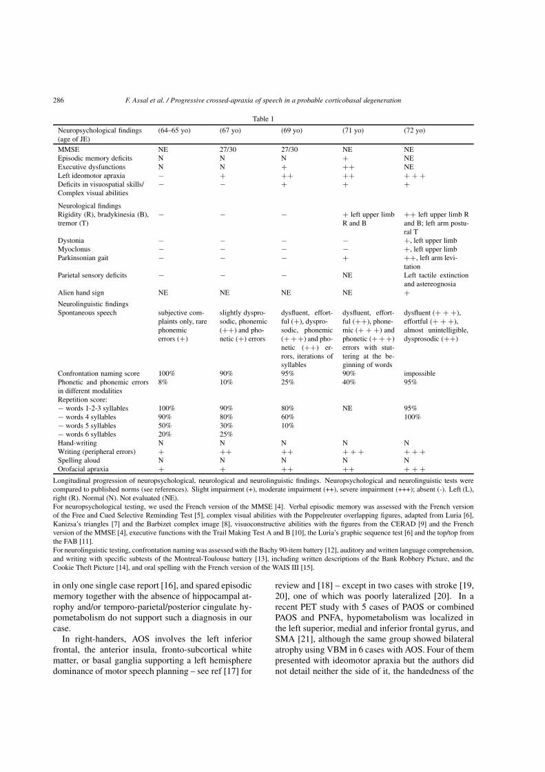
Table 1
Neuropsychological findings(age of JE)
(64–65 yo) (67 yo) (69 yo) (71 yo) (72 yo)
MMSE NE 27/30 27/30 NE NEEpisodic memory deficits N N N + NEExecutive dysfunctions N N + ++ NELeft ideomotor apraxia − + ++ ++ + + +Deficits in visuospatial skills/Complex visual abilities
− − + + +
Neurological findingsRigidity (R), bradykinesia (B),tremor (T)
− − − + left upper limbR and B
++ left upper limb Rand B; left arm postu-ral T
Dystonia − − − − +, left upper limbMyoclonus − − − − +, left upper limbParkinsonian gait − − − + ++, left arm levi-
tationParietal sensory deficits − − − NE Left tactile extinction
and astereognosiaAlien hand sign NE NE NE NE +
Neurolinguistic findingsSpontaneous speech subjective com-
plaints only, rarephonemicerrors (+)
slightly dyspro-sodic, phonemic(++) and pho-netic (+) errors
dysfluent, effort-ful (+), dyspro-sodic, phonemic(+++) and pho-netic (++) er-rors, iterations ofsyllables
dysfluent, effort-ful (++), phone-mic (+ + +) andphonetic (+++)errors with stut-tering at the be-ginning of words
dysfluent (+ + +),effortful (+ + +),almost unintelligible,dysprosodic (++)
Confrontation naming score 100% 90% 95% 90% impossiblePhonetic and phonemic errorsin different modalities
8% 10% 25% 40% 95%
Repetition score:− words 1-2-3 syllables 100% 90% 80% NE 95%− words 4 syllables 90% 80% 60% 100%− words 5 syllables 50% 30% 10%− words 6 syllables 20% 25%Hand-writing N N N N NWriting (peripheral errors) + ++ ++ + + + + + +Spelling aloud N N N N NOrofacial apraxia + + ++ ++ + + +
Longitudinal progression of neuropsychological, neurological and neurolinguistic findings. Neuropsychological and neurolinguistic tests werecompared to published norms (see references). Slight impairment (+), moderate impairment (++), severe impairment (+++); absent (-). Left (L),right (R). Normal (N). Not evaluated (NE).For neuropsychological testing, we used the French version of the MMSE [4]. Verbal episodic memory was assessed with the French versionof the Free and Cued Selective Reminding Test [5], complex visual abilities with the Poppelreuter overlapping figures, adapted from Luria [6],Kanizsa’s triangles [7] and the Barbizet complex image [8], visuoconstructive abilities with the figures from the CERAD [9] and the Frenchversion of the MMSE [4], executive functions with the Trail Making Test A and B [10], the Luria’s graphic sequence test [6] and the top/top fromthe FAB [11].For neurolinguistic testing, confrontation naming was assessed with the Bachy 90-item battery [12], auditory and written language comprehension,and writing with specific subtests of the Montreal-Toulouse battery [13], including written descriptions of the Bank Robbery Picture, and theCookie Theft Picture [14], and oral spelling with the French version of the WAIS III [15].
in only one single case report [16], and spared episodicmemory together with the absence of hippocampal at-rophy and/or temporo-parietal/posterior cingulate hy-pometabolism do not support such a diagnosis in ourcase.
In right-handers, AOS involves the left inferiorfrontal, the anterior insula, fronto-subcortical whitematter, or basal ganglia supporting a left hemispheredominance of motor speech planning – see ref [17] for
review and [18] – except in two cases with stroke [19,20], one of which was poorly lateralized [20]. In arecent PET study with 5 cases of PAOS or combinedPAOS and PNFA, hypometabolism was localized inthe left superior, medial and inferior frontal gyrus, andSMA [21], although the same group showed bilateralatrophy using VBM in 6 cases with AOS. Four of thempresented with ideomotor apraxia but the authors didnot detail neither the side of it, the handedness of the
F. Assal et al. / Progressive crossed-apraxia of speech in a probable corticobasal degeneration 287
Fig. 1. Handwriting showing letter additions (a), inversions (b), substitutions (c) and omissions (d) as well as letter-stroke reduplications (e) –together with preserved oral spelling – demonstrating peripheral agraphia.
Fig. 2. [A] Right parietal atrophy on the first CT-scan when she was 67 y.o. (top) progressing to the right frontal regions including prefrontalcortex, motor cortex and frontal poles on both sides after 5 years (bottom), without any significant hippocampal atrophy. [B] T2-MRI at the ageof 66 showing right perisylvian/right insula atrophy. [C] Coronal plane of FLAIR-MRI showing absence of significant hippocampal atrophy[D] Hypometabolism in the right hemisphere on 18F-FDG PET-scan (performed when she was 72 y.o.), extending from the parietal to frontal,temporal and occipital lobes, as well as right thalamus, putamen, pallidum and caudate nucleus; those changes are particularly severe posteriorto the right sensorimotor cortex in the parietal lobe including both superior and inferior parietal lobules, right posterior insula and the adjacentright parietal opercule. All views are axial slices except [C]. R = right; L = left. (Colours are visible in the online version of the article;http://dx.doi.org/10.3233/BEN-2012-110219)
patients, nor any peripheral agraphia [22]. In our case,(i) speech difficulties unequivocally worsened togetherwith progressive atrophy and hypometabolism in theright hemisphere (including frontal and insular corticesas well as pallidum); (ii) progression of AOS clearlyparalleled progression of peripheral agraphia – the lat-
ter being linked to left (dominant) hemisphere involve-ment in right-handed patients with progressive periph-eral agraphia [23,24]; (iii) therewas never any superim-posed atrophy or cortical/subcortical hypometabolismin the left hemisphere, even at late stages; (iv) the leftCBS reinforces the right hemisphere involvement in
288 F. Assal et al. / Progressive crossed-apraxia of speech in a probable corticobasal degeneration
our case, according to 18-FDG-PET findings where hy-pometabolic loci were contralateral to the upper limbapraxia [25], unlike in other cases with progressiveperipheral agraphia who developed right-sided CBSand disclosed left (dominant) hemisphere hypoperfu-sion [23,24].
Presence of peripheral agraphia (PA) strengthenslinks between speech and writing progressive impair-ment. Luchelli and Pagano already suggested that pe-ripheral writing deficits do not have a linguistic basisin PAOS but are also due to the involvement of motor-speech processes [26]. They based this on the obser-vation that defective articulatory rehearsal had a detri-mental effect on phonological sequencing in word pro-duction, both in the oral and written modality. Con-versely, Luzzi and Picirilli reported on a patient withprogressive dysgraphia, who consecutively developedAOS with no positive signs of aphasia [27]. Due tothe great asynchrony between the onsets of AOS andPA, these authors suggested a progressive degenera-tion of two functionally separate subsystems. Heil-mann proposed that the presence of asymmetric apraxicagraphia, or the inability to form letters, might consti-tute the beginning of CBD [28]. One of his cases thatpresented with asymmetrical limb apraxia demonstrat-ed a preserved ability to spell words that he could notwrite, providing evidence that his writing disorder wasnot induced by a language deficit, but rather a deficitin programming the movements needed to write letters.From our findings, we can at least conclude that im-pairments of motor programming of speech co-occurwith impairment of motor planning for writing.
In summary, the presentation of AOS in a right-handed female, followed by peripheral agraphia and aleft CBS, associated with right-hemisphere anomalies,constitutes a unique case of crossed-PAOS in the courseof a probable CBD/PSP pathology. It also underpinsthe neural organisation of motor planning processing inrelation with speech, as well as graphic and limb praxisimpairments.
References
[1] F.L. Darley, A.E. Aronson and J.R. Brown, Motor speechdisorders. Philadelphia: WB Saunders, 1975.
[2] M.R. McNeil, D.A. Robin and R.A. Schmidt, Apraxia ofSpeech: Definition and Differential Diagnosis. In: MR Mc-Neil, editors. Clinical Management of Sensorimotor SpeechDisorders. 2nd Ed. New York: Thieme Medical Publishers,2008.
[3] K.A. Josephs and J.R. Duffy, Apraxia of speech and nonfluentaphasia: A new clinical marker for corticobasal degenerationand progressive supranuclear palsy, Curr Opin Neurol 21(6)(2008), 688.
[4] C. Derouesne, J. Poitreneau, L. Hugonot, M. Kalafat, B.Dubois and B. Laurent, Mini-Mental State Examination: Auseful method for the evaluation of the cognitive status of pa-tients by the clinician, Consensual French version, Presse Med28(21) (1999), 1141.
[5] M. Van der Linden, F. Coyette, J. Poitrenaud, M. Kalafat, C.Calicis, S. Wyns, S. Adam, A. Agniel, C. Baisset-Mouly, F.Bardet, B. Desgranges, B. Deweer, A.M. Ergis, M.C. Gely-Nargeot, L.Grymonprez, A.C. Juillerat, C. Mouly, M.C. Gely-Nargeot, F. Sellal and C. Thomas, in: L’epreuve de rappellibre/rappel indice a 16 items (RL/RI-16). In Van der Lin-den M et les membres du GREMEM, editors. L’evaluationdes troubles de la memoire. Presentation de quatre tests dememoire episodique (avec leur etalonnage). Marseille: Solal,2004, pp. 25–47.
[6] A.R. Luria, Les fonctions corticales superieures de l’homme.Paris: Presses Universitaires de France, 1978.
[7] G. Kanizsa, Margini, quasi-percettivi in campi con stimo-lazione omogenea, Rivista di Psicologia 49(1) (1955), 7.
[8] J. Barbizet and P. Duizabo, Neuropsychologie. Paris: Masson,1985.
[9] J.C. Morris, A. Heyman, R.C. Mohs, J.P. Hughes, G. vanBelle,G. Fillenbaum, E.D. Mellits and C. Clark, The Consortium toEstablish a Registry for Alzheimer’s Disease (CERAD). PartI. Clinical and neuropsychological assessment of Alzheimer’sdisease, Neurology 39(9) (1989), 1159.
[10] T.N. Tombaugh, Trail Making Test A and B: normative datastratified by age and education, Arch Clin Neuropsychol 19(2004), 203.
[11] B. Dubois, A. Slachevsky, I. Litvan and B. Pillon, The FAB:A Frontal Assessment Battery at bedside, Neurology 55(11)(2000), 1621.
[12] N. Bachy-Langedock, Batterie d’examen des troubles endenomination. Bruxelles: Editest, 1989.
[13] J.L. Nespoulous, A.R. Lecours, D. Lafond, A. Lemay, M.Puel, Y. Joanette, F. Cot and A. Rascol, Protocole Montreal-Toulouse d’examen linguistique de l’aphasie (MT86). Isber-gues, France: L’Ortho-Edition, 1992.
[14] H. Goodglass and E. Kaplan, The Boston Diagnostic AphasiaExamination, Boston: Lea and Febiger, 1983.
[15] D. Wechsler, WAIS III. 3eme edition ed. Paris: ECPA, 1997.[16] E. Gerstner, R.M. Lazar, C. Keller, L.S. Honig, G.S. Lazar
and R.S. Marshall, A case of progressive apraxia of speech inpathologically verified Alzheimer disease, Cogn Behav Neurol20(1) (2007), 15.
[17] J. Ogar, S. Willock, J. Baldo, D. Wilkins, C. Ludy and N.Donkers, Clinical and anatomical correlates of apraxia ofspeech, Brain Lang 97(3) (2006), 343.
[18] M. Laganaro, M. Croisier, O. Bagou and F. Assal, Progres-sive apraxia of speech as a window into the study of speechplanning processes, Cortex 48(8) (2012), 963.
[19] V. Balasubramanian and L. Max, Crossed apraxia of speech:A case report, Brain Cogn 55(2) (2004), 240.
[20] T. Benke, T. Bodner and W. Ziegler, Apraxia of speech froma right frontal lesion, J Neurolinguistics 24 (2011), 268.
[21] K.A. Josephs, J.R. Duffy, T.R. Fossett, E.A. Strand, D.O.Claassen, J.L. Whitwell and P.J. Peller, FluorodeoxyglucoseF18 positron emission tomography in progressive apraxia ofspeech and primary progressive aphasia variants, Arch Neurol67(5) (2010), 596.
F. Assal et al. / Progressive crossed-apraxia of speech in a probable corticobasal degeneration 289
[22] K.A. Josephs, J.R. Duffy, E.A. Strand, J.L. Whitwell, K.F.Layton, J.E. Parisi, M.F. Hauser, R.J. Witte, B.F. Boeve,D.S. Knopman, D.W. Dickson, C.R. Jack, Jr. and R.C. Pe-tersen, Clinicopathological and imaging correlates of progres-sive aphasia and apraxia of speech, Brain 129(6) (2006), 1385.
[23] M. Grossman, D.J. Libon, X.S. Ding, B. Cloud, J. Jaggi, D.Morrison, J. Greenberg, A. Alavi and M. Reivich, Progressiveperipheral agraphia, Neurocase 7(4) (2001), 339.
[24] T. Fukui and E. Lee, Progressive agraphia can be the harbingerof degenerative dementia, Brain Lang 104(3) (2008), 201.
[25] P. Peigneux, E. Salmon, G. Garraux, S. Laureys, S. Willems,K. Dujardin, C. Degueldre, C. Lemaire, A. Luxen, G. Moo-
nen, G. Franck, A. Destee and M. Van der Linden, Neural andcognitive bases of upper limb apraxia in corticobasal degen-eration, Neurology 57(7) (2001), 1259.
[26] F. Lucchelli and C. Papagno, Is slowly progressive anarthriaa “pure” motor-speech disorder? Evidence from writing per-formance, Neurocase 11(3) (2005), 234.
[27] S. Luzzi and M. Piccirilli, Slowly progressive pure dysgraphiawith late apraxia of speech: a further variant of the focalcerebral degeneration, Brain Lang 87(3) (2003), 355.
[28] K.M. Heilman, A. Coenen and B. Kluger, Progressive asym-metric apraxic agraphia, Cogn Behav Neurol 21(1) (2008),14.

Submit your manuscripts athttp://www.hindawi.com
Stem CellsInternational
Hindawi Publishing Corporationhttp://www.hindawi.com Volume 2014
Hindawi Publishing Corporationhttp://www.hindawi.com Volume 2014
MEDIATORSINFLAMMATION
of
Hindawi Publishing Corporationhttp://www.hindawi.com Volume 2014
Behavioural Neurology
EndocrinologyInternational Journal of
Hindawi Publishing Corporationhttp://www.hindawi.com Volume 2014
Hindawi Publishing Corporationhttp://www.hindawi.com Volume 2014
Disease Markers
Hindawi Publishing Corporationhttp://www.hindawi.com Volume 2014
BioMed Research International
OncologyJournal of
Hindawi Publishing Corporationhttp://www.hindawi.com Volume 2014
Hindawi Publishing Corporationhttp://www.hindawi.com Volume 2014
Oxidative Medicine and Cellular Longevity
Hindawi Publishing Corporationhttp://www.hindawi.com Volume 2014
PPAR Research
The Scientific World JournalHindawi Publishing Corporation http://www.hindawi.com Volume 2014
Immunology ResearchHindawi Publishing Corporationhttp://www.hindawi.com Volume 2014
Journal of
ObesityJournal of
Hindawi Publishing Corporationhttp://www.hindawi.com Volume 2014
Hindawi Publishing Corporationhttp://www.hindawi.com Volume 2014
Computational and Mathematical Methods in Medicine
OphthalmologyJournal of
Hindawi Publishing Corporationhttp://www.hindawi.com Volume 2014
Diabetes ResearchJournal of
Hindawi Publishing Corporationhttp://www.hindawi.com Volume 2014
Hindawi Publishing Corporationhttp://www.hindawi.com Volume 2014
Research and TreatmentAIDS
Hindawi Publishing Corporationhttp://www.hindawi.com Volume 2014
Gastroenterology Research and Practice
Hindawi Publishing Corporationhttp://www.hindawi.com Volume 2014
Parkinson’s Disease
Evidence-Based Complementary and Alternative Medicine
Volume 2014Hindawi Publishing Corporationhttp://www.hindawi.com