Embed Size (px)
Citation preview
PRODUCTION AND IMPAIRED REGULATION
OF NEUTROPHIL EXTRACELLULAR TRAPS
FOLLOWING SEVERE THERMAL INJURY,
IMPLICATIONS FOR SEPSIS AND MULTIPLE
ORGAN FAILURE
Robert Johnathon Dinsdale
A thesis submitted to the University of Birmingham for the
degree of
DOCTOR OF PHILOSOPHY
Supervisors
Dr Paul Harrison
Professor Steve Watson
Institute of Inflammation and Ageing
College of Medical and Dental Sciences
University of Birmingham
August 2017
University of Birmingham Research Archive
e-theses repository This unpublished thesis/dissertation is copyright of the author and/or third parties. The intellectual property rights of the author or third parties in respect of this work are as defined by The Copyright Designs and Patents Act 1988 or as modified by any successor legislation. Any use made of information contained in this thesis/dissertation must be in accordance with that legislation and must be properly acknowledged. Further distribution or reproduction in any format is prohibited without the permission of the copyright holder.
Abstract
Advancements in burn care have improved immediate outcome, however, the
prevalence of sepsis and multiple organ failure (MOF) remain significant. Although
well characterised the mechanisms responsible for the pathogenesis of MOF and
increased propensity to infection are poorly understood. Neutrophil extracellular
traps (NETs) provide protection against invading pathogens but also contribute to
thrombosis.
Sepsis is required for NET generation following severe thermal injury.
Quantification of circulating NET biomarkers shows good discriminatory power for
diagnosis of sepsis. Interestingly, neutrophils isolated from 24 patients with severe
thermal injuries, ≥ 15% total body surface area, had a significantly reduced ability
to form NETs ex vivo, potentially mediated by phenotypical changes of neutrophils
and inhibitory effects of formyl peptides.
This thesis identified a major biological mechanism driving MOF after severe
thermal injury, namely the compromise to the actin scavenging system which
leads to reduced DNAse activity and a build-up of circulating DNA. Preliminary
analysis suggests that DNAse activity can be restored by prehospital use of fresh
frozen plasma following major trauma. Thus, administration of blood products or
manipulation of the actin scavenging system is a potential therapeutic target.
This thesis has identified a number of novel mechanisms responsible for the
regulation of NETs following severe thermal injuries and their implications for
sepsis and MOF.

Acknowledgements
Firstly, I must thank my supervisor Dr Paul Harrison for all support,
encouragement and opportunities provided. I will be forever grateful for his
supervision and friendship (even if he is a Blue).
I am grateful to Professor Steve Watson, Professor Janet Lord and Professor
Naiem Moiemen for their advice, discussion and mentorship. I would like to thank
all of the Lord Group members for making my time within the lab enjoyable. A
special thank you must go to Dr Jon Hazeldine and Dr Peter Hampson who have
been amazing colleagues and good friends. Although at times we disagree on
everything from food to physical activity, I am truly grateful for advice, jokes and
weekends smashing golf balls into the rough.
I would like to thank my funders the Scar Free Foundation, burns clinical team and
burns nurses at the Queen Elizabeth Hospital and all collaborating sites for their
contribution to this work. Special thank you must go to Amy Bamford and Dr
Khaled Altarrah for their help throughout my studies. Most importantly, thank you
to all patients and families who have been part of this work. Without you this could
never have happened.
To my friends, The Booze Hound Gang, Trauma Boyz and The Marangas, thank
you for always being there, the scrambled eggs, nights out in fancy dress and
unconditional friendship you have all shown me. A special thank you must be
extended to Georgia Walton who is responsible for my love of junk food and late
nights. You have always been and continue to be a source of laughter,
companionship and unwavering support. Here is to kicking Louise out of her seat.

Thank you to my greatest teacher the black dog, your intermittent barking has
fuelled my determination and passion.
I would like to thank my whole family for always being there for me and supporting
me throughout this whole process. Dad and lovely mummy, thank you for all of the
support, love and advice throughout everything. Thank you for everything you
have ever given me from the countless opportunities to the reassurance on top of
the landing. I hope this work makes you proud. Grace, Maggie and Elsie, thank
you for your companionship. To the members of my family who will never be able
to read this, I hope it would have made you proud.
Thank you.

You’ll never walk alone
Disclosures
All work presented in this thesis was performed as part of The Scientific
Investigation of the Biological Pathways Following Thermal Injury in Adults and
Children (SIFTI Study) and is therefore part of a collaboration between a number
of investigators. Where applicable, credit has been given to persons responsible
for data generation or collaboration. Importantly, all data presented in this thesis
was analysed and presented by Robert J Dinsdale.
All patients recruited within Camp Bastion, Afghanistan, were recruited by
Professor Midwinter, Dr Kirkman, Professor Woolley, Dr Watts and Dr Dalle
Lucca. Again, all laboratory experiments and data analysis was performed by
Robert J Dinsdale.
Publications arising from this thesis
1. Dinsdale RJ, Devi A, Hampson P, Wearn CM, Bamford AL, Hazeldine J,
Bishop J, Ahmed S, Watson C, Lord JM, Moiemen N, Harrison P. Changes in
novel haematological parameters following thermal injury. A prospective
observational cohort study. Scientific Reports (2017).
2. Hazeldine J, Naumann DN, Toman E, Davies D, Su Z, Hampson P, Dinsdale
RJ, Crombie N, Harrison P, Belli A and Lord JM. Immune dysfunction is
present within an hour of injury, and is associated with multiple organ
dysfunction syndrome: A prospective observational study and detailed
immunological analysis. Submitted to Plos Medicine (2017).
3. Hampson P*, Dinsdale RJ*, Wearn CJ, Hazeldine J, Moiemen N, Lord J,
Harrison P. (2016) Neutrophil dysfunction, immature granulocytes and cell-free
DNA are early biomarkers of sepsis in burn injured patients: A prospective
observational cohort study. Annals of Surgery (2016). *Joint authorship.
Publications under submission
1. Dinsdale RJ , Hazeldine J, Al Tarrah K, Hampson P, Devi A, Ermogenous C,
Bamford AL, Bishop J, Watts S, Kirkman E, Dalle Lucca JJ, Midwinter M,
Woolley T, Lord JM, Moiemen N, Harrison P. Reduced DNAse activity in burns
patients is associated with compromise to the blood-based actin scavenging
system and increased risk of multiple organ failure. Submitted to Blood (2017).
2. Hazeldine J*, Dinsdale RJ*, Harrison P, Lord JM. Mitochondrial-derived
damage-associated molecular patterns induce neutrophil dysfunction and
promote their survival in vitro. Submitted to Scientific Reports (2017). *Joint
authorship.
3. Shantsila E, Ponomaryov T, Dinsdale RJ, Harrison P, Lip G, Brill A. Plasma
cell-free DNA levels are elevated in patients with acute ischemic heart failure.
4. Montague SJ, Dinsdale RJ, Poulter NS, Andrews RK, Hampson P, Wearn
CM, Lee C, Bishop J, Bamford AL, Iqbal T, Moiemen N, Oury C, Gardiner EE,
Harrison P and Watson SP. Fibrin induced GPVI shedding is a potential
explanation behind elevated soluble GPVI observed in patients with thermal
injury and intensive care unit patients. Submitted to Blood (2017).
5. Naumann DN, Hazeldine J, Dinsdale RJ, Midwinter MM, Harrison P,
Hutchings SD, Lord JM. Endotheliopathy is associated with higher
concentrations of cell-free DNA following major trauma: a prospective
observational study. Submitted to Plos One (2017).
Published abstracts
1. Dinsdale RJ, Hampson P, Hazeldine J, Wearn CM, Brill A, Lord JM,
Moiemen NS, Harrison P. Neutrophil Extracellular Trap (NET) formation in patients
with post-burn sepsis. Journal of Thrombosis and Haemostasis 13, 140-140.
2. Montague S, Dinsdale RJ, Gardiner E, Andrews R, Wearn CM, Bishop J,
Bamford A, Nash GB, Moiemen NS, Harrison P, Watson SP. GPVI and CLEC-
2/Podoplanin axis as potential biomarkers of platelet activation in thermal injury.
Journal of Thrombosis and Haemostasis 13, 650-650.
3. Brill A, Shantsila E, Ponomaryov T, Dinsdale RJ, Harrison P, Lip G (2015).
Plasma cell-free DNA levels are elevated in patients with acute ischemic heart
failure partially due to neutrophil extracellular trap formation. Journal of
Thrombosis and Haemostasis 13, 379-380.
Conferences
1. American Burn Association Annual Meeting, Boston, March 2017
a. Dinsdale RJ, Hazeldine J, Altarrah K, Hampson P, Devi A, Wearn CM,
Bamford AL, Lord JM, Moiemen N, Harrison P. Dysregulation of
neutrophil extracellular traps (NETs) following thermal injury. (oral)
2. British Burns Association, Birmingham, May 2016
a. Dinsdale RJ, Hampson P, Hazeldine J, Wearn C, Lord JM, Moiemen
NS, Harrison P. Neutrophil Extracellular Trap (NET) formation in
patients with burn injury (oral).
b. Dinsdale RJ, Hampson P, Wearn CM, Hazeldine J, Lord JM, Moiemen
NS, Harrison P. Thermal Injury Results in the Generation of Pro-
Coagulant Microvesicles (oral).
3. Midland Academy Conference, Leicester, April 2016
a. Dinsdale RJ, Hampson P, Hazeldine J, Wearn C, Brill A, Lord JM,
Moiemen NS, Harrison P. Neutrophil Extracellular Trap (NET) formation
in patients with post-burn sepsis (oral). Second Prize for Best Oral
Presentations.
4. British Society of Haemostasis and Thrombosis Conference, London,
November 2015
a. Dinsdale RJ, Hampson P, Hazeldine J, Wearn CM, Brill A, Lord JM,
Moiemen NS, Harrison P. Neutrophil Extracellular Trap (NET) formation
in patients with post-burn sepsis (oral). Selected for the Young
Scientists in Training Symposium (Top Scoring Abstract).
5. International Society of Haemostasis and Thrombosis, Toronto, June
2015
a. Dinsdale RJ, Hampson P, Hazeldine J, Wearn CM, Brill A, Lord JM,
Moiemen NS, Harrison P. Neutrophil Extracellular Trap (NET) formation
in patients with post-burn sepsis (oral).
6. British Burns Association, Birmingham, May 2015
a. Dinsdale RJ, Hampson P, Hazeldine J, Wearn CM, Lord JM, Moiemen
NS, Harrison P. Clinical applications of novel blood counter parameters
in burn injury (poster).
7. American Burn Association Annual Meeting, Chicago, April 2015
a. Dinsdale RJ, Hampson P, Hazeldine J, Wearn CM, Lord JM, Moiemen
NS, Harrison P. Neutrophil Extracellular Trap Formation in Patients with
Post-Burn Sepsis. (poster).
8. International Society of Burn Injury Conference, Sydney, October 2014
a. Dinsdale R.J, Hampson P, Wearn CM, Hazeldine J, Ahmed S, Watson
C, Lord JM, Moieman N, Harrison P. Novel Blood Counter Parameters
in Burn Injury (oral).
b. Dinsdale RJ, Hampson P, Hazeldine J, Wearn CM, Lord JM, Moiemen
NS, Harrison P. Evidence of Neutrophil Extracellular Trap (NET)
formation in patients with post-burn sepsis (poster).
Awards
1. Top rated poster. International Society of Haemostasis and Thrombosis. July
2017.
2. Best oral presentation. British Burns Society. May 2017.
3. Second place in best oral presentation. Midlands Academy, April 2016.

4. Selected for the Young Scientists in Training Symposium (Top Scoring
Abstract). British Society of Haemostasis and Thrombosis Conference,
November 2015.
Table of Contents Chapter 1: Introduction ...................................................................................................... 2
1.1 Trauma and thermal injury ....................................................................................... 2
1.2 Pathophysiology of thermal injury ............................................................................ 3
1.3 Burn shock .............................................................................................................. 5
1.4 Improvements in critical care management of patients with thermal injuries ............ 6
1.4.1 Fluid resuscitation ............................................................................................. 6
1.4.2 Inhalation injury ................................................................................................. 7
1.4.3 Burn wound care ............................................................................................... 7
1.5 Systemic inflammatory response syndrome following thermal injury ........................ 8
1.6 Damage associated molecular patterns ................................................................. 11
1.7 Nosocomial infections, sepsis and organ dysfunction following thermal injury ....... 11
1.8 Novel biomarkers of sepsis in thermally injured patients ........................................ 14
1.8.1 Procalcitonin ................................................................................................... 17
1.8.2 C-reactive protein ............................................................................................ 19
1.8.3 Pro- and anti-inflammatory cytokines .............................................................. 19
1.8.4 Novel haematological parameters ................................................................... 20
1.8.5 Cell-free deoxyribonucleic acid ....................................................................... 23
1.9 Neutrophils ............................................................................................................ 24
1.9.1 General background ....................................................................................... 24
1.9.2 Chemotaxis and transmigration ....................................................................... 27
1.9.3 Phagocytosis .................................................................................................. 28
1.9.4 Apoptosis ........................................................................................................ 29
1.9.5 Anti-microbial actions ...................................................................................... 29
1.10 Neutrophil dysfunction post traumatic injury ........................................................ 30
1.10.1 Maturity and phenotype of neutrophils post traumatic injury .......................... 30
1.10.2 Functional dysregulation of neutrophils post traumatic injury ......................... 35
1.11 Neutrophil extracellular traps ............................................................................... 38
1.11.1 Structure and function ................................................................................... 38
1.11.2 Vital and suicidal NETosis ............................................................................. 38
1.11.3 Citrullinated histone H3 and reactive oxygen species generation .................. 43
1.11.4 Histones, DNA and nucleosomes .................................................................. 43
1.11.5 Autophagy and neutrophil extracellular traps ................................................ 46
1.12 NETs and traumatic injury ................................................................................... 46
1.13 In Vivo host defensive functions of neutrophil extracellular traps ......................... 47
1.14 Excessive NETosis, thrombosis and host tissue damage .................................... 48
1.15 Degradation of neutrophil extracellular traps ....................................................... 53
1.16 Actin .................................................................................................................... 54
1.17 Actin Scavenging System .................................................................................... 54
1.17.1 Gelsolin ......................................................................................................... 55
1.17.2 Vitamin D binding protein .............................................................................. 56
1.18 Targeting neutrophil extracellular traps ................................................................ 59
1.19 Aims and hypothesis ........................................................................................... 61
Chapter 2: Materials and Methods ................................................................................... 64
2.1 Scientific Investigation of Biological Pathways Following Thermal Injury Study ..... 64
2.1.1 Ethical approval .............................................................................................. 64
2.1.2 Study cohort .................................................................................................... 64
2.1.3 Blood sampling ............................................................................................... 65
2.1.4 Preparation of platelet free plasma ................................................................. 65
2.1.5 Preparation of plasma ..................................................................................... 66
2.1.6 Preparation of serum ...................................................................................... 66
2.1.7 Clinical diagnosis of sepsis and multiple organ failure ..................................... 66
2.2 Polytrauma patient group and study design ........................................................... 70
2.2.1 Study approval and consenting for patients injured in explosions .................... 71
2.2.2 Plasma preparation from patients injured in explosions................................... 71
2.3 Whole blood analysis using the Sysmex XN-1000 analyser................................... 74
2.4 Measurement of neutrophil reactive oxygen species generation in whole blood .... 74
2.5 Isolation of neutrophils from whole blood ............................................................... 75
2.6 Generation of neutrophil extracellular traps ........................................................... 76
2.7 Quantification of neutrophil extracellular traps ....................................................... 77
2.8 Visualisation of neutrophil extracellular traps by fluorescent microscopy ............... 78
2.9 Paraformaldehyde preparation .............................................................................. 79
2.10 Human promyelocytic leukaemia cell culture ....................................................... 79
2.11 K562 cell culture .................................................................................................. 80
2.12 Western Blot Analysis .......................................................................................... 80
2.12.1 Western blot protocol for Cit H3 .................................................................... 81
2.12.2 Western blot protocol for detection of actin ................................................... 82
2.13 Detection of cell-free DNA in plasma and serum samples using an in house
fluorometric assay ....................................................................................................... 82
2.14 Detection of cell-free DNA in plasma and serum samples using a commercial CE
marked assay .............................................................................................................. 83
2.15 Isolation of nuclear and mitochondrial DNA ......................................................... 83
2.16 Polymerase chain reaction for the quantification of plasma nuclear DNA and
mitochondrial DNA levels ............................................................................................ 84
2.17 Quantification of DNAse activity in serum samples .............................................. 85
2.18 Visualisation of neutrophil extracellular traps degradation by fluorescent
microscopy .................................................................................................................. 86
2.19 In Vitro inhibition of DNAse activity in serum samples ......................................... 87
2.20 Enzyme linked immunosorbent assay ................................................................. 87
2.20.1 Quantification of deoxyribonuclease (DNAse 1) by ELISA ............................ 87
2.20.2 Quantification of vitamin D binding protein by ELISA .................................... 89
2.20.3 Quantification of human gelsolin by ELISA .................................................... 89
2.21 Isolation of nuclei and mitochondria from K562 cells ........................................... 91
2.22 mtDAMPs effects on neutrophil function and phenotype ...................................... 91
2.22.1 Inhibition of neutrophil extracellular traps formation with isolated mtDAMPs . 92
2.22.2 Neutrophil transmigration following stimulation with mtDAMPs ..................... 93
2.22.3 Assessment of neutrophil phenotype following stimulation with mtDAMPs ....... 94
2.22.4 Activation of ERK1/2 following stimulation with mtDAMPs ............................ 94
2.22.5 Reactive oxygen species generation following stimulation with mtDAMPs .... 95
2.23 Statistical analysis ............................................................................................... 96
2.23.1 Analysis of in vitro experiments ..................................................................... 96
2.23.2 Analysis of data generated from patients with thermal injuries ...................... 96
2.23.3 Logistic regression analyses of potential biomarkers of sepsis ...................... 97
2.23.4 Analysis of data generated from patients with polytrauma ............................. 97
Chapter 3: Neutrophil extracellular trap release following thermal injury .................... 99
3.1 Introduction ......................................................................................................... 100
3.1.1 Aims .............................................................................................................. 101
3.2 Results ................................................................................................................ 102
3.2.1 Study cohort .................................................................................................. 102
3.2.2 Assessment of neutrophil extracellular trap release in healthy controls ......... 106
3.2.3 Detection of circulating cell-free DNA in healthy controls .............................. 109
3.2.4 Analysis of plasma cell-free DNA levels following thermal injury ................... 109
3.2.5 Diagnostic use of cell-free DNA in septic patients ......................................... 115
3.2.6 Longitudinal analysis of nuclear and mitochondrial DNA ............................... 115
3.2.7 Neutrophil extracellular trap formation following thermal injury ...................... 119
3.2.8 Ex vivo NETosis ............................................................................................ 121
3.2.9 Reactive oxygen species generation of neutrophils following thermal injury .. 123
3.2.10 Neutrophil function is reduced to a greater degree in septic patients ........... 123
3.2.11 White blood cell kinetics .............................................................................. 125
3.2.12 Release of immature granulocytes post-burn injury is associated with reduced
reactive oxygen species generation ....................................................................... 130
3.2.13 Immature granulocyte percentage is associated with reduced ex vivo
neutrophil extracellular trap generation .................................................................. 130
3.2.14 Thermal injury results in a change in extended neutrophil parameters ........ 133
3.2.15 Extended neutrophil parameters correlate with reduced neutrophil function 133
3.2.16 mtDAMPs activate neutrophils .................................................................... 139
3.2.17 mtDAMPs pre-treatment results in impaired neutrophil extracellular trap
generation upon secondary stimulation .................................................................. 141
3.2.18 Effect of mtDAMP pre-treatment on PMA-induced neutrophil reactive oxygen
species production and citrullination of histone H3 ................................................. 142
3.2.19 Exposure to mtDAMPs suppresses neutrophil migration to LTB4 ............... 146
3.3 Discussion ........................................................................................................... 148
Chapter 4: Reduced DNAse activity in burns patients is associated with
compromise to the blood-based actin scavenging system and increased risk of
multiple organ failure ..................................................................................................... 171
4.1 Introduction ......................................................................................................... 172
4.1.1 Aims .............................................................................................................. 173
4.2 Results ................................................................................................................ 174
4.2.1 Study cohort .................................................................................................. 174
4.2.2 Nuclear DNA levels are elevated in patients with multiple organ failure ........ 177
4.2.3 Nuclear DNA levels are elevated in patients who develop sepsis .................. 177
4.2.4 Patients with thermal injury have reduced DNAse activity ............................. 182
4.2.5 DNAse activity is lower in patients who develop multiple organ failure or sepsis
.............................................................................................................................. 182
4.2.6 DNAse-1 antigen levels are elevated following thermal injury ....................... 183
4.2.7 Actin is released following thermal injury ....................................................... 190
4.2.8 Actin inhibits DNAse activity in vitro .............................................................. 190
4.2.9 Thermal injury results in a decrease in circulating vitamin d binding protein and
gelsolin levels ........................................................................................................ 193
4.2.10 Thermal injury results in a decrease in circulating gelsolin levels ................ 193
4.2.11 In vitro actin inhibition of DNAse activity can be corrected by gelsolin ......... 194
4.2.12 Patient demographics for patients with severe injuries caused by explosions
.............................................................................................................................. 199
4.2.13 Fresh frozen plasma increases gelsolin levels and DNAse activity following
severe injury caused by explosion but has no effect on vitamin d binding protein
levels ..................................................................................................................... 199
4.3 Discussion ........................................................................................................... 205
Chapter 5: General Discussion ...................................................................................... 219
5.1 Limitations ........................................................................................................... 220
5.2 Future Work ........................................................................................................ 221
5.2.1 In vivo characterisation of NETosis ............................................................... 224
5.2.2 DNAse isoforms and their functions .............................................................. 227
5.2.3 Targeting the build-up of toxic and pro-thrombotic DNA following thermal injury
.............................................................................................................................. 228
5.2.4 DNAse as a therapy ...................................................................................... 229
5.2.5 Inhibition of PAD4 ......................................................................................... 230
5.2.6 Modulation of actin scavenging system ......................................................... 231
5.2.7 Targeting reduced neutrophil function following thermal injury ...................... 233
5.2.8 Granulocyte colony-stimulating factor and neutrophil maturity ....................... 233
5.2.9 Resolvins ...................................................................................................... 235
5.2.10 Haemoperfusion therapy ............................................................................. 236
List of Illustrations
Figure 1.1 Described model of burn zones according Page 4
to adequate and inadequate resuscitation as described
by Jackson.
Figure 1.2 Novel model of simultaneous SIRS and CARs Page 10
following major trauma.
Figure 1.3 Traditional and novel functions of neutrophils. Page 26
Figure 1.4 Administration of LPS results in distinct Page 34
phenotypical changes in circulating neutrophils.
Figure 1.5 Proposed novel mechanisms of NET release: Page 41
suicidal and vital NETosis.
Figure 1.6 Nucleosome structure. Page 45
Figure 1.7 NETs form a physical barrier and impair blood Page 52
flow through a microfluidic device.
Figure 1.8 Actin scavenging system in a healthy individual. Page 58
Figure 2.1 SIFTI Study blood sampling timeline. Page 69
Figure 3.1 CONSORT diagram showing the sub-classification Page 103
and breakdown of numbers of study subjects.
Figure 3.2 Stimulation of neutrophils with 25 nM PMA Page 107
results in robust NET formation.
Figure 3.3 Visualisation of ex vivo NET generation in response Page 108
to 25nM PMA or 100 ng/ml LPS by neutrophils isolated from a
healthy control by fluorescence microscopy.
Figure 3.4 Levels of cfDNA are significantly higher in serum Page 111
Compared to plasma samples from healthy individuals.
Figure 3.5 cfDNA is elevated post thermal injury and Page 112
elevated in septic patients.
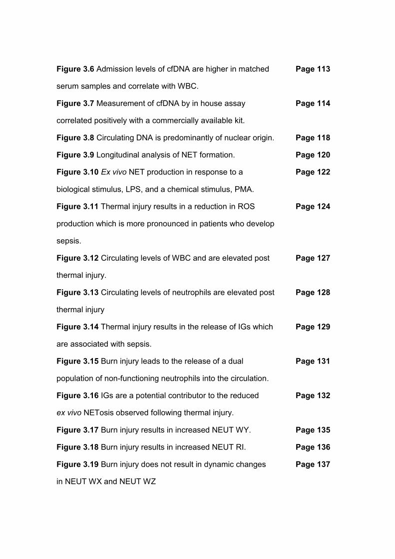
Figure 3.6 Admission levels of cfDNA are higher in matched Page 113
serum samples and correlate with WBC.
Figure 3.7 Measurement of cfDNA by in house assay Page 114
correlated positively with a commercially available kit.
Figure 3.8 Circulating DNA is predominantly of nuclear origin. Page 118
Figure 3.9 Longitudinal analysis of NET formation. Page 120
Figure 3.10 Ex vivo NET production in response to a Page 122
biological stimulus, LPS, and a chemical stimulus, PMA.
Figure 3.11 Thermal injury results in a reduction in ROS Page 124
production which is more pronounced in patients who develop
sepsis.
Figure 3.12 Circulating levels of WBC and are elevated post Page 127
thermal injury.
Figure 3.13 Circulating levels of neutrophils are elevated post Page 128
thermal injury
Figure 3.14 Thermal injury results in the release of IGs which Page 129
are associated with sepsis.
Figure 3.15 Burn injury leads to the release of a dual Page 131
population of non-functioning neutrophils into the circulation.
Figure 3.16 IGs are a potential contributor to the reduced Page 132
ex vivo NETosis observed following thermal injury.
Figure 3.17 Burn injury results in increased NEUT WY. Page 135
Figure 3.18 Burn injury results in increased NEUT RI. Page 136
Figure 3.19 Burn injury does not result in dynamic changes Page 137
in NEUT WX and NEUT WZ
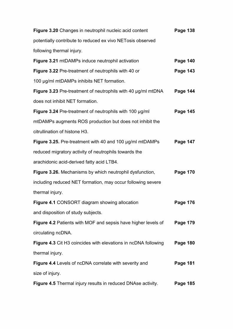
Figure 3.20 Changes in neutrophil nucleic acid content Page 138
potentially contribute to reduced ex vivo NETosis observed
following thermal injury.
Figure 3.21 mtDAMPs induce neutrophil activation Page 140
Figure 3.22 Pre-treatment of neutrophils with 40 or Page 143
100 µg/ml mtDAMPs inhibits NET formation.
Figure 3.23 Pre-treatment of neutrophils with 40 µg/ml mtDNA Page 144
does not inhibit NET formation.
Figure 3.24 Pre-treatment of neutrophils with 100 µg/ml Page 145
mtDAMPs augments ROS production but does not inhibit the
citrullination of histone H3.
Figure 3.25. Pre-treatment with 40 and 100 µg/ml mtDAMPs Page 147
reduced migratory activity of neutrophils towards the
arachidonic acid-derived fatty acid LTB4.
Figure 3.26. Mechanisms by which neutrophil dysfunction, Page 170
including reduced NET formation, may occur following severe
thermal injury.
Figure 4.1 CONSORT diagram showing allocation Page 176
and disposition of study subjects.
Figure 4.2 Patients with MOF and sepsis have higher levels of Page 179
circulating ncDNA.
Figure 4.3 Cit H3 coincides with elevations in ncDNA following Page 180
thermal injury.
Figure 4.4 Levels of ncDNA correlate with severity and Page 181
size of injury.
Figure 4.5 Thermal injury results in reduced DNAse activity. Page 185
Figure 4.6 DNAse activity within 24 hours of injury does not Page 185
correlate with severity and size of injury.
Figure 4.7 Thermal injury results in reduced DNAse activity Page 186
visualised by fluorescence microscopy.
Figure 4.8 Patients with MOF exhibited lower DNAse activity Page 187
Figure 4.9 Patients who developed sepsis have lower DNAse Page 188
activity
Figure 4.10 Thermal injury significantly increases DNAse 1 Page 189
antigen levels.
Figure 4.11 Thermal injury causes the release of circulating Page 191
actin.
Figure 4.12 Actin inhibits DNAse activity in vitro. Page 192
Figure 4.13 Levels of VDBP are reduced following thermal Page 195
injury.
Figure 4.14 Levels of GSN are reduced following thermal injury. Page 196
Figure 4.15 GSN and VDBP levels are not significantly Page 197
different between septic and non-septic patients.
Figure 4.16 GSN recovers actin inhibition of DNAse activity Page 198
in vitro.
Figure 4.17 Severe injury caused by explosion caused a Page 202
significant reduction in circulating GSN levels compared to
healthy controls.
Figure 4.18 Patients who do not receive blood products before Page 203
admission to hospital have significantly lower DNAse and GSN
levels compared to healthy individuals
Figure 4.19 Blood products increase circulating GSN levels and Page 204
protects against inhibition of DNAse activity.
Figure 4.20 Hypothesis model: The link between severe thermal Page 214
injury and pathogenesis of MOF through disruption of the
actin scavenging system.
Figure 5.1 Hypothesis model: Linking initial injury to secondary Page 223
complications following severe thermal injury.
List of Tables
Table 1.1 The ABA Consensus conference 2007 criteria for Page 16
sepsis diagnosis
Table 2.1 Inclusion and exclusion criteria for patients recruited Page 68
to the SIFTI trial.
Table 2.2 Trauma team activation criteria according to Clinical Page 72
Guidelines for Operations.
Table 2.3 Inclusion and Exclusion Criteria for with Page 73
severe injuries caused by explosions.
Table 3.1 Patient Demographics (Chapter 3). Page 104
Table 3.2 Information on first septic episode. Page 105
Table 3.3 Discriminatory power of cfDNA for predicting sepsis Page 117
at different time points was assessed through AUROC and
95% confidence intervals.
Table 4.1 Patient demographics (Chapter 4). Page 175
Table 4.2 Patient demographics for patients with severe injuries Page 201
caused by explosions (Chapter 4).
Abbreviations
ABA American Burn Association
ABSI Abbreviated burn severity index
ANOVA Analysis of variance
APC Allophycocyanin
ATCC American Type Culture Collection
ATP Adenosine triphosphate
AUROC Area under the receiver operator curve
BSA Bovine serum albumin
CARS Compensatory anti-inflammatory response syndrome
cfDNA Cell-free deoxyribonucleic acid
CGD Chronic granulomatous disease
Cit H3 Citrullinated histone H3
CLP Cecal ligation puncture
CRP C-reactive protein
DAMPs Damage associated molecular patterns
DIC Disseminated intravascular coagulation
DMSO Dimethyl sulfoxide
DNA Deoxyribonucleic acid
DNAse Deoxyribonuclease
DVT Deep vein thrombosis
E.Coli Escherichia coli
EDTA Ethylenediaminetetraacetic acid
FFP Frozen fresh plasma
FITC Fluorescein isothiocyanate
FRCs Fragmented red cells
FT Full thickness
GCS Glasgow Coma Scale
G-CSF Granulocyte-colony stimulating factor
GM-CSF Granulocyte macrophage colony-stimulating factor
GSN Gelsolin
HBSS+ Hank’s Balanced Salt Solutions containing Mg2+ and Ca2+
HL-60 Human promyelocytic leukaemia cells
HMGB1 High mobility group box 1 protein
HRP Horseradish peroxidase
IAP Inhibitor of apoptosis protein
ICU Intensive care unit
IG Immature granulocyte
IGs Immature granulocytes
IL- Interleukin
IL-4 Interleukin-4
IL-6 Interleukin-6
IL-7 Interleukin-7
IL-8 Interleukin-8
IL-10 Interleukin-10
IL-13 Interleukin-13
ISS Injury severity score
IQR Interquartile range
LPS Lipopolysaccharide
LTB4 Leukotriene B4
MDROs Multi-drug resistant organisms
MERT Medical Emergency Response Team
MFI Median fluorescence intensity
MODS Multiple organ dysfunction
MOF Multiple organ failure
mtDAMPs Mitochondrial derived damage associated molecular patterns
mtDNA Mitochondrial deoxyribonucleic acid
mTOR Mammalian target of rapamycin
fMLP N-Formylmethionine-leucyl-phenylalanine
NADPH Nicotinamide adenine dinucleotide phosphate-oxidase
ncDNA Nuclear deoxyribonucleic acid
NET(s) Neutrophil extracellular trap(s)
NEUT GI Neutrophil granularity index
NEUT RI Neutrophil reactivity index
NISS New injury severity score
OD Optical density
PAD4 Peptidylarginine deiminase 4
PBS Phosphate-buffered saline
PCR Polymerase chain reaction
PCT Procalcitonin
PE R-phycoerythrin
PFP Platelet free plasma
PKC Protein kinase C
PLT-F Platelet fluorescence count
PLT-I Platelet impedance count
PLT-O Platelet optical count
PMA Phorbol 12-phorbol myristate 13-acetate
PMX-DHP Polymixin-B immobilized fibre cartridge
PT Partial thickness
PVDF Polyvinylidene fluoride
QC Quality control
rBaux Revised Baux score
RT Room temperature
RvD2 Resolvin D2
SEM Standard error of the mean
SIFTI Scientific investigation of biological pathways following thermal injury
SIRS Systemic inflammatory response syndrome
SLE Systemic lupus erythematosus
SOFA Sequential Organ Failure Assessment
SDS Sodium dodecyl sulphate
SDS-PAGE Sodium dodecyl sulphate polyacrylamide gel electrophoresis
sp Species
STAT3 Signal transducer and activator of transcription 3
TBS Tris Buffered Saline
TBSA Total body surface area
TBST Tris Buffered Saline containing 4% Tween-20
TLR Toll-like receptor
TMB Tetramethylbenzidine
UK United Kingdom
VDBP Vitamin D binding protein
WBC White blood cell

1
Chapter 1
Introduction
2
Introduction
1.1 Trauma and thermal injury
There are approximately 5-8 million fatalities each year as a result of injuries and
trauma (1, 2). As a group alone, fatalities account for more deaths than malaria,
tuberculosis and HIV/AIDS. Trauma affects all age groups in developed and
developing countries globally and associated mortalities account for around 10%
of the deaths worldwide (1). Traumatic injury is an umbrella term which
encompasses many forms of injury, including road traffic accidents, wars, falls and
thermal injuries (1). For many years thermal injury and trauma were used
interchangeably. However, investigation of thermal injury alone is now required.
Thermal injuries are a common and debilitating form of traumatic injury which is
associated with considerable morbidity and mortality. They are among the most
expensive forms of traumatic injury due to the consequent length of
hospitalisation, rehabilitation and wound management (3). In 2015 it was reported
that approximately 13,000 injuries which required hospital attention occur every
year in England and Wales. Approximately 58% of these patients were admitted to
hospital for further medical care. Throughout this thesis the term ‘thermal injury’
will refer to injuries caused by flame, contact and scald injuries, the most
predominant causes in England and Wales (4).
Morbidity and mortality post thermal injury is heavily influenced by a plethora of
elements. Firstly, severe thermal injuries which are larger in size and depth have
an increased risk of nosocomial infection and thus an increased mortality rate (5).
More recently, studies have shown that age has a considerable effect on the
immune system (6-9) and thus ageing is a crucial confounding factor in the clinical
3
outcome. Elderly patients have a higher mortality rate, longer length of hospital
stays and more complicated outcomes (10). This may be driven by the immune
dysfunction and immunosenescence associated with ageing (7-9), or may also be
affected by pre-existing medical and premorbid conditions which are common in
elderly patients. Finally, patients with inhalation injuries have increased mortality
rates despite increased understanding and advancements within respiratory care
(11). A combination of these confounding factors in patients presenting with
severe thermal injuries increases the associated mortality and morbidity rate.
1.2 Pathophysiology of thermal injury
The pathophysiology of burn is dependent upon the degree of initial injury and
complications the patients incur. Severe thermal injury results in immediate local
and systemic responses which are extremely complex and critical to outcomes
post injury.
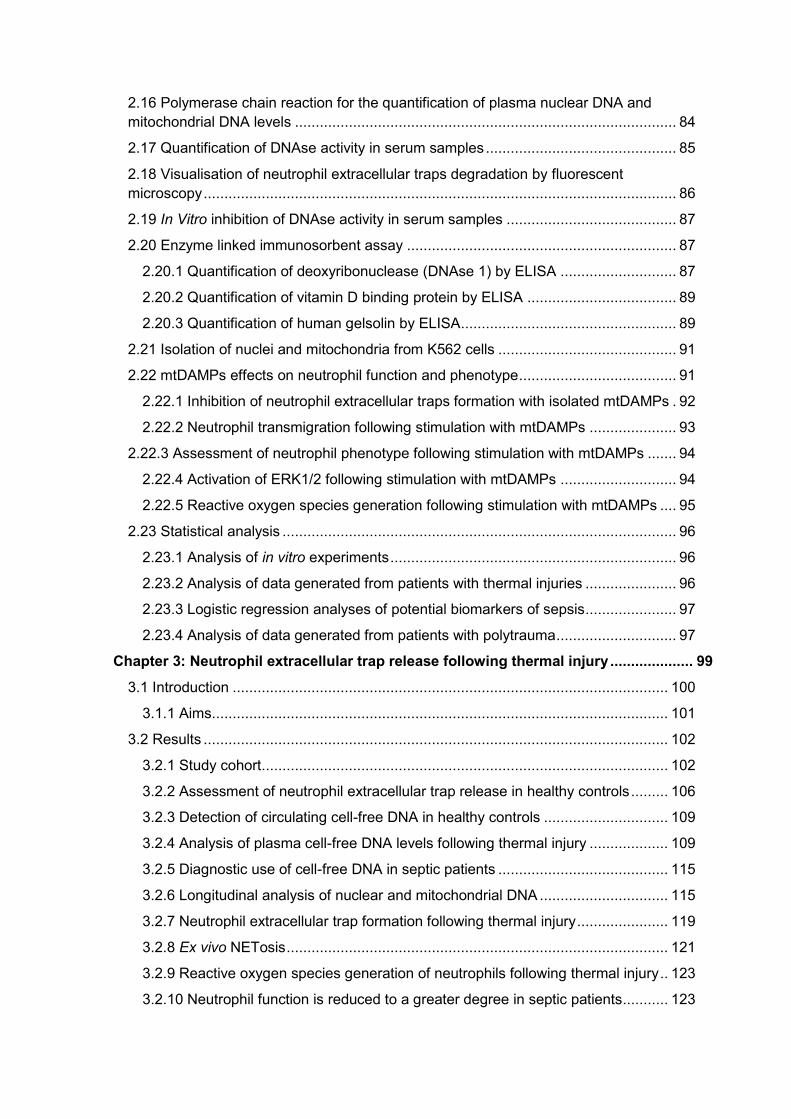
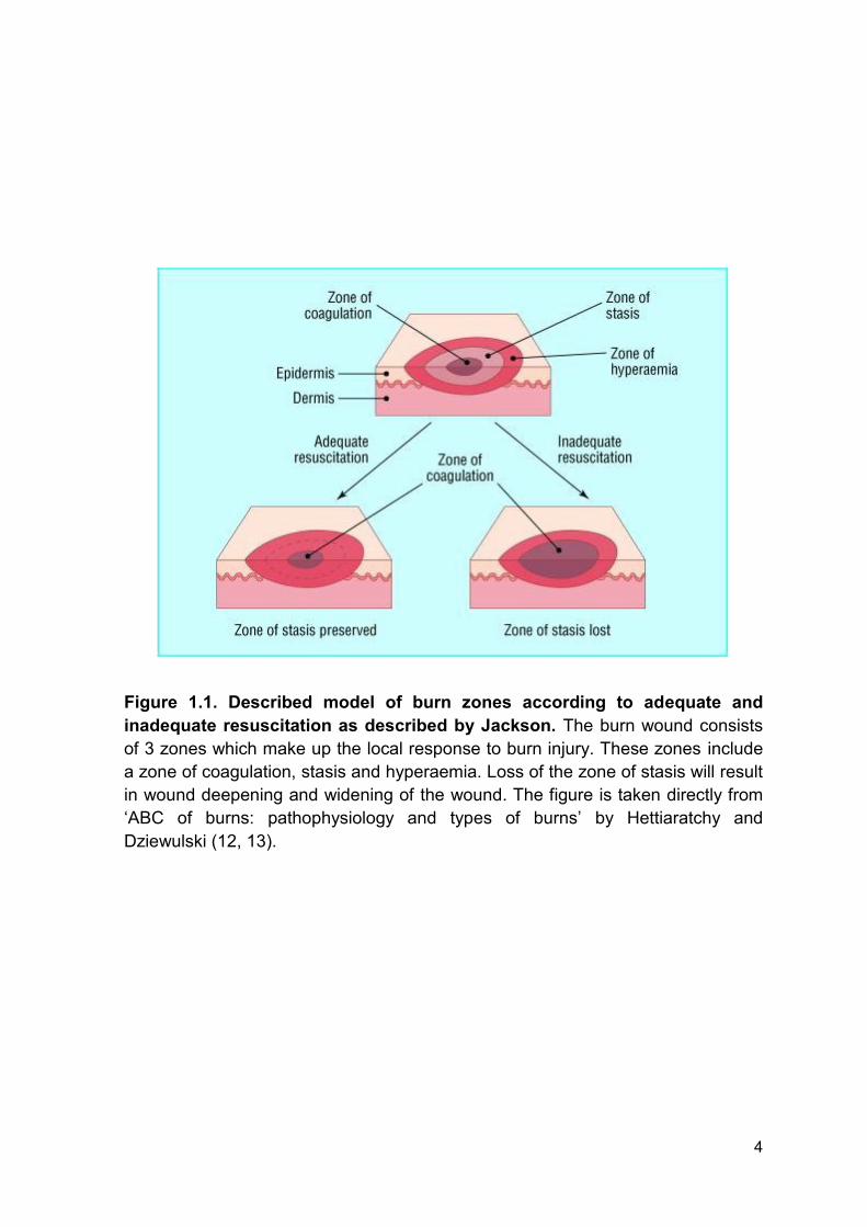
Jackson described 3 zones which make up the local response to burn injury (12).
These include the zones of coagulation, stasis and hyperaemia (Figure 1.1). The
zone of coagulation occurs at the point of maximal tissue damage and is
comprised of necrotic tissue where there is irreversible tissue loss. This is
surrounded by the zone of stasis which is characterised by decreased tissue
perfusion. The tissue in this area can be rescued however secondary
complications such as infection or ischemia may lead to further tissue necrosis.
The zone of hyperaemia has increased tissue perfusion and without secondary
complications, this will recover. The three zones of burn injury are subject to
changes and loss of the zone of stasis will result in wound deepening and
widening (12, 13).
4
Figure 1.1. Described model of burn zones according to adequate and
inadequate resuscitation as described by Jackson. The burn wound consists
of 3 zones which make up the local response to burn injury. These zones include
a zone of coagulation, stasis and hyperaemia. Loss of the zone of stasis will result
in wound deepening and widening of the wound. The figure is taken directly from
‘ABC of burns: pathophysiology and types of burns’ by Hettiaratchy and
Dziewulski (12, 13).
5
1.3 Burn shock
Following thermal injury correct diagnosis, treatment and clinical practice are
essential for positive outcomes. Smaller thermal injuries are commonly managed
by outpatient care. However, clinical treatment of severe injuries is much more
complicated and require hospitalisation, timely and accurate resuscitation,
nutritional support, and early surgical management of burn wounds. All of which is
directed at reducing morbidity and mortality (14).
Shock is a life-threatening condition characterised by reduced oxygen delivery and
circulatory failure. Shock is commonly diagnosed by a combination of
hypoperfusion and low or declining blood pressure (15). Burn shock is an umbrella
term consisting of hypovolemic and septic shock which occurs following a severe
thermal injury. Hypovolemic shock occurs immediately following severe thermal
injury and without adequate fluid resuscitation it will lead to mortality. Septic shock
is the major driver of delayed mortality associated with infection and is
characterised by shock mediated by an infectious stimuli coupled with evident
organ failure (15). The pathogenesis of burn shock is driven by the alteration in
almost all components that control both fluid and protein loss from vascular space
(16). Immediately following injury the microvasculature loses its vessel wall
integrity which results in the loss of proteins to the interstitium (17) coupled with a
decrease in the intra-vasculature colloid osmotic pressure resulting in loss of fluid,
electrolytes and further proteins from the vasculature system into the interstitium
(17). Clinically this manifests itself as hypovolemia, haemoconcentration, oedema,
reduced urine output and significant cardiovascular dysfunction (16). Timely and
6
appropriate treatment, namely fluid resuscitation, is required to prevent and
manage immediate burn shock and prevent poor outcomes post injury.
1.4 Improvements in critical care management of patients with thermal
injuries
Improvements in the immediate care of burn wounds have been made over the
past four decades which have dramatically improved survival rates of patients. It is
proposed that improvements in survival are a direct result of increased scientific
understanding of burn injury and the immediate pathophysiology (18).
In 2014, Jackson and colleagues reported data of revised estimates of mortality
from the Birmingham Burn Centre spanning from 2001 to 2010 and included 4577
patients. Although there was an increase in admissions, the overall mortality of the
cohort decreased by approximately 3-fold compared to the previous decade. The
authors concluded that improvements in outcome were multifactorial and included;
improved prehospital care, rapid referral to burns institution, early burn wound
excision and closure, and improved understanding and management of inhalation.
Furthermore, the group proposes that further research into inhalation injury, fluid
resuscitation, burn care in vulnerable populations (i.e. elderly patients), and skin
substitutes are warranted if further improvements in outcome are to be met (19).
1.4.1 Fluid resuscitation
Fundamental to the care of patients with burn injuries is accurate and timely fluid
resuscitation. Without intervention and fluid replacement in burns greater than 15-
20% total body surface area (TBSA) burn shock will occur (20). A delay of just 2
hours of accurate fluid resuscitation results in a significant increase in adverse
7
outcomes (21). Therefore the primary aim of fluid resuscitation is to prevent the
development of burn shock and to restore homeostasis during the immediate
dysregulated cellular and hormonal response (22).
1.4.2 Inhalation injury
Advancements in the management of respiratory failure caused by inhalation
injury and smoke inhalation have been fundamental in improving immediate
outcome post thermal injury (23-25). The pathogenesis of respiratory failure is
multifactorial and immediate airway management is essential (13, 26, 27).
Inhalation injuries are further sub categorised in primary and secondary which
differ by causes, progression and management. Primary inhalation injury is
caused by direct damage to the respiratory system caused by the thermal stimulus
which causes cellular damage, activation of inflammatory cells and oedema. This
can cause blockage of the airways and consequently respiratory failure, a major
clinical problem (28). Secondary injury to the respiratory system is initiated by the
inflammatory response and is amplified by complications, including sepsis and
multiple organ dysfunction (MODS) or multiple organ failure (MOF) (27, 29). Both
of which require management and treatment to ensure a positive outcome (11,
13).
1.4.3 Burn wound care
Burn wounds provide a major source of inflammatory mediators which orchestrate
the propagation and initiation of inflammation following thermal injury (30). Timely
and correct management of burn wounds is essential to prevent an uncontrolled
systemic inflammatory response, reduce infection risk, improve healing and
8
reduce the incidence of secondary complications (31, 32). Advancements in
immediate cleaning, debridement and excision of wounds have been critical in
preventing rapid colonisation of wounds, secondary complications and reducing
mortality rates (20, 32).
Early excision and skin grafting generally occur between 24 hours and 7 days
following injury to attenuate the inflammatory response, reduce rejection rate and
reduce colonisation rate of wounds (20, 32). If wounds become colonised they are
commonly treated by early debridement, wound excision and application of topical
dressings. Novel topical dressings aim to promote wound healing, reduce scarring
and identify/treat colonised wounds more efficiently (33, 34). Topical antimicrobial
agents, which are commonly used, prevent graft loss and burn wound infection
(32). Advancements in the understanding of the inflammatory response post
thermal injury, reduced infection rates of wounds and novel topical agents have
been pivotal to the improvements of burn wound care.
1.5 Systemic inflammatory response syndrome following thermal injury
Severe thermal injury results in loss of the natural defensive barrier and rapid
onset of systemic inflammatory response syndrome (SIRS) (35). SIRS is an
inflammatory response which can be initiated by a plethora of mechanisms
including injury, surgery and ischemia. It clinically manifests as elevations of body
temperature, heart rate, respiratory rate and abnormal white blood cell count or
phenotypical changes (presence of band cells) (36). Whilst similar, sepsis is a
SIRS response with a documented infection. Therefore, the difference between
SIRS and sepsis is the stimuli responsible for its initiation. SIRS is initiated by a
9
sterile stimulus, e.g. trauma, and sepsis is mediated by an infectious stimulus, e.g.
bacteria.
Many of the resultant consequences following thermal injury are mediated by the
rapid release of pro-inflammatory mediators and immune suppression (37). This
hyper-inflammatory state coupled with an increased propensity to infection often
results in sepsis which is the major cause of mortality following burn injury (38).
Systemic responses to burn injury include profound changes in the cardiovascular,
respiratory, metabolic, immunological and haematological systems (37).
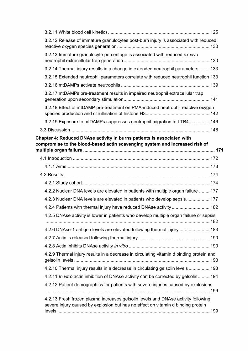
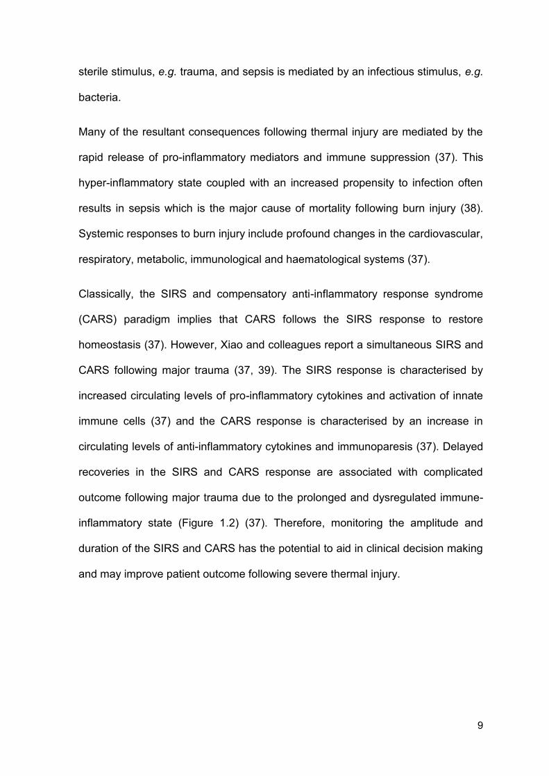
Classically, the SIRS and compensatory anti-inflammatory response syndrome
(CARS) paradigm implies that CARS follows the SIRS response to restore
homeostasis (37). However, Xiao and colleagues report a simultaneous SIRS and
CARS following major trauma (37, 39). The SIRS response is characterised by
increased circulating levels of pro-inflammatory cytokines and activation of innate
immune cells (37) and the CARS response is characterised by an increase in
circulating levels of anti-inflammatory cytokines and immunoparesis (37). Delayed
recoveries in the SIRS and CARS response are associated with complicated
outcome following major trauma due to the prolonged and dysregulated immune-
inflammatory state (Figure 1.2) (37). Therefore, monitoring the amplitude and
duration of the SIRS and CARS has the potential to aid in clinical decision making
and may improve patient outcome following severe thermal injury.
10
Figure 1.2. Novel model of simultaneous SIRS and CARs following major
trauma. (A) The traditional paradigm of immediate SIRS response followed by
CARS following major injury. A second hit can lead to secondary complications
and delayed amplification of the inflammatory response. (B) Model proposed by
Xiao and colleagues showing an immediate and simultaneous SIRS and CARS
response. The amplitude of response is associated with adverse clinical
outcomes. Figure is taken directly from ‘A genomic storm in critically injured
humans’ by Xiao et al (37).
11
1.6 Damage associated molecular patterns
Damage associated molecular patterns (DAMPs) are a collection of endogenous
cytosolic, mitochondrial and nuclear derived proteins and DNA which are detected
in high circulating concentrations following major trauma (40, 41). Elevation in
circulating levels of DAMPs is associated with secondary complications, tissue
damage and mortality (41-44). Namely, administration of mitochondrial DNA
(mtDNA) to mice resulted in organ damage which was attributed to direct
activation of neutrophils, subsequent neutrophil degranulation and local cytokine
production (42, 43). Furthermore, in prospective observational cohort studies
elevated levels of DAMPs are associated with MOF and mortality (41, 44). This
highlights the potential interaction(s) between circulating levels of DAMPs and
secondary complications following major trauma.
Exposure of monocytes to mitochondrial derived DAMPs (mtDAMPs) results in a
tolerance state in which these cells are resistant to further endotoxin stimulation
(45). It is proposed that this induced tolerance may underpin an increased
propensity to infection. Although described in monocytes, it has yet to be
examined if DAMPs induce this tolerance phenotype in neutrophils. Furthermore, it
is yet to be established if this occurs following severe thermal injury and potential
mechanistic role in the increased incidence of infection.
1.7 Nosocomial infections, sepsis and organ dysfunction following thermal
injury
Mortality rates associated with severe thermal injuries continue to steadily
decrease. This is in part caused by a multidisciplinary approach, improved burn
wound management, advancements in fluid resuscitation and identification of
12
comorbid conditions e.g. inhalation injury (31). Although immediate mortality rates
have improved, the incidence and associated mortality of secondary complications
has increased (31). Upon detection of secondary complications rapid, accurate
and timely diagnosis is required.
Sepsis is a life threatening condition characterised by an imbalance in both the
immune and haemostatic systems. Sepsis is associated with cardiac dysfunction
and acute respiratory distress syndrome which are synonymous with MOF (46).
Although the accurate and timely diagnosis is crucial for determining patient
outcomes, diagnosis remains difficult as criteria are insensitive and non-specific as
they are masked by the ongoing SIRS (47). Therefore, further research into the
pathophysiology, immune pathways and novel diagnosis criteria are required.
Whilst this is not a new concept, the prevalence of sepsis and its associated
mortality remain high. A systematic review, which included 9 studies reporting
survival data for thermal injuries, reported the prevalence of sepsis was between
8-42.5% in patients with burns. This was associated with a mortality rate (28-65%)
that exceeded that of sepsis-related deaths for traumatic injury (2.4-19.6%) or
critical care (21-53%) (38). In addition, the diagnosis of sepsis in burns patients
represents a major challenge, as many of the classical diagnostic biomarkers of
sepsis are masked by the ongoing SIRS that occurs following major burn injury
(47). Understanding the mechanisms underlying the progression and increased
propensity to sepsis is therefore critical if novel biomarkers for the accurate
prediction and/or diagnosis of sepsis in patients with thermal injuries are to be
found and novel therapeutic targets for its prevention and/or treatment identified.
13
Without intervention, sepsis can induce MOF which can be characterised by mild
through to complete and irreversible damage to vital organs (48). Whilst MOF
remains well defined the mechanisms responsible for its initiation and progression
remain poorly understood. Of such, infection and immediate traumatic injury are
attributed as initiators of MOF.
Disseminated intravascular coagulation (DIC) is characterised by abnormal and
uncontrolled coagulation and is a common and contributing factor to MODS and
MOF through thrombus formation and occlusion of blood vessels (49-52). DIC can
be classified into acute and chronic depending upon stimulating factors, length
and pathology (53-55). Trauma and thermal injury induce acute DIC which is a
consumptive coagulopathy state characterised by excessive thrombin generation
(56). DIC also causes the formation of schistocytes, due to high shear forces on
red blood cells in occluded vessels (57), which appear to have biological functions
in addition to beings potential diagnostic markers of DIC (58). However, D-dimer, a
soluble biomarker, is commonly used to diagnose DIC through evaluation of
recent thrombus formation.
MOF remains the leading cause of delayed mortality following major trauma and is
characterised by tissue hypoxia, tissue damage and organ dysfunction (59). In
2009, Nhu Nguyen reported that in 117 severely burn patients (≥40% TBSA) that
the incidence of MODS was 45% and associated with severity of injury, sepsis,
increased length of stay and importantly increased mortality rate (59). Although it
is well recognised that damage to vital organs occurs during MODS/MOF and is
associated with adverse outcome the mechanisms mediating this are unknown or
poorly understood. Therefore, understanding the mechanisms driving the
pathogenesis of MOF and organ damage has the potential to improve patient
14
outcome following thermal injury through early identification of patients at risk and
novel clinical intervention.
1.8 Novel biomarkers of sepsis in thermally injured patients
Sepsis remains a significant healthcare problem in patients following thermal injury
(38). Without timely diagnosis and treatment patients are at high risk of MOF and
consequently mortality. The Surviving Sepsis Campaign is a joint collaboration of
the Society of Critical Care Medicine and the European Society of Intensive Care
Medicine committed to reducing mortality from severe sepsis and septic shock
worldwide (60). It is advised that antibiotics should be administered to a patient
within 1 hour of clinical diagnosis of sepsis and with each hour delay in the
administration there is an associated significant increase in mortality (61-63).
A major limitation of post-burn sepsis research is the absence of an accurate
definition of sepsis as many of the classically used diagnosis criteria are
nonspecific and masked by the ongoing SIRS response (64). In 2007, a collective
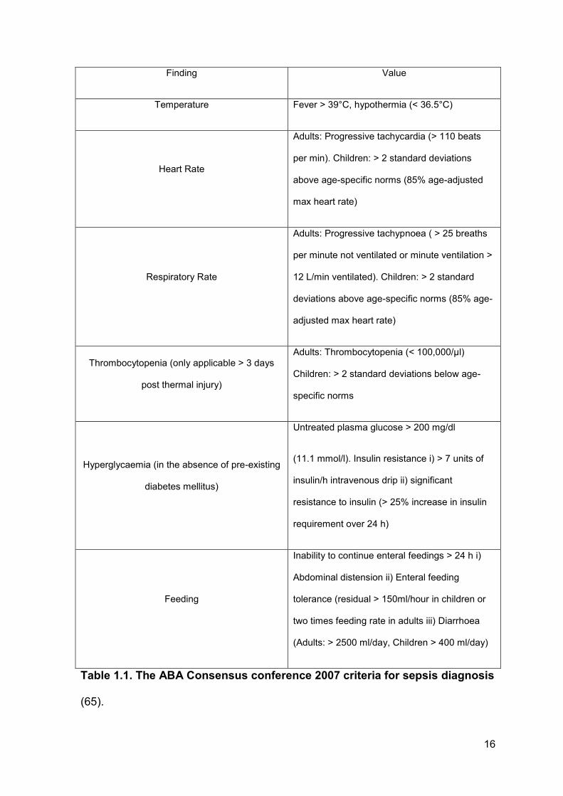
group of burns experts defined a clinical scoring criterion for the accurate
diagnosis of sepsis in patients with thermal injuries (65). According to the
American Burn Association (ABA), sepsis is diagnosed when three or more of the
following criteria listed in Table 1.1 are satisfied in addition to the clinical response
to antibiotics, positive blood or wound culture.
More recently a new sepsis diagnosis criterion was described in The Journal of the
American Medical Association (66). This study aimed to replace previous
diagnostic criteria with a new system termed the quick sequential organ failure
assessment (qSOFA) score. Whilst this tool is an excellent bedside screening tool
for identifying patients at risk of adverse outcomes it is non-specific in patients with
15
severe thermal injuries. This is due in part to the high incidence of intubation in
patients with thermal injuries and the incorporation of the Glasgow coma scale
(GCS) into the qSOFA score which is not part of the ABA scoring criteria. Thus,
diagnosis of sepsis was made using the ABA scoring criteria for all analysis within
this thesis.
16
Finding Value
Temperature Fever > 39°C, hypothermia (< 36.5°C)
Heart Rate
Adults: Progressive tachycardia (> 110 beats
per min). Children: > 2 standard deviations
above age-specific norms (85% age-adjusted
max heart rate)
Respiratory Rate
Adults: Progressive tachypnoea ( > 25 breaths
per minute not ventilated or minute ventilation >
12 L/min ventilated). Children: > 2 standard
deviations above age-specific norms (85% age-
adjusted max heart rate)
Thrombocytopenia (only applicable > 3 days
post thermal injury)
Adults: Thrombocytopenia (< 100,000/μl)
Children: > 2 standard deviations below age-
specific norms
Hyperglycaemia (in the absence of pre-existing
diabetes mellitus)
Untreated plasma glucose > 200 mg/dl
(11.1 mmol/l). Insulin resistance i) > 7 units of
insulin/h intravenous drip ii) significant
resistance to insulin (> 25% increase in insulin
requirement over 24 h)
Feeding
Inability to continue enteral feedings > 24 h i)
Abdominal distension ii) Enteral feeding
tolerance (residual > 150ml/hour in children or
two times feeding rate in adults iii) Diarrhoea
(Adults: > 2500 ml/day, Children > 400 ml/day)
Table 1.1. The ABA Consensus conference 2007 criteria for sepsis diagnosis
(65).
17
There is also the increasing problem of antibiotic resistance, a phenomenon in
which bacteria can mutate making them more invulnerable to antibiotics. This
resistance makes it harder to treat patients, increases medical costs, increases
hospital length of stay and increases mortality (67). Van Langeveld et al
investigated how multi-drug resistant organisms (MDROs) affect survival, hospital
length of stay and secondary complications in patients with thermal injuries.
Although the group found that MDROs had no effect on mortality they conclude
that their findings suggest infections caused by MDROs are associated with a
greater number of surgical procedures, longer duration of mechanical ventilation,
more antibiotic days, and longer hospitalisation (68).
Therefore, novel and accurate biomarkers are required for earlier and accurate
identification of patients at risk or who have developed sepsis following thermal
injury. This may initiate a change in clinical practice and antibiotic stewardship
which is required to overcome the growing burden of MDROs. There are a number
of novel biomarkers which may be of clinical utility in patients with thermal injuries.
However, many markers still lack sensitivity and specificity and therefore further
research is required.
1.8.1 Procalcitonin
Procalcitonin (PCT) is a naturally occurring 116-amino acid prohormone of
calcitonin produced in the thyroid, lungs and intestine (69). PCT is found in
extremely low quantities in healthy individuals but challenges such as endotoxin
insult cause a rapid upregulation of PCT production (70). Hence, the kinetics of
PCT is favourable for the development of a biomarker of sepsis. A number of
studies have reported the potential use of PCT in differentiating between
18
infectious and non-infectious systemic inflammation (71, 72). As such, PCT
remains one of the most promising biomarkers in identifying infection/sepsis (73,
74).
Current studies aim to elucidate the potential role of PCT in antibiotic stewardship,
patient stratification, diagnosis and prognostic utility following burn injury. In 2016,
Cabral and colleagues published a meta-analysis of the current understanding of
PCT as a biomarker in patients with thermal injuries. The meta-analysis included
14 studies published in both adults and children with thermal injuries (75). Pooled
area under the receiver operator curve (AUROC) for PCT in diagnosis of sepsis
was 0.83 (95% confidence intervals = 0.76, 0.90). However, substantial
differences in AUROC values are reported for individual studies with values
ranging from 0.55 – 0.98 (76, 77). Additionally, differences in the ‘cut off or
threshold value’ for distinguishing sepsis exists. Many of these differences are
attributed to the variances in methodology, heterogeneity between patient
populations and timing of samples. Nevertheless, Cabral and colleagues conclude
that PCT should be regarded as a strong diagnostic marker of sepsis in burns
patients and further work is required to standardise PCT measurements (75).
One limitation of PCT is the reported elevation that occurs postoperatively in the
absence of infectious stimuli (78). As patients with severe thermal injuries require
surgical intervention, caution must be taken when interpreting PCT data in these
patients. Furthermore, a number of studies have reported negative results for PCT
in the diagnosis of sepsis following thermal injury (47, 76). Therefore controversy
exists regarding PCT as a biomarker of sepsis in this patient population. Thus it is
suggested that further studies are required to explore PCT diagnostic utility
longitudinally compared to existing biomarkers of sepsis.
19
1.8.2 C-reactive protein
C-reactive protein (CRP) is a liver derived protein which can activate the classical
complement pathway (79). CRP has been studied as a biomarker of infection,
sepsis and mortality in a number of disease pathologies (80, 81). Of note, in a
study of 43 patients admitted to intensive care unit (ICU) with burn injury, CRP did
not correlate with severity of sepsis when PCT did (77). This further confirmed
earlier work reporting the increased diagnostic utility of PCT compared to CRP in
diagnosing sepsis (82). Furthermore, studies have questioned the ability of CRP to
distinguish between inflammation caused by infectious and non-infectious stimuli
(83, 84). As CRP is released in response to IL-6 (82), which is present in high
quantities in patients with thermal injuries (85), it is thought this may cause the
non-specific elevation in CRP and false positives reported in the context of
infection. Hence, CRP should be regarded as a marker of inflammation in patients
with thermal injuries rather than a potential biomarker of infection due to its non-
specific nature.
1.8.3 Pro- and anti-inflammatory cytokines
Thermal injury results in the simultaneous release of pro and anti-inflammatory
cytokines which are associated with severity of injury and secondary complications
(86-88). As the exaggerated immune response is proposed to underlie the
increased incidence of secondary complications post burn injury, groups have
quantified levels of pro- and anti-inflammatory cytokines and correlated levels to
clinical outcomes (89, 90).
20
In a study which included 28 children with severe burn injuries, elevated levels of a
panel of cytokines showed positive discriminatory power to identify patients who
were likely to succumb to their injuries. In this study, interleukin (IL)-4, IL-6, IL-7,
IL-10, and IL-13 were abnormal within the first 7 days post injury in patients who
did not survive their injuries when compared to patients who did survive. With
abnormalities in IL-6, IL-7 and IL-10 displaying the highest predictive power for
mortality (89). Furthermore, in a study of 468 paediatric burn patients, IL-8 serum
levels were increased in patients who developed with MOF, succumbed to their
injuries and, interestingly, sepsis (91). Hence, IL-8 may be a potential novel
biomarker to monitor infection and septic episodes.
Recent studies have investigated if a combination of biomarkers offers greater
diagnostic potential (92). For example, in a recent study of severely injured trauma
patients, a combination of patient immune status coupled with measurement of IL-
6 concentrations improved both the specificity and positive predictive value
compared to the cytokine data alone (92). However, as cytokines are released in
response to inflammation it has been questioned the specificity of cytokines in
diagnosing sepsis following burn injury. Therefore, further validation of cytokines
post burn injury is required.
1.8.4 Novel haematological parameters
Following thermal injury there is often an imbalance in the haemostatic parameters
which potentially mediates life threatening thrombotic complications, higher
incidence of MOF and increased susceptibility to sepsis. Although a thorough
analysis of cellular kinetics has not been performed longitudinally, evaluation of
21
the influence thermal injury has on individual cellular kinetics has been studied
(93-95).
Excessive stress on the bone marrow can result in emergency granulopoiesis that
is characterised by the appearance of immature precursors of neutrophils, blood
leucocytosis or neutrophilia. Of such, immature granulocytes (IGs) are a precursor
of mature neutrophils and are elevated in septic patients (96). Automated systems
now allow for the rapid quantification of IGs in human blood samples. Indeed,
quantification of IGs in neonatal sepsis has shown positive diagnostic potential of
this biomarker (97). More recently, Nierhaus et al investigated the diagnostic
potential of IGs levels in 70 consecutive patients in ICU. Quantification of IGs
could differentiate between sepsis and SIRS, within the first 48 hours after onset
of SIRS, with a sensitivity of 89.2% and a specificity of 76.4%. Although IG count
didn’t predict ICU mortality it exhibited a better discriminatory power than other
inflammatory markers studied (i.e. IL-6, CRP and lipopolysaccharide binding
protein) thus highlighting the potential predictive power of quantifying immature
precursors of neutrophils in patients with suspected infections (96).
Of note is the interplay between the haemostatic system and inflammation in burn
injuries. Interestingly, platelets are implicated in acute and chronic inflammation
due to their ability to release inflammatory mediators and their interactions with
inflammatory cells (98). Platelets are activated by a broad range of inflammatory
stimuli and are now recognised as a bridge between innate immunity and
haemostasis due to their direct interactions with pathogens and inflammatory cells
and immune-modulatory effects (99).
22
Platelet levels following severe thermal injury have been evaluated as a potential
prognostic marker for patients most at risk of complications (93). Indeed, platelet
levels fall and remain lower in patients with poorer outcomes. Recently, it has
been demonstrated that platelets can bind to and activate neutrophils, in turn,
influencing their functions (100). Therefore, abnormalities in platelet number or
function will affect host defence and response to the initial trauma and potential
infection. However, it is not known how reliable this is as a prognostic marker
given the inaccuracy of the most commonly used methodology (i.e. impedance
analysis) in determining platelet counts (101, 102).
There are a number of sources which can induce error into traditional methods of
platelet counting; via impedance or optical counting (101, 102). However, using a
novel parameter to accurately measure platelet levels, platelet fluorescence (PLT-
F) can eliminate this interference. This parameter utilises traditional fluorescence
flow cytometry in which platelets are stained with the RNA binding dye oxazine
which eliminates interference mediated by cellular debris approximately the same
size as platelets. This is especially relevant in burn injury as red cell destruction
occurs which in turn generates cellular fragments that can interfere with platelet
counting (103, 104). As their name suggests fragmented red cells (FRCs) are
products of red cell lysis or shearing present in a number of pathological
conditions (105). However, in a healthy individual FRCs are completely absent or
in extremely low quantities (106). Due to the heterogeneous nature of FRCs direct
quantification has proven difficult.
23
1.8.5 Cell-free deoxyribonucleic acid
In recent years there have been a number of studies investigating the potential of
quantifying circulating levels of cell-free deoxyribonucleic acid (cfDNA) in blood
products from patients with various disease pathologies (107-109). Although
quantification of cfDNA is non-specific, as it can be released from a number of
sources, it has shown potential to predict poor outcome following trauma (110),
thermal injury (111), cancer (112, 113) and critical illness (108).
Rhodes et al studied the significance of raised plasma cfDNA upon admission to
the ICU and its relation with clinical outcome and severity of disease (108). After
extraction, levels of plasma DNA was measured by polymerase chain reaction
(PCR) and levels were compared between septic patients, non-septic patients and
healthy volunteers. Plasma levels of cfDNA were significantly higher in septic
patients and patients who died compared to healthy volunteers, non-septic or
survivors. Thus, cfDNA may be a useful prognostic marker of sepsis and mortality
in patients admitted to ICU (108). This was further confirmed in a larger study
when Dwivedi et al demonstrated the potential prognostic utility of cfDNA levels in
a cohort of 80 patients with severe sepsis in which levels of cfDNA, IL-6, thrombin,
and protein C were measured and correlated with clinical outcome. AUROC
analysis for cfDNA to predict ICU mortality was 0.97 (95% confidence intervals,
0.93, 1.00) and to predict hospital mortality 0.84 (95% confidence intervals, 0.75,
0.94). Importantly, cfDNA exhibited improved predictive power compared to IL-6,
thrombin and protein C (109).
The recent development of fluorometric assays to measure cfDNA without a
purification step has allowed for rapid and low cost quantification. Of note,
24
Shoham et al quantified levels of serum cfDNA using a rapid fluorometric assay in
14 serum samples taken within 6 hours of thermal injury. Levels of cfDNA were
significantly raised post injury (1797 ng/mL ± 1523 ng/mL) compared to healthy
controls (374 ng/mL ± 245 ng/mL). Importantly, levels were significantly higher in
patients who died (3264 ng/ml ± 2215) compared to those who survived (1211
ng/ml ± 614). The group concluded that an admission cfDNA level equal to
1200 ng/ml represents a lethal level of admission cfDNA over which 50% of the
patients died (111).
Quantification of plasma and serum cfDNA has therefore shown potential as a
prognostic marker of outcome following trauma and thermal injury. Advancements
in technologies available to quantify levels rapidly and accurately have furthered
its application as a diagnostic and prognostic marker. However, cfDNA is non-
specific and can originate from a number of sources (114, 115). Using more
precise methods, such a PCR, the source of cfDNA can be categorized into
source of origin e.g. nuclear DNA (ncDNA) or mtDNA. Furthermore, additional
sources of elevated levels of cfDNA are from activated, apoptotic or necrotic
tissues, cells or neutrophils.
1.9 Neutrophils
1.9.1 General background
Neutrophils are the most abundant circulating leukocyte comprising approximately
50-70% of the circulating leukocyte population in healthy individuals. They are
short lived cells with an approximate half-life of 6-8 hours. Because of this they are
produced rapidly and in large quantities each day (0.5 – 1 x 10¹¹ cells per day)
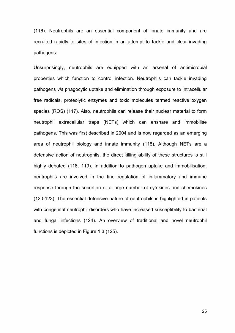
25
(116). Neutrophils are an essential component of innate immunity and are
recruited rapidly to sites of infection in an attempt to tackle and clear invading
pathogens.
Unsurprisingly, neutrophils are equipped with an arsenal of antimicrobial
properties which function to control infection. Neutrophils can tackle invading
pathogens via phagocytic uptake and elimination through exposure to intracellular
free radicals, proteolytic enzymes and toxic molecules termed reactive oxygen
species (ROS) (117). Also, neutrophils can release their nuclear material to form
neutrophil extracellular traps (NETs) which can ensnare and immobilise
pathogens. This was first described in 2004 and is now regarded as an emerging
area of neutrophil biology and innate immunity (118). Although NETs are a
defensive action of neutrophils, the direct killing ability of these structures is still
highly debated (118, 119). In addition to pathogen uptake and immobilisation,
neutrophils are involved in the fine regulation of inflammatory and immune
response through the secretion of a large number of cytokines and chemokines
(120-123). The essential defensive nature of neutrophils is highlighted in patients
with congenital neutrophil disorders who have increased susceptibility to bacterial
and fungal infections (124). An overview of traditional and novel neutrophil
functions is depicted in Figure 1.3 (125).
26
Figure 1.3. Traditional and novel functions of neutrophils. Upon arrival to the
site of infection and inflammation neutrophils can clear invading pathogens by a
number of mechanisms, including phagocytosis, NET formation and the secretion
of cytokines and chemokines. Once engulfed, neutrophils will generate toxic
compounds called ROS to kill bacteria. Upon resolution, neutrophils undergo
apoptosis and are cleared. Figure taken from ‘Diverse novel functions of
neutrophils in immunity, inflammation, and beyond’ published by Attila Mócsai
(125).
27
1.9.2 Chemotaxis and transmigration
It is essential for host defence that neutrophils can cross the endothelial barrier
and migrate towards the local site of infection. Extravasation, or transmigration, is
the process by which circulating neutrophils move into the infected tissue and
surrounding area. This process is governed by a number of complex interactions
between neutrophils and endothelium to ensure that transmigration remains
controlled and effective. For transmigration to occur the neutrophils must first
adhere to the vessel wall. Adherence occurs through transient selectin interactions
on both the neutrophil and endothelium which are upregulated in inflammatory
conditions (126). Neutrophils then move along the endothelium in processes
termed ‘rolling and crawling’ before subsequently traversing the endothelial
membrane (127, 128). Patients who have defective transmigration of neutrophils
suffer from recurring infections (129).
For the elimination of invading microbes and pathogens, neutrophils must first
migrate to the site of infection in a process termed chemotaxis. This process is the
movement of neutrophils in the direction of a chemoattractant gradient and is a
fundamental function of neutrophil biology and host defence. This directed
movement is dependent upon the regulation of intracellular signalling pathways
prompting rapid and accurate migration (130). In a healthy individual, neutrophils
will rapidly migrate towards chemokines (e.g. IL-8), bacterial components (e.g. N-
Formylmethionine-leucyl-phenylalanine (fMLP)) and complement proteins. In
support of the tight regulation of chemotaxis, there appears to be a hierarchy of
chemoattractant signals (131). The ability of neutrophils to sense
chemoattractants is acquired during terminal differentiation in the bone marrow
28
and is highly affected by both in vitro and in vivo stimuli (132). Thus, dysregulation
of this process will result in abnormal neutrophil chemotaxis.
1.9.3 Phagocytosis
Upon trafficking to a pathogen, phagocytosis and elimination can occur.
Phagocytosis is the engulfment of invading pathogens and is central to host
defence. Neutrophils express a number of complement receptors (e.g. CD35) in
addition to the Fc receptor CD16 which result in enhanced phagocytosis upon
activation (133).
Once engulfed pathogens are localised within a phagosome and subjected to
volatile components produced intracellularly by neutrophils (134). There are 2
independent mechanisms for killing engulfed pathogens. Firstly, neutrophils
generate high levels of ROS through nicotinamide adenine dinucleotide
phosphate-oxidase (NADPH) oxidase complex (135). ROS species generated
include hydroxyl radical and hydrogen peroxide which are unstable and toxic
(136). Neutrophils from patients following traumatic injury have elevated baseline
ROS production and enhanced production following secondary stimulation (137,
138). This is indicative of systemic priming of neutrophils most likely caused by the
inflammatory environment induced immediately post injury. Secondly, neutrophils
can kill pathogens via the delivery of granule products to the phagosome (139).
One such enzyme derived from azurophilic granules is neutrophil elastase which
is a serine proteinase and is known to directly digest bacteria (140). As these
compounds, ROS and granular proteins are extremely toxic, dysregulation or
abundant production is also implicated in host tissue damage which is a common
feature post traumatic and thermal injury (134).
29
1.9.4 Apoptosis
Neutrophil apoptosis is a tightly regulated pathway which prevents tissue damage
by inhibiting the release of toxic intracellular compounds of neutrophils.
Recognition and clearance of dead neutrophils, via macrophages, is an essential
step in the resolution of inflammation (141, 142). Apoptosis is controlled
predominantly by a number of bcl-2 homologues within neutrophils. Of such, MCL-
1 is a pro-survival protein which is produced and cleared rapidly and functions to
prevent apoptosis and is regarded as a major orchestrator in neutrophil circulating
life span (143, 144). In addition, neutrophils express pro-apoptotic bcl-2
homologues including, Bax (145), Bid (146) and Bim (147). Which all function to
induce apoptosis and, unlike MCL-1, take a long time to be cleared upon
generation. Severe traumatic injury results in a dysregulation of neutrophil
apoptosis which is proposed to underlie the pro-inflammatory phenotype,
increased incidence of infection and host tissue damage (148-151).
One mechanism behind dysregulated apoptosis is the elevated levels of pro-
inflammatory cytokines and/or elevated levels of ROS, which can induce apoptosis
via indirect activation of caspase 8 (152). Whilst it has been reported that
neutrophil apoptosis is dysregulated following major trauma, the mechanism
responsible and clinical implications remain unknown.
1.9.5 Anti-microbial actions
The importance of neutrophils in host defence is emphasised in disease
pathologies in which fundamental neutrophil processes are dysfunctional. One
such disease is chronic granulomatous disease (CGD) in which the individual is
30
incapable of producing ROS due to the absence of the NADPH oxidase complex
and thus suffer from recurring infections throughout their life (153). Additionally,
patients with Chediak-Higashi syndrome have a mutation in the LYST gene, which
is responsible for a lysosomal trafficking regulator (154). Patients with this
syndrome are susceptible to infections due to the inability to form functioning
phagolysosomes. Furthermore, in a murine model of Staphylococcus aureus
infection, there is a significant increase in mortality when mice are depleted of
neutrophils (155). Hence neutrophils and their components are key orchestrators
of host defence and essential for survival following bacterial challenge.
Although there is evidence showing that severe thermal injury results in altered
neutrophil function. It is currently unclear if thermal injury results in a longitudinal
disruption in neutrophil functions which may underlie the increased incidence of
infection, tissue damage and secondary complications. Hence, longitudinal and
extensive investigation of neutrophil function post thermal injury is required.
1.10 Neutrophil dysfunction post traumatic injury
Traumatic and thermal injury results in a profound immune dysregulation which is
characterised by an immediate activation closely followed by a subsequent period
of immune suppression (37). It has been proposed that this second period of
dysregulation may underlie the increased incidence of nosocomial infections.
1.10.1 Maturity and phenotype of neutrophils post traumatic injury
Of note, trauma results in profound changes in maturity, survival, phenotype and
function of neutrophils (156-161). As neutrophils are an essential component of
innate immunity, functional dysregulation has been linked to host tissue damage
31
and increased propensity to infections. However, mechanisms responsible are
poorly understood.
Maturation and production of neutrophils are controlled within the bone marrow by
a complex panel of mediators and signals. The principal regulator of
granulopoiesis is granulocyte-colony stimulating factor (G-CSF) which functions to
commit progenitor cells to myeloid lineage (162), reduce maturation time (163),
proliferation of granulocyte precursors and release of mature cells from the bone
marrow. There are secondary signals of neutrophil production and release
including; IL-6, IL-3 and granulocyte macrophage colony-stimulating factor (GM-
CSF) (164-166). In states such as severe bacterial infection, the body can adjust
to a state of ‘emergency granulopoiesis’ in which large quantities of neutrophils
are released in part mediated by G-CSF (167). Gardner and colleagues reported
in a mouse model of trauma that increased levels of G-CSF caused the release of
high levels of mature neutrophils which protected mice against a lethal Klebsiella
pneumoniae pulmonary challenge. Neutralisation of G-CSF reduced neutrophil
release and reduced survival (168). Thus one might expect that G-CSF plays a
predominantly positive role in host protection following major trauma.
However, excessive stress and stimulation of the bone marrow can result in
emergency granulopoiesis that is characterised by the appearance of immature
precursors of neutrophils, blood leucocytosis or neutrophilia (167). IGs are a
precursor of mature neutrophils and are elevated in septic patients (96). Even
though they are capable of mediating innate immune functions this is reduced
compared to mature neutrophils and may underlie the increased susceptibility to
infection following thermal injury (169). In a cohort of 83 severely injured patients,
Cook et al reported that immediate and sustained elevation in G-CSF was
32
associated with the release of immature cells from the bone marrow and an
increased risk of infection (157). Hence, the role of G-CSF in mediating survival
following thermal injury remains unclear and is most probably multi-factorial and
time dependent.
In 2011, Pillay and colleagues described a distinct change in the circulating
neutrophil phenotype following endotoxin challenge or following severe injury
(158). This group characterised neutrophil subsets by flow cytometry staining for
CD16, a low affinity Fc receptor expressed on neutrophils, and CD62L, an
adhesion molecule involved in leukocyte trafficking that is shed upon activation.
Three hours after lipopolysaccharide (LPS) administration there is a dynamic
change in neutrophil phenotype with the appearance of three subsets of
neutrophils (Figure 1.4). Two of which were detectable in challenged participants
and were absent from healthy individuals. These subsets were;
CD16dim/CD62Lbright and CD16bright/CD62Ldim neutrophils.
Firstly, CD16dim/CD62Lbright neutrophils were banded in their nuclear morphology
and most likely released from the bone marrow prematurely in response to the
LPS challenge. The second population of hyper-segmented CD16bright/CD62Ldim
neutrophils had increased maturation compared to normal blood neutrophils.
Furthermore, each of the novel subsets had differing functions with
CD16dim/CD62Lbright neutrophils having reduced apoptosis compared to the
CD16bright/CD62Ldim population. Whereas the CD16bright/CD62Ldim population
exhibited increased ROS production compared to the CD16dim/CD62Lbright
population (158).
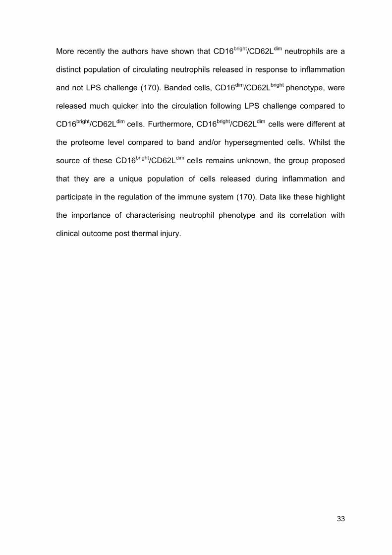
33
More recently the authors have shown that CD16bright/CD62Ldim neutrophils are a
distinct population of circulating neutrophils released in response to inflammation
and not LPS challenge (170). Banded cells, CD16dim/CD62Lbright phenotype, were
released much quicker into the circulation following LPS challenge compared to
CD16bright/CD62Ldim cells. Furthermore, CD16bright/CD62Ldim cells were different at
the proteome level compared to band and/or hypersegmented cells. Whilst the
source of these CD16bright/CD62Ldim cells remains unknown, the group proposed
that they are a unique population of cells released during inflammation and
participate in the regulation of the immune system (170). Data like these highlight
the importance of characterising neutrophil phenotype and its correlation with
clinical outcome post thermal injury.
34
Figure 1.4. Administration of LPS results in distinct phenotypical changes in
circulating neutrophils. (A) Flow cytometry of neutrophils, based on CD16 and
CD62L staining, before administration of LPS. (B) Neutrophil populations 180
minutes following LPS administration. Three populations of neutrophils are
observed based on CD16 and CD62L staining. Two populations are unique to
LPS challenge and absent from healthy individuals. Figure taken directly from ‘A
subset of neutrophils in human systemic inflammation inhibits T cell responses
through Mac-1’ by Pillay et al. (158).
35
1.10.2 Functional dysregulation of neutrophils post traumatic injury
Chemotaxis describes the movement of a cell to a stimulus in a concentration
dependent manner. Neutrophils utilise chemotaxis to migrate to sites of infection
and tackle and clear invading pathogens and bacteria. Studies dating back to
1974 have identified an inherent defect in neutrophil migration following thermal
injury (159-161). Until recently, technologies used to measure neutrophil migration
have been limited, time-consuming and require a large volume of blood.
In 2010, Butler and colleagues described a novel microfluidic device that is simple
to use, allows for precise and robust measurements of chemotaxis speed and
persistence characteristics at single-cell resolution. Importantly, this device uses
only a single drop of blood which is important to prevent anaemia in critically ill
patients (171). This group has shown that thermal injury results in a significant
reduction of directional migration speed within 24 hours of injury which reaches
maximal reduction at 72-120 hours post injury when compared to healthy
individuals. In this study one patient, with a 60% TBSA thermal injury, did not have
reduced neutrophil migration. This patient was admitted with a documented
infection unlike the rest of the cohort. The authors propose that the immediate
inflammatory cascade post thermal injury results in suppression of chemotaxis
which increases the propensity to infections and that the infection itself does not
reduce chemotaxis (171). Hence, modulation of chemotaxis remains an early
therapeutic target.
More recently the authors have described a novel phenotype of spontaneous
neutrophil migration in septic patients following severe thermal injury. This
phenotype was observed 1 – 2 days before the diagnosis of sepsis and was not
36
detectable in patients who did not develop an infection or healthy individuals
(172). Therefore quantification of chemotaxis phenotypes may aid in early
diagnosis of patients at risk of developing sepsis during their hospital stay. This
could aid in the timing of antibiotic and clinical treatment in the aim of improving
patient outcome post thermal injury (171).
Once a neutrophil has trafficked to the site of infection it functions to either
phagocytose or generate NETs to ensnare the invading pathogen. Phagocytosis is
a complex process that forms an essential component of innate immunity through
the ingestion and elimination of pathogens. As such, a dysregulation in phagocytic
function may precede the development of infections which are extremely common
in patients with thermal injuries (38). Although this has been proposed, only a
small number of studies have examined this relationship and none have studied
the dynamic longitudinal changes in relation to clinical complications post thermal
injury.
Sheng and colleagues studied neutrophil bactericidal activity in 36 patients with
thermal injuries using a chemiluminescence assay. The group demonstrated that
septic patients had lower neutrophil bactericidal function compared with healthy
controls. Bactericidal activity was significantly lower in thermal injuries above 35%
TBSA suggesting that severity of injury orchestrates the amplitude of
dysregulation consistent with previous literature examining chemotaxis (171, 173).
Alexander et al performed a prospective analysis of neutrophil function in 20
patients with severe thermal injuries (≥45% TBSA). The group reported a
significant reduction in neutrophil bactericidal activity, in response to
Staphylococcus aureus 502A, and chemotaxis in infected patients compared to
non-infected patients. Thus suggesting a broader dysregulation of neutrophil
37
functions mediated by initial injury and subsequent sepsis not previously reported
(171, 174).
ROS are essential anti-microbial compounds which function to directly kill and
eliminate engulfed pathogens within a neutrophil. Production of ROS is controlled
by NADPH oxidase complex which produces O2- and H2O2 upon activation which
are toxic compounds. It has been reported that trauma results in an increase in
ROS production (137). Conversely, multiple groups have shown that thermal injury
results in significant reduction in the ability of neutrophils to generate ROS and a
reduced phagolysosomal acidification (175-177).
Parment and colleagues in a prospective exploratory cohort study investigated
neutrophil oxidative burst in response to Escherichia coli (E.Coli), phorbol 12-
phorbol myristate 13-acetate (PMA), and fMLP. This study included 28 patients
with thermal injuries with an average TBSA of 36%. Thermal injury resulted in a
significant reduction in neutrophil oxidative burst, in response to all stimuli, from
admission up to 3.5 months post injury compared to levels seen in healthy
volunteers. Thus, thermal injury results in an immediate and sustained
immunosuppression which appears to be different from general trauma (177).
Trauma and thermal injuries result in a significant dysregulation of a number of
neutrophil functions. Although multiple groups have described the dynamic
changes in neutrophil function no group has conclusively studied the longitudinal
changes in neutrophil functions in relation to acute injury and subsequent
infections following thermal injury.
38
1.11 Neutrophil extracellular traps
1.11.1 Structure and function
NETs, although first described in 2004, are still emerging as a potentially important
area of neutrophil biology (118). NETosis (178) is a tightly regulated active cell
death pathway facilitated by neutrophil activation resulting in the extrusion of DNA
to the exterior of the cell decorated in granular proteins which can ensnare, trap or
sometimes kill bacteria (118, 119, 179). Discovery of NETs sparked an entirely
new area of neutrophil research and their association with innate immunity and
haemostasis. Systemic markers of NETosis have been reported following major
trauma, burn injury and during sepsis (180, 181).
1.11.2 Vital and suicidal NETosis
Traditionally NETs are generated by incubating neutrophils with chemical
agonists, e.g. PMA, a known potent activator of protein kinase C (PKC). In 2013,
this pathway of NET generation was termed ‘suicidal NETosis’ (178). This process
results in complete loss of the nucleus and terminal cell death hence the name.
Although NETosis was first described in 2004 there are earlier reports indicating
that PMA could cause chromatin decondensation, nuclear swelling, release of
nucleoplasm into the cytoplasm and membrane perforation (182). It wasn’t until
2004 that the process of NET release was discovered by Zychlinsky and
colleagues (118). This initial description was advanced by further studies revealing
that NETs are an important arm of innate immunity as they can bind to both gram-
positive and gram-negative bacteria (183).
39
The mechanisms causing terminal NET production are still not fully understood.
Many groups have reported that the generation of oxidants is an essential
requirement for NETosis. However, signalling components downstream of
oxidants responsible remain unknown. For instance, Fuchs et al reported that
formation of NETs is dependent upon activation of NADPH oxidase. Additionally,
the authors state that patients with CGD carry a mutation for NADPH oxidase and
therefore cannot make NETs which may be responsible for recurring infections
(184). Following activation of NADPH oxidase, ROS initiates the translocation of
neutrophil elastase to the nucleus which results in chromatin decondensation and
NETosis. Importantly, neutrophil elastase deficient mice do not form NETs in a
pulmonary model of Klebsiella pneumoniae infection thus supporting the proposed
mechanism of NADPH oxidase dependent NETosis. Hence, mobilisation of
granular proteins following ROS production are fundamental in the regulation of
chromatin density and structure (185). However, it has also been reported that
biological stimuli, e.g. Staphylococcus aureus, can result in NADPH oxidase
independent NET formation (186). Thus it is proposed that NETosis can occur
through multiple signalling pathways which may be unique to individual stimuli.
A fundamental step in suicidal NET generation is the rupture of the cellular
membrane and death of the neutrophil. Therefore one would propose that suicidal
NETosis inhibits the ability of neutrophils to perform other traditional defensive
actions, e.g. phagocytosis or chemotaxis, and may leave the host
immunocompromised which would be fatal to patients following traumatic or
thermal injury. Therefore, it is important to consider how neutrophils can generate
NETs and still perform vital defensive actions. A number of possibilities exist
including; 1) neutrophils generate NETs and survive the process; 2) neutrophils
40
are split into pools which perform different specialised functions depending upon
orchestrating signals; 3) NET formation is the the last line of defence of the
neutrophils when the host is overwhelmed.
A second pathway of NET generation has also been described and coined ‘vital
NETosis’. The first major difference between the vital and suicidal pathway is the
stimulus. Vital NETosis is mediated by naturally occurring stimuli, e.g. bacterial
components and/or activated immune cells, and is therefore seen as the NETosis
pathway occurring in vivo (178, 187). Moreover, vital NETosis occurs rapidly,
within 30 - 45 minutes, whereas suicidal NETosis is much slower, within 3 - 4
hours post stimulation. Theoretically, this allows for rapid entrapment of invading
pathogens and doesn’t compromise the host. The final difference between the two
pathways is the mechanism of release. Vital NETosis is still in part mediated by
activation of NADPH oxidase, mobilisation of granular proteins/enzymes and
histone citrullination (178). However, vital NETosis also employs vesicular
trafficking of DNA to the exterior of the cell. Vesicles co-localised with the cell
membrane and delivered the nuclear material to the outside of the cell without
membrane perforation (Figure 1.5 G, H and I) (186). This could represent a
conserved mechanism to tightly control NET release and allow additional
neutrophil functions to occur. In support of this hypothesis, Clark et al
demonstrated that neutrophils which had released NETs were impermeable to
SYTOX Green stain, suggesting that neutrophils can remain intact following
NETosis (100). The differences between ‘vital and suicidal’ NETosis are depicted
in Figure 1.5 (178).
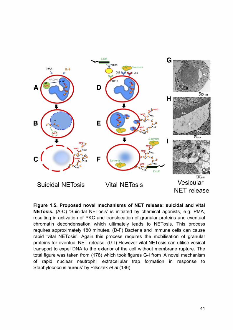
41
Figure 1.5. Proposed novel mechanisms of NET release: suicidal and vital
NETosis. (A-C) ‘Suicidal NETosis’ is initiated by chemical agonists, e.g. PMA,
resulting in activation of PKC and translocation of granular proteins and eventual
chromatin decondensation which ultimately leads to NETosis. This process
requires approximately 180 minutes. (D-F) Bacteria and immune cells can cause
rapid ‘vital NETosis’. Again this process requires the mobilisation of granular
proteins for eventual NET release. (G-I) However vital NETosis can utilise vesical
transport to expel DNA to the exterior of the cell without membrane rupture. The
total figure was taken from (178) which took figures G-I from ‘A novel mechanism
of rapid nuclear neutrophil extracellular trap formation in response to
Staphylococcus aureus’ by Pilsczek et al (186).
42
Neutrophils may be split into specialised pools which perform actions depending
upon complex orchestrating signals. In 2014, Branzk et al identified a novel
microbe sensing mechanism that tailored neutrophil anti-microbial actions
depending upon pathogen size. Neutrophils selectively released NETs in
response to large microbes, namely Candida albicans hyphae and extracellular
aggregates of Mycobacterium bovis, but did not respond to single bacteria or
yeast which were phagocytosed instead. The ‘decision’ made by neutrophils to
release a NET or undergo phagocytosis was controlled by competition for
neutrophil elastase between the two processes. Dectin-1, a pattern recognition
receptor involved in innate immunity, was the key signalling molecule in this
decision process. If phagocytosis occurred via dectin-1, neutrophil elastase
translocation was inhibited, thus preventing NET production. Conversely, knockout
of dectin-1 led to excessive NETosis and tissue damage. Thus phagocytosis
appears to act as a checkpoint to induce or prevent NET formation via dectin-1
and competition of neutrophil elastase (188). Therefore, NET formation may
represents the last resort of neutrophil defence against invading pathogens to
prevent complete loss of the circulating neutrophil pool and in turn
immunocompromising and potentially damaging the host. This is of particular
interest when studying patients with traumatic injuries as they are prone to
nosocomial infections and sepsis (38). Hence, this pathogen sensing ability may
aid patients by switching to a pro-phagocytosis phenotype, reducing the risk of the
patient becoming immunocompromised.
43
1.11.3 Citrullinated histone H3 and reactive oxygen species generation
ROS are a heterogeneous collection of molecules which are highly reactive and
formed in multiple cellular organelles as bi-products of redox reactions. NADPH
oxidases are a group of membrane bound multiprotein enzymes that generate
ROS and are expressed in large quantities in neutrophils (136). ROS play an
important link between cellular activation and NET formation as discussed above
(184). Although the full mechanisms behind ROS induced NET formation aren’t
fully understood it is known that ROS can induce histone citrullination by activation
of the enzyme peptidylarginine deiminase 4 (PAD4) which results in
decondensation of nuclear chromatin (189). PAD4 is a nuclear enzyme which is
highly abundant in human neutrophils and mediates the replacement of amino
acid arginine with citrulline (190, 191). Citrullinated histone H3 (Cit H3) is
expressed within NETs and is regarded as a specific marker of NET generation
and has recently been detected in the bloodstream of critically ill patients (181). In
addition to their role in the pathogenesis of inflammatory conditions, modified
histones are potential biomarkers of sepsis (181, 192).
1.11.4 Histones, DNA and nucleosomes
Histones are a group of small positively charged proteins including; H1, H2A, H2B,
H3 and H4. They are highly conserved and responsible for gene transcription
(193, 194). Free histones are extremely cytotoxic due to their charged nature and
administration results in rapid mortality in animal models (195-198).
DNA is the fundamental orchestrator of cellular growth, repair, function and
reproduction. DNA is released abundantly following severe tissue damage and
44
can activate a number of inflammatory cascades (199), because of this DNA is
regarded as a DAMP. A DNA molecule consists of two polynucleotide chains with
each nucleotide containing a 5-carbon sugar molecule attached to a phosphate
group (194). These phosphate groups make DNA negatively charged which
means that they can bind to histones and form nucleosome complexes (Figure
1.6).
Comprised of a nucleosome core, an octameric complex of histones and DNA,
and linker DNA, nucleosomes are the principal component of chromatin (194, 200,
201). Nucleosomes have multiple functions including signalling as a centre for
chromatin orchestrated processes and assembling higher-order structures which
aid in compaction of genomic components (200). The presence of circulating
nucleosomes is associated with NET release and cell death in patients with
chronic myeloproliferative neoplasms and deep vein thrombosis (DVT) (202, 203).
Nucleosomes have different cytotoxic and procoagulant properties compared to
free histones and DNA (198, 204-206). Despite this, many studies investigating
these components interchangeably use nucleosomes, histones and DNA to
describe in vivo associations. Therefore, characterisation of the structure and
properties of circulating nucleosomes, histones and DNA is required to
conclusively understand their roles in inflammation, coagulation and tissue
damage.
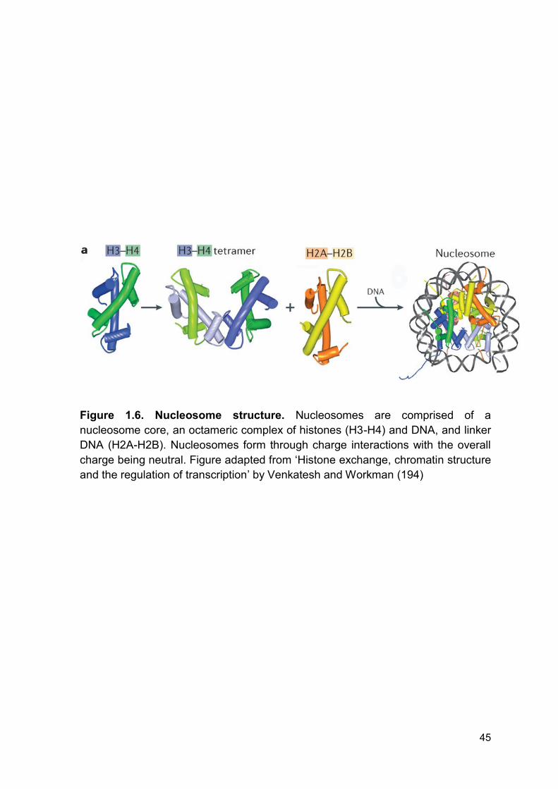
45
Figure 1.6. Nucleosome structure. Nucleosomes are comprised of a
nucleosome core, an octameric complex of histones (H3-H4) and DNA, and linker
DNA (H2A-H2B). Nucleosomes form through charge interactions with the overall
charge being neutral. Figure adapted from ‘Histone exchange, chromatin structure
and the regulation of transcription’ by Venkatesh and Workman (194)
46
1.11.5 Autophagy and neutrophil extracellular traps
Autophagy is a normal physiological response of the cell that maintains
homeostasis by protein degradation and turnover of destroyed cell organelles to
induce new cell formation (207). Recent evidence has demonstrated that NET
formation is dependent upon both ROS generation and activation of autophagy
(208). Additionally, multiple bacterial strains can activate autophagy, implicating its
activation in neutrophil biology and host defence (209-212). To date, it is currently
unknown if thermal injury and/or soluble factors released affect neutrophil function
via alteration in autophagy signalling.
1.12 NETs and traumatic injury
Traumatic and thermal injury are characterised by an initial sterile injury and
subsequent infectious stimuli. The generation of NETs is regarded as defensive
action of neutrophils to invading stimuli in an attempt to ensnare and potentially
trap bacteria. It has been demonstrated that NETs are present in septic individuals
via the measurement of non-specific circulating markers of NET formation (213,
214). Furthermore, studies have concluded that NETs are produced during
subsequent septic episodes following traumatic injury (181, 215, 216). Thus, NETs
may form an orchestrating role in the defence against invading pathogens in
patients with thermal injuries. However, it has yet to be conclusively established if
NETs are generated following thermal injury.
Interestingly there is an emerging body of evidence to implicate NET formation
and non-infectious stimuli (217-219). With elevations in circulating NET
biomarkers reported post traumatic and thermal injury (215, 220). It is proposed
that the severe tissue damage is enough to induce rapid NET formation within
47
minutes of injury through the release of inflammatory mediators. In a cohort of
patients with burn injury, plasma cfDNA are increased compared to healthy
individuals and thus suggests immediate NET formation (220). Although this study
reports elevation in NET components it does not measure any specific markers of
the process. Quantification of cfDNA alone is non-specific to NETosis, with plasma
cfDNA originating from a number of sources including apoptotic, necrotic or
NETosing cells and bacteria themselves (115). Therefore, to our knowledge, it has
yet to be conclusively established whether NETosis is occurring in patients with
burn injury.
1.13 In Vivo host defensive functions of neutrophil extracellular traps
Despite extensive studies of NETs, their direct in vivo interactions and effects are
still being investigated with multiple groups reporting contradicting results (119,
221). Therefore, how NETs contribute to host defence post thermal injury is poorly
understood and warrants further investigation.
Meng and colleagues provided evidence that NETs were responsible for in vivo
killing of bacteria. Administration of deoxyribonuclease (DNAse) to mice in a cecal
ligation puncture (CLP) model of sepsis resulted in reduced in vivo NET markers
coupled with increased colony forming units of bacteria and an increased pro-
inflammatory response (221). Hence, in this study NETs are directly responsible
for killing bacteria and host protection. However, in 2012 Menegazzi challenged
the current paradigm of NETs directly killing bacteria. The group reports that NETs
ensnared bacteria but did not kill them as incubation of NETs with DNAse freed
bacteria which were still alive (119). Therefore, NETs may have more of a physical
bacteriostatic role in ensnaring bacteria and preventing dissemination rather than
48
a direct bactericidal activity. Though the potential implications this has in severe
infections, e.g. sepsis following severe thermal injury, are currently unknown and
seem rather redundant if neutrophil function is already reduced.
Furthermore, in a murine model of sepsis, NETs accumulate within various organs
and interact with the vascular endothelium. Within the microvasculature NETs
colocalised with inflammatory cells, including platelets, and may be contributing to
thrombosis which is commonly observed post infection (222).
1.14 Excessive NETosis, thrombosis and host tissue damage
Advancements in the treatment of thermal injuries have improved initial survival
following severe thermal injury. However, delayed mortality remains a significant
health care issue with sepsis and MOF being the leading causes of mortality (4,
5). Even though MOF is well characterised the mechanisms mediating the
pathogenesis are poorly understood. Therefore advancements in understanding
the pathogenesis of MOF may improve patient outcome.
Although neutrophils are an essential component of innate immunity, recent
evidence has shown that NETs can initiate a procoagulant phenotype, tissue and
organ damage (195, 221, 223-225). Prolonged or uncontrolled NET generation
results in damage to host tissues and significantly contributes to the pathogenesis
of inflammatory conditions (195, 198, 226, 227). DNA and histones are key
components of NETs, but elevation in circulating levels may also mediate organ
damage through their cytotoxic nature (228). As such, NETs can provide a bridge
between the innate immunity and haemostatic systems. For example, they can
directly bind to and activate platelets along with various coagulation system
components (100, 229). These interactions during excessive NETosis may then
49
promote thrombosis, DIC (52) and potentially mediate the pathogenesis of MOF
following sepsis (230, 231).
Elevated levels of NET components have been implicated in thrombosis formation
in a murine model of DVT induced stenosis in the inferior vena cava. 6 hours after
stenosis, levels of plasma cfDNA were significantly elevated compared to sham-
treated animals and neutrophils were visible in both the red (red blood cell rich)
and white (platelet rich) thrombi. Moreover, Cit H3, a specific marker of NETs,
colocalised with neutrophils in red thrombi. This data is consistent with a previous
publication which identified nucleosomes in thrombi of baboons following induction
of DVT (232). Infusion of DNAse protected animals against flow restriction induced
by DVT regardless of the length of stenosis. Furthermore, DNAse treated mice
had no visible thrombi suggesting DNAse disrupts the mechanisms leading to
thrombosis. The group hypothesised that ‘the anti-thrombotic effect of DNAse is
likely to be mediated by removal of NETs generated locally at the site of stenosis’
(233).
The procoagulant effect of NET components and cfDNA, is well documented (230,
234). However, the role of cfDNA on fibrinolysis is poorly understood. Fibrinolysis
is the enzymatic breakdown of fibrin in blood clots predominantly by the enzyme
plasmin. In 2015, Gould and colleagues investigated the effect cfDNA has on
fibrinolysis in sepsis. The group quantified levels of cfDNA in plasma obtained
from 400 patients with severe sepsis and clot lysis was measured in an isolated
system and breakdown of formed clots was visualised using electron microscopy.
Clots formed in plasma from septic patients had decreased permeability,
decreased susceptibility to fibrinolysis and increased clot stability which could be
reversed by the addition of DNAse. It is proposed that this impairment of
50
fibrinolysis was caused by cfDNA forming a ternary complex between plasmin,
cfDNA and fibrin that inhibits the activity of plasmin (235). Hence, this study
highlights the multifaceted actions of NET components in the production and
maintenance of thrombosis and potentially the pathogenesis of MOF following
sepsis.
MOF is characterised by the formation of thrombi in the microvasculature resulting
in tissue hypoxia, damage and ischemia. Boneschansker et al have demonstrated,
using a microfluidic device replicating capillary plexuses, that NETs are capable of
perturbing blood flow in capillary networks (Figure 1.7 A-B) (236). NETs generated
from a very small number of neutrophils resulted in more than half of the
microfluidic device being obstructed from red blood cell flow and this inhibition
lasted for hours post NET formation despite the presence of naturally occurring
DNAse in the blood. Administration of Turbo™ DNAse, a more potent analogue of
naturally occurring DNAse 1, degraded NETs and restored blood flow within 30
minutes of infusion (Figure 1.7 B). Importantly, digestion products of NETs did not
impair blood flow within the microfluidic device (236). Complete breakdown of the
NET structures and removal of DNA from the channels was therefore sufficient to
prevent this obstruction. This study highlighted a potential mechanism in which the
physical properties of NETs may result in the pathogenesis of MOF by obstructing
blood flow through capillary networks.
NETs and their components have the potential to not only induce thrombosis but
can also propagate further detrimental effects by impairing fibrinolysis and
obstructing blood flow. The formation of excessive or uncontrolled NETosis may
result in tissue hypoxia, the onset of DIC, cause host tissue damage and
potentially mediate the pathogenesis of MOF in patients with thermal injuries.

51
Importantly, digestion or breakdown of NETs via DNAse may be a therapeutic
target to prevent NET mediated pathogenesis of MOF.
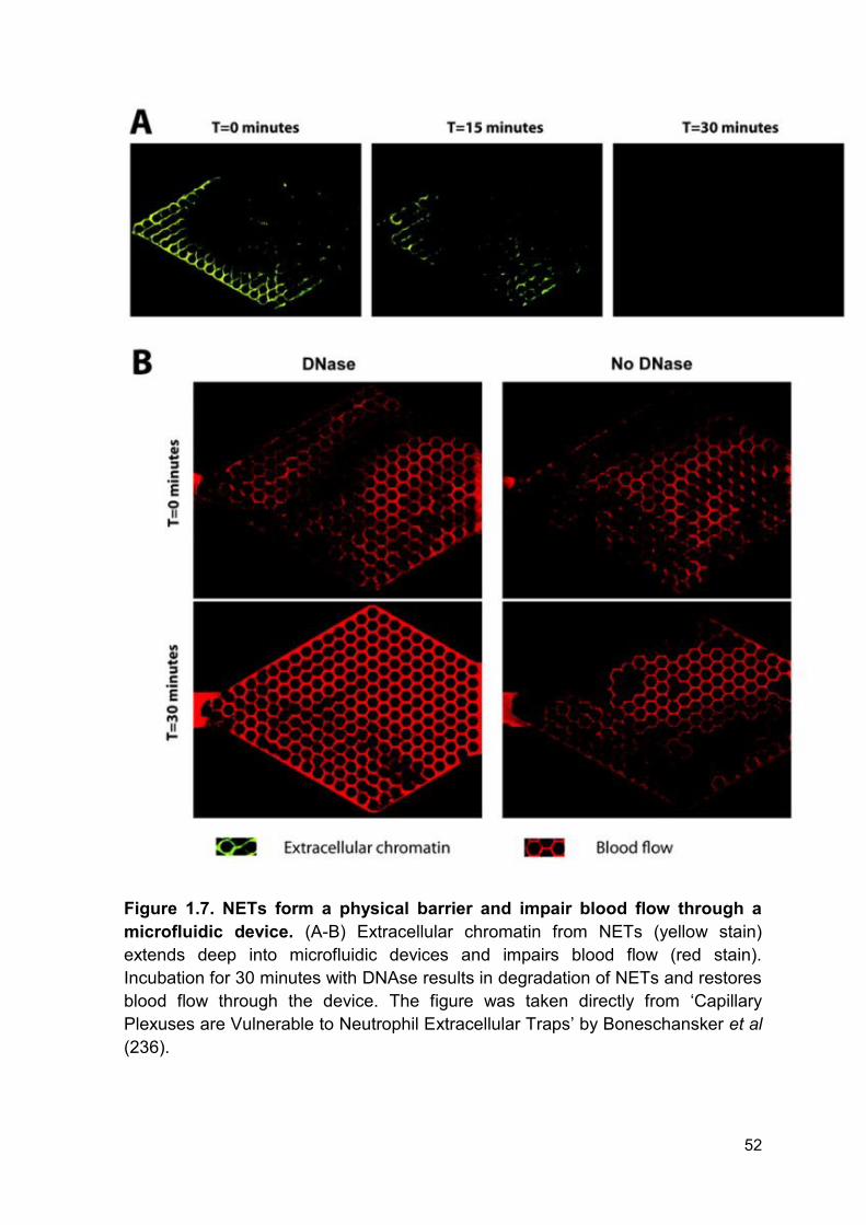
52
Figure 1.7. NETs form a physical barrier and impair blood flow through a
microfluidic device. (A-B) Extracellular chromatin from NETs (yellow stain)
extends deep into microfluidic devices and impairs blood flow (red stain).
Incubation for 30 minutes with DNAse results in degradation of NETs and restores
blood flow through the device. The figure was taken directly from ‘Capillary
Plexuses are Vulnerable to Neutrophil Extracellular Traps’ by Boneschansker et al
(236).
53
1.15 Degradation of neutrophil extracellular traps
DNAse is the major extracellular endonuclease found in a number of bodily fluids
and functions to breakdown chromatin and DNA which are released following
cellular damage, cell death and NETosis. Levels of circulating cfDNA are elevated
in a number of disease pathologies but the mechanisms behind uncontrolled
elevation in this marker are poorly understood. Of note, elevated levels of plasma
cfDNA are associated with and contribute to the progression of autoimmune
diseases (237, 238). In a subpopulation of patients with systemic lupus
erythematosus (SLE), DNAse activity was impaired by DNAse inhibitors or
inhibitory antibodies (237). Although a reduction in DNAse activity and build-up of
cfDNA has been shown to be associated with the development of autoimmune
diseases, to date no group has investigated DNAse activity following thermal injury
(237).
To date, only one study has investigated DNAse levels post traumatic injury. This
study recruited 39 patients with traumatic injury and quantified levels of DNAse 1.
Whilst there was a significant elevation in circulating DNA levels post-injury, only
on the day of injury was a difference observed in DNAse 1 levels, with patients
having significantly higher circulating DNAse 1 levels. At all other study time-
points, DNAse 1 levels in patients were comparable to that for healthy controls
(239). Thus, the accumulation of circulating NET markers may be due to a
reduction in DNAse activity rather than quantity and may be one mechanism for
the pathogenesis of MOF following thermal injury.
54
1.16 Actin
Actin is the most abundant protein in mammalian cells and is a fundamental
protein for cell motility (240). Actin exists in a balance between monomeric and
filamentous actin. Processes such as cell motility are dependent upon the rapid
polymerisation of monomeric actin into filamentous actin (241, 242). However, this
predisposition to polymerise is of detrimental consequences if actin is released
into the circulation, as extracellular actin can cause direct damage to the
microvasculature capillaries, impair clot lysis and activate platelets (240, 243,
244). Elevation in circulating actin is attributed to a saturation of the actin
scavenging system.
Interestingly, DNAse activity is indirectly controlled by actin scavenging system
(240, 245) as actin can bind to DNAse and inhibit its enzymatic activity (237, 245).
Due to the extensive amount of tissue damage post severe thermal injury one
might hypothesise that actin is released following severe thermal injury and results
in the inhibition of DNAse activity and build-up of circulating cfDNA. Data reported
by Lee et al support this hypothesis as elevated levels of actin have been detected
in a cohort of septic patients (246). Therefore, DNAse activity may be inhibited in
septic patients predisposing them to risk of actin toxicity, accumulation of
circulating DNA, host tissue damage, a pro-thrombotic phenotype and MOF.
1.17 Actin Scavenging System
Control of the actin scavenging system is mediated by two key proteins; gelsolin
(GSN) and vitamin D binding protein (VDBP) (240). Both proteins functions to
clear monomeric and filamentous actin from the circulation through the liver. The
actin scavenging system is summarised in Figure 1.8.
55
1.17.1 Gelsolin
Plasma GSN is found in high quantities in healthy individuals and can bind to both
monomeric and filamentous actin (246). Levels of circulating GSN in healthy
individuals range from 150 – 300 µg/ml and plasma GSN has a half-life of
approximately 2 days (247). GSN is a 90 kDa cytoplasmic actin binding protein
which also exists as a plasma variant. The plasma variant of GSN is larger, 93
kDa, and more positively charged compared to cytoplasmic GSN. Although both
variants are similar in structure, their processing and eventual actions are
independent (248).
GSN functions in collaboration with VDBP to rapidly depolymerise and reduce
circulating levels of actin. GSN has three actin binding sites and functions to clear
actin in two distinct processes (249-251). Firstly, GSN functions to prevent any
further addition of monomeric actin to existing chains by binding to the barbed end
of actin (250). Secondly, GSN binds to two distinct sites on the side of polymerised
actin and severs the filaments resulting in depolymerisation (240, 251). As GSN
binds to circulating actin it is not surprising that studies have reported a decrease
in circulating levels of GSN in a number of disease pathologies connected with
actin release (252-259). Furthermore, it has recently been reported that
administration of GSN can reverse actin inhibition of lung macrophage binding and
uptake of bacteria (260). In addition to being part of the actin scavenging system,
GSN has direct interactions in modulating the immune response. Thus, reduced
levels of GSN will have a multifaceted role in host response.
Huang et al reported in 95 patients with thermal injuries that plasma GSN levels
were reduced and associated with mortality, development of sepsis and MODS
56
(254). Furthermore, in a rat burn model (40% TBSA) plasma GSN levels decrease
12 hours post injury and remain lower for up to 6 days post injury. This was
accompanied by increased pulmonary microvasculature permeability in burn
treated animals. Importantly, administration of recombinant plasma GSN prior to
burn injury protected against this pulmonary microvasculature dysfunction (261).
Therefore plasma GSN may not only serve as a biomarker of poor outcome but
could also be a potential therapeutic target to prevent secondary complications.
However, the potential translation of GSN and its action in human burn injury have
not been fully invesitgated.
1.17.2 Vitamin D binding protein
VDBP, also referred to as Gc-globulin, is a 58-kd alpha-2-globulin which is a key
component of vitamin D transport (262). VDBP is found in abundant quantities in
healthy individuals with levels comparable to GSN (263). Unlike GSN, VDBP binds
to monomeric actin only allowing GSN to bind to polymeric actin (264). This
relationship suggests an orchestrated response to enhance actin clearance in vivo
by VDBP and GSN (265).
Binding of VDBP to actin prevents further polymerisation by clearing residual
monomeric actin from the circulation. The clearance of actin-vitamin D binding
protein complexes is rapid and is mediated through the liver primarily but this can
also occur through the spleen and lung (266, 267). It is important to note that
clearance of actin via GSN and VDBP results in consumption with a transient
decrease in their circulating levels.
57
Levels of VDBP have also been suggested as a prognostic marker of outcome
and organ damage following severe traumatic injury (268-271). Interestingly, it has
been reported that trauma results in an eventual over production of VDBP (271).
This implies a reactive response of the body to saturation or dysregulation of the
actin scavenging system. However, this has not been studied longer than 1 week
post trauma.
To date, no group has studied the relationship between VDBP, GSN and DNAse
post thermal injury. As VDBP and GSN are responsible for actin clearance one
might hypothesise that in thermal injury low plasma VDBP and GSN levels will be
associated with dysregulated DNAse activity. Therefore dysregulation in the actin
scavenging system may precede actin inhibition of DNAse activity and is
potentially contributing to the complicated outcomes associated with MOF post
thermal injury.
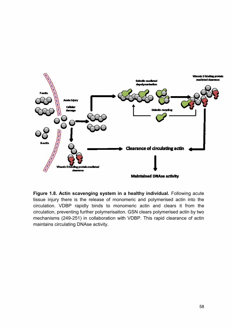
58
Figure 1.8. Actin scavenging system in a healthy individual. Following acute
tissue injury there is the release of monomeric and polymerised actin into the
circulation. VDBP rapidly binds to monomeric actin and clears it from the
circulation, preventing further polymerisaiton. GSN clears polymerised actin by two
mechanisms (249-251) in collaboration with VDBP. This rapid clearance of actin
maintains circulating DNAse activity.
59
1.18 Targeting neutrophil extracellular traps
It is proposed that targeting the formation or breakdown of NETs in various
diseases pathologies may have positive effects on disease progression (236, 237,
272, 273). One proposed mechanism for targeting NETs is the breakdown of pre-
formed and excessive NETosis via the administration of DNAse to reduce
thrombotic complications, host tissue damage and pro-inflammatory response
during infection (230, 232, 235, 236, 274). However, administration of DNAse in a
murine model of sepsis, induced by CLP, resulted in increases in the pro-
inflammatory response, the colonisation of bacteria and mortality (221). This was
attributed to increased bacterial dissemination caused by early and inappropriate
degradation of NETs. This study highlights the potential importance of time
dependent treatment as NETs should only be targeted when in excess and
contributing to the pro-inflammatory and thrombotic phenotype.
NET production may also be inhibited via the inhibition of PAD4. In a murine
model of sepsis, PAD4 deficient mice had comparable mortality to wild-type mice.
Similarly, PAD4 knockout mice were partially protected against LPS induced
shock, suggesting that knock-out of PAD4 and elimination of NETs may protect
against the pro-inflammatory and toxic effects of NETs during sepsis. The authors
conclude ‘preventing NET formation by PAD4 inhibition in inflammatory or
thrombotic diseases is not likely to increase host vulnerability to bacterial
infections’ (275). However, this model does not take into account pre-existing
immunosuppression which is observed post burn injury (159-161, 173, 175-177).
Moreover, in a murine model of infection, using necrotizing fasciitis, PAD4
knockout mice were more susceptible to infection than wild-type mice. The authors

60
attributed this susceptibility to the inability to form NETs in response to the
infection (276). Therefore full characterisation of NET formation kinetics, functions
and the relationship with other neutrophil functions is required to understand if
targeting NETs post thermal injury would provide any host protection.
61
1.19 Aims and hypothesis
Advancements in the initial care of burn wounds and immediate survival have
increased dramatically over the past four decades (3, 18). However, sepsis and,
consequently, MOF remain leading causes of delayed mortality following severe
thermal injury (47). Although sepsis and MOF are well characterised, the
mechanisms driving the increased incidence of infection and pathogenesis of MOF
are poorly understood.
Evidence has implicated abnormal neutrophil function in the increased propensity
to infection. However, this has not been studied longitudinally (159-161, 173, 175-
177). The recent discovery of NETs has identified a novel paradigm in which
neutrophil functions may be dysregulated post thermal injury and contributing to
the pathogenesis of MOF (230, 232, 235, 236, 274). To date, no group has
conclusively shown that NETs are produced following thermal injury and no group
has examined the relationship between NETs, MOF and sepsis following severe
thermal injury.
Accordingly, the aims of this thesis were to:
1. Determine the effect severe thermal injury has on neutrophil function and
understand potential mechanisms mediating the dysfunction.
2. Investigate if thermal injury initiates NETosis and to investigate if NETosis
is induced by sterile injury and/or infectious stimuli.
3. Determine if NET generation is associated with secondary complications,
MOF and sepsis, and to investigate potential mechanisms and therapeutic
targets.
62
4. Investigate if clearance of DNA was dysfunctional and associated with
MOF.
5. Determine if reduced DNAse activity is a potential therapeutic target to
reduce secondary complications.

63
Chapter 2
Materials and Methods
64
Materials and Methods
2.1 Scientific Investigation of Biological Pathways Following Thermal Injury
Study
2.1.1 Ethical approval
Ethical approval for the study was granted by a UK NHS research ethics
committee (Reference 12/EM/0432). Where possible, written informed consent
was received from participants prior to their inclusion in the study. Due to the
severe nature of the injuries being studied, the ethics committee approved the use
of a legal consultee, either personal or nominated, if the patient was not initially
able to consent for inclusion in the study themselves. When the patient was able,
they were approached to give written consent to continue to participate in the
study.
2.1.2 Study cohort
Scientific Investigation of the Biological Pathways Following Thermal Injury in
Adults and Children (SIFTI) study is a multi-centre prospective observational
cohort study. 150 patients were recruited in total to the study from January 2013 to
October 2015. In addition, blood samples were collected from 10 healthy
volunteers after obtaining written informed consent from the donors, who at the
time of participation were in good health, free of immunological illness and
significant co-morbidity, and were not taking any medication known to interfere
with immunity. To be included in the SIFTI Trial the patient must meet the
inclusion criteria summarised in Table 2.1. Further inclusion and exclusion criteria
65
are listed below Table 2.1. The study aimed to evaluate changes in the
inflammatory, immune, endocrine, metabolic and coagulation responses to severe
thermal injury in children, adults and the elderly. This study is the first multi-centre
observational study of patients with thermal injuries to be conducted in the United
Kingdom (UK) and Europe.
2.1.3 Blood sampling
Blood samples from patients and healthy individuals were collected at fixed
intervals following injury (day 1 [< 24 hours post-injury], day 3 [+/- 1 day], day 7
[+/- 1 day], day 14 [+/- 3 days], day 21 [+/- 3 days], day 28 [+/- 3 days], month 2
[+/- 3 days], month 3 [+/- 7 days], month 6 [+/- 7 days] and month 12 [+/- 7 days])
as shown in Figure 2.1. Blood samples were collected into BD Vacutainers®
(Becton Dickinson, UK) containing either ethylenediaminetetraacetic acid (EDTA),
z-serum clotting activator or 1/10 volume of 3.2% trisodium citrate.
2.1.4 Preparation of platelet free plasma
Citrate anticoagulated blood was centrifuged at 2000 x g for 20 minutes at 4oC
and the top 2/3rds of plasma carefully removed and placed into a new sterile
600 µl Eppendorf tube. Plasma was then centrifuged at 13,000 x g for 20 minutes
at 4oC and the top 2/3rds of the platelet free plasma (PFP) removed and stored at
-80°C ahead of further analysis.
66
2.1.5 Preparation of plasma
Heparin or citrate anticoagulated blood was centrifuged at 1500 x g for 8 minutes
at 4oC and the top 2/3rds of plasma carefully removed and stored at -80°C ahead
of analysis.
2.1.6 Preparation of serum
For serum, blood samples were collected into BD Vacutainers® containing z-
serum clotting activator and allowed to clot for 30 minutes at room temperature
(RT). Samples were centrifuged at 1500 x g for 10 minutes at RT. The top 2/3rds
of serum was carefully removed and stored at -80°C before analysis.
2.1.7 Clinical diagnosis of sepsis and multiple organ failure
A diagnosis of sepsis was made when at least 3 of the sepsis trigger criteria
agreed in 2007 by the ABA (65) were met along with either a positive bacterial
culture or when a clinical response to antibiotics was observed. Sepsis criteria
were assessed on a daily basis. The source of sepsis for each episode was made
through the prospective recording of adverse events during the study. Pneumonia
and ventilator associated pneumonia, urinary tract infection and central line
associated blood stream infection were diagnosed according to the US Centres for
Disease Control criteria. This method has been published (180).
The presence of MOF was assessed daily using the Denver post-injury MOF
score and was defined as a score of >3 on two consecutive days with contribution
from two organ systems (277).The APACHE II score (278) and SOFA score (279)
were also evaluated for the first 24 hours of admission. The abbreviated burn

67
severity index (ABSI) (280) and the revised Baux score (rBaux) (281) were also
calculated for each patient. Clinical methodology has been published (180, 282).
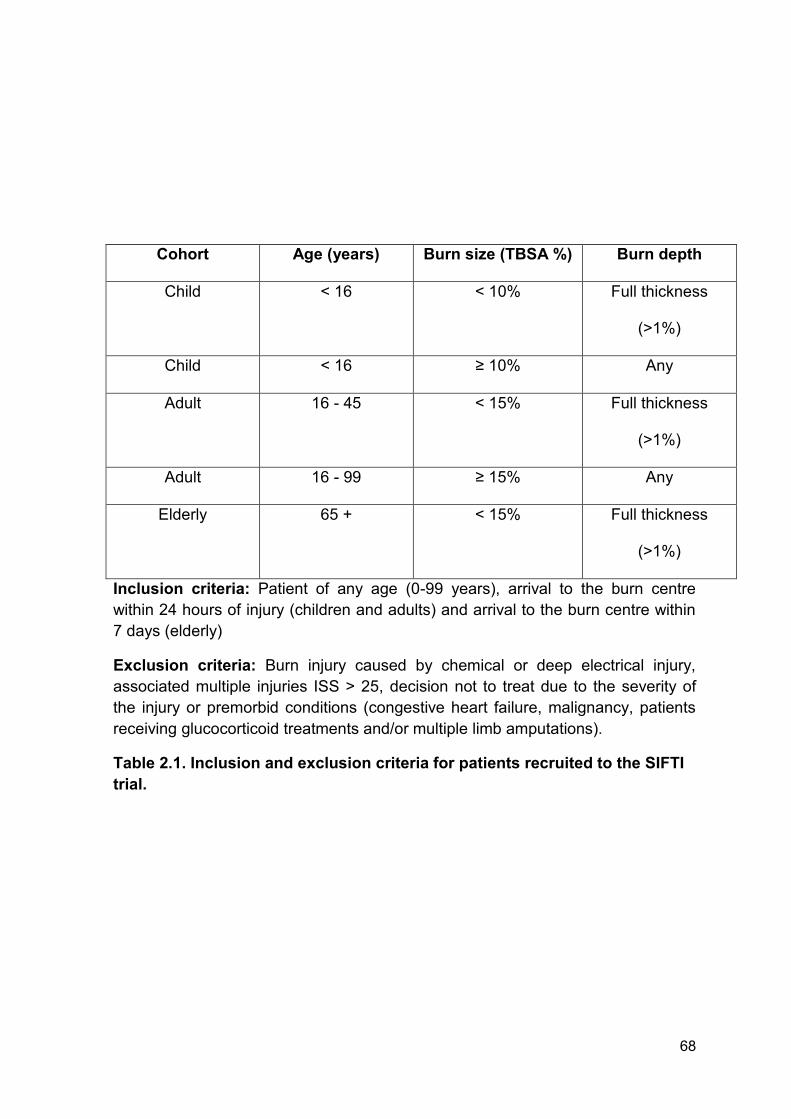
68
Cohort Age (years) Burn size (TBSA %) Burn depth
Child < 16 < 10% Full thickness
(>1%)
Child < 16 ≥ 10% Any
Adult 16 - 45 < 15% Full thickness
(>1%)
Adult 16 - 99 ≥ 15% Any
Elderly 65 + < 15% Full thickness
(>1%)
Inclusion criteria: Patient of any age (0-99 years), arrival to the burn centre
within 24 hours of injury (children and adults) and arrival to the burn centre within
7 days (elderly)
Exclusion criteria: Burn injury caused by chemical or deep electrical injury,
associated multiple injuries ISS > 25, decision not to treat due to the severity of
the injury or premorbid conditions (congestive heart failure, malignancy, patients
receiving glucocorticoid treatments and/or multiple limb amputations).
Table 2.1. Inclusion and exclusion criteria for patients recruited to the SIFTI
trial.
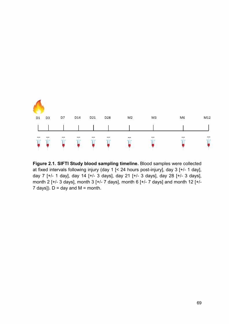
69
Figure 2.1. SIFTI Study blood sampling timeline. Blood samples were collected
at fixed intervals following injury (day 1 [< 24 hours post-injury], day 3 [+/- 1 day],
day 7 [+/- 1 day], day 14 [+/- 3 days], day 21 [+/- 3 days], day 28 [+/- 3 days],
month 2 [+/- 3 days], month 3 [+/- 7 days], month 6 [+/- 7 days] and month 12 [+/-
7 days]). D = day and M = month.
70
2.2 Polytrauma patient group and study design
Data regarding recruitment, bleeding and ethics were provided by Professor
Woolley as he was the senior investigator responsible for management of the
polytrauma cohort. This information is also presented within his doctorate thesis.
This was a prospective observational study which recruited trauma casualties (≥18
years of age) requiring full trauma team activation that presented to the Joint
Force Role 3 Field Hospital in Camp Bastion, Afghanistan, between November
2011 and August 2013 with injuries caused by explosions. A full Trauma team
activation occurs for any patients triaged pre-hospital as T1 (the most severe
triage category) or meeting the activation criteria, Table 2.2. Due to the logistical
constraints of performing research in a deployed military environment, and due to
a lack of full time dedicated researchers, it was not possible to include all trauma
patients. All patients met the inclusion and exclusion criteria described in Table
2.3.
By the end of the conflict in Afghanistan, casualties were evacuated from the
battle field either by the UK Medical Emergency Response Team (MERT) or via
the USA forces PEDRO or Dustoff. Each of these evacuation platforms had
different medical and military capabilities. Of note, the MERT and PEDRO could
administer prehospital blood products to multiple casualties by the end of the
conflict.
Clinical details, including injury severity score (ISS) and new injury severity score
(NISS), were collected by the on-site investigator where possible or retrospectively
analysed. Due to the nature of military trauma and sensitivity of data, some
information was not available for analysis.
71
2.2.1 Study approval and consenting for patients injured in explosions
Due to the nature of major and massive haemorrhage after military trauma a
formal ethics submission was not required by the Ministry of Defence Research
Ethics Committee as the analysis was performed using leftover plasma (waste)
which is a negligible amount in relation to the amount of clinical blood loss. The
US ethical chain however granted ethical approval (log number M-10242).
Informed consent was not required as this represented a minimal risk study.
2.2.2 Plasma preparation from patients injured in explosions
4.5 mls of blood was collected into tri-sodium citrate collection tubes (3.2%), with a
final ratio of blood: anticoagulant 9:1. Blood was centrifuged at 3000 RPM for 20
minutes (Heraeus ® Megafuge 16 series®, Thermoscientific). The plasma was
removed and frozen at -30°C. Samples were transported to the UK using boxes
on cardice and stored until analysis at -80°C.
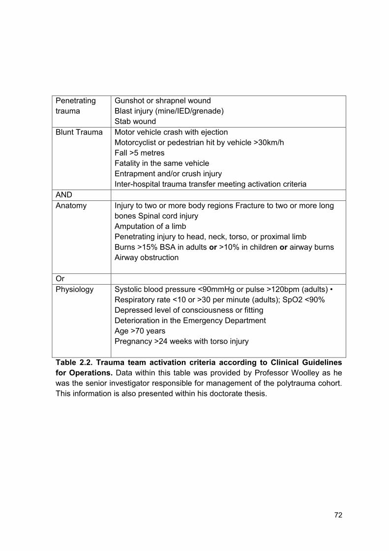
72
Penetrating
trauma
Gunshot or shrapnel wound
Blast injury (mine/IED/grenade)
Stab wound
Blunt Trauma Motor vehicle crash with ejection
Motorcyclist or pedestrian hit by vehicle >30km/h
Fall >5 metres
Fatality in the same vehicle
Entrapment and/or crush injury
Inter-hospital trauma transfer meeting activation criteria
AND
Anatomy Injury to two or more body regions Fracture to two or more long
bones Spinal cord injury
Amputation of a limb
Penetrating injury to head, neck, torso, or proximal limb
Burns >15% BSA in adults or >10% in children or airway burns
Airway obstruction
Or
Physiology Systolic blood pressure <90mmHg or pulse >120bpm (adults) •
Respiratory rate <10 or >30 per minute (adults); SpO2 <90%
Depressed level of consciousness or fitting
Deterioration in the Emergency Department
Age >70 years
Pregnancy >24 weeks with torso injury
Table 2.2. Trauma team activation criteria according to Clinical Guidelines
for Operations. Data within this table was provided by Professor Woolley as he
was the senior investigator responsible for management of the polytrauma cohort.
This information is also presented within his doctorate thesis.
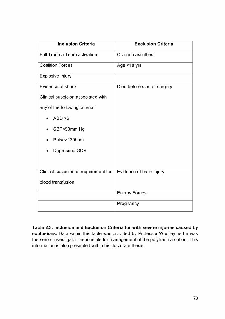
73
Inclusion Criteria Exclusion Criteria
Full Trauma Team activation Civilian casualties
Coalition Forces Age <18 yrs
Explosive Injury
Evidence of shock:
Clinical suspicion associated with
any of the following criteria:
ABD >6
SBP<90mm Hg
Pulse>120bpm
Depressed GCS
Died before start of surgery
Clinical suspicion of requirement for
blood transfusion
Evidence of brain injury
Enemy Forces
Pregnancy
Table 2.3. Inclusion and Exclusion Criteria for with severe injuries caused by
explosions. Data within this table was provided by Professor Woolley as he was
the senior investigator responsible for management of the polytrauma cohort. This
information is also presented within his doctorate thesis.
74
2.3 Whole blood analysis using the Sysmex XN-1000 analyser
The Sysmex XN-1000 (Sysmex UK, UK) is a state of the art whole blood analyser
that is capable of producing a full blood cell differential count from 88 µl of human
blood. The analyser has a set of unique parameters including, IGs, red cell
reticulocytes, accurate platelet counts and the immature platelet fraction. The
instrument also produces three platelet counts: platelet impedance (PLT-I),
platelet optical (PLT-O) and PLT-F count. The instrument was utilised to study the
immaturity index and kinetics of various cell counts affected by thermal injury.
Cellular kinetics was determined in EDTA anticoagulated blood immediately
following collection of sample. To ensure reproducibility the analyser was quality
checked every day before analysis of clinical samples using an internal quality
control (QC) (XN Check Reagent™, (Sysmex UK, UK)). The instrument was also
enrolled into a national external QC scheme (UKNEQAS, UK). To determine
normal ranges for each cellular parameter, EDTA anticoagulated blood from 40
healthy volunteers was analysed. Analysis of healthy volunteers was performed in
collaboration with the Department of Pathology at the Queen Elizabeth Hospital in
Birmingham (UK), by Chris Watson and Miss Sumiya Ahmed (Aston University,
Birmingham, UK).
2.4 Measurement of neutrophil reactive oxygen species generation in whole
blood
Analysis of ROS production was primarily performed by Dr Peter Hampson as part
of the SIFTI study. The methodology has subsequently been published (180).
75
Neutrophil ROS production in response to 1.62 µM PMA stimulation was assessed
using the commercially available PhagoBURST® kit according to manufacturer’s
instructions (BD Biosciences, UK). 100 µl of heparinised blood was aliquoted in a
fresh flow cytometry analysis tube, ensuring no blood was on the side of the tubes.
To the ‘negative control’ tube, 20 µl of wash buffer was added. To the ‘test’ tube
20 µl PMA (1.62 µM) was added after gentle mixing. Samples were mixed gently
and incubated at 37oC for 10 minutes. After incubation, 20 µl of substrate solution
was added to each well and samples were incubated at 37oC for a further 10
minutes. The whole blood samples were lysed and fixed with 2 ml of RT 1 x lysing
solution. Samples were vortexed and incubated for 20 mins at RT. Cells were
pelleted by centrifugation (5 min, 250 x g, 2 - 8 °C) and the supernatant was
discarded. 3 ml of wash solution was added to the tubes and samples were
washed by centrifugation (5 min, 250 x g, 2 - 8 °C) and the supernatant was
discarded. 200 µl of DNA staining solution was added to the tubes, mixed and
incubated for 10 minutes on ice (protected from light). ROS production was
measured within 30 minutes. 10,000 neutrophils, gated according to their forward
scatter/sideward scatter properties, were analysed on an Accuri C6 flow cytometer
and data evaluated using CFlow software. Data are presented as median
fluorescence intensity (MFI) values, which reflect the enzymatic activity per cell
(BD Biosciences, UK).
2.5 Isolation of neutrophils from whole blood
EDTA Anticoagulated blood was transferred to a sterile 50 ml Falcon tube
(Thermo Fisher, USA) and 1 ml of 2% Dextran (Sigma-Aldrich, UK) was added for
every 6 ml of blood. This was performed to cause sedimentation of the
76
erythrocytes so that the top layer, containing white blood cells (WBC), could be
removed following 30 minutes of incubation at RT. After 30 minutes, the leukocyte-
rich plasma was removed from the separated whole blood and was carefully
layered onto a Percoll (Scientific Lab Supplies, UK) density gradient, which
consisted of 2.5 ml of 80% Percoll and 5 ml 56% Percoll and centrifuged for 20
minutes at 218 x g with no brake at RT. Following centrifugation, the neutrophils
that are located at the 56-80% Percoll interface were removed and placed into a
sterile 50 ml Falcon tube and resuspended in RPMI-1640 media supplemented
with 2 mM L-glutamine, 100 U/ml penicillin and 100 μg/ml streptomycin (Thermo
Fisher, USA). Neutrophils were then centrifuged at 461 x g for 10 minutes at RT.
Following centrifugation, the supernatant was discarded and the cells were
resuspended in RPMI-1640 media and diluted to an appropriate final
concentration. Cell count was determined using a Sysmex XN-1000 whole blood
analyser (Section 2.3) with the aspiration sensor turned off. Following
quantification, cellular concentration was adjusted for individual experiments.
Viability of neutrophil preparations was analysed by cytospin and differential
staining using a commercially available Giemsa stain (Diff-Qik; Gentaur Europe,
Belgium). Neutrophils preparations contained ≥ 98% of neutrophil in the total cell
population.
2.6 Generation of neutrophil extracellular traps
To induce NET formation, 2 x 10⁵ of isolated neutrophils (200 µl total volume)
were seeded into wells of a clear flat bottomed 96 well plate (Sigma-Aldrich, UK).
Neutrophils were then stimulated with 25 nM PMA or 100 ng/ml LPS (Both: Sigma-
Aldrich, UK). Both PMA and LPS were diluted from stock solutions with RPMI-
77
1640 media supplemented with 2 mM L-glutamine which alone served as a buffer
control:
PMA stock solution (1.6 mM) was diluted 1:640 (dilution 1). Next, dilution 1
was further diluted 1:100 directly into the well containing 200 µl of isolated
neutrophils. Final dilution was equal to 1:64000
LPS stock solution (1 mg/ml) was diluted 1:100 (dilution 1). Next, dilution 1
was further diluted 1:100 directly into the well containing 200 µl of isolated
neutrophils. Final dilution was equal to 1:10000
Neutrophils were incubated for 3 hours at 37oC and 5% CO2 atmosphere with
either PMA or LPS to induce NET formation. Following 3 hours, NETs were
quantified by a fluorometric assay (Section 2.7).
2.7 Quantification of neutrophil extracellular traps
Following stimulation, the supernatant was removed from wells and placed into
sterile 600 µl eppendorf tubes and centrifuged at 2200 x g for 10 minutes at 4oC to
pellet cells and ‘non-NET’ material. Whilst it is not possible to guarantee that all
NETs were removed, the level of DNA was comparable between individuals
suggesting low variation between experiments. Furthermore, NET release was
confirmed by fluorescence microscopy. Following centrifugation 100 µl of cell free
supernatant was removed and placed into a black 96 well plate (Corning, USA).
The supernatant was incubated with 1 µM SYTOX® Green Dye (Thermo Fisher,
Life Technologies, UK) for 10 minutes at RT and in the dark. Fluorescence was
measured using a BioTek® Synergy 2 fluorometric plate reader (NorthStar
Scientific Ltd, UK) with excitation and emission set at 485 nm and 528 nm
respectively. All samples were analysed in duplicate. As a control/calibration step,
78
cell free supernatant from unstimulated neutrophils and buffer controls were
analysed in duplicate. For calibration of samples, a λ-DNA (ThermoFischer
Scientific, UK) standard curve was utilised ranging from 1000 ng/ml down to 0
ng/ml. λ-DNA (stock concentration = 0.3 µg/µl) was diluted 1:20 in sterile
phosphate buffered saline (PBS) (Sigma-Aldrich, UK). The top standard was then
serially diluted 7 times and finally diluted 1:15 in the plate with 1 µM SYTOX®
Green Dye.
2.8 Visualisation of neutrophil extracellular traps by fluorescent microscopy
This methodology was adapted from a previous publication (283) and has been
previously described by Dr Jon Hazeldine within his doctorate thesis.
To visualise NET generation by fluorescence microscopy, 2 x 10⁵ isolated
neutrophils, resuspended in 2 ml of RPMI media supplemented with 2 mM L-
glutamine, 100 U/ml penicillin and 100 μg/ml streptomycin, were seeded onto
13 mm circular glass coverslips (VWR International, UK) and incubated for 30
minutes at 37oC in a 5% CO2 atmosphere to allow cell adherence.
Post incubation, neutrophils were stimulated with 25 nM PMA or 100 ng/ml LPS
for 3 hours (37oC and 5% CO2 atmosphere). All agonists were prepared from
stock solutions in RPMI media, which alone served as a buffer control. Following
stimulation, cells were fixed with an additional 30 minute incubation with 4%
paraformaldehyde (PFA) (Section 2.9) at 37oC in a 5% CO2 atmosphere. Once
fixed, slides were processed for fluorescence microscopy as detailed below:
5 minute wash in sterile PBS at RT (3 times)
1 minute wash with 0.1% Triton X-100 at RT
79
5 minute wash in sterile PBS at RT
5 minute incubation with 1 µM SYTOX® Green Dye at RT
5 minute wash in sterile PBS at RT
Slides were then mounted in fluoromount medium (Sigma-Aldrich, UK) and
imaged using a LEICA DMI 6000 B microscope at X20 or X40 objective.
2.9 Paraformaldehyde preparation
To make 1 L of 4% PFA, 800 ml of 1 x PBS was heated to 60oC and stirred
continuously. Next, 40 g of PFA powder (Sigma-Aldrich, UK) was added to the
heated 1 x PBS and stirred. The pH of the mixture was adjusted with 1 N NaOH
dropwise until the solution cleared. Once dissolved, the volume was adjusted to 1
L and pH was adjusted to pH 6.9. Finally, solution was filtered sterilised and stored
at 4oC ahead of use in visualisation of NETs and degradation assays.
2.10 Human promyelocytic leukaemia cell culture
Human promyelocytic leukaemia cells (HL-60) (American Type Culture Collection
(ATCC), USA) were cultured to produce a positive control, of Cit H3, for use on the
sodium dodecyl sulphate polyacrylamide gel electrophoresis (SDS-PAGE)
analysis. HL-60 cells were cultured in RPMI-1642 media (Thermo Fisher, USA)
supplemented with 10% (volume/volume (v/v)) heat-inactivated fetal calf serum
(FCS; Sera Laboratories International, UK), 2 mM L-glutamine, 100 U/ml penicillin
and 100 μg/ml streptomycin. HL-60 cells were cultured until there was a suitable
number to differentiate into granulocyte cells.
To differentiate, cells were cultured for 3 days in RPMI-1642 media containing
1.25% dimethyl sulfoxide (DMSO). After 3 days, cells were re-suspended in
80
Locke's Solution and stimulated with 4 µM calcium ionophore (A23187, Sigma-
Aldrich, UK) for 15 minutes at 37oC and 5% CO2. Following stimulation, 1 x 10⁶
cells, determined by cell counts using a standard haemocytometer, were pelleted
by centrifugation at 461 x g for 8 minutes at RT. Cells were then re-suspended in 1
mL of loading buffer ahead of SDS-PAGE analysis. HL-60 positive controls were
further diluted 1:50 before use in western blot analysis.
2.11 K562 cell culture
K562 cells (ATCC number CCL-243) were cultured to create mtDAMPs (Section
2.21). Cells were purchased from the American Type Culture Collection (ATCC,
USA) and were maintained in RPMI-1642 media supplemented with 10%
(volume/volume (v/v)) heat-inactivated fetal calf serum, 2 mM L-glutamine, 100
U/ml penicillin and 100 μg/ml streptomycin in 75 cm2 cell culture flasks at 37oC in
a humidified 5% CO2 atmosphere. Cells were split 1 in 3 on the day preceding
experimentation and washed once (220 x g, 8 minutes, RT) in PBS prior to use.
Cultures of K562 cells were replaced on a monthly basis.
2.12 Western Blot Analysis
3 µl of patient plasma, 20 µl of in vitro preparations or 8 µl of molecular weight
marker (New England Biolabs, UK) were diluted in hot sodium dodecyl sulphate
(SDS) sample buffer (4% SDS (v/v), 0.1 M dithiothreitol, 20% glycerol (v/v), 0.0625
M Tris-HCL and 0.004% bromophenol blue (w/v)). Samples were then heated at
100oC for 10 minutes before being loaded onto a 5% acrylamide stacking gel/15%
acrylamide running SDS-PAGE gel. Gels were run for 90 minutes at 33 mAmps.
The gel was transferred to a polyvinylidene fluoride (PVDF) membrane (GE
81
Healthcare Life Sciences, UK) by wet transfer for 90 minutes (BioRad, UK).
Following transfer the membrane was blocked, at RT for 60 minutes, with 25 ml
1 x Tris Buffered Saline (TBS) containing 4% Tween-20 (TBST), 5% milk (Marvel,
UK), 2% bovine serum albumin (BSA) (Fisher Scientific, UK) and 276 µl of 35%
hydrogen peroxide (to block the endogenous peroxide activity in the plasma
samples). Blocking buffer was removed after 60 minutes and the membrane was
washed 3 times (15 minute washes) with 1 x TBST.
2.12.1 Western blot protocol for Cit H3
Cit H3 in PFP was measured using SDS-PAGE and Western blotting, as
described in Section 2.12. Following the final wash, primary antibody to Cit H3,
diluted in 1 x TBST containing 2.5% BSA to a working concentration of 1 µg/ml,
(ab5103, Abcam UK) was applied to the membrane overnight at 4oC with gentle
shaking. Primary antibody was then removed and the membrane was washed
three times (15 minutes each) with 1 x TBST. The secondary antibody (ECL™
Donkey Anti-Rabbit, Horseradish Peroxidase (HRP)-Linked Whole Antibody (GE
Healthcare Life Sciences)) diluted 1 in 4000 in 1 x TBST containing 2% BSA, was
then applied for 1 hour at RT. Following three washes of 15 minutes each with 1 x
TBST, Amersham™ ECL Plus Western Blotting Detection System (GE Healthcare
Life Sciences) was applied directly to the membrane for 3 minutes before it was
developed using ChemiDoc™ Technology (BioRad, UK). Cit H3 is represented as
a positive band at 17 kDa, calibrated using a loaded molecular weight marker.
82
2.12.2 Western blot protocol for detection of actin
Actin in PFP was measured using SDS-PAGE and Western blotting, as described
in Section 2.12. Membranes were incubated with 1 µg/ml primary antibody anti-
actin, N-terminal antibody produced in rabbit (A2103, Sigma-Aldrich, UK)
overnight at 4oC. Primary antibody was then removed and the membrane was
washed three times (15 minutes each) with 1 x TBST. Membranes were incubated
with secondary antibody HRP-linked anti-rabbit IgG for 1 hour (GE Healthcare Life
Sciences, USA) at RT. Following three washes of 15 minutes each with 1 x TBST,
Amersham™ ECL Plus Western Blotting Detection System (GE Healthcare Life
Sciences, USA) was applied directly to the membrane for 3 minutes before it was
developed using ChemiDoc™ Technology. Actin is represented as a positive band
at 42 kDa, calibrated using a loaded molecular weight marker.
2.13 Detection of cell-free DNA in plasma and serum samples using an in
house fluorometric assay
PFP or serum samples were thawed at 37oC for 10 minutes before being gently
mixed. Next, 10 µl of PFP or serum sample was added directly to a 96-well plate
(Corning, USA) and incubated with 140 µl of cell-impermeable dye (1 µM)
SYTOX® Green Dye for 10 minutes in the dark at RT, in order to stain cfDNA.
Fluorescence was measured using a BioTek® Synergy 2 fluorometric plate reader
with excitation and emission set at 485 nm and 528 nm respectively. All samples
were analysed in duplicate. As a control/calibration step, plasma from healthy
volunteers was included on every plate and a buffer control was included. For
calibration of samples, a λ-DNA standard curve was utilised ranging from 1000
ng/ml down to 0 ng/ml.
83
2.14 Detection of cell-free DNA in plasma and serum samples using a
commercial CE marked assay
Levels of cfDNA were measured in plasma using a commercially available
fluorometric assay (Trillium Diagnostics, USA) to compare to our in-house assay
of cfDNA quantification. Plasma samples were thawed at 37oC for 10 minutes
before being gently mixed ahead of analysis. Next, 10 µl of thawed PFP samples
were incubated with 90 µl reagent A, which binds to cfDNA, at RT for 15 minutes.
Fluorescence was measured using a BioTek® Synergy 2 fluorometric plate reader
with excitation and emission set at 485 nm and 528 nm respectively. All samples
were analysed in duplicate. As a control/calibration step, plasma from healthy
volunteers was included on every plate and a buffer control was included. For
calibration of samples, a λ-DNA standard curve was utilised ranging from 800
ng/ml down to 0 ng/ml
2.15 Isolation of nuclear and mitochondrial DNA
This work was performed in collaboration with Dr Peter Hampson and Dr Jon
Hazledine. The method has subsequently been published (180).
To isolate ncDNA or mtDNA from plasma or cellular preparations a Qiagen DNA
isolation kit was used (Qiagen, Germany). Briefly, 200 µl of plasma or 200 µl
cellular preparations were transferred into a sterile 1.5 ml Eppendorf tube which
was centrifuged at 3000 x g for 10 minutes at 4oC. Following centrifugation, 150 µl
was transferred to a new sterile Eppendorf containing 20 µl of Protease reagent.
Next, 150 µl of Buffer AL was added to all samples and mixed by pulse vortexing
for 15 seconds before being incubated for 10 minutes at 56oC. 150 µl of 100%
ethanol was added to all samples and mixed by pulse vortexing for 15 seconds.
84
Following which, the mixture was carefully transferred to the QIAamp Mini spin
column and centrifuged at 5500 x g for 1 minute at RT. The tube containing the
filtrate was discarded and the QIAamp Mini spin column was transferred to a new
2 ml collection tube. Next, 500 µl of Buffer AW1 was added and the column was
centrifuged at 5500 x g for 1 minute at RT. Again the QIAamp spin column was
placed into a clean 2 ml collection tube and the tube containing the filtrate was
removed. Next, 500 µl of Buffer AW2 was added to the QIAamp spin column and
was centrifuged at 8000 x g for 3 minutes at RT. Following centrifugation, the
QIAamp spin column was removed and centrifuged at 14500 x g for 1 minute to
dry the column. Finally, 50 µl of Buffer AE was added to the QIAamp spin column
and incubated for 5 minutes at RT before being centrifuged at 5500 x g for 1
minute. The liquid in the bottom of the tube contained the DNA which was
quantified using the Nanodrop (Thermo Fisher, USA). DNA had a 260/280 ratio of
1.8 – 2 and was stored at -80oC ahead of use in PCR or DAMPs experiments.
2.16 Polymerase chain reaction for the quantification of plasma nuclear DNA
and mitochondrial DNA levels
This work was performed in collaboration with Dr Peter Hampson and Dr Jon
Hazledine. The method has subsequently been published (180).
To determine the source of the circulating cfDNA, PCR analysis was performed.
DNA was isolated from 150 µl of PFP using a QIAamp DNA Blood Mini Kit
(Qiagen, Germany) and eluted in 50 µl of nuclease-free water of which 5 µl was
used in the PCR reaction. Plasma ncDNA was measured by PCR using the SYBR
Green 480 Probes Master kit and analysed using a Light Cycler 480 (Roche,
Switzerland).
85
Primer sets used to ncDNA was specific for the genes encoding β-globin:
Forward 5’-GTGCACCTGACTCCTGAGGAGA-3’ and reverse 5’-
CCTTGATACCAACCTGCCCAG-3’
Primer sets used to mtDNA was specific for the genes encoding cytochrome b:
Forward 5’-GTGCACCTGACTCCTGAGGAGA-3’ and reverse 5’-
CCTTGATACCAACCTGCCCAG-3’
Primers were synthesized by Eurofins MWG. Primer sequences have no
significant homology with DNA found in any bacterial species published on
BLAST. For concentration determination, a standard curve was created using
purified ncDNA, isolated from K562 cells. Samples that produced no PCR
products after 40 cycles were considered undetectable and the Ct number set to
40 for statistical purposes (180).
2.17 Quantification of DNAse activity in serum samples
The protocol was adapted from a previous publication (237). Neutrophils were
isolated from whole EDTA anticoagulated blood samples, from healthy volunteers,
by Percoll density gradient centrifugation (Section 2.5). 5 x 10⁴ neutrophils were
seeded into each well of a 96 well flat bottom clear tissue culture plate.
Neutrophils were stimulated with 25 nM PMA for 3 hours at 37oC in a 5% CO2
atmosphere. Formed NETs were stored at 4oC for up to 24 hours ahead of
degradation with serum, plasma or DNAse 1.
Following stimulation, neutrophils were incubated for 6 hours with 5% serum or
plasma from healthy volunteers or patients at 37oC in a 5% CO2 environment.
86
Serum was pre-diluted (1:4) in Hank’s Balanced Salt Solutions containing Mg2+
and Ca2+ (HBSS+) (Thermo Fisher, USA) before being diluted 1:5 directly into the
NET containing wells. Following 6 hour incubation wells were fixed by adding 4%
PFA for 30 minutes at 37oC in a 5% CO2 environment. Wells were washed gently
with sterile PBS to remove residual serum and non-fixed cellular debris. NETs
were incubated with 1 µM SYTOX® Green Dye for 10 minutes at RT in the dark.
Fluorescence was measured using a BioTek® Synergy 2 fluorometric plate reader
with excitation and emission set at 485 nm and 528 nm respectively. For analysis
of DNAse activity in patient’s sera, NET degradation by the pooled-serum from 9
healthy donors was defined as 100% activity. A calibration serum from a healthy
individual and a buffer control were included on each assay plate.
2.18 Visualisation of neutrophil extracellular traps degradation by
fluorescent microscopy
To visualise NET degradation by fluorescence microscopy, 5 x 10⁴ isolated
neutrophils, resuspended in 2 ml of RPMI media supplemented with 2 mM L-
glutamine, 100 U/ml penicillin and 100 μg/ml streptomycin, were seeded onto 13
mm circular glass coverslips and incubated for 30 minutes at 37oC in a 5% CO2
atmosphere to allow cell adherence.
Post incubation neutrophils were stimulated with 25 nM PMA for 3 hours (37oC
and 5% CO2 atmosphere). All agonists were prepared from stock solutions in
RPMI media, which alone served as buffer controls. Formed NETs were stored at
4oC for up to 24 hours ahead of degradation with serum or plasma.
Neutrophils were then incubated for 6 hours with 10 U/ml rhDNAse 1 (positive
control), 5% serum from healthy volunteers or patients with thermal injuries.
87
rhDNAse 1 was chosen over other isoforms of DNAse as it is the predominant
enzyme responsible for the degradation of circulating DNA. Serum was pre-diluted
(1:4) in HBSS+ before being diluted 1:5 directly into the NET containing wells.
Following stimulation cells were fixed with an additional 30 minute incubation with
4% PFA (37oC and 5% CO2 atmosphere). Once fixed, slides were processed as
detailed in Section 2.8.
2.19 In Vitro inhibition of DNAse activity in serum samples
NETs were generated as described in Section 2.17. Following generation NETs
were stored at 4oC ahead of degradation with serum. To investigate the inhibition
of DNAse activity, sera from healthy volunteers was incubated with either 1 µM, 2
µM or 5 µM actin from rabbit muscle (Sigma-Aldrich, UK) to cause inhibition of
serum DNase activity. Actin was diluted from a stock (11.63 µM) in HBSS+ which
also served as a buffer control. Actin was added directly into wells with serum
making a total well volume of 250 µl and protocol was followed as previously
described (Section 2.17).
2.20 Enzyme linked immunosorbent assay
All enzyme linked immunosorbent assays (ELISA) were performed as per
manufacturer’s instructions.
2.20.1 Quantification of deoxyribonuclease (DNAse 1) by ELISA
As DNAse 1 is the predominant enzyme responsible for the degradation of
circulating DNA levels were quantified in PFP using a human DNASE1/DNAse I
ELISA Kit, catalogue number LS-F4463 (LifeSpan BioSciences, Inc). Briefly,
88
samples were thawed at 37oC, 5% CO2 for 10 minutes before being gently mixed.
Next, 10 µl of sample was added to 90 µl of Sample diluent (1:10 dilution) in a
clear 96 well plate. Following initial dilution, 10 µl of diluted sample was added
directly to assay plate which contained 90 µl of Sample diluent (1:10 dilution)
making a final dilution of 1:100. Sample, standards and blanks were incubated for
1 hour at 37oC. Wells were then aspirated and 100 µl of a biotin-conjugated
detection antibody (Detection reagent A) is then added which binds to captured
antigen. The plate was then aspirated and washed three times with 300 µl of wash
buffer to remove any unbound Detection reagent A. Following washing, 100 µl of
an avidin-HRP (Detection reagent B) conjugate which binds to the biotin added to
each well. Plate was incubated for 30 minutes at 30oC. The plate was then
aspirated and washed five times with 300 µl of wash buffer to remove any
unbound Detection reagent B. Following washing, 90 µl of tetramethylbenzidine
(TMB) substrate solution was added to each well and incubated for 10 minutes at
37oC before adding 50 µl of a sulfuric acid stop solution to terminate the colour
change. Optical density (OD) of each well is measured at a wavelength of 450 nm
± 2 nm. In all assays samples were analysed in singlet, standards (ranging from
5000 – 0 pg/ml) were analysed in duplicate and plasma from a known healthy
volunteer was included to ensure reproducibility between plates. DNAse levels
were extrapolated from standard curve values using GraphPad Prism® software
(GraphPad Software Limited, USA). OD, for all assays, was measured using a
BioTek® Synergy HT.
89
2.20.2 Quantification of vitamin D binding protein by ELISA
VDBP levels were quantified in serum samples using a VDBP ELISA kit
(catalogue number K2314, ImmunoDiagnostik, Germany). This assay does not
measure VDBP complexed with actin. All serum samples were diluted 1:40000
with SAMPLEBUF (sample dilution buffer). Firstly, 20 µl sample was added to 980
µl SAMPLEBUF and mixed well (1:50 dilution 1). Next, 20 µl of dilution 1 was
added to 980 µl SAMPLEBUF and mixed well (1:50 dilution 2). Finally, 20 µl of
dilution 2 was added to 300 µl SAMPLEBUF and mixed well (1:16 dilution 3). Final
dilution of 1:40000. For analysis, 100 µl of dilution 3 was added to each well.
Before analysis each well was washed 5 times with 250 µl of wash buffer.
Following washing, 100 µl of prediluted sample was added to each well and
incubated at RT for 1 hour with shaking. Following incubation contents of the well
were discarded and washed 5 times with 250 µl of wash buffer to remove any
unbound material. Next, 100 µl of peroxidase-labelled antibody was added to each
well and incubated for 1 hour at RT with shaking. Again, wells were washed 5
times with 250 µl of wash buffer to remove unbound antibody. 100 µl of TMB
solution was added to each well and incubated at RT for 10 minutes. Colour
change was stopped by the addition of 100 µl of stop solution which was mixed
briefly. Absorption was determined immediately with an ELISA reader at 450 nm
against 620 nm (or 690 nm) as a reference. VDBP levels were extrapolated from
standard curve values using GraphPad Prism® software.
2.20.3 Quantification of human gelsolin by ELISA
GSN levels in serum, from patients with thermal injuries, and plasma samples,
patients with injuries caused by explosions, were quantified using LSBioTM
90
Human GSN/Gelsolin ELISA Kit, catalogue number LS-F5675 (LifeSpan
BioSciences Inc, UK). Briefly, samples were thawed at 37oC, 5% CO2 for 10
minutes before being gently mixed. Next, 1 µl of sample was added to 49 µl of
Sample diluent (1:50 dilution) in a clear 96 well plate. Next, 1 µl of diluted sample
was further diluted in 99 µl of assay buffer and mixed gently (1:100 dilution).
Following dilution, 10 µl of diluted sample was added directly to assay plates
which contained 90 µl of Sample diluent (1:10 dilution) making a final dilution of
1:50000. Sample, standards and blanks were incubated for 2 hours at 37oC. Wells
were then aspirated and 100 µl of a biotin-conjugated detection antibody
(Detection reagent A) was then added which binds to captured antigen for 1 hour
at 37oC. The plate was then aspirated and washed three times with 300 µl of wash
buffer to remove any unbound Detection reagent A. Following washing, 100 µl of
an avidin-HRP (Detection reagent B) conjugate which binds to the biotin was
added to each well and plate was incubated for 60 minutes at 37oC. The plate was
then aspirated and washed five times with 350 µl of wash buffer to remove any
unbound Detection reagent B. Following washing, 90 µl of TMB substrate solution
was added to each well and incubated for 20 minutes at 37oC before adding 50 µl
of a sulphuric acid stop solution to terminate the colour change. OD of each well
was measured at a wavelength of 450 nm ± 2 nm. In all assays samples were
analysed in singlet, standards (ranging from 4000 – 0 pg/ml) were analysed in
duplicate and plasma from a known healthy volunteer was included to ensure
reproducibility between plates. GSN levels were extrapolated from standard curve
values using GraphPad Prism® software. OD, for all assays, was measured using
a BioTek® Synergy HT.
91
2.21 Isolation of nuclei and mitochondria from K562 cells
Dr Jon Hazeldine isolated nuclei and mitochondria from K562 cells ahead of use
during in vitro experiments.
Firstly, RIPA lysis buffer (Thermo Fisher, USA) was diluted 1:10 in distilled water
containing protease inhibitory cocktail to make 1 x RIPA buffer. Cells were
transferred into a sterile 1.5 ml Eppendorf tube and pelleted by centrifugation at
1500 x g for 2 minutes at RT. Post centrifugation, the supernatant was removed
and cellular pellet was re-suspended in 1 ml 1 x RIPA lysis buffer. Eppendorf was
left on ice for 10 minutes before the solution was transferred to a tight fitting
homogeniser using a Pasteur pipette and 120 strokes were performed to lyse
cells. Next, the solution was transferred to a fresh sterile 1.5 ml Eppendorf tube
using a Pasteur pipette and centrifuged at 800 x g for 10 minutes at 4oC to pellet
the nucleus. Post spin, the supernatant was carefully removed and placed into a
new sterile 1.5 ml Eppendorf tube using a Pasteur pipette and centrifuged at 3000
x g for 30 minutes at 4oC to pellet the mitochondria. Both the nuclear and
mitochondria pellet were resuspended in 200 µl of sterile PBS. mtDNA and ncDNA
were then isolated using a QIAamp DNA isolation kit (Section 2.15).
2.22 mtDAMPs effects on neutrophil function and phenotype
All experiments were performed in collaboration with Dr Jon Hazeldine (Institute of
Inflammation and Ageing, University of Birmingham, UK). Dr Hazeline performed
western blot analysis and assessment of neutrophil activation by surface
phenotyping using flow cytometry. All other measurements and experiments were
performed in collaboration between Dr Jon Hazeldine and Robert J Dinsdale.
92
2.22.1 Inhibition of neutrophil extracellular traps formation with isolated
mtDAMPs
Neutrophils were isolated from fresh blood by Percoll density gradient (Section
2.5). Next, 2 x 10⁵ were treated for 15 minutes, at 37oC and 5% CO2, with 40 or
100 µg/ml mtDAMPs or vehicle control. Following incubation neutrophils (2 x 10⁵)
were then stimulated for 3 hours with 25 nM PMA. Post-treatment, supernatants
were carefully collected and centrifuged at 2200 x g for 10 minutes at RT, after
which the DNA content of cell-free supernatants was analysed using the SYTOX™
Green dye assay. Background fluorescence values acquired from SYTOX™
Green staining of mtDAMPs in the absence of neutrophils were subtracted from
test readings. NET production is expressed as a fold increase compared to
unstimulated neutrophils alone.
For fluorescent microscopy, 2 x 10⁵ neutrophils were seeded onto glass coverslips
and stimulated with 40 or 100 µg/ml mtDAMPs, 40 µg/ml mtDNA or vehicle control
for 15 minutes at 37oC in a humidified 5% CO2 atmosphere. Post treatment,
samples were stimulated with 25 nM PMA for 3 hours (37oC, 5% CO2), after which
samples were fixed for 30 minutes with 4% PFA (37oC, 5% CO2) and
permeabilized with 0.1% Triton X-100. NETs were stained with 1 µg/ml of a rabbit
polyclonal anti-histone H3 (citrulline R2, R8 and R17) antibody for 90 minutes,
washed three times with PBS and stained for 90 minutes with 10 µg/ml of a goat
anti-rabbit ALEXA Fluro-555 conjugated antibody. Following three washes in PBS,
cells were stained with 1 µM SYTOX Green dye. Once stained, slides were
mounted in fluoromount medium and visualized using a LEICA DMI 6000 B
microscope at x20 objective.
93
2.22.2 Neutrophil transmigration following stimulation with mtDAMPs
Neutrophils were isolated from whole blood by Percoll density gradient as
previously described (Section 2.5). Freshly isolated neutrophils (1 x 10⁷/ml) in
HEPES buffer containing 1 mM Ca2+ were incubated for 30 minutes in a 370C
water bath with 3 µg/ml calcein-acetoxmethyl ester (calcein-AM, Fisher Scientific
UK, UK). Post incubation, neutrophils were treated for 15 minutes (370C, 5% CO2)
with 40 or 100 µg/ml mtDAMPs or vehicle control, after which cells were pelleted
by centrifugation at 1500 x g for 2 minutes at RT, supernatants removed and
neutrophils re-suspended to a concentration of 1 x 10⁷/ml in HEPES buffer
containing 1 mM Ca2+. A total of 100 µl, containing 1 x 10⁶ neutrophils, were then
dispensed into the upper chambers of cell culture inserts (polycarbonate
membrane, 3 µM pores, Corning, USA) that had been pre-loaded into wells of a
24-well flat bottomed plate (BD Biosciences, UK) that contained pre-warmed
phenol red free RPMI-1640 media supplemented with GPS, 10% heat inactivated-
FCS and 1 ng/ml leukotriene B4 (LTB4; R&D Systems, UK).
Following a 90-minute incubation at 37oC in the dark, cell culture inserts were
removed from plates without any attempt to dislodge adherent neutrophils and
plates read immediately for calcein fluorescence using a BioTek Synergy 2
fluorometric plate reader with excitation and emission set at 485 nm and 528 nm
respectively. Fluorescent intensities were converted into neutrophil numbers via
the use of a standard curve that was generated from calcein-AM loaded
neutrophils that had been incubated alongside the test samples in the conditions
described above. The number of neutrophils measured in media in which no
chemokine was added was subtracted from the numbers calculated for wells that
94
contained 1 ng/ml LTB4 in order to determine specific chemokine-mediated
migration.
2.22.3 Assessment of neutrophil phenotype following stimulation with
mtDAMPs
Freshly isolated neutrophils (1 x 10⁵) were stimulated with either 40 or 100 µg/ml
of mtDAMPs or vehicle control for 15 minutes at 37oC in a humidified 5% CO2
atmosphere. Post treatment, samples were placed on ice and stained for 20
minutes with the following mouse anti-human monoclonal antibodies or their
concentration-matched isotype controls: 2 µg/ml fluorescein isothiocyanate
(FITC)-labelled CD62L (clone DREG56; eBioscience, UK), 1 µg/ml CXCR1-FITC
(clone eBIO8F1-1-4; eBioscience, UK), 0.5 µg/ml R-phycoerythrin (PE)-labelled
CXCR2-PE (clone eBio5E8-C7-F10; eBioscience, UK), 2.5 µg/ml allophycocyanin
(APC)-labelled CD11b (clone ICRF44, BioLegend, UK) or 2 µg/ml CD16-APC
(Clone 3G8; BD Biosciences, UK). Post incubation, cells were pelleted (250 x g, 5
minutes, 4oC), supernatants discarded and neutrophils washed once in PBS
containing 1% BSA. Following resuspension in PBS, samples were transferred to
polypropylene FACS tubes for flow cytometric analysis, which was performed on
an Accuri-C6TM bench top cytometer. 10,000 neutrophils, gated according to their
forward scatter/sideward scatter properties were acquired for analysis, where
receptor expression was measured as MFI values.
2.22.4 Activation of ERK1/2 following stimulation with mtDAMPs
To study mitogen activated protein kinase (MAPK) activation following mtDAMP
stimulation, freshly isolated neutrophils (1 x 10⁶ in RPMI + GPS + FCS media)
were treated for 0, 2, 5, 10, 15 or 30 minutes (37oC, 5% CO2) with 100 µg/ml
95
mtDAMPs or vehicle control, after which cells were pelleted (1500 x g, 2 minutes,
4oC) and supernatants removed.
Cell lysates prepared by the addition of SDS sample buffer and boiled for 10
minutes (Section 2.12). Lysates were separated on 12% SDS-PAGE and
membranes probed with pERK1/2 (Cell Signalling Technology, USA) and ERK1/2
antibodies overnight at 4oC (diluted 1:1000). Primary antibody was then removed
and the membrane was washed three times (15 minutes each) with 1 x TBST.
Membranes were incubated with secondary antibody goat anti-rabbit secondary
antibody conjugated to HRP (diluted 1:4000 in TBST; GE Healthcare, UK) at RT.
Following three washes of 15 minutes each with 1 x TBST, Amersham™ ECL Plus
Western Blotting Detection System was applied directly to the membrane for 3
minutes before it was developed using ChemiDoc™ Technology.
2.22.5 Reactive oxygen species generation following stimulation with
mtDAMPs
100 µl aliquots of heparinised whole blood were treated for 15 minutes (37oC, 5%
CO2) with 40 or 100 µg/ml mtDAMPs or vehicle control. Post-treatment, neutrophil
ROS production in response to 1.62 µM PMA stimulation was assessed using the
commercially available PhagoBURST® kit according to manufacturer’s
instructions (Section 2.4). 10,000 neutrophils, gated according to their forward
scatter/sideward scatter properties, were analysed on an Accuri C6 flow cytometer
and data evaluated using CFlow software. Data are presented as MFI values,
which reflect the enzymatic activity per cell.
96
2.23 Statistical analysis
Data were analysed using GraphPad Prism® (GraphPad Software, Inc), SPSS
(IBM) and R version 3.0.1 (http://www.r-project.org) together with the Ime4,
effects, rms and pROC packages. All data within this thesis was checked for
normality using the Kolmogorov-Smirnov test.
2.23.1 Analysis of in vitro experiments
If data was normally distributed the following statistical tests were performed;
Unpaired t test was performed to analyse two independent groups. A paired t test
was performed to analyse groups from paired samples. Not normally distributed
paired groups were analysed by a Wilcoxon matched pairs signed rank test. If
data was normally distributed and there was more than one matched group within
the analysis a one-way analysis of variance (ANOVA) followed by the Bonferroni
post hoc test was used. If data was not normally distributed and there was more
than one matched group within the analysis a one-way analysis of variance
(ANOVA) followed by the Friedman’s test followed by Dunn's Multiple Comparison
Test post hoc test was used.
2.23.2 Analysis of data generated from patients with thermal injuries
When comparing to healthy individuals the level of statistical significance was
adjusted manually for all analysis due to data not being equally distributed across
all longitudinal time points. The threshold for significance was set at p < 0.005,
unless otherwise stated within figure legend, as there are 10 longitudinal time
points measured within this analysis. If data was normally distributed data was
97
analysed using an unpaired t test. If data was not normally distributed data was
analysed using a Mann Whitney test. When analysis was performed on patient
groups, e.g. sepsis and no sepsis, the threshold for significance was not adjusted
(p < 0.05). If data was normally distributed data was analysed using an unpaired t
test. If data was not normally distributed data was analysed using a Mann Whitney
test. Categorical variables were compared using a Chi-squared test.
2.23.3 Logistic regression analyses of potential biomarkers of sepsis
Logistic regression analyses were conducted to examine the relationships
between biomarkers at pre-specified sample times (e.g. day 7) and the presence
of sepsis. Discriminatory power was assessed through the area under the receiver
operator characteristic curve (AUROC). Longitudinal analyses were performed
using linear mixed-effects models. Sample day was included in these models as a
restricted cubic spline to allow for a flexible non-linear relationship between time
and the response variable. Analysis was performed using the statistical software
packages SPSS (IBM) and R version 3.0.1 (http://www.r-project.org) together with
the Ime4, effects, rms and pROC packages. Analysis was performed by Dr
Jonathon Bishop (University of Birmingham, UK).
2.23.4 Analysis of data generated from patients with polytrauma
As data was equally distributed amongst analysis groups the threshold for
significance did not need to be adjusted manually (p < 0.05). When comparing to
healthy controls, a one-way ANOVA followed by Bonferroni’s post hoc test was
performed when data was normally distributed. If data was not normally distributed
a one-way ANOVA followed by Dunn's Multiple Comparison Test was performed.
98
When analysis was performed between patient cohorts (blood products and no
blood products) the threshold for significance was not adjusted (p < 0.05). If data
was normally distributed an unpaired t test was performed. If data was not
normally distributed a Mann Whitney test was performed. Categorical variables
were compared using a Chi-squared test.
99
Chapter 3
Neutrophil extracellular trap release following thermal
injury
100
3.1 Introduction
Neutrophils are essential effectors cells of the innate immune system and are
equipped with a number of antimicrobial functions. In 2004, Brinkmann and
colleagues defined a novel anti-microbial process in which neutrophils release
their nuclear material to the outside of the cell in a ‘net-like’ structure which is
designed to ensnare and trap bacteria (118, 119). This process is termed NET
release or NETosis (178).
The majority of research within the field of NETosis has focussed around their role
during infection and sepsis. This is in part due to the specific functions of NETs in
orchestrating the response to infection and due to their components being
potential biomarkers (181, 192). Sepsis is a life threatening condition
characterised by an imbalance in both the immune and haemostatic systems. It
often results in cardiac dysfunction and acute respiratory distress syndrome, which
are associated with MOF (46). Although accurate and early diagnosis of sepsis is
crucial for determining patient outcomes, this is challenging as the criteria used
are insensitive and nonspecific (47).
A number of studies have concluded that NET production occurs in septic patients
following traumatic injury (181, 215, 216). Furthermore, it has also been suggested
that NETs can also be produced in response to sterile thermal and traumatic injury
(111, 181, 215). However, many of the studies use quantification of plasma cfDNA
alone as a marker of in vivo NETosis. This method is non-specific to NETosis as
plasma cfDNA can originate from a number of sources including bacteria
themselves (115). Therefore, to our knowledge, it has yet to be conclusively
established whether NETosis occurs in patients following burn injury.
101
There is emerging evidence that neutrophil function is dysregulated following
severe thermal injury (156, 159-161, 173, 174, 177). It has been proposed that
this dysregulation of neutrophil function may underlie the increased incidence of
nosocomial infections. Parment and colleagues have reported a reduced ability of
neutrophils to generate ROS, an essential precursor of NETosis, following thermal
injury (177). Although precursors of NET formation are reduced no group has
investigated the effect severe thermal injury has on NET formation. Furthermore, if
dysfunction exists no group has fully investigated the potential mechanisms
responsible. Potential causes of neutrophil dysfunction include non-functioning
immature neutrophils and circulating soluble mediators (40, 41, 45, 169).
3.1.1 Aims
The aims of this chapter were:
Conclusively investigate NET formation following severe thermal injury by
the measurement of specific circulating biomarkers.
Investigate the association and predictive power of circulating NET
components and secondary complications, e.g. sepsis.
Investigate the ability of neutrophils to form NETs ex vivo to understand if
thermal injury results in impaired neutrophil function.
Investigate potential mechanisms responsible for the dysregulation of
neutrophil function.
102
3.2 Results
3.2.1 Study cohort
All analysis was performed on adult patients (≥ 18 years of age) with burns ≥ 15%
TBSA. The consort diagram for this analysis is included (Figure 3.1). Analysis was
restricted to burns ≥ 15% TBSA to minimise the confounding bias of age and
severity of injury in analysis. Furthermore, this has reduced the span and
confounding bias of fluid resuscitation. Importantly, all patients were admitted
within 24 hours of their injuries and all received standardised burn resuscitation
protocols as per Parkland’s formula and as such have received equivalent fluid
resuscitation: average = 5.4 mls/kg/%TBSA (Standard deviation = 2.1) (284, 285).
In total there are 63 patients included in this analysis. The median age of the
cohort was 39 years of age (range 16 - 88) and the median TBSA was 30% (range
15 - 95%). Patients were further subcategorised into septic and non-septic
according to the ABA sepsis scoring criteria (65). Of note, 6 patients died within 7
days of injury from non-septic causes and were removed from the analysis, as it
was not possible to determine whether these patients would have developed
sepsis. Septic patients had significantly larger and more severe injuries (p <
0.001) and consequently a higher mortality rate (p = 0.002). Full patient
demographics are reported in Table 3.1.
The incidence of sepsis was 61.4 % and the median time to first septic episode
was 4 days (Interquartile range (IQR): 4, 8). The major source of sepsis in this
cohort was pulmonary infection, accounting for 69%, and the most common
colonising organism was Bacillus sp, accounting for 20%. Full details of colonising
organisms and source can be found in Table 3.2.
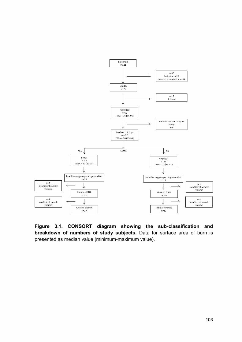
103
Figure 3.1. CONSORT diagram showing the sub-classification and
breakdown of numbers of study subjects. Data for surface area of burn is
presented as median value (minimum-maximum value).
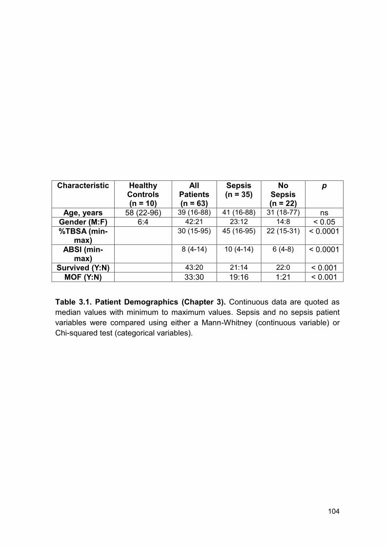
104
Characteristic Healthy Controls (n = 10)
All Patients (n = 63)
Sepsis (n = 35)
No Sepsis (n = 22)
p
Age, years 58 (22-96) 39 (16-88) 41 (16-88) 31 (18-77) ns
Gender (M:F) 6:4 42:21 23:12 14:8 < 0.05
%TBSA (min-max)
30 (15-95) 45 (16-95) 22 (15-31) < 0.0001
ABSI (min-max)
8 (4-14) 10 (4-14) 6 (4-8) < 0.0001
Survived (Y:N) 43:20 21:14 22:0 < 0.001
MOF (Y:N) 33:30 19:16 1:21 < 0.001
Table 3.1. Patient Demographics (Chapter 3). Continuous data are quoted as
median values with minimum to maximum values. Sepsis and no sepsis patient
variables were compared using either a Mann-Whitney (continuous variable) or
Chi-squared test (categorical variables).
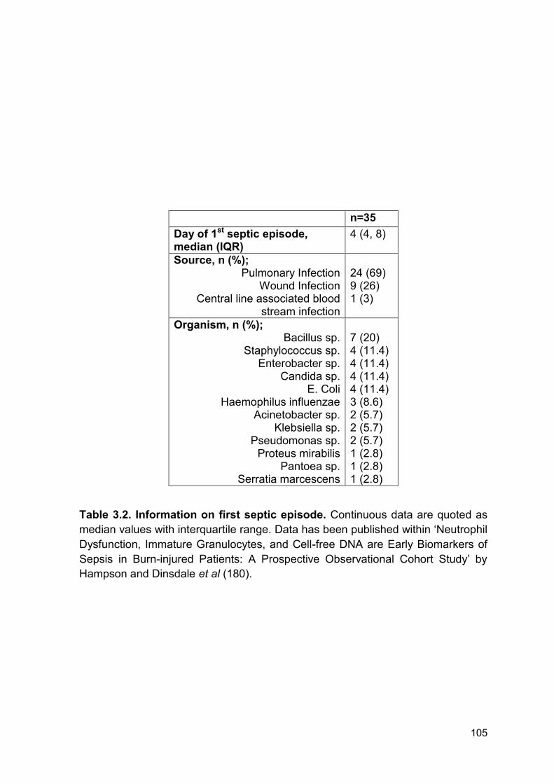
105
n=35
Day of 1st septic episode, median (IQR)
4 (4, 8)
Source, n (%); Pulmonary Infection
Wound Infection Central line associated blood
stream infection
24 (69) 9 (26) 1 (3)
Organism, n (%); Bacillus sp.
Staphylococcus sp. Enterobacter sp.
Candida sp. E. Coli
Haemophilus influenzae Acinetobacter sp.
Klebsiella sp. Pseudomonas sp.
Proteus mirabilis Pantoea sp.
Serratia marcescens
7 (20) 4 (11.4) 4 (11.4) 4 (11.4) 4 (11.4) 3 (8.6) 2 (5.7) 2 (5.7) 2 (5.7) 1 (2.8) 1 (2.8) 1 (2.8)
Table 3.2. Information on first septic episode. Continuous data are quoted as
median values with interquartile range. Data has been published within ‘Neutrophil
Dysfunction, Immature Granulocytes, and Cell-free DNA are Early Biomarkers of
Sepsis in Burn-injured Patients: A Prospective Observational Cohort Study’ by
Hampson and Dinsdale et al (180).
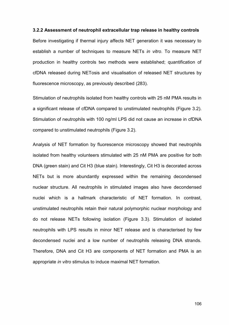
106
3.2.2 Assessment of neutrophil extracellular trap release in healthy controls
Before investigating if thermal injury affects NET generation it was necessary to
establish a number of techniques to measure NETs in vitro. To measure NET
production in healthy controls two methods were established; quantification of
cfDNA released during NETosis and visualisation of released NET structures by
fluorescence microscopy, as previously described (283).
Stimulation of neutrophils isolated from healthy controls with 25 nM PMA results in
a significant release of cfDNA compared to unstimulated neutrophils (Figure 3.2).
Stimulation of neutrophils with 100 ng/ml LPS did not cause an increase in cfDNA
compared to unstimulated neutrophils (Figure 3.2).
Analysis of NET formation by fluorescence microscopy showed that neutrophils
isolated from healthy volunteers stimulated with 25 nM PMA are positive for both
DNA (green stain) and Cit H3 (blue stain). Interestingly, Cit H3 is decorated across
NETs but is more abundantly expressed within the remaining decondensed
nuclear structure. All neutrophils in stimulated images also have decondensed
nuclei which is a hallmark characteristic of NET formation. In contrast,
unstimulated neutrophils retain their natural polymorphic nuclear morphology and
do not release NETs following isolation (Figure 3.3). Stimulation of isolated
neutrophils with LPS results in minor NET release and is characterised by few
decondensed nuclei and a low number of neutrophils releasing DNA strands.
Therefore, DNA and Cit H3 are components of NET formation and PMA is an
appropriate in vitro stimulus to induce maximal NET formation.
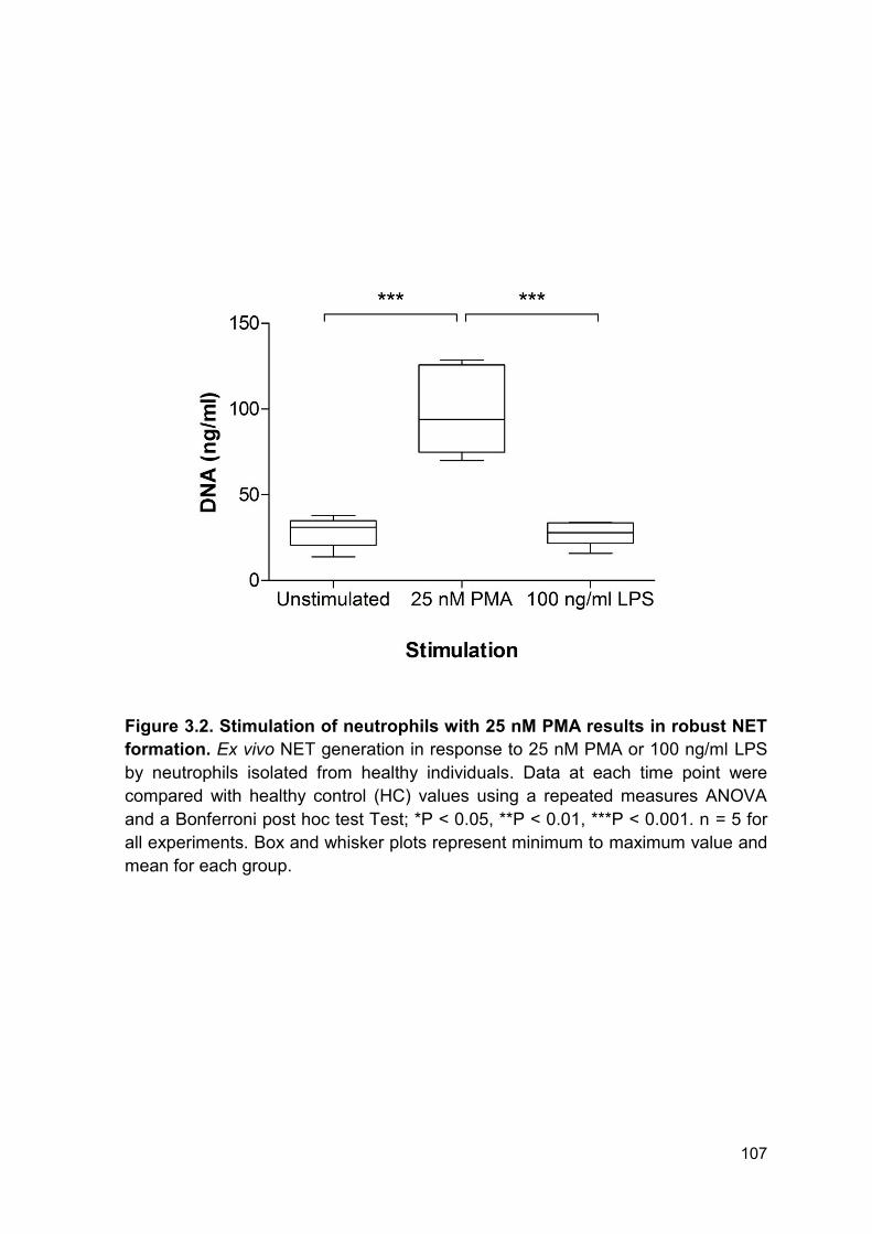
107
Figure 3.2. Stimulation of neutrophils with 25 nM PMA results in robust NET
formation. Ex vivo NET generation in response to 25 nM PMA or 100 ng/ml LPS
by neutrophils isolated from healthy individuals. Data at each time point were
compared with healthy control (HC) values using a repeated measures ANOVA
and a Bonferroni post hoc test Test; *P < 0.05, **P < 0.01, ***P < 0.001. n = 5 for
all experiments. Box and whisker plots represent minimum to maximum value and
mean for each group.
108
Figure 3.3. Visualisation of ex vivo NET generation in response to 25 nM
PMA or 100 ng/ml LPS by neutrophils isolated from a healthy control by
fluorescence microscopy. DNA was stained by 1 µM SYTOX Green Dye (green
stain) and Cit H3 was stained using 1 µg/ml AB5103 in addition to secondary
antibody ALEXA Fluor 555 (blue stain). Slides were mounted in fluoromount
medium and visualized using a LEICA DMI 6000 B microscope at X20 objective.
Data shown is representative of 5 individual experiments.
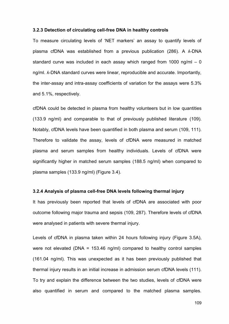
109
3.2.3 Detection of circulating cell-free DNA in healthy controls
To measure circulating levels of ‘NET markers’ an assay to quantify levels of
plasma cfDNA was established from a previous publication (286). A ʎ-DNA
standard curve was included in each assay which ranged from 1000 ng/ml – 0
ng/ml. ʎ-DNA standard curves were linear, reproducible and accurate. Importantly,
the inter-assay and intra-assay coefficients of variation for the assays were 5.3%
and 5.1%, respectively.
cfDNA could be detected in plasma from healthy volunteers but in low quantities
(133.9 ng/ml) and comparable to that of previously published literature (109).
Notably, cfDNA levels have been quantified in both plasma and serum (109, 111).
Therefore to validate the assay, levels of cfDNA were measured in matched
plasma and serum samples from healthy individuals. Levels of cfDNA were
significantly higher in matched serum samples (188.5 ng/ml) when compared to
plasma samples (133.9 ng/ml) (Figure 3.4).
3.2.4 Analysis of plasma cell-free DNA levels following thermal injury
It has previously been reported that levels of cfDNA are associated with poor
outcome following major trauma and sepsis (109, 287). Therefore levels of cfDNA
were analysed in patients with severe thermal injury.
Levels of cfDNA in plasma taken within 24 hours following injury (Figure 3.5A),
were not elevated (DNA = 153.46 ng/ml) compared to healthy control samples
(161.04 ng/ml). This was unexpected as it has been previously published that
thermal injury results in an initial increase in admission serum cfDNA levels (111).
To try and explain the difference between the two studies, levels of cfDNA were
also quantified in serum and compared to the matched plasma samples.
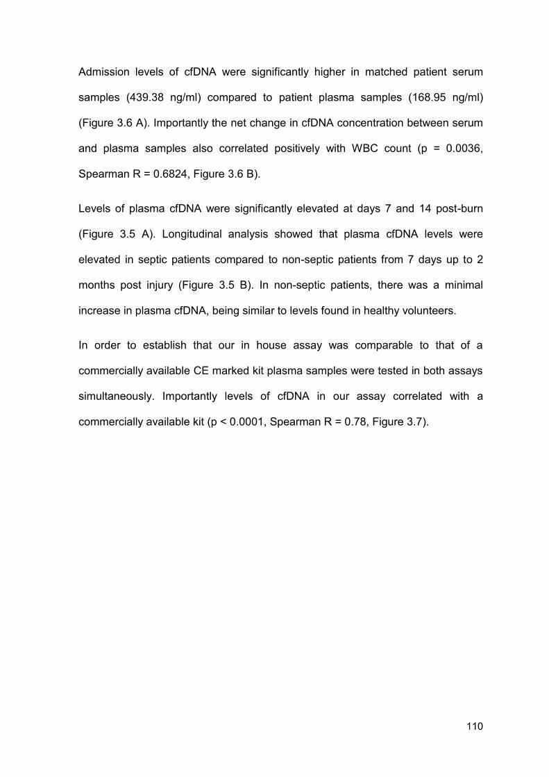
110
Admission levels of cfDNA were significantly higher in matched patient serum
samples (439.38 ng/ml) compared to patient plasma samples (168.95 ng/ml)
(Figure 3.6 A). Importantly the net change in cfDNA concentration between serum
and plasma samples also correlated positively with WBC count (p = 0.0036,
Spearman R = 0.6824, Figure 3.6 B).
Levels of plasma cfDNA were significantly elevated at days 7 and 14 post-burn
(Figure 3.5 A). Longitudinal analysis showed that plasma cfDNA levels were
elevated in septic patients compared to non-septic patients from 7 days up to 2
months post injury (Figure 3.5 B). In non-septic patients, there was a minimal
increase in plasma cfDNA, being similar to levels found in healthy volunteers.
In order to establish that our in house assay was comparable to that of a
commercially available CE marked kit plasma samples were tested in both assays
simultaneously. Importantly levels of cfDNA in our assay correlated with a
commercially available kit (p < 0.0001, Spearman R = 0.78, Figure 3.7).
111
Figure 3.4. Levels of cfDNA are significantly higher in serum compared to
plasma samples from healthy individuals. Levels of cfDNA were quantified in
plasma and serum samples from healthy individuals (n = 12). Data was compared
by a Wilcoxon matched pairs signed rank test. ***P < 0.001. Data is presented as
before and after to depict changes within individual donors.
112
Figure 3.5. cfDNA is elevated post thermal injury and elevated in septic
patients. (A) Plasma cfDNA levels (log scale) across time following burn injury
(n = 50). Data at each time point were compared with healthy control (HC) (n = 10)
values using a Mann-Whitney test; *P < 0.005. (B) Levels of cfDNA compared
between septic and non-septic patients. Data was analysed using a Mann-
Whitney test; *P < 0.05. Box and whisker plots represent minimum to maximum
value and median for each group. HC = healthy control, D = day, M = month.
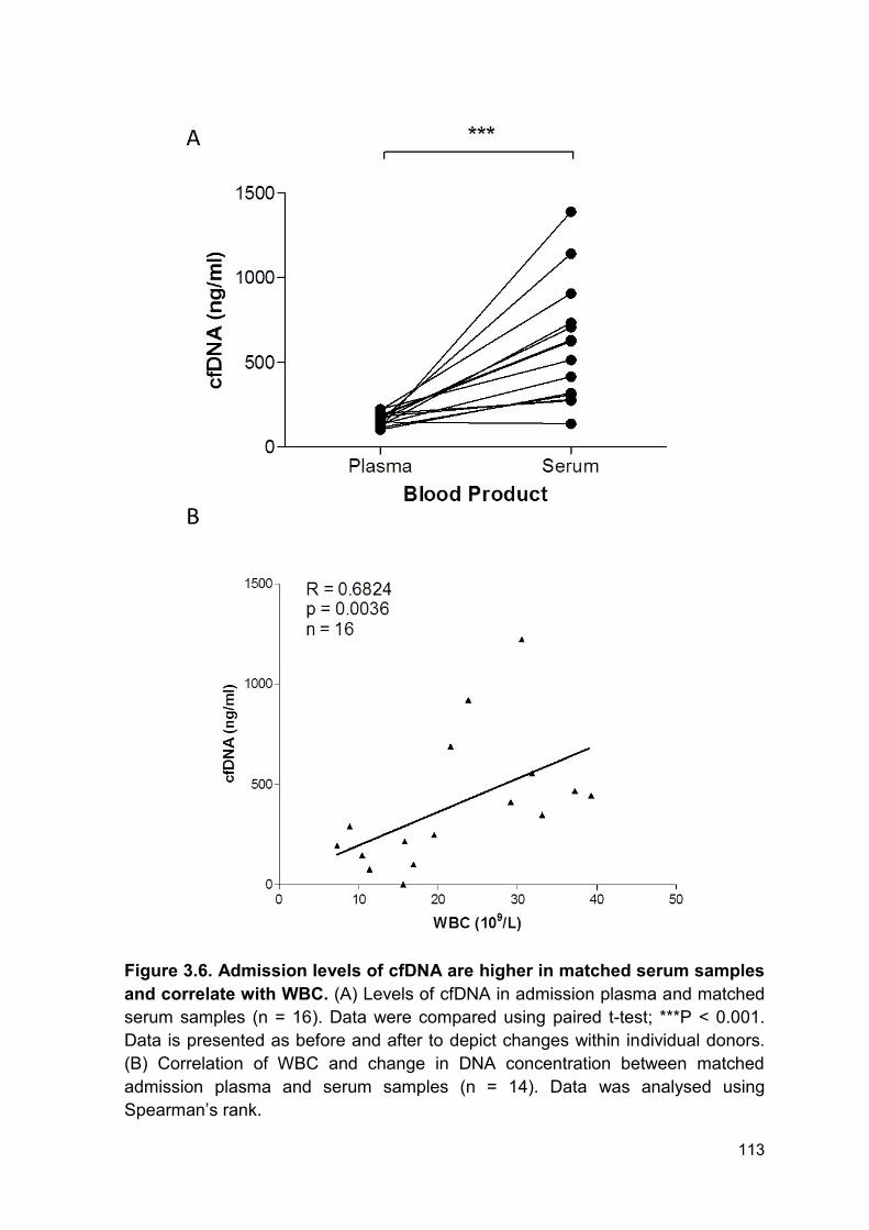
113
Figure 3.6. Admission levels of cfDNA are higher in matched serum samples
and correlate with WBC. (A) Levels of cfDNA in admission plasma and matched
serum samples (n = 16). Data were compared using paired t-test; ***P < 0.001.
Data is presented as before and after to depict changes within individual donors.
(B) Correlation of WBC and change in DNA concentration between matched
admission plasma and serum samples (n = 14). Data was analysed using
Spearman’s rank.
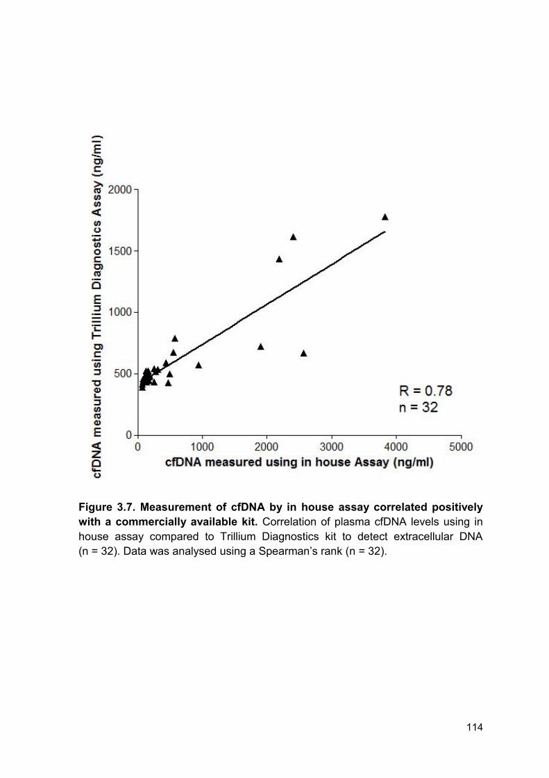
114
Figure 3.7. Measurement of cfDNA by in house assay correlated positively
with a commercially available kit. Correlation of plasma cfDNA levels using in
house assay compared to Trillium Diagnostics kit to detect extracellular DNA
(n = 32). Data was analysed using a Spearman’s rank (n = 32).
115
3.2.5 Diagnostic use of cell-free DNA in septic patients
In order to investigate the potential diagnostic utility of plasma cfDNA during
sepsis, we performed statistical regression analysis on the longitudinal kinetics for
septic and non-septic patients (Table 3.3). There was a clear significant difference
in plasma cfDNA trajectories following thermal injury between septic and non-
septic patients (Figure 3.5 B). On days 1 and 3 post injury the model gave
reasonable discriminatory power between septic and non-septic patients with a c-
statistic of 0.691 (95% CI (0.542, 0.840)) and 0.640 (95% CI (0.479, 0.802))
respectively. Discriminatory power of the model increased with time with a c-
statistic of 0.775 (95% CI (0.604, 0.947)) on day 7 post injury. However, the
maximal difference between the two patients groups was observed at day 14
following injury. Levels of plasma cfDNA at day 14 following thermal injury provide
good discriminatory power of whether an individual was in the septic group or not
with a c-statistic of 0.883 (95% CI (0.767, 1.000)).
3.2.6 Longitudinal analysis of nuclear and mitochondrial DNA
In order to investigate the potential source(s) of circulating cfDNA, quantitative
PCR analysis was performed to determine the concentration of ncDNA and
mtDNA. Analysis of plasma levels of mtDNA revealed no significant elevation
following thermal injury compared to healthy volunteers (Figure 3.8 A). However,
there was a significant elevation of ncDNA between day 1 and day 28 post-injury
(Figure 3.8 B). Plasma ncDNA levels had comparable kinetics to cfDNA, with
levels peaking at day 7 and 14, and there was a strong positive correlation
between the two measurements (r= 0.763, p < 0.001). However, there was a
significant elevation of ncDNA at day 1-3 post-injury unlike plasma cfDNA. This is

116
most likely due to the higher sensitivity of the PCR assay compared to the
fluorometric assay (lower limit of detection; 0.1 ng/ml vs.60 ng/ml).
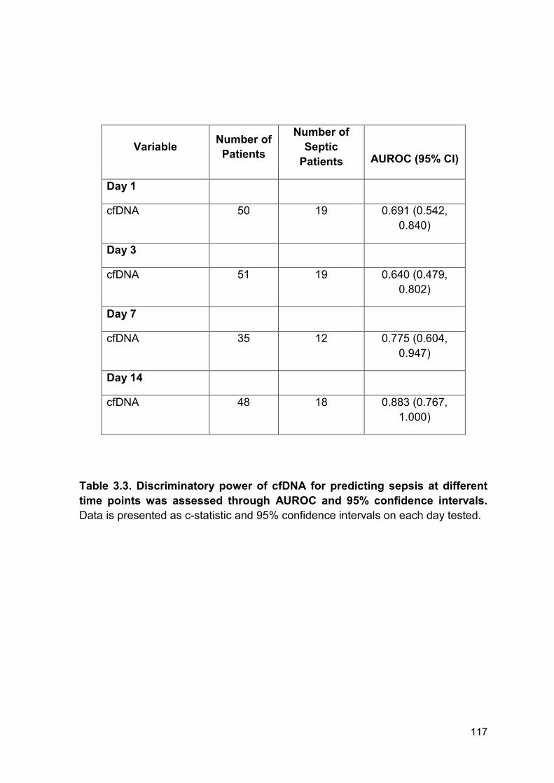
117
Variable Number of
Patients
Number of
Septic
Patients
AUROC (95% CI)
Day 1
cfDNA 50 19 0.691 (0.542,
0.840)
Day 3
cfDNA 51 19 0.640 (0.479,
0.802)
Day 7
cfDNA 35 12 0.775 (0.604,
0.947)
Day 14
cfDNA 48 18 0.883 (0.767,
1.000)
Table 3.3. Discriminatory power of cfDNA for predicting sepsis at different
time points was assessed through AUROC and 95% confidence intervals.
Data is presented as c-statistic and 95% confidence intervals on each day tested.
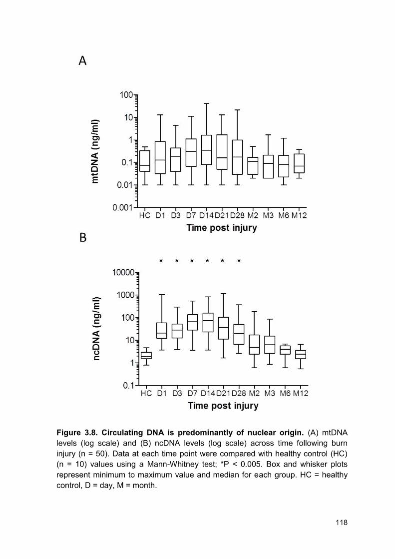
118
Figure 3.8. Circulating DNA is predominantly of nuclear origin. (A) mtDNA
levels (log scale) and (B) ncDNA levels (log scale) across time following burn
injury (n = 50). Data at each time point were compared with healthy control (HC)
(n = 10) values using a Mann-Whitney test; *P < 0.005. Box and whisker plots
represent minimum to maximum value and median for each group. HC = healthy
control, D = day, M = month.
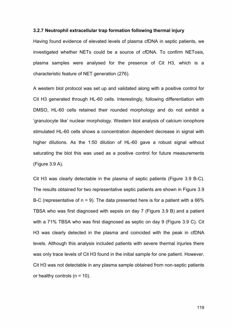
119
3.2.7 Neutrophil extracellular trap formation following thermal injury
Having found evidence of elevated levels of plasma cfDNA in septic patients, we
investigated whether NETs could be a source of cfDNA. To confirm NETosis,
plasma samples were analysed for the presence of Cit H3, which is a
characteristic feature of NET generation (276).
A western blot protocol was set up and validated along with a positive control for
Cit H3 generated through HL-60 cells. Interestingly, following differentiation with
DMSO, HL-60 cells retained their rounded morphology and do not exhibit a
‘granulocyte like’ nuclear morphology. Western blot analysis of calcium ionophore
stimulated HL-60 cells shows a concentration dependent decrease in signal with
higher dilutions. As the 1:50 dilution of HL-60 gave a robust signal without
saturating the blot this was used as a positive control for future measurements
(Figure 3.9 A).
Cit H3 was clearly detectable in the plasma of septic patients (Figure 3.9 B-C).
The results obtained for two representative septic patients are shown in Figure 3.9
B-C (representative of n = 9). The data presented here is for a patient with a 66%
TBSA who was first diagnosed with sepsis on day 7 (Figure 3.9 B) and a patient
with a 71% TBSA who was first diagnosed as septic on day 9 (Figure 3.9 C). Cit
H3 was clearly detected in the plasma and coincided with the peak in cfDNA
levels. Although this analysis included patients with severe thermal injuries there
was only trace levels of Cit H3 found in the initial sample for one patient. However,
Cit H3 was not detectable in any plasma sample obtained from non-septic patients
or healthy controls (n = 10).
120
Figure 3.9. Longitudinal analysis of NET formation. In all experiments, Cit H3
was detected in the plasma of septic patients as a 17kDa band by Western
Blotting. (A) HL-60 positive control diluted 1:20, 1:50, 1:100, 1:150. (B-C) Two
representative septic patients following thermal injury (data is representative of 9).
Data presented includes longitudinal plasma cfDNA measurements (log scale) and
corresponding Cit H3 blots. A positive control (generated in HL-60 cells) was
included on all blots. HC = healthy control, D = day, MWM = molecular weight
marker.
121
3.2.8 Ex vivo NETosis
There is an increasing amount of evidence that thermal injury results in
suppression or dysregulation of neutrophil functions (156, 173, 174). However, to
date, no group has investigated if thermal injury also disrupts NET formation.
Therefore, to investigate whether thermal injury affected NET production by
circulating neutrophils, cfDNA release by isolated neutrophils stimulated with
25nM PMA or 100 ng/ml LPS was quantified by fluorescent microscopy and
fluorometry.
Firstly, to investigate if neutrophils from patients with thermal injuries were primed
and in turn released higher levels of NETs, neutrophils were stimulated with 100
ng/ml LPS. In healthy individuals, neutrophils stimulated with LPS do not result in
a significant increase in NET release compared to unstimulated neutrophils
(Figure 3.2). Interestingly, we found that neutrophils from burn injury patients did
not release more NETs when stimulated with 100 ng/ml LPS compared to healthy
controls at all time points tested (Figure 3.10 A).
To confirm maximal NET generation, neutrophils were stimulated with PMA for 3
hours. Interestingly, neutrophils from burn injury patients released significantly
lower levels of DNA compared to neutrophils isolated from healthy controls on
days 3 and 7 post injury (Figure 3.10 B). Fluorescence microscopy imaging
confirmed that neutrophils isolated from burn injury patients are partially resistant
to NETosis induced by 25nM PMA, with the majority of the neutrophils from burn
injury patients retaining their natural cell and nuclear morphology following
stimulation (Figure 3.10 C). However, in healthy controls nearly all cells release
NETs and have decondensed nuclei.
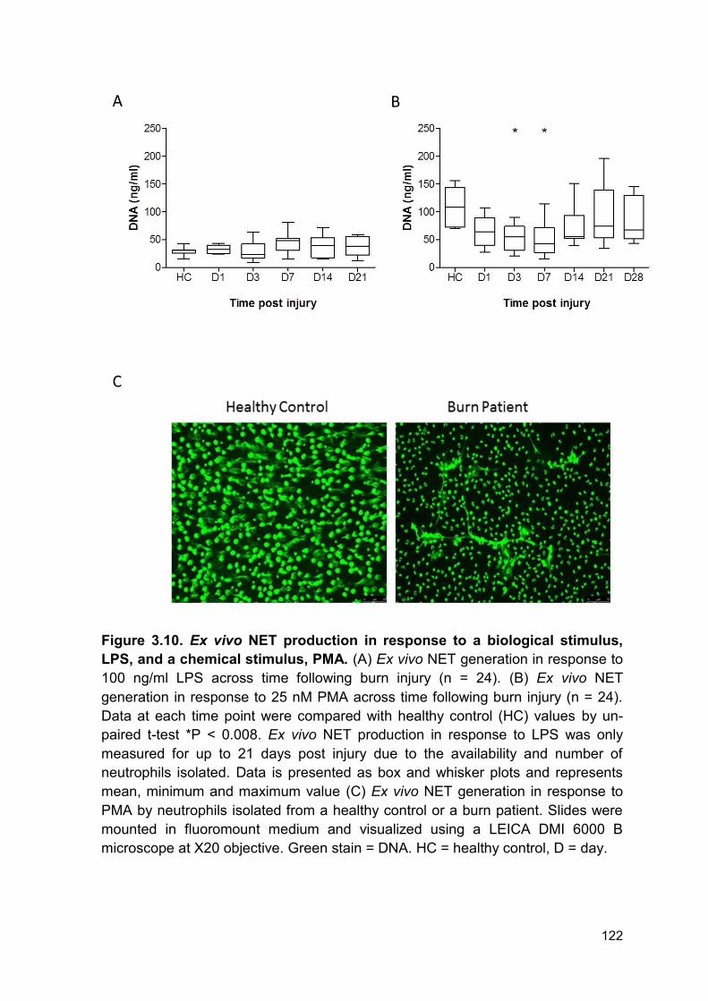
122
Figure 3.10. Ex vivo NET production in response to a biological stimulus,
LPS, and a chemical stimulus, PMA. (A) Ex vivo NET generation in response to
100 ng/ml LPS across time following burn injury (n = 24). (B) Ex vivo NET
generation in response to 25 nM PMA across time following burn injury (n = 24).
Data at each time point were compared with healthy control (HC) values by un-
paired t-test *P < 0.008. Ex vivo NET production in response to LPS was only
measured for up to 21 days post injury due to the availability and number of
neutrophils isolated. Data is presented as box and whisker plots and represents
mean, minimum and maximum value (C) Ex vivo NET generation in response to
PMA by neutrophils isolated from a healthy control or a burn patient. Slides were
mounted in fluoromount medium and visualized using a LEICA DMI 6000 B
microscope at X20 objective. Green stain = DNA. HC = healthy control, D = day.

123
3.2.9 Reactive oxygen species generation of neutrophils following thermal
injury
Analysis of ROS generation was performed in collaboration with Dr Peter
Hampson as part of the SIFTI Trial (University of Birmingham, UK). Data has been
published (180).
Of note, we have shown that there is a significant reduction in the ability of
neutrophils to phagocytose and ex vivo NETosis following thermal injury compared
to healthy individuals (180). Thus, we investigated if neutrophils were
dysfunctional in ROS production, suggesting a broader inhibition of neutrophil
antibacterial functions. Compared to the levels measured in healthy volunteers,
neutrophil ROS production was significantly reduced in patients with severe
thermal injuries (Figure 3.11 A). This reduction ensued 3 days following injury and
was lowest at 7 days post injury. The reduction in ROS formation persisted for up
to a month following thermal injury (Figure 3.11 A).
3.2.10 Neutrophil function is reduced to a greater degree in septic patients
Analysis of ROS production in relation to sepsis status was performed to
understand if dysregulated ROS production was responsible for the increased
incidence of sepsis post thermal injury. ROS was reduced to a similar degree over
the first 7 days following injury in both patient cohorts. However, ROS production
was significantly lower in septic patients from day 14 – month 2 compared to non-
septic individuals (Figure 3.11 B).
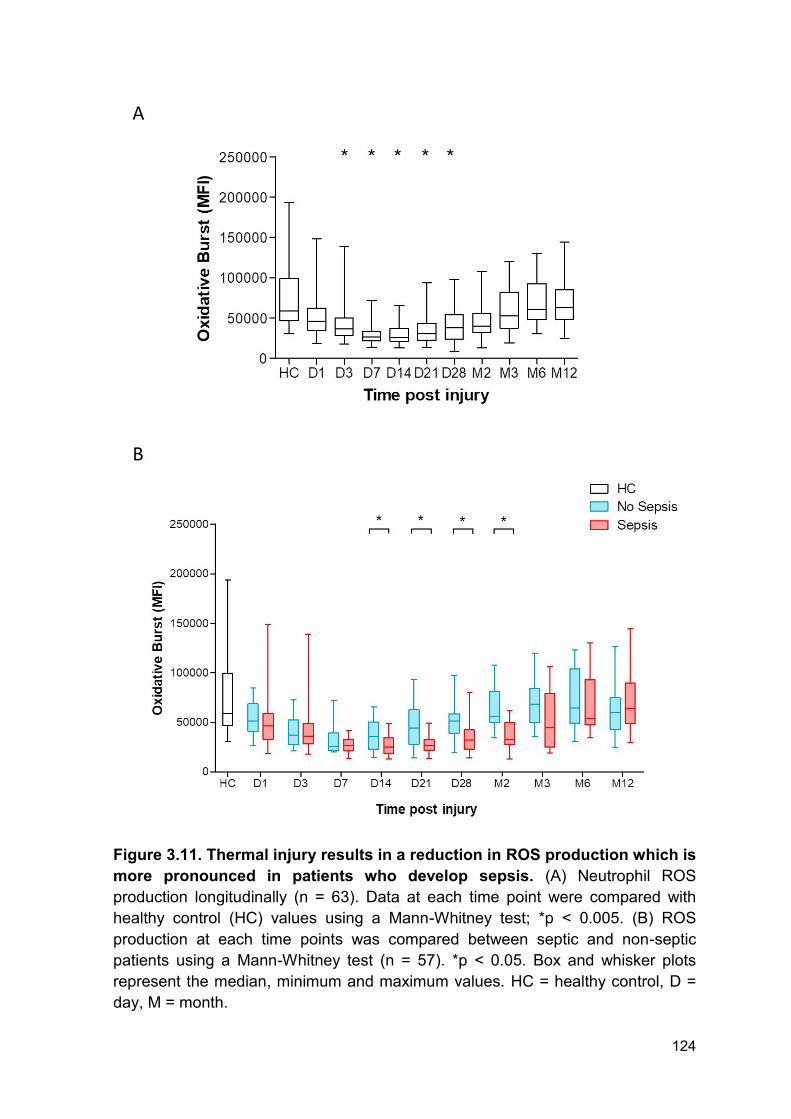
124
Figure 3.11. Thermal injury results in a reduction in ROS production which is
more pronounced in patients who develop sepsis. (A) Neutrophil ROS
production longitudinally (n = 63). Data at each time point were compared with
healthy control (HC) values using a Mann-Whitney test; *p < 0.005. (B) ROS
production at each time points was compared between septic and non-septic
patients using a Mann-Whitney test (n = 57). *p < 0.05. Box and whisker plots
represent the median, minimum and maximum values. HC = healthy control, D =
day, M = month.
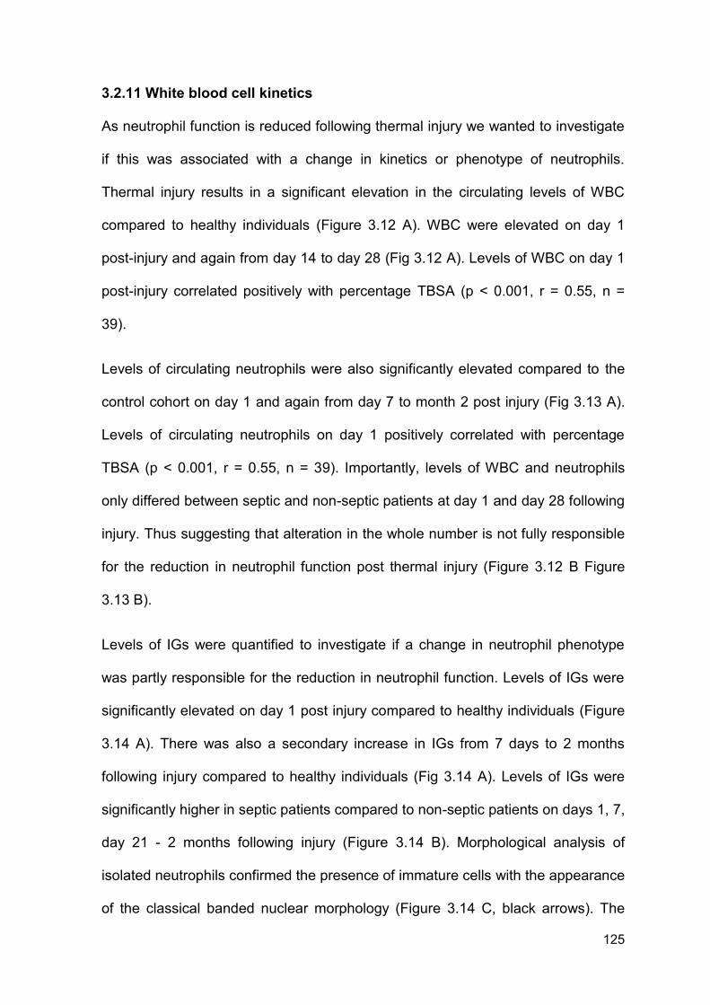
125
3.2.11 White blood cell kinetics
As neutrophil function is reduced following thermal injury we wanted to investigate
if this was associated with a change in kinetics or phenotype of neutrophils.
Thermal injury results in a significant elevation in the circulating levels of WBC
compared to healthy individuals (Figure 3.12 A). WBC were elevated on day 1
post-injury and again from day 14 to day 28 (Fig 3.12 A). Levels of WBC on day 1
post-injury correlated positively with percentage TBSA (p < 0.001, r = 0.55, n =
39).
Levels of circulating neutrophils were also significantly elevated compared to the
control cohort on day 1 and again from day 7 to month 2 post injury (Fig 3.13 A).
Levels of circulating neutrophils on day 1 positively correlated with percentage
TBSA (p < 0.001, r = 0.55, n = 39). Importantly, levels of WBC and neutrophils
only differed between septic and non-septic patients at day 1 and day 28 following
injury. Thus suggesting that alteration in the whole number is not fully responsible
for the reduction in neutrophil function post thermal injury (Figure 3.12 B Figure
3.13 B).
Levels of IGs were quantified to investigate if a change in neutrophil phenotype
was partly responsible for the reduction in neutrophil function. Levels of IGs were
significantly elevated on day 1 post injury compared to healthy individuals (Figure
3.14 A). There was also a secondary increase in IGs from 7 days to 2 months
following injury compared to healthy individuals (Fig 3.14 A). Levels of IGs were
significantly higher in septic patients compared to non-septic patients on days 1, 7,
day 21 - 2 months following injury (Figure 3.14 B). Morphological analysis of
isolated neutrophils confirmed the presence of immature cells with the appearance
of the classical banded nuclear morphology (Figure 3.14 C, black arrows). The

126
image shown is from a patient with 45% TBSA thermal injury with neutrophils
isolated on day 7 post injury during a known septic episode.
127
Figure 3.12. Circulating levels of WBC are elevated post thermal injury. (A)
WBC across time (n = 39) compared to healthy controls (n = 40) using a Mann-
Whitney test *p < 0.005. (B) WBC count at each time points was compared
between septic and non-septic patients using a Mann-Whitney test (n = 39)
*p < 0.05. Box and whisker plots represent median, minimum and maximum
values. HC = healthy control, D = day, M = month.
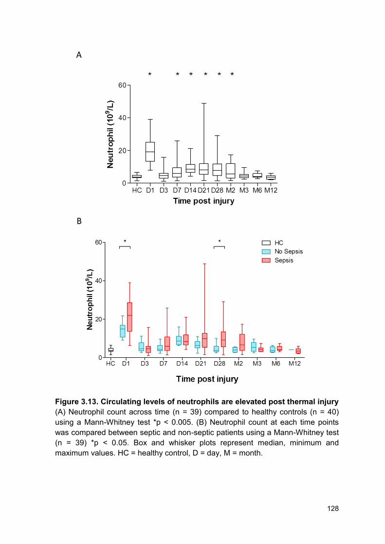
128
Figure 3.13. Circulating levels of neutrophils are elevated post thermal injury
(A) Neutrophil count across time (n = 39) compared to healthy controls (n = 40)
using a Mann-Whitney test *p < 0.005. (B) Neutrophil count at each time points
was compared between septic and non-septic patients using a Mann-Whitney test
(n = 39) *p < 0.05. Box and whisker plots represent median, minimum and
maximum values. HC = healthy control, D = day, M = month.
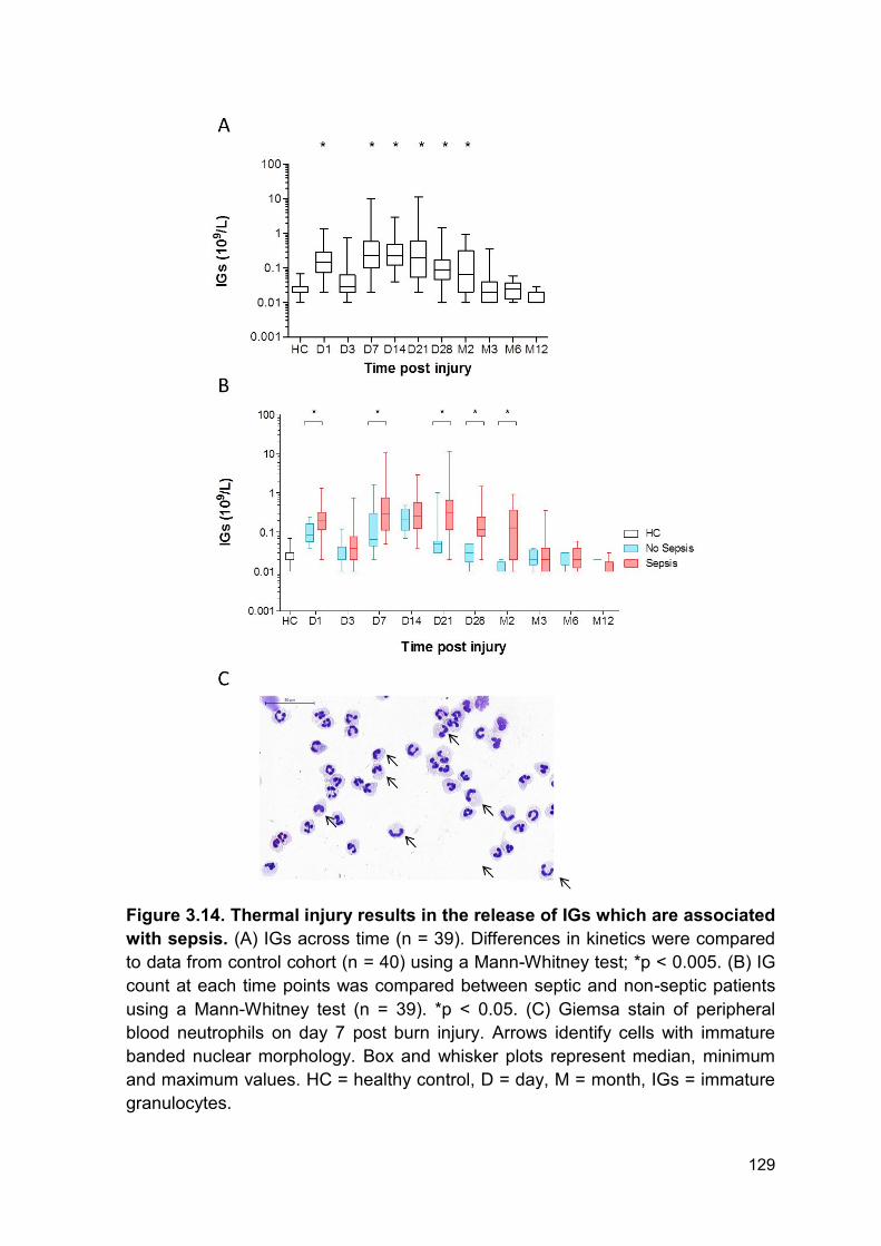
129
Figure 3.14. Thermal injury results in the release of IGs which are associated
with sepsis. (A) IGs across time (n = 39). Differences in kinetics were compared
to data from control cohort (n = 40) using a Mann-Whitney test; *p < 0.005. (B) IG
count at each time points was compared between septic and non-septic patients
using a Mann-Whitney test (n = 39). *p < 0.05. (C) Giemsa stain of peripheral
blood neutrophils on day 7 post burn injury. Arrows identify cells with immature
banded nuclear morphology. Box and whisker plots represent median, minimum
and maximum values. HC = healthy control, D = day, M = month, IGs = immature
granulocytes.
130
3.2.12 Release of immature granulocytes post-burn injury is associated with
reduced reactive oxygen species generation
This data was performed in collaboration with Dr Hampson as part of the SIFTI
Trial. Flow cytometric analysis of neutrophils following severe injury revealed a
subset of neutrophils that exhibited greater forward-scatter and reduced sideward-
scatter properties when compared to the ‘normal’ neutrophils (Figure 3.15 A). The
second population of neutrophils was termed a ‘dual population’. Of note, this dual
population of neutrophils produced lower amount of ROS compared to ‘normal
neutrophils’ (Figure 3.15 A).
The ‘dual population’ of neutrophils were significantly elevated from day 3 – day
28 post injury compared to healthy individuals (Figure 3.15 B). The kinetics of
release were comparable to IG release, emphasised by the significant correlation
(r = 0.65, p < 0.0001) between the percentage of ‘dual population’ neutrophils
and percentage of IGs (Figure 3.15 C).
3.2.13 Immature granulocyte percentage is associated with reduced ex vivo
neutrophil extracellular trap generation
The ability of the ‘dual population’ to generate NETs could not be investigated
directly. However, we used IGs percentage as a surrogate marker as there was a
significant correlation between the percentage of neutrophils that fell within the
‘dual population’ and IG frequency (Figure 3.15 C). As the appearance of IGs is in
part responsible for the reduction in ROS generation, the ability to undergo ex vivo
NETosis was correlated with IG percentage. Ex vivo NET production correlated
negatively with IG percentage (n = 56, r = - 0.3517, p = 0.0079, Figure 3.16).
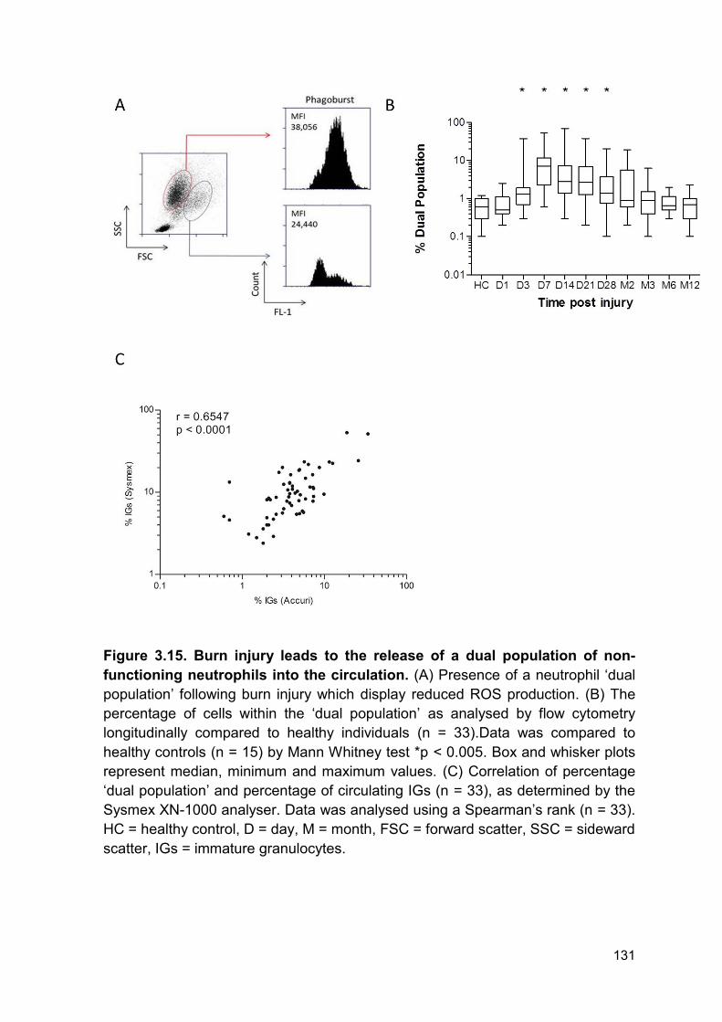
131
Figure 3.15. Burn injury leads to the release of a dual population of non-
functioning neutrophils into the circulation. (A) Presence of a neutrophil ‘dual
population’ following burn injury which display reduced ROS production. (B) The
percentage of cells within the ‘dual population’ as analysed by flow cytometry
longitudinally compared to healthy individuals (n = 33).Data was compared to
healthy controls (n = 15) by Mann Whitney test *p < 0.005. Box and whisker plots
represent median, minimum and maximum values. (C) Correlation of percentage
‘dual population’ and percentage of circulating IGs (n = 33), as determined by the
Sysmex XN-1000 analyser. Data was analysed using a Spearman’s rank (n = 33).
HC = healthy control, D = day, M = month, FSC = forward scatter, SSC = sideward
scatter, IGs = immature granulocytes.
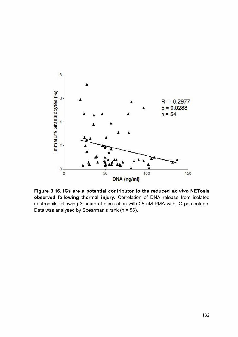
132
Figure 3.16. IGs are a potential contributor to the reduced ex vivo NETosis
observed following thermal injury. Correlation of DNA release from isolated
neutrophils following 3 hours of stimulation with 25 nM PMA with IG percentage.
Data was analysed by Spearman’s rank (n = 56).
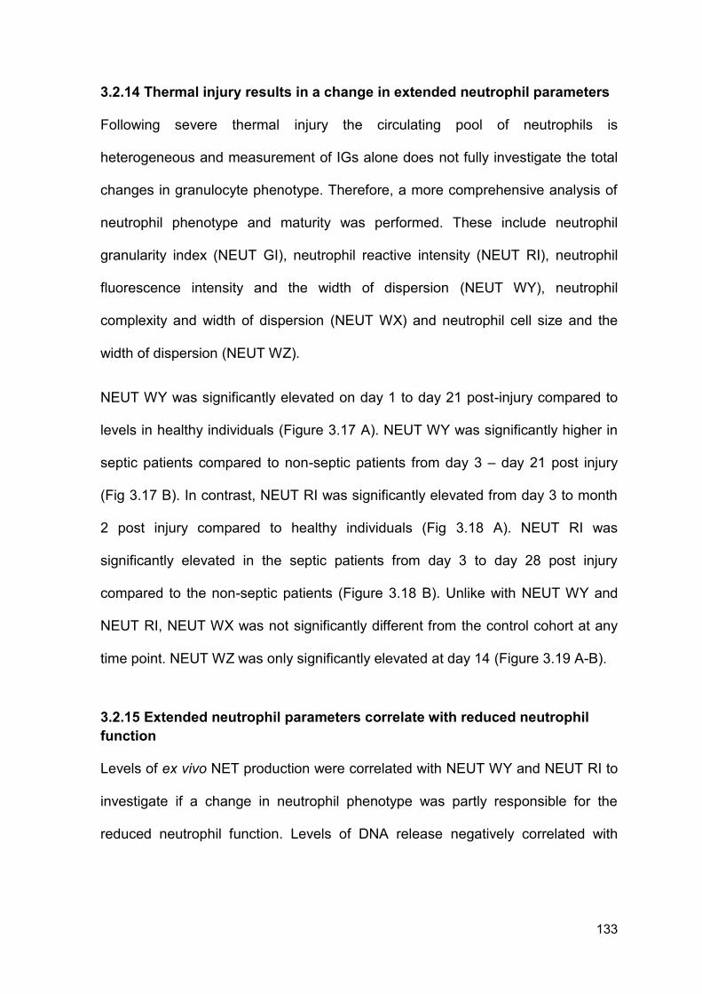
133
3.2.14 Thermal injury results in a change in extended neutrophil parameters
Following severe thermal injury the circulating pool of neutrophils is
heterogeneous and measurement of IGs alone does not fully investigate the total
changes in granulocyte phenotype. Therefore, a more comprehensive analysis of
neutrophil phenotype and maturity was performed. These include neutrophil
granularity index (NEUT GI), neutrophil reactive intensity (NEUT RI), neutrophil
fluorescence intensity and the width of dispersion (NEUT WY), neutrophil
complexity and width of dispersion (NEUT WX) and neutrophil cell size and the
width of dispersion (NEUT WZ).
NEUT WY was significantly elevated on day 1 to day 21 post-injury compared to
levels in healthy individuals (Figure 3.17 A). NEUT WY was significantly higher in
septic patients compared to non-septic patients from day 3 – day 21 post injury
(Fig 3.17 B). In contrast, NEUT RI was significantly elevated from day 3 to month
2 post injury compared to healthy individuals (Fig 3.18 A). NEUT RI was
significantly elevated in the septic patients from day 3 to day 28 post injury
compared to the non-septic patients (Figure 3.18 B). Unlike with NEUT WY and
NEUT RI, NEUT WX was not significantly different from the control cohort at any
time point. NEUT WZ was only significantly elevated at day 14 (Figure 3.19 A-B).
3.2.15 Extended neutrophil parameters correlate with reduced neutrophil
function
Levels of ex vivo NET production were correlated with NEUT WY and NEUT RI to
investigate if a change in neutrophil phenotype was partly responsible for the
reduced neutrophil function. Levels of DNA release negatively correlated with

134
NEUT WY (R = -0.4494, p = 0.0036, n = 41, Figure 3.20 A) and NEUT-RI (R = -
0.7368, p < 0.0001, n = 41, Figure 3.20 B).
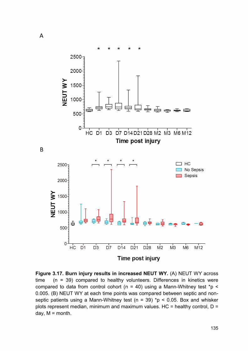
135
Figure 3.17. Burn injury results in increased NEUT WY. (A) NEUT WY across
time (n = 39) compared to healthy volunteers. Differences in kinetics were
compared to data from control cohort (n = 40) using a Mann-Whitney test *p <
0.005. (B) NEUT WY at each time points was compared between septic and non-
septic patients using a Mann-Whitney test (n = 39) *p < 0.05. Box and whisker
plots represent median, minimum and maximum values. HC = healthy control, D =
day, M = month.
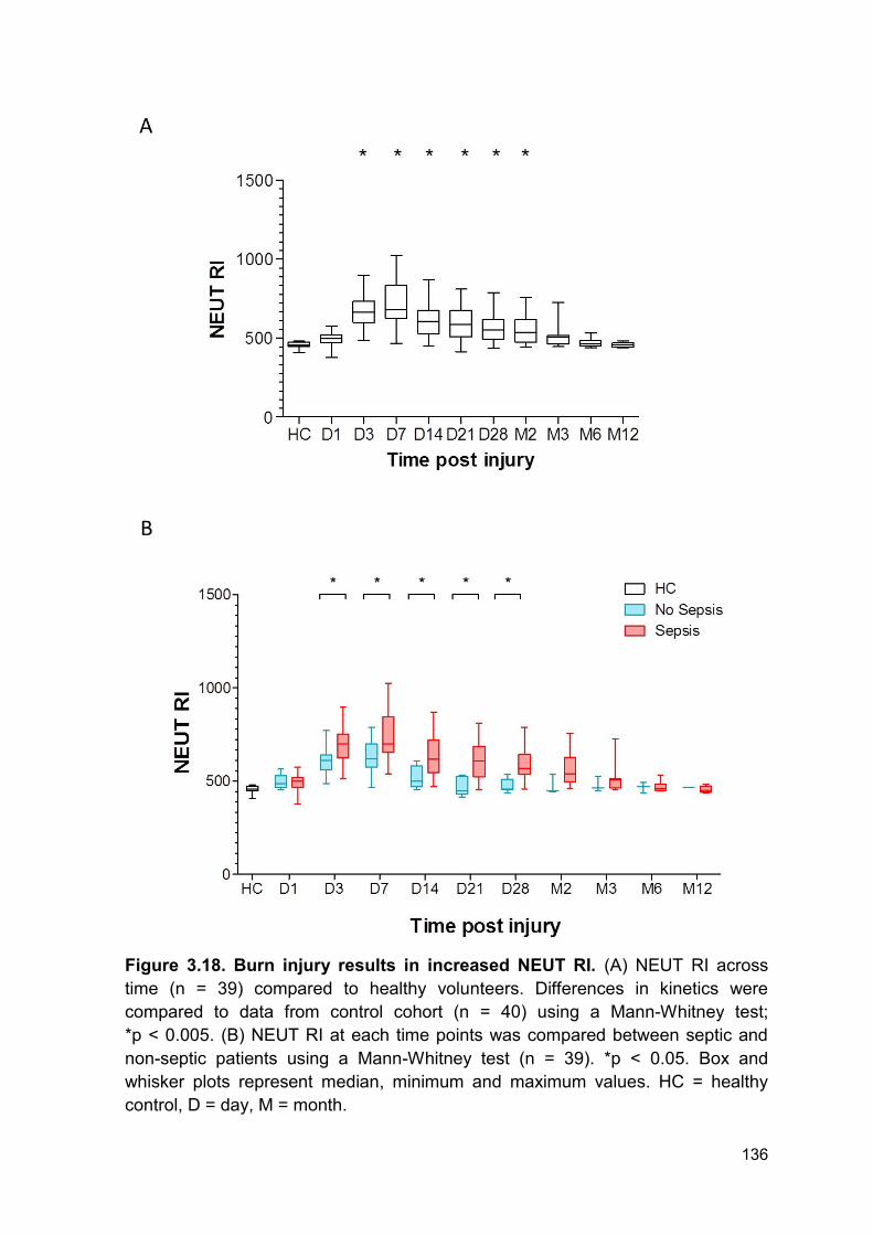
136
Figure 3.18. Burn injury results in increased NEUT RI. (A) NEUT RI across
time (n = 39) compared to healthy volunteers. Differences in kinetics were
compared to data from control cohort (n = 40) using a Mann-Whitney test;
*p < 0.005. (B) NEUT RI at each time points was compared between septic and
non-septic patients using a Mann-Whitney test (n = 39). *p < 0.05. Box and
whisker plots represent median, minimum and maximum values. HC = healthy
control, D = day, M = month.
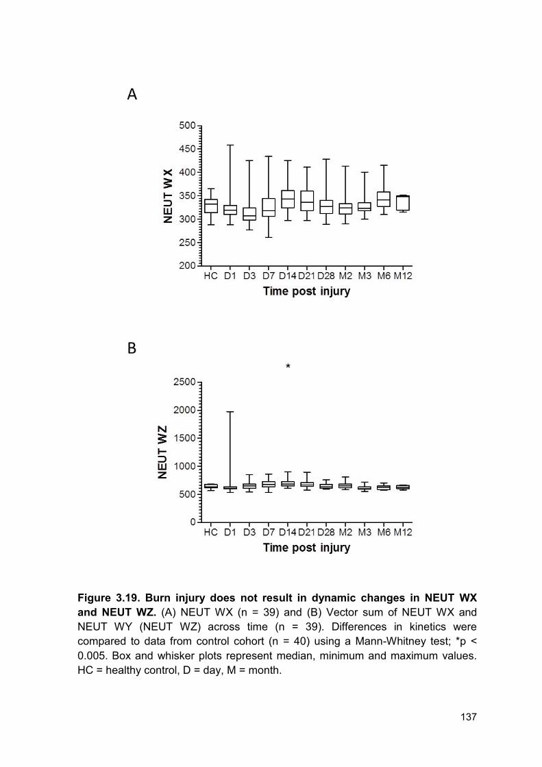
137
Figure 3.19. Burn injury does not result in dynamic changes in NEUT WX
and NEUT WZ. (A) NEUT WX (n = 39) and (B) Vector sum of NEUT WX and
NEUT WY (NEUT WZ) across time (n = 39). Differences in kinetics were
compared to data from control cohort (n = 40) using a Mann-Whitney test; *p <
0.005. Box and whisker plots represent median, minimum and maximum values.
HC = healthy control, D = day, M = month.
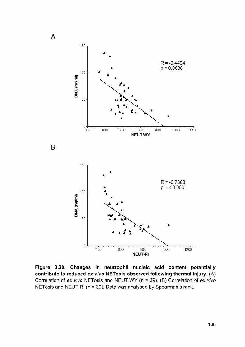
138
Figure 3.20. Changes in neutrophil nucleic acid content potentially
contribute to reduced ex vivo NETosis observed following thermal injury. (A)
Correlation of ex vivo NETosis and NEUT WY (n = 39). (B) Correlation of ex vivo
NETosis and NEUT RI (n = 39). Data was analysed by Spearman’s rank.
139
3.2.16 mtDAMPs activate neutrophils
This work was performed by Dr Jon Hazeldine (University of Birmingham, UK) as
part of a collaboration and joint first authorship manuscript (in preparation).
An additional mechanism for reduced neutrophil function post thermal injury is
direct immune suppression by mtDAMPs. To confirm previous observations that
mtDAMPs could induce neutrophil activation (43), we investigated the surface
expression of CD62L, CD11b, CXCR1 and CRX2 on human neutrophils following
15-minute stimulation with 40 or 100 µg/ml mtDAMPs.
Treatment of neutrophils with both 40 and 100 µg/ml of mtDAMPs resulted in a
significant reduction in surface density of CXCR1, CXCR2 and CD62L, compared
to control neutrophils stimulated with vehicle buffer (Figure 3.21 A-C). Neutrophils
treated with 40 and 100 µg/ml mtDAMPs had significantly higher levels of CD11b
surface expression compared to control neutrophils (Figure 3.21 D). Activation of
neutrophils was further confirmed by western blotting for MAPK ERK1/2.
Neutrophils simulated for up to 30 minutes with 100 µg/ml mtDAMPs had
detectable phosphorylation of ERK1/2 within 2 minutes of stimulation (Figure 3.21
E). Total EKR1/2 was included as a loading control for all experiments (Figure
3.21 E).
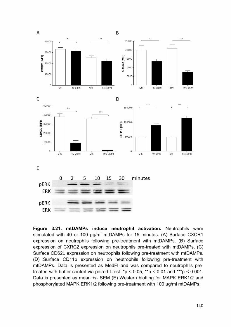
140
Figure 3.21. mtDAMPs induce neutrophil activation. Neutrophils were
stimulated with 40 or 100 µg/ml mtDAMPs for 15 minutes. (A) Surface CXCR1
expression on neutrophils following pre-treatment with mtDAMPs. (B) Surface
expression of CXRC2 expression on neutrophils pre-treated with mtDAMPs. (C)
Surface CD62L expression on neutrophils following pre-treatment with mtDAMPs.
(D) Surface CD11b expression on neutrophils following pre-treatment with
mtDAMPs. Data is presented as MedFI and was compared to neutrophils pre-
treated with buffer control via paired t test. *p < 0.05, **p < 0.01 and ***p < 0.001.
Data is presented as mean +/- SEM (E) Western blotting for MAPK ERK1/2 and
phosphorylated MAPK ERK1/2 following pre-treatment with 100 µg/ml mtDAMPs.
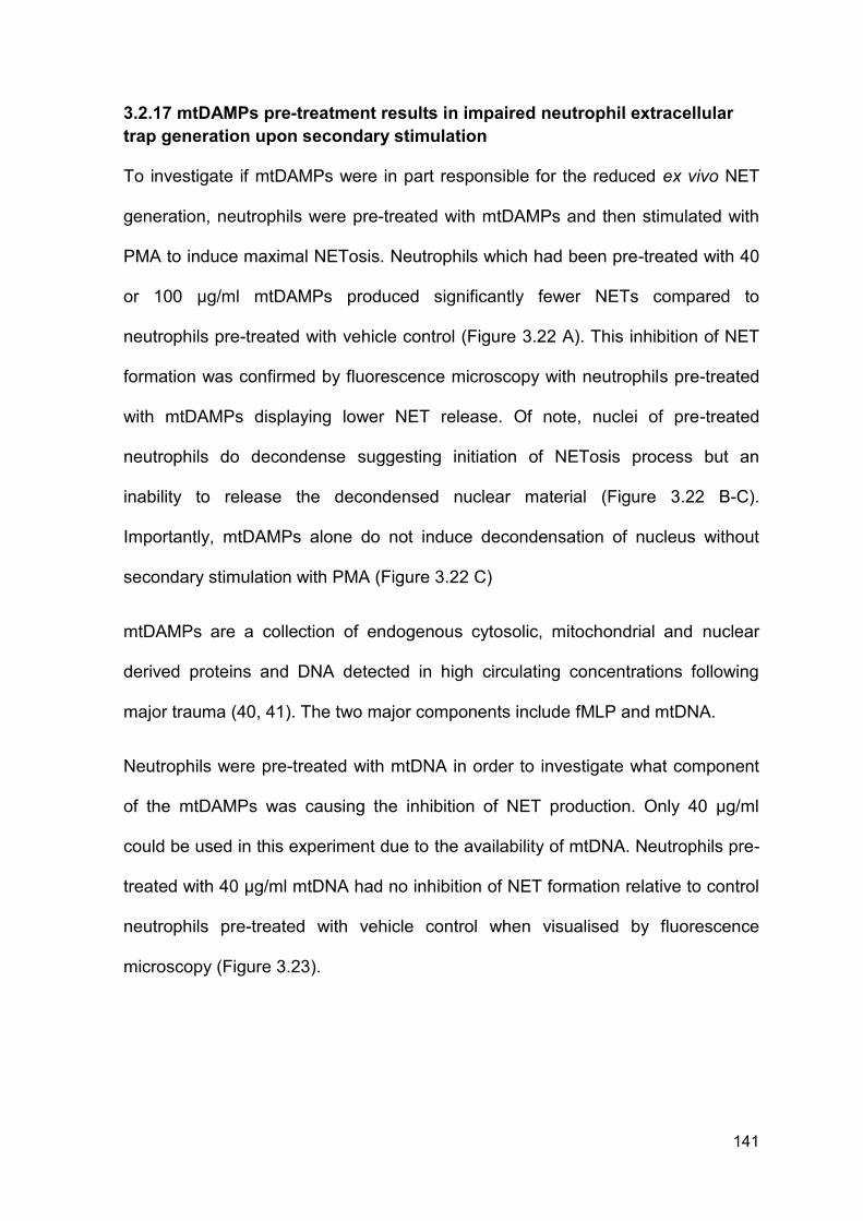
141
3.2.17 mtDAMPs pre-treatment results in impaired neutrophil extracellular
trap generation upon secondary stimulation
To investigate if mtDAMPs were in part responsible for the reduced ex vivo NET
generation, neutrophils were pre-treated with mtDAMPs and then stimulated with
PMA to induce maximal NETosis. Neutrophils which had been pre-treated with 40
or 100 µg/ml mtDAMPs produced significantly fewer NETs compared to
neutrophils pre-treated with vehicle control (Figure 3.22 A). This inhibition of NET
formation was confirmed by fluorescence microscopy with neutrophils pre-treated
with mtDAMPs displaying lower NET release. Of note, nuclei of pre-treated
neutrophils do decondense suggesting initiation of NETosis process but an
inability to release the decondensed nuclear material (Figure 3.22 B-C).
Importantly, mtDAMPs alone do not induce decondensation of nucleus without
secondary stimulation with PMA (Figure 3.22 C)
mtDAMPs are a collection of endogenous cytosolic, mitochondrial and nuclear
derived proteins and DNA detected in high circulating concentrations following
major trauma (40, 41). The two major components include fMLP and mtDNA.
Neutrophils were pre-treated with mtDNA in order to investigate what component
of the mtDAMPs was causing the inhibition of NET production. Only 40 µg/ml
could be used in this experiment due to the availability of mtDNA. Neutrophils pre-
treated with 40 µg/ml mtDNA had no inhibition of NET formation relative to control
neutrophils pre-treated with vehicle control when visualised by fluorescence
microscopy (Figure 3.23).
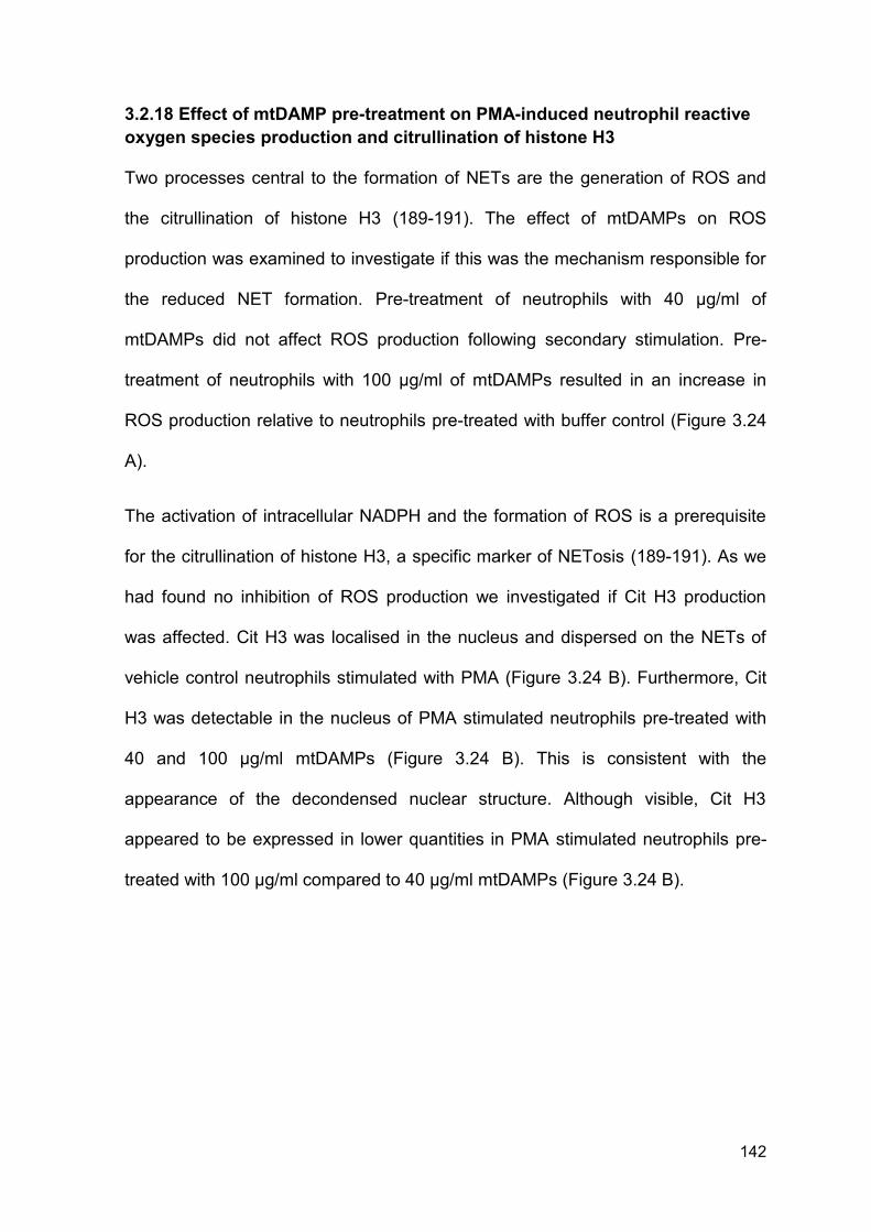
142
3.2.18 Effect of mtDAMP pre-treatment on PMA-induced neutrophil reactive
oxygen species production and citrullination of histone H3
Two processes central to the formation of NETs are the generation of ROS and
the citrullination of histone H3 (189-191). The effect of mtDAMPs on ROS
production was examined to investigate if this was the mechanism responsible for
the reduced NET formation. Pre-treatment of neutrophils with 40 µg/ml of
mtDAMPs did not affect ROS production following secondary stimulation. Pre-
treatment of neutrophils with 100 µg/ml of mtDAMPs resulted in an increase in
ROS production relative to neutrophils pre-treated with buffer control (Figure 3.24
A).
The activation of intracellular NADPH and the formation of ROS is a prerequisite
for the citrullination of histone H3, a specific marker of NETosis (189-191). As we
had found no inhibition of ROS production we investigated if Cit H3 production
was affected. Cit H3 was localised in the nucleus and dispersed on the NETs of
vehicle control neutrophils stimulated with PMA (Figure 3.24 B). Furthermore, Cit
H3 was detectable in the nucleus of PMA stimulated neutrophils pre-treated with
40 and 100 µg/ml mtDAMPs (Figure 3.24 B). This is consistent with the
appearance of the decondensed nuclear structure. Although visible, Cit H3
appeared to be expressed in lower quantities in PMA stimulated neutrophils pre-
treated with 100 µg/ml compared to 40 µg/ml mtDAMPs (Figure 3.24 B).
143
Figure 3.22. Pre-treatment of neutrophils with 40 or 100 µg/ml mtDAMPs
inhibits NET formation. (A) Fold increase in DNA release following pre-treatment
with 40 or 100 µg/ml mtDAMPs and secondary stimulation with PMA. Data was
compared to unstimulated neutrophils via paired t test (**p < 0.01). Data is
presented as mean +/- SEM (B) Fluorescence microscopy displaying neutrophils
pre-treated with, either, buffer control or 40 or 100 µg/ml mtDAMPs followed by
secondary stimulation with PMA. Images at 20x (C) Fluorescence microscopy
displaying neutrophils pre-treated with, either, buffer control or 40 or 100 µg/ml
mtDAMPs followed by secondary stimulation with. Images are taken at 63x
magnification. (B-C) Data is representative of 3 independent experiments.
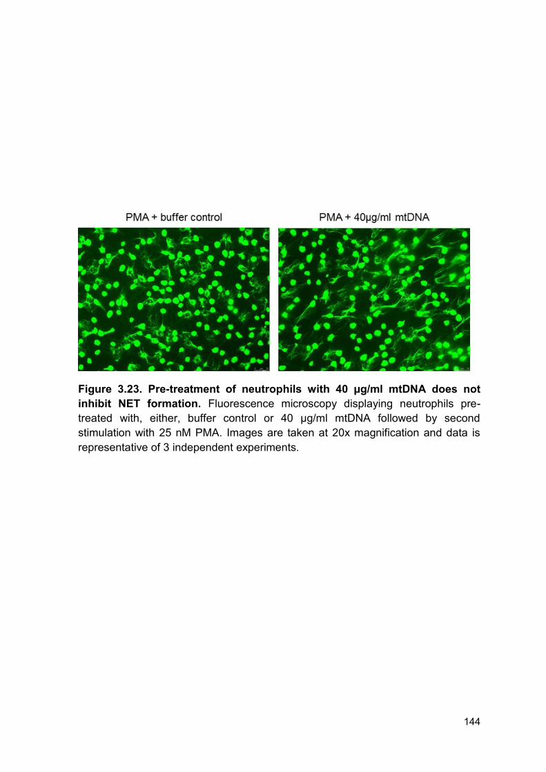
144
Figure 3.23. Pre-treatment of neutrophils with 40 µg/ml mtDNA does not
inhibit NET formation. Fluorescence microscopy displaying neutrophils pre-
treated with, either, buffer control or 40 µg/ml mtDNA followed by second
stimulation with 25 nM PMA. Images are taken at 20x magnification and data is
representative of 3 independent experiments.
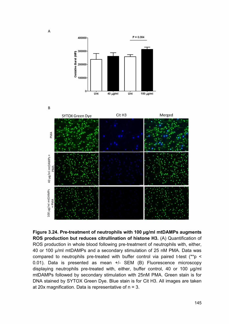
145
Figure 3.24. Pre-treatment of neutrophils with 100 µg/ml mtDAMPs augments
ROS production but reduces citrullination of histone H3. (A) Quantification of
ROS production in whole blood following pre-treatment of neutrophils with, either,
40 or 100 µ/ml mtDAMPs and a secondary stimulation of 25 nM PMA. Data was
compared to neutrophils pre-treated with buffer control via paired t-test (**p <
0.01). Data is presented as mean +/- SEM (B) Fluorescence microscopy
displaying neutrophils pre-treated with, either, buffer control, 40 or 100 µg/ml
mtDAMPs followed by secondary stimulation with 25nM PMA. Green stain is for
DNA stained by SYTOX Green Dye. Blue stain is for Cit H3. All images are taken
at 20x magnification. Data is representative of n = 3.
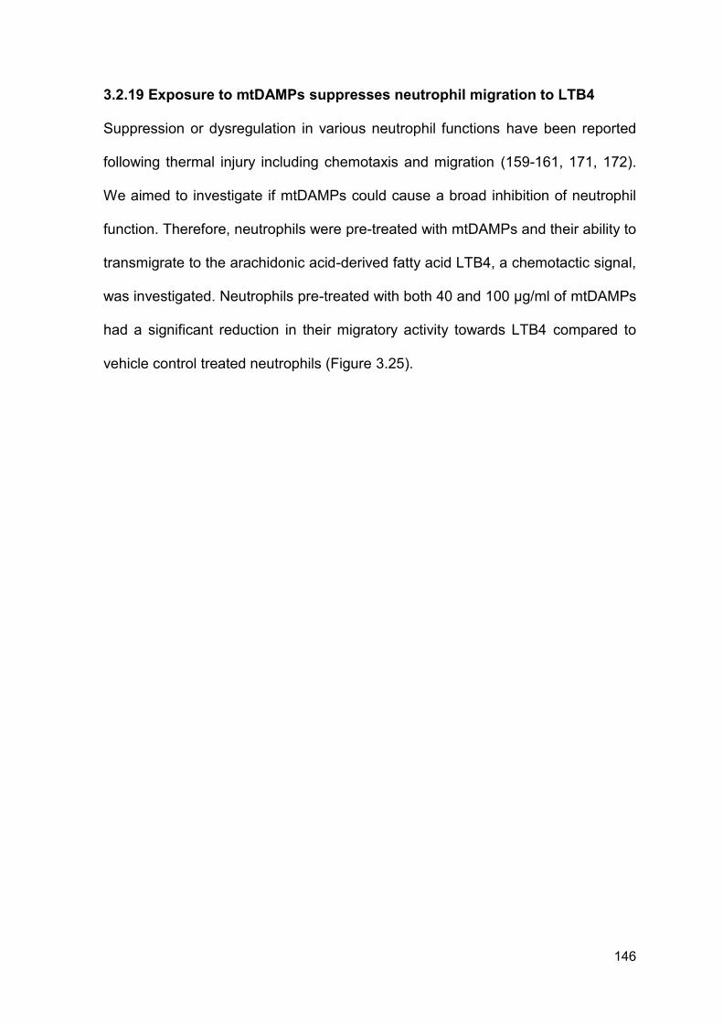
146
3.2.19 Exposure to mtDAMPs suppresses neutrophil migration to LTB4
Suppression or dysregulation in various neutrophil functions have been reported
following thermal injury including chemotaxis and migration (159-161, 171, 172).
We aimed to investigate if mtDAMPs could cause a broad inhibition of neutrophil
function. Therefore, neutrophils were pre-treated with mtDAMPs and their ability to
transmigrate to the arachidonic acid-derived fatty acid LTB4, a chemotactic signal,
was investigated. Neutrophils pre-treated with both 40 and 100 µg/ml of mtDAMPs
had a significant reduction in their migratory activity towards LTB4 compared to
vehicle control treated neutrophils (Figure 3.25).
147
Figure 3.25. Pre-treatment with 40 and 100 µg/ml mtDAMPs reduced
migratory activity of neutrophils towards the arachidonic acid-derived fatty
acid LTB4. Neutrophils were pre-treated with 40 or 100 µg/ml mtDAMPs and the
amount of neutrophils which transmigrated towards to LTB4 was quantified. Data
was analysed via repeated measures ANOVA and Bonferroni test compared to
vehicle control neutrophils (Unt). ***p < 0.001
148
3.3 Discussion
When first described, NETs sparked a change in the traditional paradigm of
neutrophil biology (118). NETs are comprised of DNA, granular proteins and
modified histones and function to ensnare, trap and potentially kill invading
pathogens (118, 119). Multiple groups have used quantification of DNA and
histones as a method for quantifying NET release in vivo and in vitro (288). Here
we report, consistent with previous literature, neutrophils stimulated in vitro with
PMA results in maximal NET formation which can be characterised by increased
release of DNA from neutrophils. One limitation of studying DNA release as a
single entity of NET release is the lack of specificity as DNA can originate from a
number of sources including apoptosis and necrosis (115). Therefore, specific
markers of NETosis, e.g. Cit H3 which decorates the remaining decondensed
nuclear structure and extruded NET structures, need to be investigated to confirm
NETosis (289). In this thesis, it has been shown that NETs are decorated with Cit
H3, consistent with previous literature (181, 190, 233).
In vitro NETosis is further limited by the stimulus used. The use of PMA is
commonly criticised because it lacks biological relevance as a stimulus. Therefore,
additional stimuli including IL-8, fMLP and LPS have been shown to induce NET
formation in vitro following initial priming of neutrophils (9). Here we report that
LPS alone does not induce NETosis in isolated neutrophils from healthy
individuals. Therefore, stimulation of neutrophils isolated from patients with
thermal injuries with LPS allows the investigation of priming status of neutrophils
and their propensity to undergo NETosis.
NETosis is a conserved and active cell death pathway which forms a fundamental
part of innate immunity. NETs are released from neutrophils in an attempt to
149
ensnare, trap and potentially kill invading bacteria (118, 119). Previous groups
have shown that NETs are implicated in the response to sterile traumatic injury,
consequent infection and sepsis in various disease pathologies (111, 290). It has
been proposed that NETosis can be induced rapidly by a number of soluble
mediators released following tissue damage (9). Moreover, Branzk and colleagues
demonstrated that neutrophils were able to adapt and tailor their host defensive
functions based upon the pathogenic challenge. If the challenge is too large in
size to be engulfed by phagocytosis, then a pro-NETosis phenotype was adopted
(188). In our patient cohort the most colonising bacteria was Bacillus sp.
Interestingly, when neutrophils are incubated with Mycobacterium bovis bacillus
Calmette-Guérin they formed NETs in preference to phagocytosis (188).
Therefore, given the nature and sources of infections in patients with thermal
injuries it is not unreasonable to hypothesise that patients will have a pro-NETosis
phenotype.
Elevation in circulating cfDNA post thermal injury has been attributed to in vivo
NET formation (111, 215, 290). However, this method alone is non-specific and
circulating DNA can originate from a number of sources including cellular damage,
apoptotic cells, necrosing cells, NETosis and bacteria themselves (115). A specific
marker of NET formation is the presence of Cit H3 which is facilitated by the
activation of the enzyme PAD4. This mediates the decondensation of nuclear
chromatin and extrusion of NETs to the outside of the cell decorated in Cit H3
(189).
Hirose and colleagues detected the presence of Cit H3 in the blood stream of
critically ill patients following traumatic injury (181) thus confirming NET formation.
Here, we expand upon this and demonstrate that Cit H3 is detectable in the
150
plasma of patients following thermal injury during sepsis and coinciding with
elevated levels of cfDNA. Thus, we propose that NETosis is a contributing factor
to the increase in cfDNA detectable in septic patients. Importantly, Cit H3 was only
detectable in trace quantities immediately following injury in one patient. Hence,
the initial sterile injury does not induce NET formation following thermal injury.
Data presented in this thesis would suggest that NETs are a defensive action of
neutrophils to control and eliminate infection post thermal injury rather than a
response initiated by the sterile injury.
In contrast, Hazeldine et al report NET formation within 60 minutes of sterile
traumatic injury. This occurs in the absence of an infectious stimulus and appears
to be mediated by a different mechanism of NET generation following thermal
injury. Thus, there appears to be a difference between burn and general traumatic
injury in the generation of NETs (291). However, variations in results may be
attributed to differences and difficulties in methodologies used to study NET
formation. Namely, current Western blotting assays, for Cit H3, are limited due to
its semi-quantitative nature and trace quantities of Cit H3 may be below the limit of
detection in plasma samples of patients with thermal injuries. Advancements in
ELISA based technologies may aid in the sensitive quantification of Cit H3 and
further its validation as a biomarker of sepsis, NETosis and secondary
complications (292). Nevertheless, measurements of Cit H3 in conjunction with
cfDNA have provided important information on the kinetics and timing of NETosis
following thermal injury.
NETosis was long regarded as process localised to tissues at sites of infection
and injury (293-295). However, recent evidence has implicated NETosis as a
process which can occur within the circulation, resulting in formation and
151
propagation of thrombosis in a mouse model of DVT (233). Hence, it is plausible
that thermal injury results in both NET formation within the tissues and circulation.
This is one potential explanation for the detection of NET components in plasma
following thermal injury.
NETosis can be initiated in vivo by a plethora of mechanisms. One of which is
neutrophils directly interacting with circulating bacteria and the formation of NETs
during sepsis (225). Furthermore, neutrophils can also bind to inflammatory cells
which initiate and amplify neutrophil response and function. In addition to their
central role in haemostasis, platelets are key players in host defence against
infection in a process termed ‘immunothrombosis’ (296, 297). Of particular interest
is ‘vital NETosis’ governed by the interaction between activated platelets and
neutrophils (178). In our cohort of patients, Montague et al report activation of
platelets, by measurement of circulating sGPVI, at the later time points correlating
with NET production (Montague et al, manuscript submitted). Thus, it is
reasonable to hypothesise that activated platelets are also in part responsible for
the NETosis in patients following thermal injury.
In addition to their procoagulant activity, it has been reported that heme-related
molecules can induce rapid NETosis in vitro (58). In a murine model of sickle cell
anaemia, heme causes NET release and is associated with increased mortality
(298). Thermal injury results in the production of FRCs (282), which will potentially
carry heme-related molecules, which could bind to and activate neutrophils to
induce NETosis. Future studies should focus on the mechanism(s) driving
NETosis post thermal injury to better understand their role during in vivo
responses.
152
Severe sepsis and septic shock remain a global health care challenge with a
systematic review, published in 2012, reporting sepsis as the major cause of
mortality in burn injury patients (38). Accurate and early diagnosis of sepsis still
remains challenging with many of the diagnostic criteria being non-specific in
patients with burns. This is due to many of the classically used diagnosis criteria
being masked by the ongoing SIRS which occurs immediately following severe
thermal injury. The Surviving Sepsis Campaign published guidelines, in 2012,
which recommends administration of antibiotics within 60 minutes of diagnosis of
sepsis (299). Furthermore, a delay in diagnosis of sepsis is associated with
significantly increased mortality (300). In 2016, the Surviving Sepsis Campaign
stressed the importance of antibiotic stewardship aided by further testing to ensure
infection and in turn reduce overuse of antibiotics (301). This is in an effort to
overcome the growing problem of antibiotic resistance. Blood cultures remain the
gold standard for diagnosis of sepsis even though many problems including
specificity and time required to process have been reported (302, 303). Hence, the
identification and development of novel and accurate biomarkers of sepsis is
imperative.
Recent literature has shown a potential role for cfDNA in the diagnosis of sepsis
(109, 304-306). Quantification of cfDNA in serum samples, taken within 6 hours of
injury, from patients with burns correlated with injury severity and outcome (111).
Surprisingly, we report no elevation in plasma cfDNA within 24 hours of injury
compared to levels detected in healthy individuals. There are a number of possible
explanations for these discordant observations. Firstly, a difference in the timing of
the first sample could explain the results. In our analysis, the median time to first
blood sample following burn injury was 9 hours. As it has previously been shown
153
that cfDNA is cleared within 30 minutes of appearance in circulation this may
result in clearance and reduced levels of cfDNA (307-309). Another key difference
between the two studies is in the preparation of blood samples. In our analysis
cfDNA was quantified in plasma samples were as Shoham and colleagues
measured cfDNA in serum (111). Analysis of matched plasma and serum samples
shows that levels of cfDNA are significantly higher in admission serum samples
compared to matched plasma samples from burn injury patients. Furthermore, the
change in DNA between matched samples positively correlated with WBC count.
In addition, levels of cfDNA were significantly higher in serum samples compared
to matched plasma samples from healthy volunteers. Interestingly, the ‘net’
change in DNA was much lower in healthy individuals compared to the ‘net’
change observed in patients. As the ‘net’ change of DNA correlates with WBC, we
propose that this increase is mediated by the significant increase in WBC count in
patients following thermal injury compared to healthy individuals. Therefore, the
false elevation in serum could be a consequence of in vitro clotting, cell death or
non-specific binding of SYTOX™ Green dye to any residual WBC. This is not only
an important standardization issue but is also in agreement with previously
published data (310).
It has been suggested that plasma cfDNA is a potential novel biomarker of sepsis.
Consistent with previous publications (109, 220), longitudinal analysis of cfDNA
levels in our cohort of patients revealed a potential diagnostic use for this
biomarker. Levels of cfDNA are significantly higher in septic individuals and
AUROC analysis showed good discriminatory at days 7 and day 14 post injury.
Quantification of cfDNA by fluorometric analysis has a number of advantages.
Firstly, the process is rapid and requires no isolation of DNA. Unlike Cit H3,
154
plasma cfDNA can be quantified which allows for evaluation of the marker in
multiple clinical settings. Finally, the assay is relatively cheap, requires a small
volume of blood and is rapid to perform. In all, this makes the assay applicable as
a point of care test to quickly identify patients with infection. Conversely, there are
a number of limitations of the assay and cfDNA as a biomarker. It is possible that
DNA binding dyes such as SYTOX Green™ could bind non-specifically to other
charged molecules in the plasma samples causing false elevation. Additionally,
cfDNA lacks specificity as it can originate from a number of sources (115). Hence,
it is important to consider the source of circulating DNA.
Using a murine cecal ligation model of sepsis it has been reported that the major
source of elevated cfDNA during sepsis is host tissue damage and not NETs
(311). Thus, the elevation in cfDNA reported in our cohort could be caused by an
accumulation of DNA from initial tissue damage and consequential tissue damage
that occurs during sepsis and MOF. Hamaguchi and colleagues went on to show
that depletion of neutrophils only had a minor effect upon cfDNA levels confirming
an insignificant role of neutrophils in mediating the elevation in cfDNA (311). Thus,
the contribution of NETs in the elevation of cfDNA may be minor following thermal
injury but still may explain some of the later elevation in cfDNA reported within this
thesis.
Circulating DNA can also be of bacterial origin (312, 313). Here, PCR analysis
was performed to determine the source of cfDNA. Analysis shows that the
predominant source of circulating DNA is nuclear in origin and not mitochondrial
following thermal injury. Importantly, the primers used shared no homology with
any bacterial species and therefore the ncDNA must, therefore, be host derived;
either from host tissue damage, apoptosis, necrosis or NETosis. However, we
155
cannot exclude the possibility that bacterial DNA is a contributing factor to the total
cfDNA measured.
ncDNA levels were significantly elevated within 24 hours of injury relative to
healthy individuals which was contradictory to the cfDNA results. The significant
elevation of ncDNA at day 1 – 3 following injury is in part due to the higher
sensitivity of the PCR assay. As the lower limit of detection of the PCR assay was
0.1 ng/ml and the lower limit of detection for the fluorometric based assay was 60
ng/ml. Still, ncDNA followed comparable kinetics to total cfDNA and they
correlated strongly with each other (r = 0.763).
mtDNA is derived from the mitochondria of cells and can act as a DAMP (314).
Elevated levels of mtDNA are associated with secondary complications following
traumatic injury (314-317). This is attributed to their direct interactions with
immune cells and damage to host tissue due to their cytotoxic nature (314, 317,
318). Itagaki and colleagues reported an association between mtDNA levels and
NET formation following traumatic injury. Consistent with their previous findings,
trauma patients had a significant elevation in mtDNA compared to healthy
individuals and incubation of isolated neutrophils with mtDNA resulted in the
induction of NET release (315). This may in part be attributable to DAMP induced
activation of neutrophils, however the exact mechanism remains unknown (319).
Conversely, in our analysis we report no significant elevation in mtDNA
concentrations in patients with severe thermal injuries compared to healthy
individuals. It is currently unknown if this is a contradictory finding perhaps due to
the different mechanism(s) of injury. However, it could be in part explained by
differences in timing of blood sampling resulting in clearance of mtDNA or pre-
analytical variables by which centrifugation has removed the larger microvesicles
156
containing mitochondria and hence mtDNA (320). Finally, higher levels of mtDNA
are detected in elderly trauma patients compared to young trauma patients (315).
As the mean age of our cohort was 39 years this may result in the lower levels of
mtDNA detected. As we have few elderly patients with burns ≥ 15% TBSA it was
not possible to investigate this hypothesis within our cohort. Thus, further research
is required to investigate mtDNA concentrations following thermal injury in relation
to outcome, age and secondary complications.
Itagaki and colleagues report preliminary evidence showing a reduced ability of
neutrophils from elderly trauma patients to form NETs ex vivo relative to age
matched controls and young trauma patients (315). Given the high incidence of
infections post burn injury and apparent reduced neutrophil function it has led to
the investigation of this relationship (38, 172, 174, 321).
As early as 1985, Arturson et al reported, in a cohort of 50 patients with thermal
injuries, a reduction in chemotaxis, phagocytosis and impaired killing of bacteria
(175). As recently as 2014, a spontaneous migratory phenotype for neutrophils
isolated from patients with severe thermal injuries was observed. This migratory
phenotype was present 1 - 2 days prior to clinical diagnosis of sepsis and was not
present in healthy or non-septic individuals (172). Whilst these studies
documented a dysregulation in neutrophil function following severe thermal injury,
they have only studied this in the early days post injury. Thus, the longitudinal
neutrophil response has not been thoroughly examined. Whilst the ability of
neutrophils to generate NETs post trauma has been investigated no group has
investigated the effect thermal injury has on this NET production (315).
157
Traumatic injury results in the systemic priming of neutrophils which has been
attributed to the enormous release of soluble mediators (322, 323). Zhang et al
reported a link between initial traumatic injury and consequent immune cell
activation and inflammation (42). Further research has shown that adenosine
triphosphate (ATP) and N-formyl peptides can stimulate ROS production,
degranulation and cytokine secretion in neutrophils (43, 324, 325). Due to the
elevated levels of NET components in the circulation we had hypothesised that
neutrophils from patients following thermal injury would be primed and in turn
would overproduce NETs rapidly with ex vivo stimulation. To test this hypothesis,
neutrophils isolated from patients with burn injury were stimulated with LPS alone
to investigate their priming status and ability to form NETs ex vivo (9, 293).
However, neutrophils from patients following thermal injury did not generate NETs
when stimulated with 100 ng/ml LPS. This would suggest that neutrophils are not
primed or have a reduced ability to form NETs following thermal injury.
We expanded upon this finding and report that neutrophils stimulated with 25 nM
PMA release significantly lower amounts of NETs on day 3 – 7 post thermal injury
relative to healthy individuals. This was further confirmed by fluorescent
microscopy in which minimal release of DNA was observed from neutrophils
isolated from patients. These findings are consistent with previous literature which
reports a reduction in maximal NET generation in patients with traumatic injuries
(326). In a number of patients it was clear that neutrophils also retained their
natural morphology following stimulation without significant decondensation of
nuclei. As PMA is not receptor dependent this indicates a clear defect in later
signalling cascades. Thus, there appears to be a reduction in the ability to form
NETs following thermal injury which may leave patients at risk of nosocomial
158
infections. One limitation of this data is the absence of a second marker of NET
formation, e.g. neutrophil elastase, in our fluorescence microscopy based assay.
However, within this analysis we use an isolated in vitro system to induce maximal
NET formation. Thus, DNA released from neutrophils following stimulation with
PMA can be regarded as a surrogate marker of NET formation. Therefore, we
would propose that the lack of a more comprehensive staining panel does not
affect our conclusions.
Initially a reduction in ex vivo NETosis appears to contradict our in vivo data.
However, there are a number of possible explanations for NET markers detected
in vivo and reduced ex vivo NETosis. One possible explanation is that functional
neutrophils have migrated and are generating NETs in the damaged tissues and
thus leaving non-functional neutrophils in the circulation. Secondly, there may be a
dysregulation in the breakdown and clearance of NETs allowing for the build-up of
NET components. One limitation of our current data is the inability to investigate
the relationship between ex vivo NETosis and susceptibility to infection post
thermal injury due to limited sample size. Thus, further studies are required to fully
understand the clinical relevance of this finding post thermal injury and to
investigate if reduced ex vivo NETosis is associated with an increased incidence
of infection and a potential therapeutic target.
Whilst a reduction in NET formation has been shown post traumatic injury (315), to
our knowledge no groups have studied potential mechanisms for this
dysregulation in the same patient cohort. ROS are a heterogeneous collection of
molecules which form an important link between cellular activation and NET
formation. Production of ROS is controlled by NADPH oxidase complex which
159
produces O2- and H2O2 upon activation. ROS induced decondensation of nuclear
chromatin precedes NETosis and is facilitated by the activity of PAD4 (189-191).
There are contradicting data regarding ROS production following traumatic injury.
Namely, it has been shown that baseline ROS production is increased and may be
contributing to host tissue damage (137, 138). Conversely, Parment and
colleagues report a reduction in oxidative capacity for up to 3.5 months post
thermal injury (177). In agreement with Parment and colleagues, we report a
significant reduction in ROS formation following thermal injury relative to healthy
individuals. Furthermore, in our analysis septic patients had significantly lower
levels of ROS production than non-septic patients. This highlights the immediate
and sustained immunosuppression induced by thermal injury in septic patients.
Given that ROS production is regarded as a prerequisite of NETosis, this may be
a potential mechanism by which neutrophils have a reduced ability to generate
NETs ex vivo. However, ROS independent mechanisms of NET formation must
also be considered. Hence, given the limited sample size, further research is
required to fully study in vivo reduced ROS formation and ex vivo NETosis
following thermal injury.
Independent of NETosis, ROS also forms a key component of host defence
against engulfed pathogens (134). Neutrophils are essential effector cells of innate
immunity and function to clear invading pathogens. Dysfunction or dysregulation
of neutrophil functions may leave the host immunocompromised as highlighted in
CGD patients who are incapable of forming ROS due to the absence of NADPH
(153). Moreover, phagocytosis is significantly reduced following thermal injury and
is more pronounced in septic individuals (180). This may be contributing to the
increased incidence of infection as septic patients with thermal injuries have
160
reduced ROS formation and phagocytosis following thermal injury. In addition, it
has recently been suggested that pathogens can use evasion strategies to avoid
killing by neutrophils. Laskay et al suggest an evasion strategy, by Leishmania,
that allows pathogens to use granulocytes as host cells and replicate. As ROS is a
critical effector of intracellular killing and reduced post thermal injury, this process
may be occurring. Therefore, pathogens may be using neutrophils as a ‘Trojan
horse’ to aid in their dissemination due to the inability to kill engulfed pathogens
(327).
Given that NETosis can also be mediated through ROS independent mechanisms
it is important to consider additional mechanisms by which dysregulation in NET
formation may occur (186). IGs are released prematurely from the bone marrow in
response to stress and infection (96). Although they can still phagocytose and
migrate, it is reduced compared to mature neutrophils (169). In addition, immature
cells are termed ‘band cells’ due to their classical banded morphology and
increased nuclear content. Here we show that neutrophils which are banded in
morphology are present in isolated neutrophil preparations from patients. Analysis
of neutrophils following severe thermal injury also revealed a ‘dual population’ of
neutrophils which have distinct forward and sideward scatter properties
comparable to data published by Pillay et al in 2011 (158). The kinetics of the
‘dual population’ of neutrophils correlated with the appearance of IGs and in our
analysis this ‘dual population’ of neutrophils was defective in generating ROS and
phagocytosis (180). IG frequency correlated with reduced ex vivo NETosis and
therefore immature cells may be contributing to the reduced neutrophil function
post burn.
161
It is important to consider the heterogeneous nature of the circulating population of
neutrophils. Quantification of IGs alone is therefore limited as it does not account
for phenotypical changes in the total neutrophil population. We performed a more
comprehensive analysis of neutrophil phenotype by quantifying five additional
novel parameters; NEUT WY, NEUT WX, NEUT WZ, NEUT GI and NEUT RI.
Here we report a significant increase in NEUT WY and NEUT RI post thermal
injury. The elevation is potentially caused by the cumulative release of IGs and
promyelocytes, myelocytes or metamyelocytes. Moreover, the presence of IGs
and extended measurements of neutrophil phenotype correlated with reduced ex
vivo NETosis. Thus, changes in neutrophil maturity and morphology could be in
part responsible for the reduced neutrophil function observed following severe
thermal injury.
Quantification of circulating IGs is able to discriminate between septic and non-
septic patients with a sensitivity of 89.2% and a specificity of 79.4% (96). Here we
show that in addition to their potential mechanistic role in sepsis, IGs have a
potential diagnostic use as they are significantly higher in septic patients
compared to non-septic individuals. Further investigation and formal statistical
analysis on the kinetics of IGs release in relation to infection is required to fully
understand the diagnostic potential of this marker.
Recently it has been reported that quantification of extended neutrophil
parameters could predict an increase in circulating IGs and discriminate between
IG release from infectious or non-infectious causes (328). Therefore a combination
of IG number and extended neutrophil parameters may provide a novel panel of
biomarkers to discriminate between sterile injury and infection following burn. In a
recent study by Luo et al, NEUT X, NEUT Y and NEUT Z could identify septic
162
patients (329). In our analysis only NEUT WY and NEUT RI, measurements of
nucleic acid content, demonstrated any significant and dynamic changes in
thermal injury patients compared to control cohort. The differences may be
mediated by methodology as, NEUT-Y, NEUTX and NEUT-Z do not account for
width of dispersion unlike NEUT-WY. Stiel et al have reported that an increase in
nucleic acid content had a strong discriminatory power for diagnosing sepsis with
sensitivity equal to 90.91% and specificity equal to 80.60% (330). In agreement,
septic individuals had higher NEUT WY and NEUT RI values compared to non-
septic patients. Quantification of immature neutrophils and measurement of
nucleic acid content may be a potential novel biomarker of sepsis following
thermal injury. Furthermore, measurement of the extended neutrophil parameters
of maturity and phenotype may have better potential compared to quantifying IGs
alone.
Although quantification of novel biomarkers is showing promise in the early
diagnosis of sepsis in patients with thermal injuries, understanding the
mechanisms responsible for the immunosuppression offers the opportunity to
explore novel therapeutic interventions. However, current mechanisms
responsible for immunosuppression are poorly understood. One potential source
of immediate immunosuppression is the release of mtDAMPs.
mtDAMPs are comprised of a heterogeneous mixture of molecules including;
mtDNA, cytochrome C, N-formyl peptides and ATP (331). However, fMLP and
mtDNA are regarded as the two major effectors on neutrophils. This was
demonstrated in 2015 by Hazeldine et al who report neutrophil activation through
N-Formyl peptides within mtDAMPs (43).
163
Once released into the circulation from damaged tissues and cells, mtDAMPs can
activate and induce a state of tolerance in immune cells (42, 45, 332). In 2014,
Fernández-Ruiz et al defined endotoxin tolerance in monocytes induced by
circulating mtDAMPs. Notably, this phenomenon of tolerance was associated with
an increased infection rate (45). In a more recent study, the authors report that
patients with ischemic stroke display this endotoxin tolerance phenotype and it is
association with elevated circulating mtDNA. Furthermore, monocytes from
healthy individuals developed this endotoxin tolerance phenotype when cultured
with serum from patients with ischemic stroke or elevated levels of mtDNA (332).
Whilst this refractory state has been described in monocytes it is possible that
circulating mtDAMPs induce a comparable phenotype in neutrophils following
thermal injury and maybe responsible for the reported dysfunction.
Following severe traumatic injury mtDAMPs, including N-formyl peptides and
mtDNA, are released into the circulation (42, 333). Whilst here we report no
significant elevation in mtDNA following thermal injury the difference may be
caused by a number of factors including pre-analytical variables, time to blood
sampling and mechanism of injury. Furthermore, we have been unable to measure
circulating levels of fMLP due to lack of sample availability. Therefore, further
study is warranted to fully investigate mtDAMP kinetics post thermal injury and
their association with neutrophil function. Of note, in septic patients or patients
with DIC, elevated levels of circulating high mobility group box 1 protein (HMGB1)
have been observed (334, 335). Thus, one would hypothesise that fMLP is
released in response to consequent host tissue damage and infection following
thermal injury in a similar manner to HMGB1 (42, 333).
164
In 2015, Hazeldine et al reported activation of human neutrophils following
stimulation with 40 or 100 µg/ml mtDAMPs. The authors showed for the first time
that activation of neutrophils by mtDAMPs was dependent upon signalling through
the MAPK p38 and ERK 1/2. Selective inhibition of the formyl peptide receptor-1
inhibited this activation and the group concluded that N-formyl peptides are the
main constituents driving mtDAMP-induced neutrophil activation (43). Consistent
with their findings we report that mtDAMPs result in activation of neutrophils
characterised by CD62L shedding and phosphorylation of ERK 1/2 (43). Here we
expand upon this observation by investigating CXCR1 and CXCR2 surface
expression on neutrophils following 15 minutes incubation with mtDAMPs.
Following activation of neutrophils, CXCR1 and CXCR2 are internalised resulting
in a decrease in their surface expression (336, 337). Here, we report that
stimulation with mtDAMPs results in a rapid decrease in both CXCR1 and CXCR2
surface expression. The decrease in CXCR2 is more pronounced than CXCR1
which may be explained by CXCR2 being rapidly internalised following activation
compared to CXCR1 (337).
Various endogenous and exogenous inhibitors of NET release have been reported
(338-341). Although dysregulation in NET formation has been reported following
major trauma (180, 315), the mechanisms driving this dysregulation are poorly
understood. Furthermore, the role mtDAMPs and inhibition NET formation has yet
to be fully explored. Pre-treatment of neutrophils with either 40 or 100 µg/ml of
mtDAMPs results in a significant reduction in NET formation following secondary
stimulation with PMA. To investigate the component responsible for inhibition of in
vitro NET formation neutrophils were pre-treated with mtDNA. Consistent with
previous findings we report no inhibition of NET formation when neutrophils are
165
pre-treated with mtDNA (315). Hence, an additional component of mtDAMPs that
is not mtDNA is potentially responsible for the inhibition of PMA induced NET
formation. Thus data presented within this thesis does not contradict previous
observations (315).
Neutrophils pre-treated with mtDAMPs and then stimulated with PMA had
decondensed chromatin and nuclear structure and did not retain their natural
polymorphic nuclear structure. This is indicative of dysregulation of NET release
rather than recognition of stimulus. Since PMA activity is not dependent upon
receptor activation it appears that inability to release NETs is mediated by
dysregulation in a downstream cascade. This is different to our ex vivo data in
which neutrophils do not decondense following stimulation with PMA. However,
this difference may be explained by cumulative dysregulation in our ex vivo data
caused by a change in phenotype and abnormal ROS production. Importantly, this
is not accounted for in our in vitro analysis of mtDAMPs functions.
Formation of NETs is generally NADPH oxidase-dependent with many activators
of ROS being reported as inducers of NETosis (189-191, 342). In addition, mice
with NADPH oxidase deficient neutrophils are not able to form NETs (342). To
investigate if a dysregulation in ROS production was responsible for inhibition of
NETosis, levels of ROS were quantified following pre-treatment with mtDAMPs
and secondary stimulation with PMA. Of note, Hazeldine et al have previously
reported that stimulation of neutrophils with mtDAMPs results in a significant
increase in ROS production from resting neutrophils (43). Here we expand upon
this and show that pre-treatment of neutrophils with mtDAMPs and a secondary
stimulation with PMA results in a significant increase in ROS production compared
to vehicle control neutrophils. ROS induced decondensation of nuclear chromatin
166
precedes NETosis and is facilitated by the activity of enzyme PAD4 (189-191,
343). Here we report, although reduced, neutrophils still have Cit H3 detectable
and a decondensed nuclear structure. As ROS formation can mediate citrullination
of histones by PAD4 and eventual chromatin decondensation this may explain our
results (135, 189, 208, 343).
It is important to consider the limitations of these experiments. As we are
measuring ROS production in whole blood we cannot eliminate that mtDAMPs are
indirectly increasing neutrophil ROS production via activation and interaction with
other inflammatory cells. However, our data suggest that a reduction in ROS is not
causing inhibition of NETosis following pre-treatment with mtDAMPs.
Multiple groups, including ourselves, have reported a reduction in neutrophil ROS
production following severe thermal injury (175-177). Here we report that pre-
treatment of neutrophils with mtDAMPs increases ROS production following
secondary stimulation. Therefore, mtDAMPs are probably not responsible for the
in vivo reduction in ROS production reported in this thesis. As discussed earlier,
this reduction in ROS formation is most likely caused by a change in neutrophil
maturity, phenotype or an unknown soluble mediator.
Ramijsen et al report that the formation of NETs is dependent upon both ROS
generation and autophagy (208). Autophagy is an essential cellular mechanism for
cellular homeostasis and is regulated by activation of the mammalian target of
rapamycin (mTOR). Pharmacological inhibition of mTOR accelerated NETosis
following stimulation with fMLP and is therefore a negative regulator of autophagy.
Inhibition of autophagy by wortmannin abolishes citrullination of histones induced
by co-treatment of neutrophils with rapamycin and fMLP (344). We hypothesise
167
that mtDAMPs may be activating mTOR which inhibits autophagy and in turn
prevents NET production. Therefore, one may hypothesise that the reduction in Cit
H3 staining observed is in part caused by mtDAMP inhibition of autophagy.
However, this is only a hypothesis and further study is required to fully understand
the effect mtDAMPs have on autophagy and consequently NET formation.
For neutrophils to function in tissues they must migrate towards the site of
infection before phagocytosing or generating NETs against the pathogen. One
hypothesis speculates if the dysfunctional pool of circulating neutrophils consists
of cells which are unable to migrate to sites of injury and in turn leaving the patient
immunocompromised. Hence this may manifest as the clear dysregulation in
migratory phenotype of neutrophils observed following severe thermal injury (159-
161, 171, 172). In addition, studies have reported impaired migration, of
neutrophils isolated from trauma patients, towards IL-8 and LTB4 (326, 345). In
this thesis, pre-treatment of neutrophils with mtDAMPs causes a reduction in
neutrophils ability to migrate towards LTB4. Suggesting that mtDAMPs may be a
contributory factor to the dysregulated chemotaxis and migration reported
following severe thermal and traumatic injury.
We propose that the reduced migration of neutrophils is mediated by both mtDNA
and fMLP. Firstly, Li et al have reported using an ex vivo model that pre-treatment
of neutrophils with 10 nM fMLP resulted in reduced transmigration in response to
both 1 and 10 nM LTB4 (326). This was earlier shown by Campbell et al who
reported decreased migration towards IL-8 following pre-treatment with 10 nM
fMLP (346). Furthermore, mtDNA contains CpG DNA repeats as mitochondria
evolved from bacteria (347, 348). Incubation of neutrophils with CpG DNA
significantly impairs migration towards LTB4 which occurred simultaneously to a
168
reduction in BLT1, a high affinity receptor for LTB4 (349). In addition, the authors
show a reduction in migration towards IL-8 following pre-treatment with CpG DNA.
Again, this reduction occurred simultaneously to a reduction in the IL-8 specific
receptor, CXCR1 (349). In our studies, pre-treatment of neutrophils with mtDAMPs
results in a significant reduction in both CXCR1 and CXCR2. Therefore, whilst we
have yet to investigate an ability to migrate towards IL-8, it is possible that
mtDAMPs may result in disrupted transmigration towards this chemokine. Finally,
reduced transmigration towards LTB4 may be caused by internalisation of BLT1
receptors following treatment with mtDAMPs.
In conclusion, this data supports the hypothesis that mtDAMPs may induce a state
of neutrophil dysregulation and may be responsible, in part, for the reduced ex
vivo NETosis and dysregulated migration reported following thermal injury (159,
161, 171, 172). These results may provide a novel area of research linking the
initial traumatic injury, subsequent suppression of neutrophil function and
increased incidence of nosocomial infections. Following further studies, this may
identify therapeutic targets to manipulate and correct the reduced neutrophil
function following thermal injury. However, it is important to consider that all of
these experiments are performed in isolation without any potential cellular
interactions or additional soluble mediators. However, we propose that the
reduced neutrophil function is in part mediated by the initial and rapid release of
mtDAMPs.
In summary, data presented in this chapter show NETs are generated following
thermal injury. Consistent with previous literature (181, 350, 351), NETs are
generated during the infectious phase and are most likely forming a key part of
host defence against invading pathogens. Shoham et al have previously reported
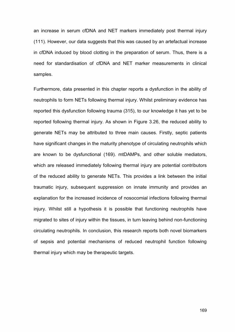
169
an increase in serum cfDNA and NET markers immediately post thermal injury
(111). However, our data suggests that this was caused by an artefactual increase
in cfDNA induced by blood clotting in the preparation of serum. Thus, there is a
need for standardisation of cfDNA and NET marker measurements in clinical
samples.
Furthermore, data presented in this chapter reports a dysfunction in the ability of
neutrophils to form NETs following thermal injury. Whilst preliminary evidence has
reported this dysfunction following trauma (315), to our knowledge it has yet to be
reported following thermal injury. As shown in Figure 3.26, the reduced ability to
generate NETs may be attributed to three main causes. Firstly, septic patients
have significant changes in the maturity phenotype of circulating neutrophils which
are known to be dysfunctional (169). mtDAMPs, and other soluble mediators,
which are released immediately following thermal injury are potential contributors
of the reduced ability to generate NETs. This provides a link between the initial
traumatic injury, subsequent suppression on innate immunity and provides an
explanation for the increased incidence of nosocomial infections following thermal
injury. Whilst still a hypothesis it is possible that functioning neutrophils have
migrated to sites of injury within the tissues, in turn leaving behind non-functioning
circulating neutrophils. In conclusion, this research reports both novel biomarkers
of sepsis and potential mechanisms of reduced neutrophil function following
thermal injury which may be therapeutic targets.
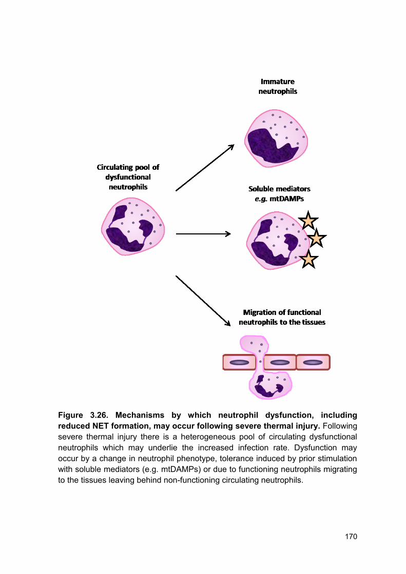
170
Figure 3.26. Mechanisms by which neutrophil dysfunction, including
reduced NET formation, may occur following severe thermal injury. Following
severe thermal injury there is a heterogeneous pool of circulating dysfunctional
neutrophils which may underlie the increased infection rate. Dysfunction may
occur by a change in neutrophil phenotype, tolerance induced by prior stimulation
with soluble mediators (e.g. mtDAMPs) or due to functioning neutrophils migrating
to the tissues leaving behind non-functioning circulating neutrophils.
171
Chapter 4
Reduced DNAse activity in burns patients is associated
with compromise to the blood-based actin scavenging
system and increased risk of multiple organ failure
172
4.1 Introduction
Sepsis is a life threatening condition characterised by an imbalance in both the
immune and haemostatic systems, which results in cardiac dysfunction and acute
respiratory distress syndrome. Without intervention, sepsis can lead to MOF,
which is a leading cause of delayed mortality following major trauma and is
characterised by tissue hypoxia, tissue damage and organ dysfunction (59).
Although it is well recognised that damage to vital organs occurs during MOF, the
mechanisms mediating this damage and progression remain poorly understood
(352). Understanding the pathogenesis of MOF and organ damage has the
potential to improve patient outcome following major injury including burns.
Immunothrombosis is a recently described phenomenon in which thrombosis
forms a vital part of host protection against invading pathogens (296). This
process is supported by innate immune cells and forms a matrix upon which
recognition and elimination of pathogens can occur. Of note, neutrophils have a
multifaceted role in coagulation and are implicated in thrombosis, tissue damage
and various disease pathologies (229, 230, 237, 353). One proposed mechanism
by which this occurs is through the generation of NETs which are comprised of
DNA, histones, granule-derived peptides and enzymes and have been shown both
in vitro and in vivo to ensnare, trap and in some instances directly eliminate
pathogens (119).
Recent studies have shown that NETs and their components can initiate a
procoagulant phenotype and cause tissue and organ damage (195, 223, 224).
NET induced thrombi have decreased permeability, decreased susceptibility to
fibrinolysis and increased clot stability (235). Build-up of circulating NET
components may occur due to overproduction of NETs, due to systemic priming,
173
or a reduced ability to degrade and breakdown NETs. As described in an earlier
chapter (3.2.8 Ex vivo NETosis, Figure 3.10), neutrophils isolated from burn-
injured patients are dysfunctional in producing NETs ex vivo compared to healthy
individuals. Therefore, we hypothesise that the accumulation of NET components
following injury is likely caused by impairment in clearance mechanisms.
DNAse 1 is the major extracellular endonuclease found in blood and functions to
break down chromatin and DNA that are released following cellular damage, cell
death and NETosis. Reduction in DNAse activity is associated with the
pathogenesis of autoimmune disease through physical inhibition of the enzyme by
circulating antibodies (237). DNAse activity is also indirectly regulated by the actin
scavenging system that prevents the formation of DNAse and actin complexes.
The system is comprised of two key proteins; GSN and VDBP (237, 240, 245,
246). Whilst it has been shown that reduced levels of GSN are associated with
mortality, development of sepsis and MOF (254). No group has studied dynamic
changes in GSN and VDBP levels in relation to DNAse activity and build-up of
circulating toxic NET components following severe thermal injury.
4.1.1 Aims
The aims of this chapter were:
Investigate if clearance of DNA was dysfunctional following thermal injury.
Investigate if a reduction in DNAse activity was associated with secondary
complications namely, sepsis and MOF.
Investigate if reduced DNAse activity is a potential therapeutic target to
reduce secondary complications.
174
4.2 Results
4.2.1 Study cohort
Consistent with Chapter 3, all analysis was performed on adult patients (≥ 18
years of age) with burns ≥ 15% TBSA. The consort diagram for this analysis is
included (Figure 4.1). Analysis was restricted to burns ≥ 15% TBSA to minimise
the confounding bias of age and severity of injury in analysis. Furthermore, this
has reduced the span and confounding bias of fluid resuscitation (284, 285).
Patient and healthy controls demographics are shown in Table 4.1. A total of 64
adult burns patients were included in the study, with a mean age of 43 years
(range 16 - 88) and mean burn size of 35% TBSA (range 15 - 95). In addition, 19
healthy controls were included in this analysis with a mean age of 44.5 years
(range 20 - 95.9). The incidence of sepsis was 59%, with 38 patients experiencing
one or more episodes during their hospital stay. The median time to first septic
episode in this cohort was 5 days post-injury (IQR 4 - 7). The incidence of MOF in
the cohort was 37.5% (24/64), with the median time to first episode 4 days post-
injury (IQR 2 - 11). A significantly greater injury severity, incidence of sepsis and
mortality rate was recorded in those patients who developed MOF (p < 0.05). All
patients received standardised burn resuscitation protocols as per Parkland
Formula and as such received equivalent fluid resuscitation (average = 5.4
mls/kg/%TBSA, standard deviation = 2.1).
Different from Chapter 3, all patients were included in the analysis within this
chapter irrelevant of time to mortality as no attempt to evaluate the diagnostic or
prognostic accuracy of biomarkers using AUROC was made.
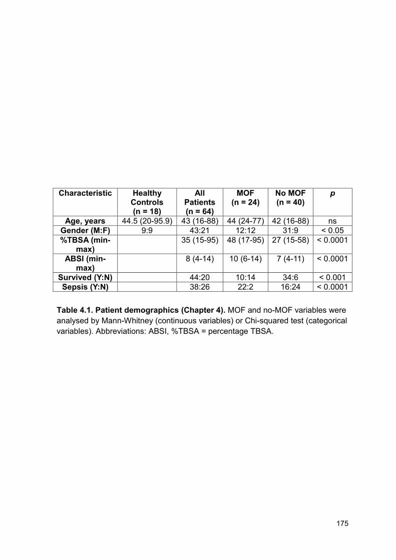
175
Characteristic Healthy Controls (n = 18)
All Patients (n = 64)
MOF (n = 24)
No MOF (n = 40)
p
Age, years 44.5 (20-95.9) 43 (16-88) 44 (24-77) 42 (16-88) ns
Gender (M:F) 9:9 43:21 12:12 31:9 < 0.05
%TBSA (min-max)
35 (15-95) 48 (17-95) 27 (15-58) < 0.0001
ABSI (min-max)
8 (4-14) 10 (6-14) 7 (4-11) < 0.0001
Survived (Y:N) 44:20 10:14 34:6 < 0.001
Sepsis (Y:N) 38:26 22:2 16:24 < 0.0001
Table 4.1. Patient demographics (Chapter 4). MOF and no-MOF variables were
analysed by Mann-Whitney (continuous variables) or Chi-squared test (categorical
variables). Abbreviations: ABSI, %TBSA = percentage TBSA.
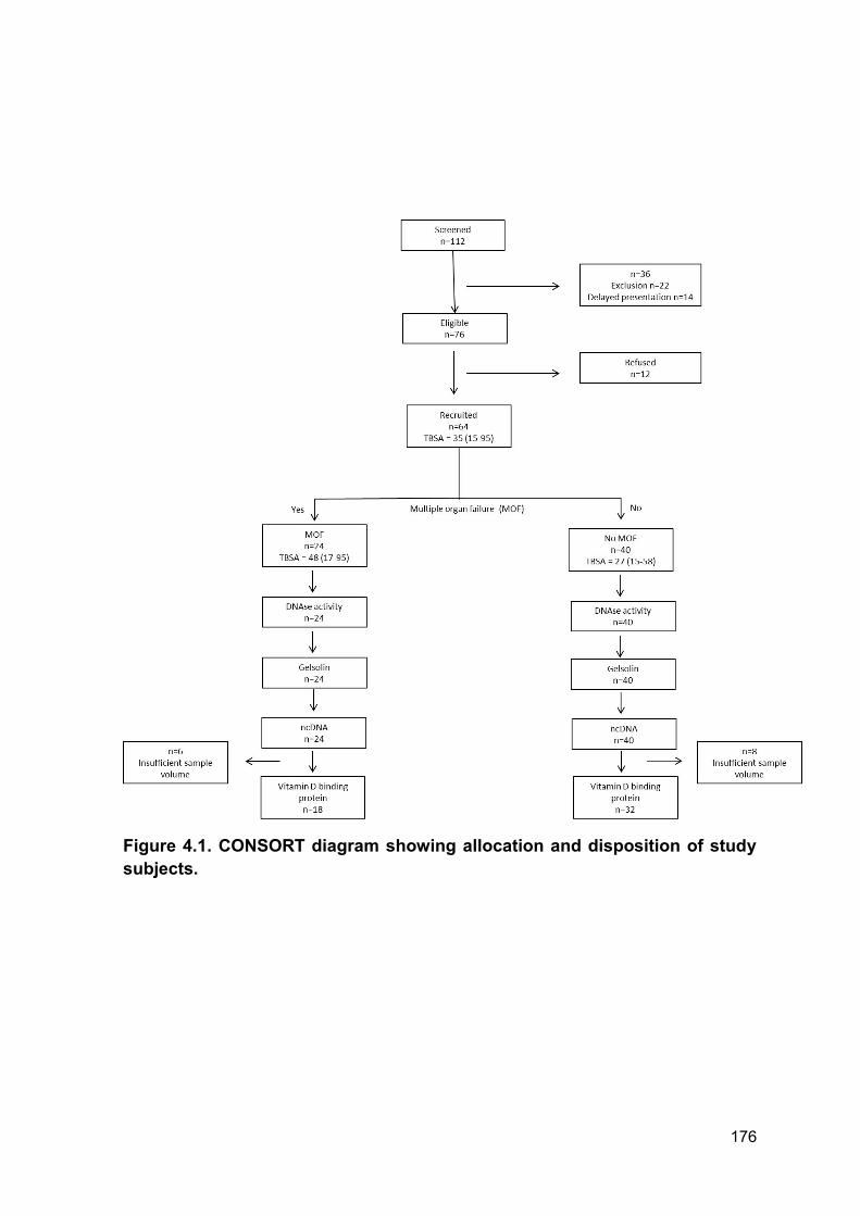
176
Figure 4.1. CONSORT diagram showing allocation and disposition of study
subjects.
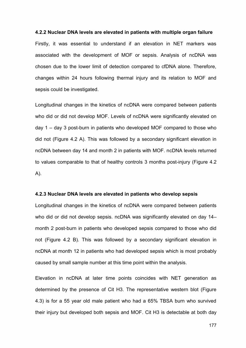
177
4.2.2 Nuclear DNA levels are elevated in patients with multiple organ failure
Firstly, it was essential to understand if an elevation in NET markers was
associated with the development of MOF or sepsis. Analysis of ncDNA was
chosen due to the lower limit of detection compared to cfDNA alone. Therefore,
changes within 24 hours following thermal injury and its relation to MOF and
sepsis could be investigated.
Longitudinal changes in the kinetics of ncDNA were compared between patients
who did or did not develop MOF. Levels of ncDNA were significantly elevated on
day 1 – day 3 post-burn in patients who developed MOF compared to those who
did not (Figure 4.2 A). This was followed by a secondary significant elevation in
ncDNA between day 14 and month 2 in patients with MOF. ncDNA levels returned
to values comparable to that of healthy controls 3 months post-injury (Figure 4.2
A).
4.2.3 Nuclear DNA levels are elevated in patients who develop sepsis
Longitudinal changes in the kinetics of ncDNA were compared between patients
who did or did not develop sepsis. ncDNA was significantly elevated on day 14–
month 2 post-burn in patients who developed sepsis compared to those who did
not (Figure 4.2 B). This was followed by a secondary significant elevation in
ncDNA at month 12 in patients who had developed sepsis which is most probably
caused by small sample number at this time point within the analysis.
Elevation in ncDNA at later time points coincides with NET generation as
determined by the presence of Cit H3. The representative western blot (Figure
4.3) is for a 55 year old male patient who had a 65% TBSA burn who survived
their injury but developed both sepsis and MOF. Cit H3 is detectable at both day
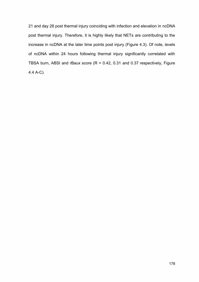
178
21 and day 28 post thermal injury coinciding with infection and elevation in ncDNA
post thermal injury. Therefore, it is highly likely that NETs are contributing to the
increase in ncDNA at the later time points post injury (Figure 4.3). Of note, levels
of ncDNA within 24 hours following thermal injury significantly correlated with
TBSA burn, ABSI and rBaux score (R = 0.42, 0.31 and 0.37 respectively, Figure
4.4 A-C).
179
Figure 4.2. Patients with MOF and sepsis have higher levels of circulating
ncDNA. (A) ncDNA across time (n = 64) differences in kinetics were compared
between patients who did and did not develop MOF using a Mann-Whitney test;
*p < 0.05. (B) ncDNA across time (n = 64) differences in kinetics were compared
between patients who did and did not develop sepsis using a Mann-Whitney test;
*p < 0.05. Both panels are presented on a log scale. Box and whisker plots
represent median, minimum and maximum values. HC = healthy control, D = day,
M = month.
180
Figure 4.3. Cit H3 coincides with elevations in ncDNA following thermal
injury. ncDNA (log scale) and Cit H3 western for a representative patient: 55 year
old male patient who had a 65% TBSA burn who survived their injury but
developed both sepsis and MOF. Positive control = Calcium ionophore stimulated
HL-60 cells (Section 2.10), HC = healthy control, D = day.
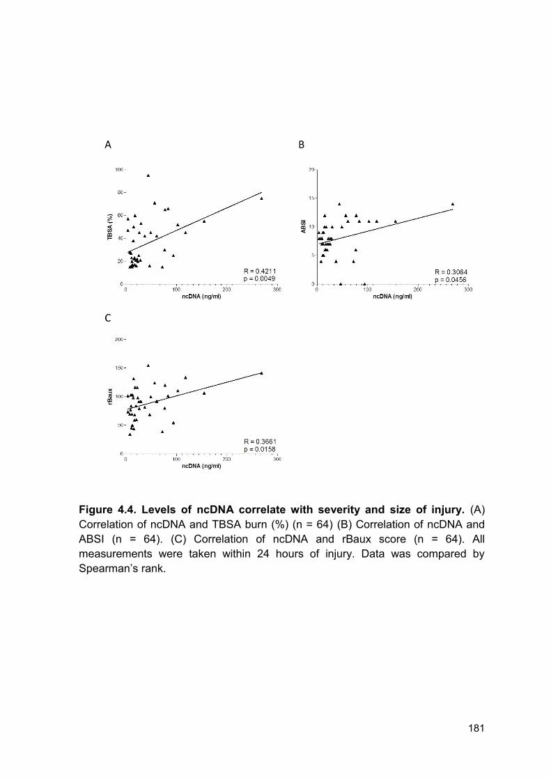
181
Figure 4.4. Levels of ncDNA correlate with severity and size of injury. (A)
Correlation of ncDNA and TBSA burn (%) (n = 64) (B) Correlation of ncDNA and
ABSI (n = 64). (C) Correlation of ncDNA and rBaux score (n = 64). All
measurements were taken within 24 hours of injury. Data was compared by
Spearman’s rank.
182
4.2.4 Patients with thermal injury have reduced DNAse activity
To investigate if the accumulation of ncDNA was associated with reduced
clearance, DNAse activity was quantified in serum samples. There was a
significant reduction in DNAse activity, from day 1 – day 28 post-injury compared
to healthy individuals (Figure 4.5). Interestingly, DNAse activity on day of injury did
not correlate with size of thermal injury (TBSA %), severity of injury (ABSI) or
rBaux (Figure 4.6 A-C).
Reduced DNAse activity was confirmed by fluorescence microscopy with NETs
still visible in slides treated with patient serum (Figure 4.7). Conversely, all healthy
control serum samples digested NETs completely (Figure 4.7). Data presented is
from 5 independent experiments and is representative of 10 experiments. DNAse
activity significantly and inversely correlated with ncDNA levels across all time
points (n = 64, r = -0.3574, p < 0.0001).
4.2.5 DNAse activity is lower in patients who develop multiple organ failure
or sepsis
To understand if a reduced DNAse activity was associated with MOF, by
facilitating the accumulation of ncDNA, patients were split into those who did and
those who did not develop MOF. Importantly, patients who developed MOF had
significantly lower DNAse activity at day 28 and month 2 post injury compared to
patients who did not develop MOF (Figure 4.8).
Patients were further subcategorised according to their sepsis status to investigate
if infection had any effect on DNAse activity. Patients who developed sepsis had
significantly lower DNAse activity at day 21 to month 2 post injury compared to
patients who did not develop sepsis (Figure 4.9).
183
4.2.6 DNAse-1 antigen levels are elevated following thermal injury
As we have reported a sustained elevation in NET markers in the presence of an
inability to produce NETs in vivo we investigated if clearance was impaired. Levels
of DNAse 1 were quantified to understand if the reduction in DNAse activity from
day 1 – day 28 post injury was mediated by a reduction in circulating DNAse
antigen. Levels were quantified in 24 patients who had reduced DNAse activity
(below 50% compared to healthy controls) within 24 hours of injury. Interestingly,
there was no significant difference in DNAse antigen levels from day 1 – day 3
post injury. However, there was a significant increase in DNAse antigen levels
from day 7 – day 14 and on day 28 post injury compared to levels measured in
healthy volunteers (Figure 4.10).
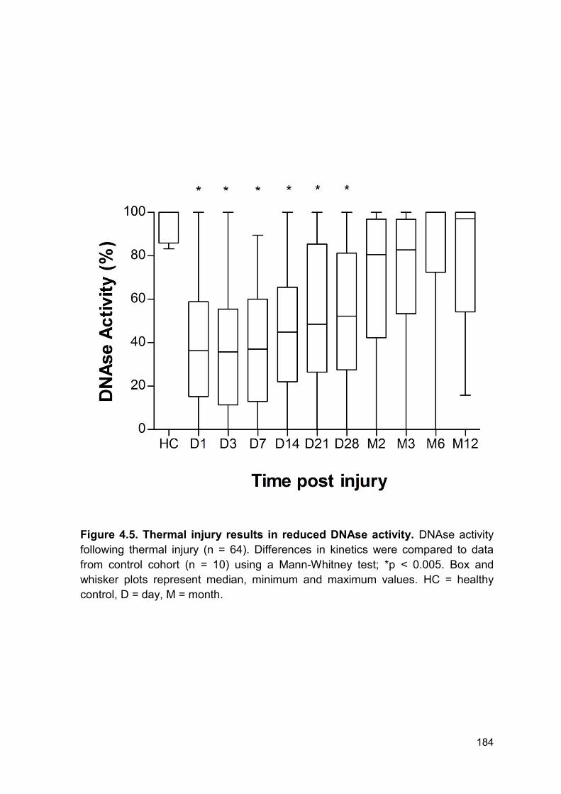
184
Figure 4.5. Thermal injury results in reduced DNAse activity. DNAse activity
following thermal injury (n = 64). Differences in kinetics were compared to data
from control cohort (n = 10) using a Mann-Whitney test; *p < 0.005. Box and
whisker plots represent median, minimum and maximum values. HC = healthy
control, D = day, M = month.
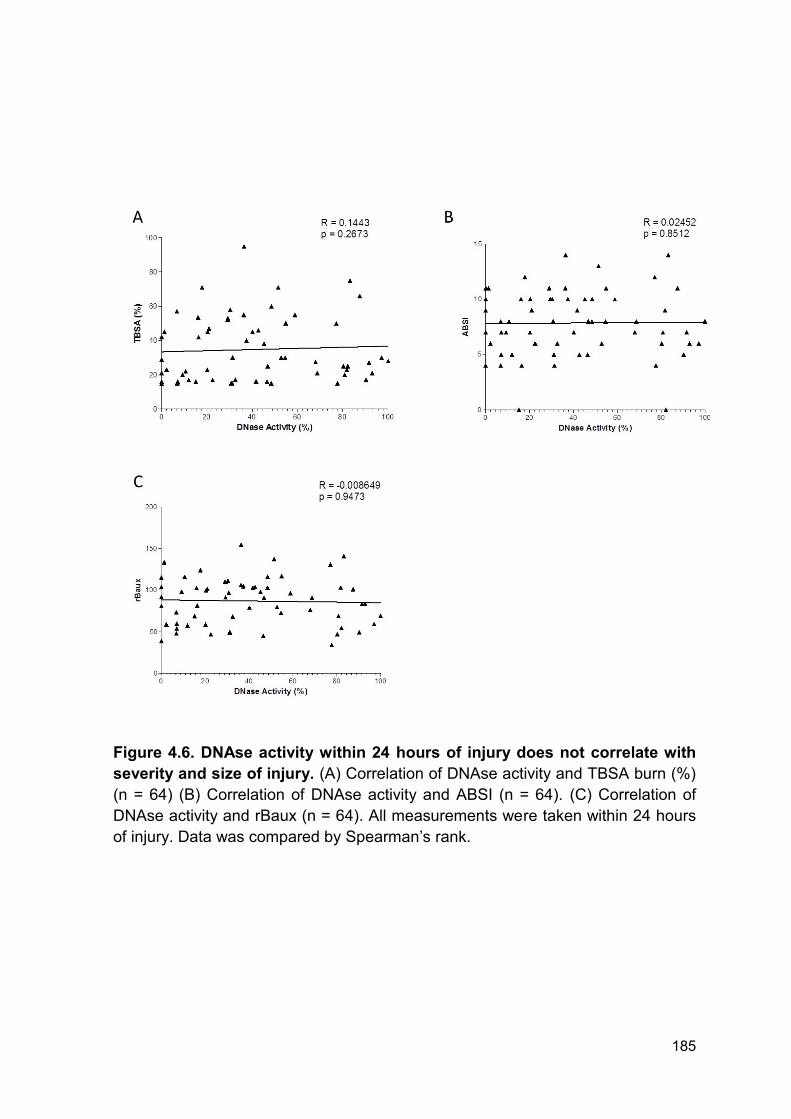
185
Figure 4.6. DNAse activity within 24 hours of injury does not correlate with
severity and size of injury. (A) Correlation of DNAse activity and TBSA burn (%)
(n = 64) (B) Correlation of DNAse activity and ABSI (n = 64). (C) Correlation of
DNAse activity and rBaux (n = 64). All measurements were taken within 24 hours
of injury. Data was compared by Spearman’s rank.
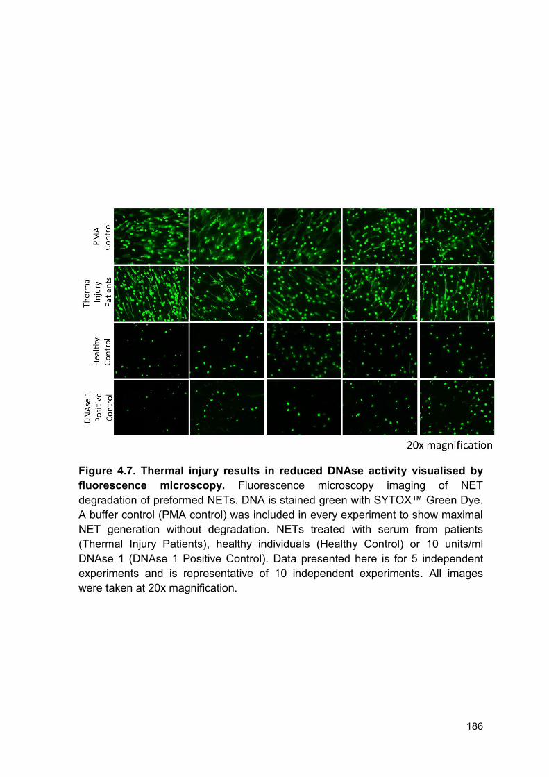
186
Figure 4.7. Thermal injury results in reduced DNAse activity visualised by
fluorescence microscopy. Fluorescence microscopy imaging of NET
degradation of preformed NETs. DNA is stained green with SYTOX™ Green Dye.
A buffer control (PMA control) was included in every experiment to show maximal
NET generation without degradation. NETs treated with serum from patients
(Thermal Injury Patients), healthy individuals (Healthy Control) or 10 units/ml
DNAse 1 (DNAse 1 Positive Control). Data presented here is for 5 independent
experiments and is representative of 10 independent experiments. All images
were taken at 20x magnification.
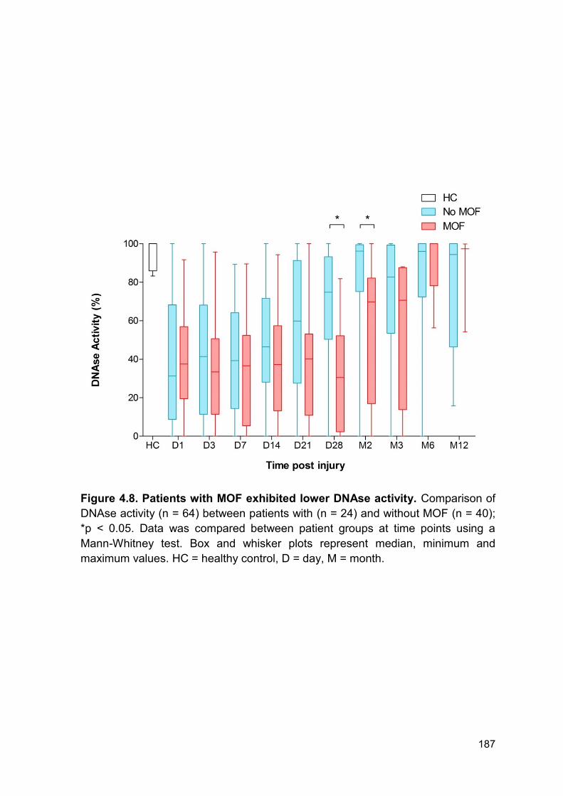
187
Figure 4.8. Patients with MOF exhibited lower DNAse activity. Comparison of
DNAse activity (n = 64) between patients with (n = 24) and without MOF (n = 40);
*p < 0.05. Data was compared between patient groups at time points using a
Mann-Whitney test. Box and whisker plots represent median, minimum and
maximum values. HC = healthy control, D = day, M = month.
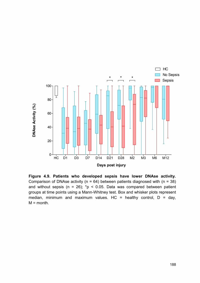
188
Figure 4.9. Patients who developed sepsis have lower DNAse activity.
Comparison of DNAse activity (n = 64) between patients diagnosed with (n = 38)
and without sepsis (n = 26); *p < 0.05. Data was compared between patient
groups at time points using a Mann-Whitney test. Box and whisker plots represent
median, minimum and maximum values. HC = healthy control, D = day,
M = month.
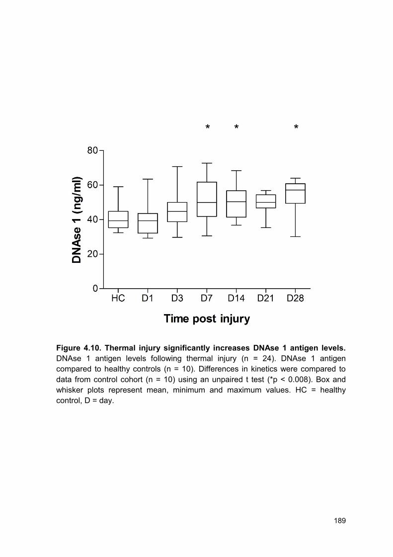
189
Figure 4.10. Thermal injury significantly increases DNAse 1 antigen levels.
DNAse 1 antigen levels following thermal injury (n = 24). DNAse 1 antigen
compared to healthy controls (n = 10). Differences in kinetics were compared to
data from control cohort (n = 10) using an unpaired t test (*p < 0.008). Box and
whisker plots represent mean, minimum and maximum values. HC = healthy
control, D = day.
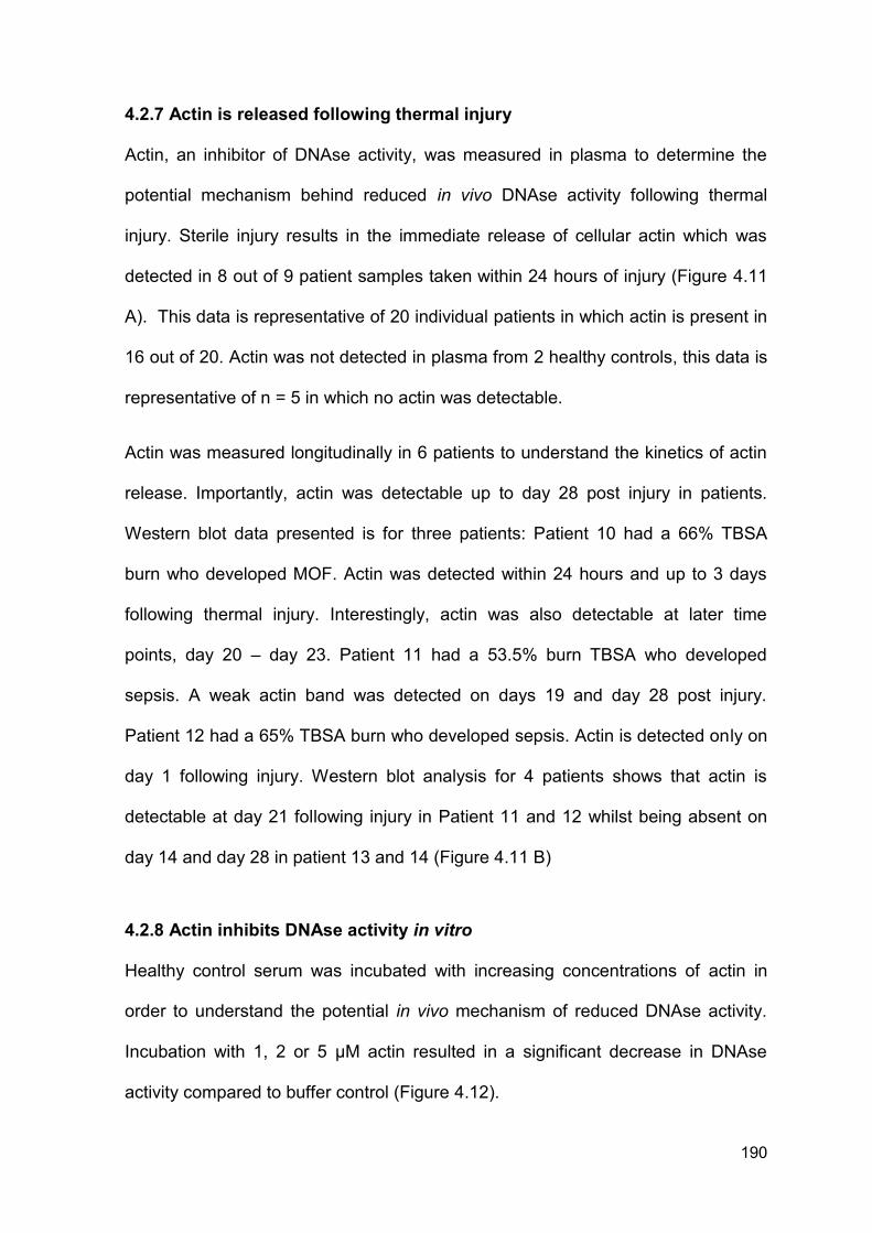
190
4.2.7 Actin is released following thermal injury
Actin, an inhibitor of DNAse activity, was measured in plasma to determine the
potential mechanism behind reduced in vivo DNAse activity following thermal
injury. Sterile injury results in the immediate release of cellular actin which was
detected in 8 out of 9 patient samples taken within 24 hours of injury (Figure 4.11
A). This data is representative of 20 individual patients in which actin is present in
16 out of 20. Actin was not detected in plasma from 2 healthy controls, this data is
representative of n = 5 in which no actin was detectable.
Actin was measured longitudinally in 6 patients to understand the kinetics of actin
release. Importantly, actin was detectable up to day 28 post injury in patients.
Western blot data presented is for three patients: Patient 10 had a 66% TBSA
burn who developed MOF. Actin was detected within 24 hours and up to 3 days
following thermal injury. Interestingly, actin was also detectable at later time
points, day 20 – day 23. Patient 11 had a 53.5% burn TBSA who developed
sepsis. A weak actin band was detected on days 19 and day 28 post injury.
Patient 12 had a 65% TBSA burn who developed sepsis. Actin is detected only on
day 1 following injury. Western blot analysis for 4 patients shows that actin is
detectable at day 21 following injury in Patient 11 and 12 whilst being absent on
day 14 and day 28 in patient 13 and 14 (Figure 4.11 B)
4.2.8 Actin inhibits DNAse activity in vitro
Healthy control serum was incubated with increasing concentrations of actin in
order to understand the potential in vivo mechanism of reduced DNAse activity.
Incubation with 1, 2 or 5 µM actin resulted in a significant decrease in DNAse
activity compared to buffer control (Figure 4.12).
191
Figure 4.11. Thermal injury causes the release of circulating actin. (A)
Western blot of actin in plasma samples from 9 patients (P 1 - 9) taken within 24
hours of injury. Actin was not detected in plasma from healthy controls (HC) n = 2.
(B) Western blot for Patient 10 with a 66% TBSA who developed MOF. Patient 11
had a 53.5% burn TBSA who developed sepsis. Patient 12 had a 65% TBSA burn
who developed sepsis. Western blot analysis for 4 individual patients (P13-16).
HC = healthy control, D = day.
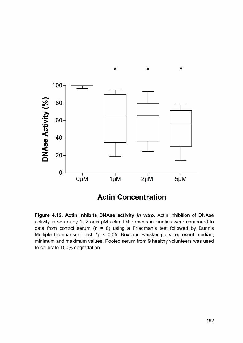
192
Figure 4.12. Actin inhibits DNAse activity in vitro. Actin inhibition of DNAse
activity in serum by 1, 2 or 5 µM actin. Differences in kinetics were compared to
data from control serum (n = 8) using a Friedman’s test followed by Dunn's
Multiple Comparison Test; *p < 0.05. Box and whisker plots represent median,
minimum and maximum values. Pooled serum from 9 healthy volunteers was used
to calibrate 100% degradation.
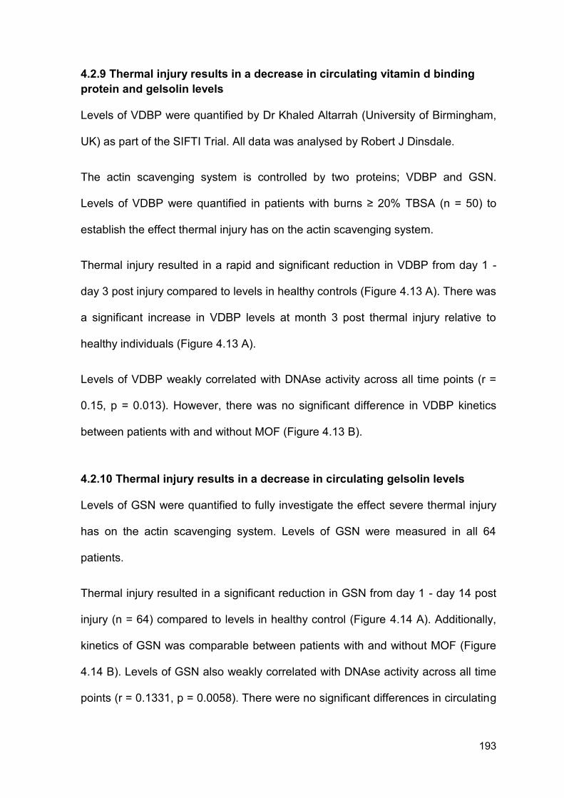
193
4.2.9 Thermal injury results in a decrease in circulating vitamin d binding
protein and gelsolin levels
Levels of VDBP were quantified by Dr Khaled Altarrah (University of Birmingham,
UK) as part of the SIFTI Trial. All data was analysed by Robert J Dinsdale.
The actin scavenging system is controlled by two proteins; VDBP and GSN.
Levels of VDBP were quantified in patients with burns ≥ 20% TBSA (n = 50) to
establish the effect thermal injury has on the actin scavenging system.
Thermal injury resulted in a rapid and significant reduction in VDBP from day 1 -
day 3 post injury compared to levels in healthy controls (Figure 4.13 A). There was
a significant increase in VDBP levels at month 3 post thermal injury relative to
healthy individuals (Figure 4.13 A).
Levels of VDBP weakly correlated with DNAse activity across all time points (r =
0.15, p = 0.013). However, there was no significant difference in VDBP kinetics
between patients with and without MOF (Figure 4.13 B).
4.2.10 Thermal injury results in a decrease in circulating gelsolin levels
Levels of GSN were quantified to fully investigate the effect severe thermal injury
has on the actin scavenging system. Levels of GSN were measured in all 64
patients.
Thermal injury resulted in a significant reduction in GSN from day 1 - day 14 post
injury (n = 64) compared to levels in healthy control (Figure 4.14 A). Additionally,
kinetics of GSN was comparable between patients with and without MOF (Figure
4.14 B). Levels of GSN also weakly correlated with DNAse activity across all time
points (r = 0.1331, p = 0.0058). There were no significant differences in circulating
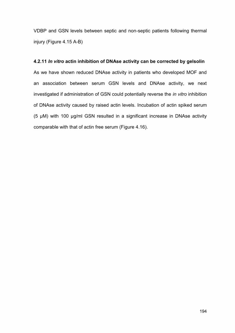
194
VDBP and GSN levels between septic and non-septic patients following thermal
injury (Figure 4.15 A-B)
4.2.11 In vitro actin inhibition of DNAse activity can be corrected by gelsolin
As we have shown reduced DNAse activity in patients who developed MOF and
an association between serum GSN levels and DNAse activity, we next
investigated if administration of GSN could potentially reverse the in vitro inhibition
of DNAse activity caused by raised actin levels. Incubation of actin spiked serum
(5 µM) with 100 µg/ml GSN resulted in a significant increase in DNAse activity
comparable with that of actin free serum (Figure 4.16).
195
Figure 4.13. Levels of VDBP are reduced following thermal injury. (A) VDBP
across time (n = 50). Differences in kinetics were compared to data from 10
healthy volunteers using a Mann-Whitney test; *p < 0.005. (B) VDBP across time
(n = 50) between patients with (n = 18) and without MOF (n = 32) using a Mann-
Whitney test; *p < 0.05. Data is presented as a box and whisker plots and
represents median, minimum and maximum values. HC = healthy control,
D = day, M = month.
196
Figure 4.14. Levels of GSN are reduced following thermal injury. (A) GSN
levels across time (n = 64) compared to healthy controls (n = 10). Data was
analysed by Mann-Whitney test compared to healthy controls *p < 0.005. (B) GSN
levels across time comparing those who did (n = 24) and those who did not
(n = 40) develop MOF. Differences in kinetics were compared between patient
groups using a Mann-Whitney test; *p < 0.05. Data is presented as a box and
whisker plots and represents median, minimum and maximum values. HC =
healthy control, D = day, M = month.
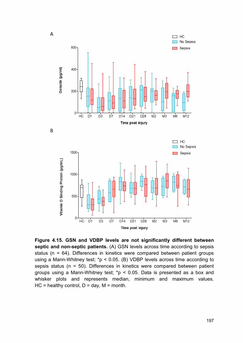
197
Figure 4.15. GSN and VDBP levels are not significantly different between
septic and non-septic patients. (A) GSN levels across time according to sepsis
status (n = 64). Differences in kinetics were compared between patient groups
using a Mann-Whitney test; *p < 0.05. (B) VDBP levels across time according to
sepsis status (n = 50). Differences in kinetics were compared between patient
groups using a Mann-Whitney test; *p < 0.05. Data is presented as a box and
whisker plots and represents median, minimum and maximum values.
HC = healthy control, D = day, M = month.
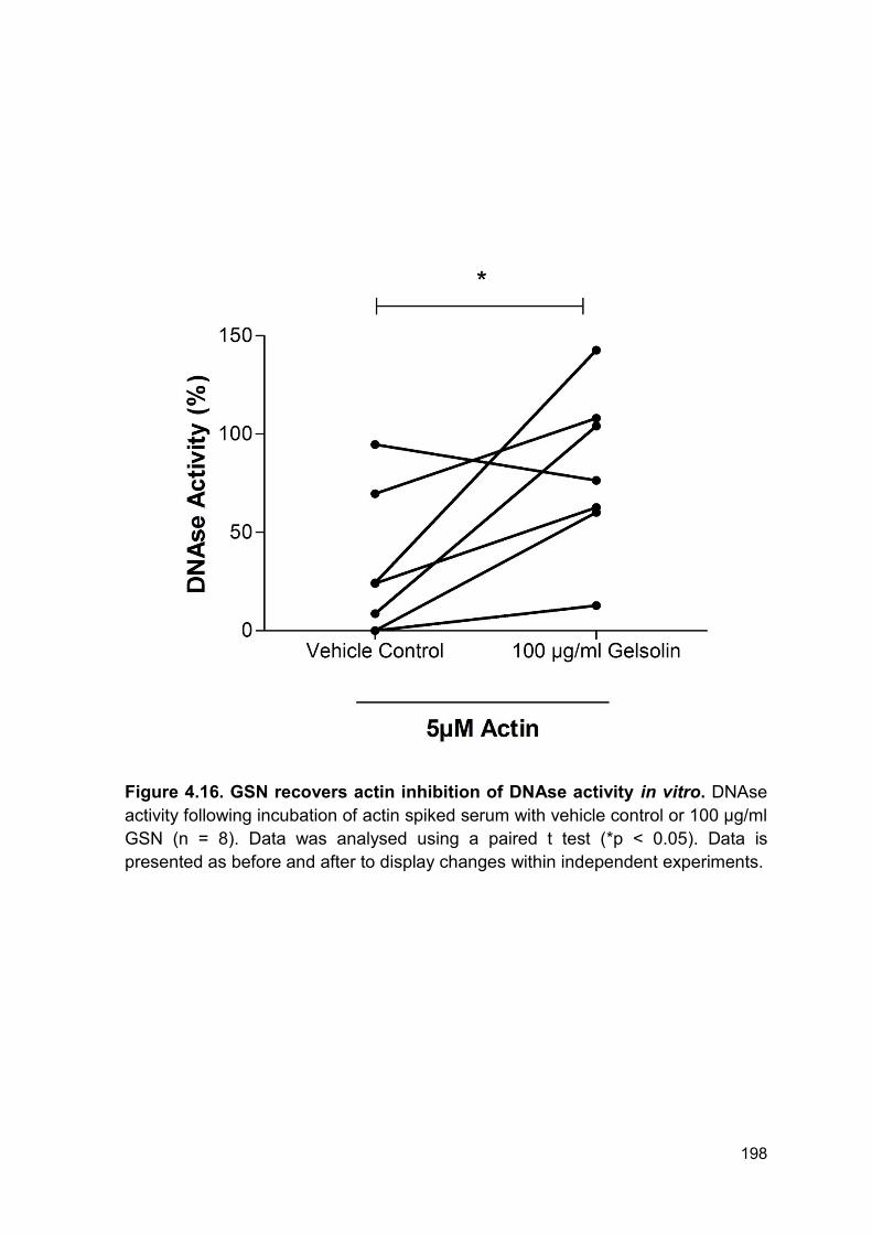
198
Figure 4.16. GSN recovers actin inhibition of DNAse activity in vitro. DNAse
activity following incubation of actin spiked serum with vehicle control or 100 µg/ml
GSN (n = 8). Data was analysed using a paired t test (*p < 0.05). Data is
presented as before and after to display changes within independent experiments.
199
4.2.12 Patient demographics for patients with severe injuries caused by
explosions
To investigate if blood products, namely fresh frozen plasma (FFP), can potentially
increase DNAse activity by correcting the blood based actin scavenging system,
levels of GSN, VDBP and DNAse activity were quantified in plasma from patients
following severe traumatic injury caused by explosions. This cohort was split into
patients who had (n = 6) or had not received FFP (n = 6) prior to hospital
admission and blood sampling. There was no significant difference in ISS, NISS
and time to admission following injury between the 2 groups. Full patient
demographics can be found in Table 4.2. On average patients received 3 units of
blood products before admission to hospital. Due to the nature of this work and
the cohort it was not possible to obtain clinical data on outcomes or secondary
complications.
4.2.13 Fresh frozen plasma increases gelsolin levels and DNAse activity
following severe injury caused by explosion but has no effect on vitamin d
binding protein levels
All patients in this analysis were matched for clinical scores of severity and
admission times. In the total cohort (n = 12), DNAse activity and VDBP levels were
comparable between healthy controls and patients with severe injuries caused by
explosions (Figure 4.17 A, C). However, GSN levels were significantly lower in
patients with severe injuries caused by explosions when compared to healthy
individuals (Figure 4.17 B).
Patients who did not receive blood products before hospital admission had
significantly lower DNAse activity and plasma GSN levels compared to healthy
controls (Figure 4.18 A-B). However, there was no difference when comparing
200
plasma VDBP levels (Figure 4.18 C). DNAse activity, GSN and VDBP were
comparable between patients who received blood products and healthy controls
(Figure 4.18 A-C).
Administration of blood products and FFP before admission to hospital
significantly increased DNAse activity compared to patients who had not received
blood products upon admission to hospital (Figure 4.19 A). Furthermore,
administration of blood products and FFP before admission to hospital significantly
increased circulating GSN levels compared to patients who had not received blood
products and FFP (Figure 4.19 B). Finally, administration of blood products and
FFP did not affect circulating levels of VDBP in both patient groups (Figure 4.19
C).
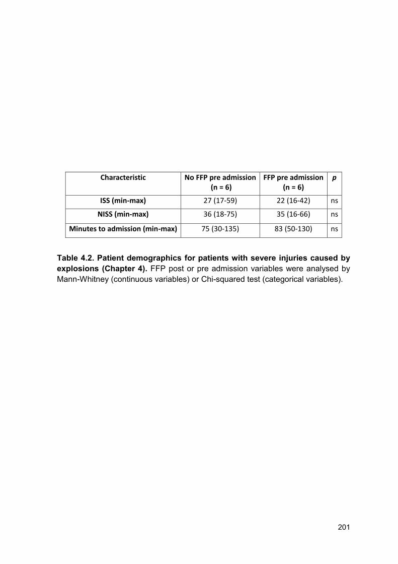
201
Characteristic No FFP pre admission (n = 6)
FFP pre admission (n = 6)
p
ISS (min-max) 27 (17-59) 22 (16-42) ns
NISS (min-max) 36 (18-75) 35 (16-66) ns
Minutes to admission (min-max) 75 (30-135) 83 (50-130) ns
Table 4.2. Patient demographics for patients with severe injuries caused by
explosions (Chapter 4). FFP post or pre admission variables were analysed by
Mann-Whitney (continuous variables) or Chi-squared test (categorical variables).
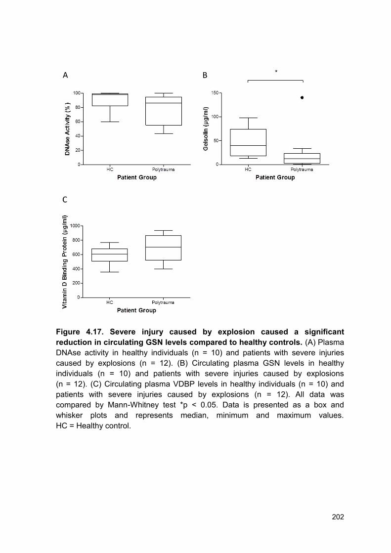
202
Figure 4.17. Severe injury caused by explosion caused a significant
reduction in circulating GSN levels compared to healthy controls. (A) Plasma
DNAse activity in healthy individuals (n = 10) and patients with severe injuries
caused by explosions (n = 12). (B) Circulating plasma GSN levels in healthy
individuals (n = 10) and patients with severe injuries caused by explosions
(n = 12). (C) Circulating plasma VDBP levels in healthy individuals (n = 10) and
patients with severe injuries caused by explosions (n = 12). All data was
compared by Mann-Whitney test *p < 0.05. Data is presented as a box and
whisker plots and represents median, minimum and maximum values.
HC = Healthy control.
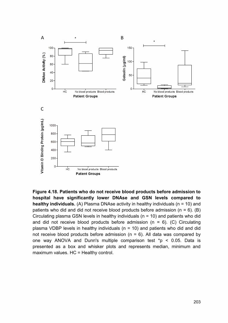
203
Figure 4.18. Patients who do not receive blood products before admission to
hospital have significantly lower DNAse and GSN levels compared to
healthy individuals. (A) Plasma DNAse activity in healthy individuals (n = 10) and
patients who did and did not receive blood products before admission (n = 6). (B)
Circulating plasma GSN levels in healthy individuals (n = 10) and patients who did
and did not receive blood products before admission (n = 6). (C) Circulating
plasma VDBP levels in healthy individuals (n = 10) and patients who did and did
not receive blood products before admission (n = 6). All data was compared by
one way ANOVA and Dunn's multiple comparison test *p < 0.05. Data is
presented as a box and whisker plots and represents median, minimum and
maximum values. HC = Healthy control.
204
Figure 4.19. Blood products increase circulating GSN levels and protects
against inhibition of DNAse activity. (A) Comparison of DNAse levels at
hospital admission between patients with (n = 6) or without (n = 6) prior blood
product administration. (B) GSN levels at hospital admission between patients with
(n = 6) or without (n = 6) prior blood product infusion at time of blood sampling. (C)
Comparison of VDBP activity levels at hospital admission from patients with
(n = 6) or without (n = 6) prior blood product infusion at time of blood sampling. For
panel A and C data was analysed by unpaired t-test. For panel B data was
analysed by Mann-Whitney test. *p < 0.05. For panel A and C, data is presented
as a box and whisker plots and represents mean, minimum and maximum values.
For panel B, data is presented as a box and whisker plots and represents median,
minimum and maximum values. HC = Healthy control.
205
4.3 Discussion
Circulating levels of cfDNA have been described as a novel biomarker of
secondary complications and mortality with studies reporting positive prognostic
and diagnostic utility (109, 180, 220, 304-306). In a cohort of 67 patients with
severe sepsis, quantification of ncDNA showed positive prognostic utility for
predicting 24 hours mortality in patients who presented to the emergency
department (354).
Circulating DNA can arise from a number of sources including tissue damage,
apoptosis, necrosis and NETosis (114). In our analysis levels of ncDNA within 24
hours of injury correlated with measurements of burn size and severity. Therefore,
we propose that the initial elevation in ncDNA is originating from tissue damage
caused by the burn injury. Due to the cytotoxic nature of DNA (355, 356), the initial
elevation in circulating ncDNA may be contributing to the immediate host tissue
and organ damage which occurs in patients with burn injuries. Consistent with
previous literature (354), ncDNA levels are significantly higher in septic patients
compared to non-septic individuals. We propose that tissue damage, surgery and
NETosis may also be contributing to this secondary increase. Interestingly, levels
of ncDNA are significantly higher at 12 months following injury in patients who
developed sepsis compared to those who didn’t. This elevation is either caused by
lower sample numbers in the analysis for 12 month samples, a reactive response
by the body in which DNA cannot be cleared or further release from tissue
remodelling and scar formation.
NETs and histones form key components of host defence and innate immunity
(118, 355, 357). Engelmann and Massberg described an innate immune response
206
in which coagulation is activated in an attempt to ensnare, recognise and remove
bacteria (296, 358, 359). This process is termed immunothrombosis and is in part
mediated by the production of NETs (296). NETs are capable of providing both a
stimulus to and scaffold for thrombus formation. Uncontrolled or excessive NET
generation and immunothrombosis can result in increased risk of thrombosis, host
tissue damage and the accumulation of pathogenic components, including DNA,
in the microvasculature (226).
Fuchs and colleagues demonstrated that NETs perfused with blood caused
recruitment of red cells, increased fibrin deposition, platelet adhesion and
activation. Importantly, this could be prevented by incubation with DNAse which
promotes NET breakdown (232). In a murine model of DVT, Cit H3 was found in
close proximity to thrombi. Furthermore, DNA and histones form components of
the scaffold required for the pathogenesis of DVT (233). In addition to promoting
thrombosis, elevated levels of NET components are cytotoxic and can result in
host tissue damage. For example, incubation of activated endothelial cells with
NETs results in cell damage which, again, can be prevented by degrading NETs
with DNAse (360).
Here, we report that ncDNA was elevated from day 1 to day 3 and again at day 14
to 2 months post-injury in patients who developed MOF compared to those who
did not. Here we report an accumulation of circulating ncDNA for up to 28 days
following injury. We hypothesised that clearance of host and NET derived DNA
was dysregulated. Multiple groups have shown that degradation of NETs with
DNAse can protect against tissue damage, thrombosis and procoagulant
interactions (232, 360). Furthermore, elevated levels of circulating DNA are also
associated with and contribute to the progression of autoimmune diseases,
207
notably SLE (237, 238). This build-up of circulating DNA can be explained in part
to the impaired DNAse activity caused by the presence of ‘DNAse inhibitors’ or
inhibitory antibodies in these patients (237).
As we report a build-up of circulating DNA and reduced ex vivo NET formation we
next investigated DNAse activity in patient samples. Here we report a reduction in
DNAse activity from day 1 – day 28 post injury in the total cohort. Of note, there
was large variation in DNAse activity within the whole cohort at all time points.
Furthermore, levels of DNAse activity did not correlate with injury severity. This
may be explained by timing of sample, clinical intervention or heterogeneity within
the patient cohort. Importantly, DNAse activity was lower in patients who
developed MOF or sepsis compared to those who did not develop either. Both
groups had comparable reduction in DNAse activity for the first 14 days following
thermal injury. Therefore, one might hypothesise that the initial injury causes the
immediate reduction in activity reported in both groups and the later reduction may
be caused by further tissue damage or surgical procedures. Within this cohort the
average amount of individual septic episodes was 2.5 episodes per patient. The
average time to first episode was 5 days following injury (range 3 – 70 days) and
the time to last episode was 23 days following injury (range 3 – 130 days).
Furthermore, whilst most patients underwent surgery within the first 5 days for
debridement and immediate treatment of burn wounds, patients did have further
surgical procedures if required, e.g. skin graft rejection. Thus, the variation within
DNAse activity at the later time points following thermal injury may be explained by
the ongoing tissue damage from surgical procedures and sporadic infectious
episodes.
208
It is important to consider the mechanism by which DNAse activity inhibition
occurs. Here, we report that levels of total DNAse antigen are increased post
thermal injury, which is consistent with existing literature (239). Therefore, the
apparent reduction in DNAse activity is indicative of inhibition of enzymatic activity.
In this thesis, only levels of DNAse 1 antigen were quantified as this is the
predominant enzyme responsible for the degradation of circulating DNA. However,
there are different isoforms of DNAse, including DNAse-γ, therefore we cannot
exclude the possibility that the reduction in DNAse activity may be mediated in
part by reduced DNAse-γ antigen levels. Thus, further study of DNAse isoforms
and their contribution to reduced DNAse activity following injury is required.
A naturally occurring inhibitor of DNAse activity is actin, which is an abundant
protein in mammalian cells (240). Actin exists in a balance between monomeric
and filamentous actin, which is essential for cellular function (241, 242). However,
this predisposition of actin to rapidly polymerise is extremely dangerous if it occurs
in the circulation. Actin is recognised as a DAMP due to its rapid release,
immunostimulatory actions and conserved structure (361). Polymerised actin, F-
actin, binds to the DNGR-1 receptor (CLEC9A) and primes cytotoxic T-cells
against dead cell antigens (362-364). In addition to its DAMP properties,
extracellular actin can cause direct damage to the microvasculature, impair clot
lysis and activate platelets (240, 243, 244).
Actin can bind to DNAse forming a stoichiometric 1:1 complex which inhibits
enzymatic activity (245, 365). In this analysis circulating actin is detected in
patients immediately following and for up to 28 days post-injury. The initial release
of actin most likely originates from the extensive tissue damage occurring
following severe thermal injury. This will not only inhibit DNAse activity but may
209
also be responsible for the immediate stimulation of the immune system and host
tissue damage that occurs following thermal injury due to its DAMP properties and
cytotoxic nature (240, 243, 244, 361-364).
Here we show that actin is detectable for up to 28 days post injury. In Patient 10
(Figure 4.11 B) actin is cleared at day 10 and is then detectable at day 20 - day
23. Therefore, this secondary appearance of actin is not from the initial tissue
damage and may be caused by further host tissue damage during surgery,
infection or MOF. Patient 10 has been chosen as a representative of this study
due to quality of blot. However, this appearance of actin at the later time points is
present in multiple patients. This is consistent with previous literature reporting
elevated levels of actin in septic individuals (246). This prolonged appearance of
actin may be in part responsible for the prolonged inhibition of DNAse activity and
may also be responsible for host tissue damage and a pro-thrombotic phenotype
associated with actin toxicity. One limitation of these data is the inability to quantify
circulating actin by an ELISA based method. Thus, within this analysis we make
no attempt to relate circulating levels of actin with DNAse activity, nor do we
suggest there is an increase in circulating levels at different time points tested.
Further studies are warranted to investigate the relationship between exact actin
concentrations and secondary complications in order to study the dynamic kinetic
changes and interactions. Furthermore, performing western blot analysis on
plasma is extremely difficult and challenging due to the increased concentration of
high molecular weight proteins which distort gels and cause high background
signal. This has affected the quality of western blot produced and prevents further
analysis of blots by densitometry. Therefore, novel and more accurate
210
methodologies are required to quantify levels of actin in blood products from
patients.
Many of the detrimental effects of circulating levels of actin are attributed to
saturation of the actin scavenging system. Control of the actin scavenging system
is mediated by two key proteins; GSN and VDBP (240). GSN is normally found in
high quantities in healthy individuals and can bind to both monomeric and
filamentous actin (246, 247). GSN functions to clear circulating actin via 2 distinct
processes; preventing further polymerisation or severing existing filamentous actin
(240, 249-251). It is therefore not surprising that studies have reported a decrease
in circulating levels of GSN in a number of disease pathologies connected with
actin release (252-259). Of note, Huang et al reported in 95 patients with thermal
injuries that plasma GSN levels were reduced and associated with mortality,
development of sepsis and MODS (254). In a rat burn model, plasma GSN levels
decreased within 12 hours to 6 days post-injury. This was accompanied by
increased pulmonary microvasculature permeability, which was corrected by
administration of recombinant plasma GSN prior to burn injury (261). Moreover,
administration of GSN can also reverse actin inhibition of lung macrophage
binding and uptake of bacteria (260). As GSN functions may extend beyond
simply actin scavenging, GSN levels may not only serve as a biomarker of poor
outcomes but could also be a potential therapeutic target to reduce or prevent
secondary complications.
VDBP is an abundant circulating protein that is a key component of vitamin D
transport (262, 263). Unlike GSN, VDBP binds to monomeric actin only (264, 265).
Binding of VDBP to actin prevents further polymerisation by rapidly clearing
residual monomeric actin from the circulation (266, 267). Levels of VDBP have
211
been suggested as good prognostic markers of outcome and organ damage
following severe trauma (268-271).
Following severe thermal injury there is an immediate and transient decrease in
both VDBP, up to 3 days post injury, and GSN, up to 14 days post injury,
compared to levels quantified in healthy volunteers. Thus, severe thermal injury
results in dysregulation of the actin scavenging system which will facilitate the
accumulation of monomeric and polymerised circulating actin which can then
cause host damage and inhibit DNAse activity (240, 243, 244, 361-364). Of note,
patients with thermal injuries have significantly higher levels of VDBP at 3 months
following injury compared to healthy individuals. This overproduction is consistent
with previous publications (271) and suggests a reactive response mediated
potentially by the injury, recovery, medications or diet.
The decrease in both VDBP and GSN may be caused by a number of
mechanisms which include; loss of protein through endothelium dysfunction,
dilution during fluid resuscitation or saturation through elevated levels of actin.
Severe thermal injury results in systemic endothelial dysfunction and capillary leak
(366). Like albumin, it is possible that VDBP and GSN may be lost by this
mechanism. However, it is reported that endothelial dysfunction and capillary leak
are only present for up to 5 hours post-injury (367). Therefore, this would not
explain the decrease seen at 48 hours post-injury in this analysis as capillary leak
and endothelial dysfunction will have been corrected. It is also possible that fluid
resuscitation may cause the immediate decrease in both GSN and VDBP. All
patients received a standardised burn resuscitation protocol as per the Parkland
Formula thus this would not fully explain the difference in the later kinetics of GSN
and VDBP. Of note, the VDBP ELISA does not bind to VDBP complexed with
212
actin. Hence, this may explain the initial reduction in VDBP reported within this
thesis.
In addition to their mechanistic functions, GSN and VDBP have been proposed as
biomarkers of poor outcomes following trauma and burns (254, 268-271). In our
cohort, there are no differences in VDBP and GSN levels or kinetics between
patients who did and did not develop MOF or sepsis. This is surprising and
contradictory to existing literature; however, there are a number of potential
explanations. The significant reduction in VDBP and GSN occurred before the
onset of sepsis and MOF in this cohort. Therefore, the decrease may solely be
caused by immediate actin release following injury due to the severe nature of the
injuries included within this analysis. Furthermore, differences may not have been
seen due to the severity of injuries included within this analysis, low numbers of
patients or differences in timing of sample between studies.
From this analysis, we have developed a hypothesis model depicting how patients
are predisposed to the pathogenesis of MOF following severe thermal injury
(Figure 4.20). We hypothesise that following severe thermal injury there is the
initial release of both monomeric and polymeric actin which results in the rapid
consumption of circulating GSN and VDBP, potentially due to extensive tissue
damage and actin release. This reduction facilitates the rapid accumulation of
circulating actin and binding to DNAse and inhibition of enzymatic activity. All
patients are then predisposed to the development of MOF following severe
thermal injury but that a second stimulus is required to elevate DNA and reveal
this susceptibility. This could arise through repeated surgery with resultant tissue
damage or through NETosis in septic patients. This is supported by the growing
amount of evidence reporting the involvement of NETs in immunothrombosis (273,
213
368). This phenotype will facilitate the build-up of circulating DNA and NET
components which further the pathogenesis of MOF by promoting impaired
fibrinolysis, thrombotic complications, tissue damage and occlusion of the
microvasculature (195, 224, 235, 236). This hypothesis model provides a novel
mechanistic link between the initial traumatic injury and subsequent infection in
potentially mediating the pathogenesis of MOF.
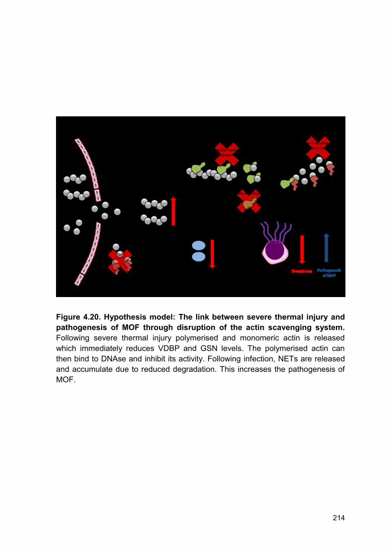
214
Figure 4.20. Hypothesis model: The link between severe thermal injury and
pathogenesis of MOF through disruption of the actin scavenging system.
Following severe thermal injury polymerised and monomeric actin is released
which immediately reduces VDBP and GSN levels. The polymerised actin can
then bind to DNAse and inhibit its activity. Following infection, NETs are released
and accumulate due to reduced degradation. This increases the pathogenesis of
MOF.
215
In 2011, Cohen and colleagues demonstrated the therapeutic potential of GSN in
a rat model of sepsis caused by double puncture of the cecum. Administration of 1
mg/ml recombinant human GSN, only once, resulted in significantly improved
survival and reduced tissue damage compared to sham animals (369). The group
concluded that administration of GSN is a potential therapy to reduce the severity
of illness caused by infection and sepsis. They propose that GSN may have an
effect on expression and regulation of pro-inflammatory cytokines. However, we
would suggest that GSN is having a multifaceted role by regulating cytokine
release (369), inflammatory cell function (260) and readdressing the overwhelmed
actin scavenging system. We show that administration of GSN in vitro can restore
DNAse activity in actin spiked serum, providing further evidence that GSN is a
potential therapy. However, this experiment is performed in vitro with no
contribution from VDBP or any other cellular interactions. Therefore, further
experiments are required to investigate the potential of GSN and VDBP combined
in vivo.
To investigate this possibility we performed a preliminary retrospective analysis in
samples from military patients following severe injury caused by explosions
receiving FFP prior to first blood sampling and hospital admission. Severe trauma
caused by explosions is becoming an increasingly common form of injury due to
conflicts and terrorism. Injuries can be classified into primary to quaternary injuries
depending upon severity and mechanism of blast injury (370). Burn injuries are
also common among patients with injuries caused by explosions and therefore
clinical treatment is comparable.
Patients with severe injury caused by explosions who are hemodynamically
unstable receive packed red blood cells and FFP in a 1:1 ratio immediately
216
following their injuries. By definition, FFP contains high levels of GSN and VDBP
which may explain some of the known therapeutic potential in the context of
traumatic injury by boosting the depleted levels of the actin scavenging proteins
(371). In our preliminary analysis, we have split our cohort into two groups who did
or did not receive blood products before admission to hospital. The decision to
give blood products was based upon resources available during transportation
from the battlefield and not injury severity or mechanism of injury. Importantly,
subgroups within this analysis are matched for ISS, NISS and time to admission
following injury. Theoretically, the only difference between the two groups is the
infusion of blood products.
Here we provide preliminary data showing that early administration of blood
products significantly increases GSN levels immediately post severe trauma
caused by explosion. This increase in GSN was also accompanied by a significant
increase in DNAse activity. However, VDBP levels were not affected. This data
would suggest that an early increase in circulating GSN is able to improve DNAse
activity rapidly following injury independently of VDBP. In 2005, Chhabra et al
showed that the N-terminal fragment of GSN could bind to and disrupt actin-
DNAse complexes, in turn, restoring enzymatic activity (372). Therefore, this may
be a potential mechanism by which GSN can rapidly restore or enhance DNAse
activity independently of VDBP. However, it is important to note that blood
products will contain many other soluble factors which may enhance DNAse
activity, including DNAse 1 itself. Therefore, the restoration of DNAse activity may
not be solely attributed to the increases in GSN. Thus, further studies are required
to investigate all components within blood products to fully understand if they are
contributing any therapeutic benefit.
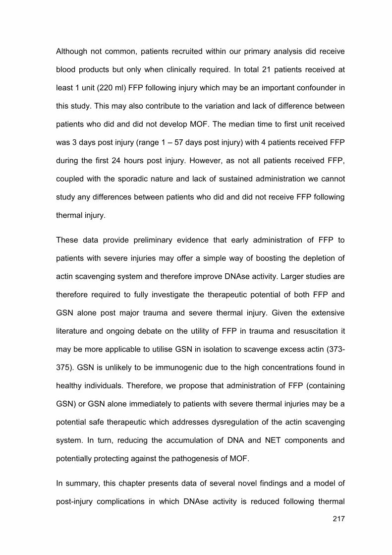
217
Although not common, patients recruited within our primary analysis did receive
blood products but only when clinically required. In total 21 patients received at
least 1 unit (220 ml) FFP following injury which may be an important confounder in
this study. This may also contribute to the variation and lack of difference between
patients who did and did not develop MOF. The median time to first unit received
was 3 days post injury (range 1 – 57 days post injury) with 4 patients received FFP
during the first 24 hours post injury. However, as not all patients received FFP,
coupled with the sporadic nature and lack of sustained administration we cannot
study any differences between patients who did and did not receive FFP following
thermal injury.
These data provide preliminary evidence that early administration of FFP to
patients with severe injuries may offer a simple way of boosting the depletion of
actin scavenging system and therefore improve DNAse activity. Larger studies are
therefore required to fully investigate the therapeutic potential of both FFP and
GSN alone post major trauma and severe thermal injury. Given the extensive
literature and ongoing debate on the utility of FFP in trauma and resuscitation it
may be more applicable to utilise GSN in isolation to scavenge excess actin (373-
375). GSN is unlikely to be immunogenic due to the high concentrations found in
healthy individuals. Therefore, we propose that administration of FFP (containing
GSN) or GSN alone immediately to patients with severe thermal injuries may be a
potential safe therapeutic which addresses dysregulation of the actin scavenging
system. In turn, reducing the accumulation of DNA and NET components and
potentially protecting against the pathogenesis of MOF.
In summary, this chapter presents data of several novel findings and a model of
post-injury complications in which DNAse activity is reduced following thermal
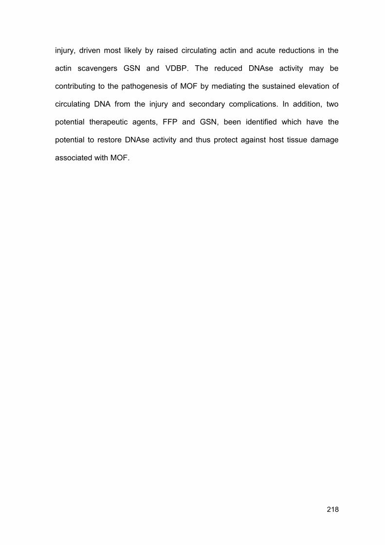
218
injury, driven most likely by raised circulating actin and acute reductions in the
actin scavengers GSN and VDBP. The reduced DNAse activity may be
contributing to the pathogenesis of MOF by mediating the sustained elevation of
circulating DNA from the injury and secondary complications. In addition, two
potential therapeutic agents, FFP and GSN, been identified which have the
potential to restore DNAse activity and thus protect against host tissue damage
associated with MOF.

219
Chapter 5
General Discussion
220
General Discussion
5.1 Limitations
Despite showing for the first time the association between and potential
mechanisms responsible for dysregulated NET release following severe thermal
injury, there are a number of limitations which must be considered.
This study was designed to be exploratory and hypothesis generating in nature
and not confirmatory. Hence we cannot estimate a specific outcome or obtain a
pre-determined level of precision from our results. Data generated should be
regarded as a pilot to obtain data to inform a potential larger confirmatory study.
Furthermore, in this thesis, clinical samples were obtained and analysed at fixed
time points. Whilst this remains a strength of the current study it limits the ability to
study the full kinetics of many in vivo markers and their relation to secondary
complications or clinical treatments. A second study has been established in which
blood samples are taken daily over the first 14 days which will allow us to
investigate and characterise the daily kinetics of neutrophil function, NET release
and diagnostic utility of described biomarkers.
Whilst we have shown the protective role of immediate administration of blood
products in patients with severe injuries caused by explosions it remains extremely
preliminary data in a small population of patients. Due to the nature of injuries and
sensitivity of data it has not been possible to correlate increases in DNAse activity
and GSN with improved outcome in this patient cohort. In addition, in this analysis
only GSN, VDBP and DNAse activity have been quantified and investigated.
Blood products contain many soluble mediators, including DNAse, and therefore
221
the protection against reduced DNAse activity cannot be solely attributed to
increases in GSN levels. Therefore, further characterisation of blood products or
the use of GSN and/or VDBP in isolation is required.
5.2 Future Work
Severe thermal injuries constitute a major form of traumatic injury (1).
Advancements in the initial care of patients with burns have dramatically improved
immediate survival (18, 19, 31). However, delayed mortality associated with sepsis
and MOF remain a significant health care problem (38, 47). Although sepsis and
MOF are well characterised the mechanisms mediating the increased
susceptibility to infection and the pathogenesis of MOF are poorly understood.
This thesis has investigated and identified a number of potential mechanisms,
therapeutic targets and biomarkers which have the potential to improve patient
outcome following severe thermal injury.
Infection and sepsis remain a major clinical burden in patients with severe thermal
injuries. Whilst this has been reported, the mechanisms responsible for the
increased susceptibility and incidence are poorly understood. Data from this thesis
reports a reduction in ex vivo NET production and intracellular ROS formation
which may underlie the inability to clear pathogens and be in part responsible for
the increased incidence of infections (Figure 5.1 A). The exact mechanism by
which this occurs is yet to be described but possible factors include; abnormal
neutrophil maturity phenotype or release of soluble mediators (e.g. mtDAMPs)
(Figure 5.1 B). Thus, understanding the mechanisms responsible for reduced
neutrophil function may aid in both the early identification of at risk patients and
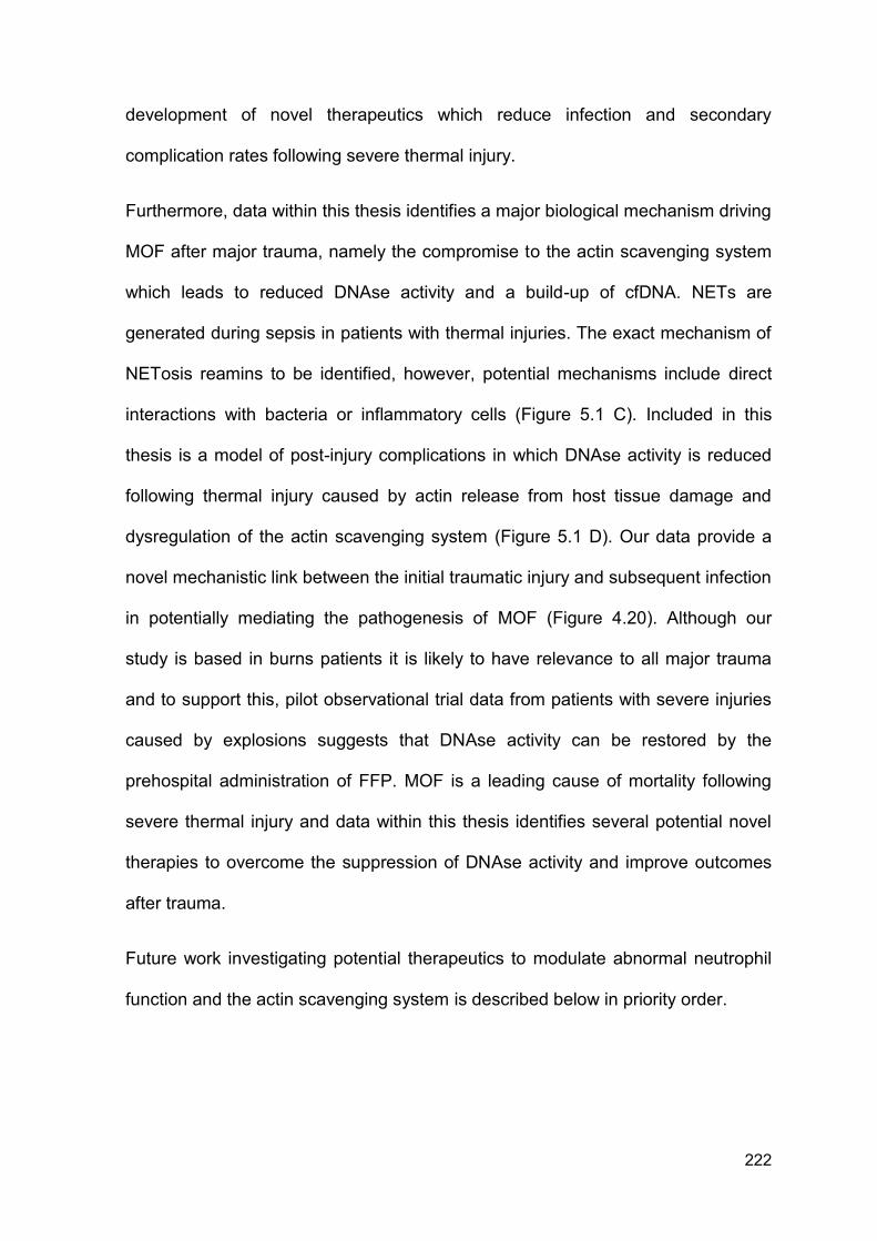
222
development of novel therapeutics which reduce infection and secondary
complication rates following severe thermal injury.
Furthermore, data within this thesis identifies a major biological mechanism driving
MOF after major trauma, namely the compromise to the actin scavenging system
which leads to reduced DNAse activity and a build-up of cfDNA. NETs are
generated during sepsis in patients with thermal injuries. The exact mechanism of
NETosis reamins to be identified, however, potential mechanisms include direct
interactions with bacteria or inflammatory cells (Figure 5.1 C). Included in this
thesis is a model of post-injury complications in which DNAse activity is reduced
following thermal injury caused by actin release from host tissue damage and
dysregulation of the actin scavenging system (Figure 5.1 D). Our data provide a
novel mechanistic link between the initial traumatic injury and subsequent infection
in potentially mediating the pathogenesis of MOF (Figure 4.20). Although our
study is based in burns patients it is likely to have relevance to all major trauma
and to support this, pilot observational trial data from patients with severe injuries
caused by explosions suggests that DNAse activity can be restored by the
prehospital administration of FFP. MOF is a leading cause of mortality following
severe thermal injury and data within this thesis identifies several potential novel
therapies to overcome the suppression of DNAse activity and improve outcomes
after trauma.
Future work investigating potential therapeutics to modulate abnormal neutrophil
function and the actin scavenging system is described below in priority order.
223
Figure 5.1. Hypothesis model: Linking initial injury to secondary
complications following severe thermal injury. (A) Severe thermal injury
significantly reduces, both, ex vivo NET formation and ROS generation. (B) This
reduction is potentially mediated by a phenotypical change in the circulating
neutrophil pool, soluble mediators e.g. mtDAMPs or functional neutrophils have
potentially migrated to the tissues leaving a non-functioning pool of circulating
neutrophils. (C) During infection, NETs are generated following thermal injury.
Potential mechanisms include direct induction through bacteria or through
neutrophil interactions with inflammatory cells. (D) Thermal injury results in
dysregulation of the actin scavenging system which facilitates the build-up of
circulating DNA, from initial injury and NETs, which are potentially responsible for
the pathogenesis of MOF. Panel D has been presented earlier in this thesis
(Figure 4.20).
224
5.2.1 In vivo characterisation of NETosis
Levels of cfDNA are associated with cellular damage and secondary complications
in a number of disease pathologies (108, 376). However, it is important to consider
the nature and structure of circulating DNA as it is released, complexed with
histones, in nucleosomes following NETosis. Whilst this is known, the broad
effects of free histones, free DNA and nucleosomes are described interchangeably
whereas there is a clear difference in their biological activity (377).
Free histones are cationic, highly conserved between species and orchestrate
gene transcription. Intravenous administration of free histones results in rapid
mortality of animals which can be prevented using an anti-histone antibody (198).
Histones can also mediate cytotoxic damage to endothelial and epithelial cells via
activation of TLRs (197). Whilst the mechanism by which histones are cytotoxic
remains unclear, studies have proposed that their cationic nature facilitates the
direct binding of histones to cells resulting in perforation, cell death and damage
caused by calcium influx (195, 196).
One potential therapeutic to reduce histone cytotoxicity is the administration of
negatively charged molecules such as heparins (378, 379). The non-anticoagulant
form of unfractionated heparin, for example, has been trialled as a novel
therapeutic in sepsis (378). Whilst this study did not report an improvement in 28
day mortality, Wildhagen and colleagues report a beneficial effect of this novel
therapy in mediating the inhibition of histone cytotoxicity (378). During sepsis there
is an abnormal consumption coagulopathy and thus an increased risk of bleeding.
Hence, administration of a non-anticoagulated form of heparin may have
promising effects on reducing tissue damage mediated by histones during sepsis
without potentiating bleeding.
225
DNA is regarded as a DAMP due to its rapid release and stimulation of immune
processes (199). The origin of DNA, nuclear, mitochondrial or bacterial, affects its
biological activity. In 2015, Bhagirath and colleagues isolated DNA from septic
patients and determined the different function of nuclear, mitochondrial and
bacterial DNA on coagulation and inflammation. All three sources of DNA were
capable of inducing thrombin formation, by an intrinsic dependent pathway
mechanism, and activation of platelets by platelet integrin αIIbβ3. Although, all
three have similar procoagulant properties, their functions in initiating inflammation
differ. Whilst, ncDNA and mtDNA prolonged in vitro neutrophil life, bacterial DNA
did not. However, only bacterial DNA promoted in vitro secretion of IL-6 (206).
Due to their opposing charges, DNA and histones will bind together and form a
nucleosome complex with an overall neutral charge. Like free histones and DNA,
nucleosomes can activate neutrophils (380). However, nucleosomes have
different cytotoxic properties compared to free histones and DNA. Unlike free
histones, administration of nucleosomes does not result in a significant increase in
mortality (204, 205). This may be explained by the inability of nucleosomes, unlike
histones, to cause direct damage or cell death to cultured endothelial cells.
However, subsequent physical or enzymatic degradation of nucleosomes then
resulted in damage to endothelial cells (381). Thus components of nucleosomes,
histones and DNA, are then responsible for the cellular damage and this effect is
nullified when components are complexed. In this thesis, we have investigated
three potential sources of DNA; nuclear, mitochondrial, and NET derived.
However, the status by which they exist, free or complexed, has not been fully
investigated. Thus, further study is required to investigate the relationship between
226
free histones, DNA and nucleosome complexes with secondary complications and
host tissue damage.
In 2013, Abrams et al reported the potential therapeutic benefit of CRP in
neutralising in vivo and in vitro histone induced endothelial damage, enhanced
coagulation and increased vascular permeability (381). Supported by earlier work
(382, 383), CRP is able to compete with phospholipid-containing liposomes and
form CRP-histone complexes, confirmed using immunofluorescence staining and
a gel overlay assay. The formation of CRP-histone complexes reduced integration
of histones into the cellular wall of endothelial cells and in turn cellular damage.
The exact mechanism of binding between CRP and histones remains to be
identified, however, the authors propose that the positively charged histones are
likely binding the negatively charged central pore of the CRP pentamer (381).
Interestingly, it has yet to be established if CRP-histone complexes have
increased clearance. Thus, Abrams and colleagues demonstrated, for the first
time, that CRP is a conserved mechanism which protects the body against histone
toxicity in the acute phase following trauma and illness (381). Hence, it may be
possible to use CRP, or a therapeutic mimicking CRPs mechanism of action, as
an intervention to neutralise the cytotoxic effect of circulating histones following
severe thermal injury.
As described above cfDNA can originate from a number of sources (114). In 2016,
Lehmann-Werman and colleagues described a novel technique by which the
tissue specific origin of cfDNA could be elucidated (384). It is likely that following
thermal injury circulating DNA is heterogeneous in origin given the severe nature
of injury, surgical procedures, infection, host tissue damage and NET release.
Despite the shared homology in nucleotide sequences, DNA also has unique
227
methylation patterns depending upon the cell or tissue of origin (385). Thus, it is
possible to infer cell death within specific organs and its contribution to the
circulating cfDNA. As proof of principle, Lehmann-Werman and colleagues
detected pancreatic β-cell DNA in patients with diagnosed type-1 diabetes and
islet-graft recipients, neuronal or glial DNA in patients with traumatic brain injury or
cardiac arrest and exocrine pancreas DNA in pancreatic cancer or pancreatitis
patients (384). Given the exploratory nature of this study, however, they were
unable to conclusively evaluate the diagnostic or prognostic utility of methylation
patterns. However, investigation of methylation patterns in patients following
thermal injury may have the potential to aid in the accurate diagnosis of specific
organ dysfunction and responses to clinical treatment.
5.2.2 DNAse isoforms and their functions
In 1997, Rodriguez and colleagues described three members of a DNAse 1 family.
All three displayed similarity in their nucleotide, amino acid sequences (386) and
biochemical properties, dependency on Ca2+ and Mg2+ (387). DNAse 1 is the
major endonuclease responsible for the breakdown of circulating nucleic acids.
However, in recent years groups have begun investigating the physiological roles
of DNAse-γ (386-388). Like our analysis, many assays established to measure
DNAse activity, therefore, quantify total DNAse. Furthermore, most groups focus
upon the role of DNAse 1 alone and thus do not investigate the role of DNAse-γ
(237, 353, 389).
As it functions internucleosomally and degrades nuclear chromatin, DNAse-γ was
originally reported to be involved in apoptotic DNA fragmentation (390-392).
However, cells transfected with DNAse-γ can secrete this enzyme thus suggesting
228
a mechanism by which exocytosis can occur (393). Consequently, DNAse-γ,
potentially from monocytes (394), may contribute to total circulating DNAse
activity.
DNAse 1 and –γ function synergistically to clear total circulating DNA through
distinct substrate consumption and degradation. DNAse 1 has a higher affinity and
accelerated clearance of free DNA compared to DNAse-γ which is more efficient
at degrading chromatin (387, 395).
Recent studies report a clear link between genetic mutations of DNAse-γ and
development multiple autoimmune diseases (396-398). Highlighted in DNAse-γ
mice which develop an SLE phenotype associated with the presence of
autoantigens against chromatin (399). Although DNAse-γ has been related to
autoimmunity, there have there have been no investigations into its function and
potential role in secondary complications following trauma or severe burn. Thus,
further work could investigate the combined and individual functions of DNAse-1
and –γ activity in the degradation of chromatin and DNA release following severe
thermal injury. This may reveal additional mechanistic information and/or novel
therapeutics to reduce secondary complications driven by elevated circulating
DNA or chromatin.
5.2.3 Targeting the build-up of toxic and pro-thrombotic DNA following
thermal injury
Following thermal injury, there is the disruption of the actin scavenging system
which predisposes patients to the accumulation of tissue derived and NET derived
DNA (Figure 4.20). This can then facilitate further host tissue damage, obstruct
blood flow and induce thrombosis (233, 236, 360). Modulation of this system by
229
targeting DNAse, GSN or VDBP has the potential to protect against this pro-
thrombotic and host cytotoxic phenotype.
5.2.4 DNAse as a therapy
DNAse is the major extracellular endonuclease found in a number of bodily fluids
and is responsible for the breakdown and clearance of circulating chromatin and
DNA. Severe thermal injury results in the immediate release of DNA which is
followed by a secondary release during infection. Both populations of DNA are
implicated in the generation of thrombosis, host tissue damage, endothelial
damage and secondary complications in trauma patients and animal models of
thrombosis (232, 233, 356, 400). Thus, targeting total DNA accumulation by
administration of DNAse may provide therapeutic benefit.
Using a murine model of DVT, extracellular chromatin formed a scaffold upon
which venous thrombosis could occur and was involved in the pathogenesis of the
DVT. Administration of DNAse 1 was capable of protecting animals from DVT
(233). Furthermore, PAD4 deficient mice form fewer thrombi after inferior vena
cava stenosis compared to wild-type animals (401). Thus confirming the role of
NET derived DNA in the propagation of venous thrombus formation. In addition to
their procoagulant activity, DNA, derived from NETs, can also cause direct
damage to endothelial cells (360). Interestingly, coculture of neutrophils with
activated endothelial cells results in NETosis which caused direct cellular damage
thus it appears to be self-propagating. This could also be abrogated by the
degradation of NETs using DNAse (360). These data provide evidence that
administration of DNAse to target host derived and NET derived DNA may have
potential to alleviate host tissue damage and reduce the pro-thrombotic
230
phenotype. Importantly, administration of DNAse 1 is a licenced therapeutic in
patients with cystic fibrosis and used to reduce the viscosity of sputum through
direct degradation of NET derived DNA.
It is also very important to consider the potential complications associated with
DNAse. Administration of DNAse in a murine model of sepsis resulted in an
increased pro-inflammatory response along with increased colonisation of bacteria
and mortality (221). This was further confirmed by Mai et al who highlighted the
importance of timing when targeting NETs. Early administration of DNAse resulted
in increased mortality, whereas later administration reduced tissue damage and
decreased circulating DNA levels resulting in improved survival (402). These data
provide evidence that DNA should only be broken down when in excess and
contributing an enhanced pro-inflammatory and thrombotic phenotype. Given the
clinical complexity and heterogeneous nature of patients following severe burns
this may be challenging. Furthermore, as actin is released following severe
thermal injury this may inhibit any administered DNAse, rendering the therapy
potentially non-effective. Hence, a more targeted approach towards NETs or
modulation of the actin scavenging system may be more applicable.
5.2.5 Inhibition of PAD4
NETosis can also be prevented by inhibition of PAD4. Indeed, PAD4 knockout
mice are totally protected against venous thrombosis formation and tissue damage
following liver injury (225, 275). However, these studies do not take into account
the pre-existing immunosuppression which occurs following thermal injury.
Therefore, elimination of NETs completely could leave, an already
immunocompromised patient, susceptible to further infection. Moreover, PAD4
231
deficient mice are more susceptible to necrotizing fasciitis infection than wild-type
mice and this susceptibility is attributed to the inability to form NETs (276).
5.2.6 Modulation of actin scavenging system
The actin scavenging system is comprised of two key proteins; VDBP and GSN.
Following severe thermal injury, there are reductions in circulating levels of both
proteins which predispose patients to reduce DNAse activity and the build-up of
circulating DNA (Figure 4.13 - 4.14). As both VDBP and GSN are extremely
abundant within the blood they are unlikely to be toxic if used as direct
therapeutics.
One potential mechanism to replenish depleted VDBP and GSN is through
administration of blood products, namely FFP. Although not known, replenishment
of the actin scavenging system may play a role in a number of the reported
therapeutic benefits of using blood products following major trauma (371). In this
thesis, administration of FFP during transportation from site of injury to hospital in
major trauma victims improved DNAse activity in addition to increasing circulating
GSN levels but without increasing circulating VDBP levels (Figure 4.19). Whilst
this data suggests a role of GSN in recovery of DNAse activity any improvement
by FFP will be multifactorial and caused by a number of soluble factors which may
enhance DNAse activity, including DNAse 1 itself. Therefore, the restoration of
DNAse activity cannot be solely attributed to the increase in GSN. Thus, further
studies are required to investigate whether other components within blood
products are contributing.
Whilst VDBP remains a potential therapy, to date, no group has investigated the
effect of administration of VDBP on outcome following trauma or burns. Despite
232
this, large quantities of protein can be produced from plasma fractions (403).
Following thermal injury GSN and VDBP both become saturated, most likely
caused by the release of monomeric and polymeric actin. Administration of GSN
and VDBP as a combination therapy may therefore aid in the rapid recovery of the
actin scavenging system.
There is growing evidence for the use of GSN in the treatment of infection, trauma
and burns (261, 404-406). In 2007, Lee and colleagues reported a reduction in
mortality and a dynamic change in cytokine release in GSN depleted mice that
were given 20 mg/mL recombinant human plasma GSN after challenge with
endotoxin. Although the mechanism responsible remains unclear, improvements
in outcome were associated with depolymerisation of actin in these animals (404).
Interestingly, levels of total circulating actin did not decrease which suggests that
GSN can modulate the activity of actin independent of VDBP which is consistent
with previous literature (407, 408).
A clinical trial has been established to assess the pharmacokinetics and safety of
recombinant plasma GSN given to patients admitted to an ICU, including patients
with trauma, infection and burns. The hypothesis of the study states that
administration of GSN will increase circulating GSN levels and decrease the
incidence of secondary complications, e.g. MOF or death (Trial Number:
NCT00671307). Results for this study are not currently available, however, a
second study may be warranted to focus upon patients admitted to an ICU with a
dysregulated actin scavenging system.
Whilst the beneficial effects of GSN alone have been described (261, 404-406),
the cumulative benefit of GSN with VDBP or DNAse have not. Given the
233
saturation and dysregulation of the actin scavenging system, one might
hypothesise that a combination therapy may be more appropriate. Furthermore, it
may be possible to produce novel therapies which contain the active N-terminal
fragment of GSN which is responsible for its ability to restore DNAse activity
independently of VDBP (372). Therefore, one might propose the development of a
novel compound which mimics the N-terminal fragment of GSN for its use in a
clinical trial investigating early and sustained administration of GSN and its effects
on DNAse activity and outcome following severe thermal injury. Of note, GSN
function also extends beyond control of actin homeostasis and as a potential use
as a biomarker of poor outcome as it also exhibits various anti-inflammatory
activities (260, 408). Thus, understanding the mechanism(s) by which GSN
provides benefit is fundamental to its development as a therapy.
5.2.7 Targeting reduced neutrophil function following thermal injury
The association between severe thermal injury and abnormal neutrophil function
has been well reported (156, 159-161, 173, 174, 177). Furthermore, this reduction
or dysregulation in neutrophil function may underlie the increased incidence of
nosocomial infections reported within these patient cohorts. In this thesis, it has
been shown that measurements of abnormal neutrophil function are potential
biomarkers of sepsis, in addition to being novel therapeutic targets.
5.2.8 Granulocyte colony-stimulating factor and neutrophil maturity
Traumatic injury induces marked alterations in haematopoiesis which is
characterised by a repolarisation towards a myeloid lineage (409, 410). Production
and maturation of neutrophils is multifaceted with the principle regulator of
granulopoiesis being G-CSF (162, 163) which causes activation of signal
234
transducer and activator of transcription 3 (STAT3), via MEK-1/-2 and ERK-1/-2
(411, 412).
Severe thermal injury results in the release of IGs/neutrophils which are
associated with sepsis status and reduced neutrophil function (Chapter 3, Figure
3.14, 3.17 and 3.18). One limitation of the work presented in this thesis is the lack
of mechanistic insight responsible for the release of IGs following thermal injury.
This release may be induced by emergency granulopoiesis caused by excessive
stress from immediate injury and consequent infectious episodes. In a study which
included 83 severely injured patients, sustained elevation in G-CSF was
associated with the release of immature cells and increased risk of infection (157).
In addition, Ertel and colleagues report an inhibition of neutrophil apoptosis
caused by local G-CSF which propagates damage caused by neutrophils at sites
of injury (413). Therefore, one might propose that inhibition of G-CSF can
potentially reduce circulating IGs and protect against, neutrophil mediated,
immunosuppression.
Conversely, administration of G-CSF has positive effects in mice (168, 414).
Namely, an increase in neutrophil chemotaxis in animals who received
recombinant G-CSF following burn injury which was attributed to a combination of
myeloid expansion and direct enhancement of neutrophil function (414). Gardner
et al report a key role for G-CSF/STAT3 axis in providing protection against post-
traumatic infection which was consistent with human gene analysis. Paradoxically,
thermal injury resulted in protection from a lethal Klebsiella pneumoniae
pulmonary challenge. Inhibition of G-CSF reduced STAT3 activation and
diminished this protective response by preventing myeloid differentiation and
neutrophil release (168). Thus, caution must be advised when targeting G-CSF
235
following severe thermal injury as it potentially exhibits multiple roles in host
protection and innate immunity (157, 168, 413, 414). Therefore a more targeted
approach modulating neutrophil maturation may be more applicable.
5.2.9 Resolvins
Acute inflammation is necessary for the host to respond to initial tissue damage or
invading pathogens and is required to maintain host protection and homeostasis.
However, if inflammation is prolonged or uncontrolled it can lead to host tissue
damage and secondary complications (415). Resolution of inflammation was long
characterised as an eventual reduction of pro-inflammatory mediators by a
passive process. As reviewed by Serhan and colleagues, resolution of
inflammation is now considered an active and responsive process controlled by
the release of a number of negative regulators (416). Resolvins, protectins and
lipoxins are three examples of anti-inflammatory mediators which induce
resolution and containment of acute inflammation (417, 418).
Resolvins are named after their role in the resolution of inflammation and
produced from eicosapentaenoic or docosahexaenoic acid, the most abundant
omega-3 polyunsaturated fatty acids (417, 419). Administration of resolvin D2
(RvD2) can enhance both neutrophil phagocytosis and killing of engulfed E.Coli
(420). In a cecal ligation and puncture model of sepsis, treatment with RvD2
causes a reduction in leukocyte-endothelial interactions, cytokine production, and
a reduction in local and systemic bacterial burden (420). Furthermore, Kurihara et
al reported a restoration of neutrophil chemotaxis following burn injury when
animals were administered RvD2 which was attributed to the direct action of RvD2
on neutrophils (421). Whilst it is known that resolvin D1 can act through ALX/FPR2
236
and GPR32 G-protein coupled receptors (422), the receptors responsible for RvD2
actions are currently unknown. Hence, further characterisation of RvD2 and its
receptors is essential to understand the mechanisms by which immunoregulation
and enhancement of neutrophil function occur. Nevertheless, RvD2 may be a
potential therapeutic to reduce both the uncontrolled inflammation and reduced
neutrophil function which occurs following severe thermal injury.
5.2.10 Haemoperfusion therapy
Direct haemoperfusion therapy can filter cells or soluble mediators from the blood
of patients. This results in reduced uncontrolled activation of the immune system
and a reduction in host tissue damage (423, 424). Whilst direct haemoperfusion
therapy is a recognised treatment for septic shock in Japan, only parts of Western
Europe currently use this technology (425). Direct haemoperfusion therapy using a
polymixin-B immobilized fibre cartridge (PMX-DHP) primarily works by removal of
endotoxin from the blood. However, recent evidence has shown a broader
mechanistic role for PMX-DHP in the removal of activated neutrophils which can
cause endothelial damage (424). Whilst the mechanism responsible for selective
removal of activated cells remains unclear the authors propose that it is caused by
a direct interaction between activated neutrophils and polymixin-B located on the
fibres of the filter (424). Thus, depletion of activated neutrophils by PMX-DHP may
help to control inflammation in patients with septic shock.
More recently, Lee and colleagues developed a microfibre mesh decorated in
polythylenimine and polyamidoamine dendrimers which can capture DAMPs,
including extracellular DNA. In vitro immobilisation of DAMPs located within blood
from trauma patients results in reduced activation of toll-like receptor (TLR) -2, 3, 4
237
and 9 compared to unfiltered serum in a monocyte derived reporter cell line.
Furthermore, immobilisation of DAMPs in hearts transplanted in vivo abrogated
thrombus formation (423). In this thesis, DAMPs cause dysregulation of neutrophil
function in vitro which mimics our in vivo findings in patients with severe thermal
injuries (Figure 3.21 - 3.25). Hence, immobilisation of DAMPs using the microfibre
mesh decorated in polythylenimine and polyamidoamine dendrimers has the
potential to protect against dysregulation of neutrophil function following thermal
injury and reduce thrombotic complications associated with elevated DNA and
histones. However, direct haemoperfusion therapy using polythylenimine and
polyamidoamine dendrimers remains experimental with limited translation to
clinical practice due to difficulty in integrating these microfibre meshes into
available technology.
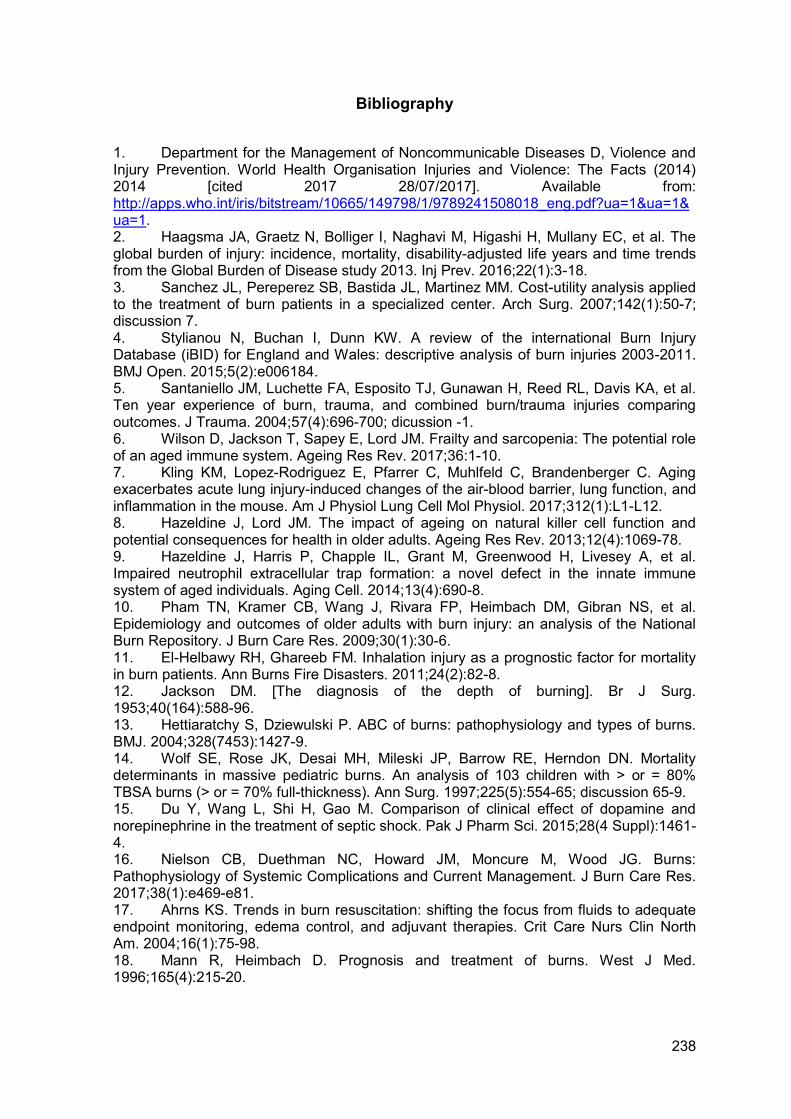
238
Bibliography
1. Department for the Management of Noncommunicable Diseases D, Violence and Injury Prevention. World Health Organisation Injuries and Violence: The Facts (2014) 2014 [cited 2017 28/07/2017]. Available from: http://apps.who.int/iris/bitstream/10665/149798/1/9789241508018_eng.pdf?ua=1&ua=1&ua=1. 2. Haagsma JA, Graetz N, Bolliger I, Naghavi M, Higashi H, Mullany EC, et al. The global burden of injury: incidence, mortality, disability-adjusted life years and time trends from the Global Burden of Disease study 2013. Inj Prev. 2016;22(1):3-18. 3. Sanchez JL, Pereperez SB, Bastida JL, Martinez MM. Cost-utility analysis applied to the treatment of burn patients in a specialized center. Arch Surg. 2007;142(1):50-7; discussion 7. 4. Stylianou N, Buchan I, Dunn KW. A review of the international Burn Injury Database (iBID) for England and Wales: descriptive analysis of burn injuries 2003-2011. BMJ Open. 2015;5(2):e006184. 5. Santaniello JM, Luchette FA, Esposito TJ, Gunawan H, Reed RL, Davis KA, et al. Ten year experience of burn, trauma, and combined burn/trauma injuries comparing outcomes. J Trauma. 2004;57(4):696-700; dicussion -1. 6. Wilson D, Jackson T, Sapey E, Lord JM. Frailty and sarcopenia: The potential role of an aged immune system. Ageing Res Rev. 2017;36:1-10. 7. Kling KM, Lopez-Rodriguez E, Pfarrer C, Muhlfeld C, Brandenberger C. Aging exacerbates acute lung injury-induced changes of the air-blood barrier, lung function, and inflammation in the mouse. Am J Physiol Lung Cell Mol Physiol. 2017;312(1):L1-L12. 8. Hazeldine J, Lord JM. The impact of ageing on natural killer cell function and potential consequences for health in older adults. Ageing Res Rev. 2013;12(4):1069-78. 9. Hazeldine J, Harris P, Chapple IL, Grant M, Greenwood H, Livesey A, et al. Impaired neutrophil extracellular trap formation: a novel defect in the innate immune system of aged individuals. Aging Cell. 2014;13(4):690-8. 10. Pham TN, Kramer CB, Wang J, Rivara FP, Heimbach DM, Gibran NS, et al. Epidemiology and outcomes of older adults with burn injury: an analysis of the National Burn Repository. J Burn Care Res. 2009;30(1):30-6. 11. El-Helbawy RH, Ghareeb FM. Inhalation injury as a prognostic factor for mortality in burn patients. Ann Burns Fire Disasters. 2011;24(2):82-8. 12. Jackson DM. [The diagnosis of the depth of burning]. Br J Surg. 1953;40(164):588-96. 13. Hettiaratchy S, Dziewulski P. ABC of burns: pathophysiology and types of burns. BMJ. 2004;328(7453):1427-9. 14. Wolf SE, Rose JK, Desai MH, Mileski JP, Barrow RE, Herndon DN. Mortality determinants in massive pediatric burns. An analysis of 103 children with > or = 80% TBSA burns (> or = 70% full-thickness). Ann Surg. 1997;225(5):554-65; discussion 65-9. 15. Du Y, Wang L, Shi H, Gao M. Comparison of clinical effect of dopamine and norepinephrine in the treatment of septic shock. Pak J Pharm Sci. 2015;28(4 Suppl):1461-4. 16. Nielson CB, Duethman NC, Howard JM, Moncure M, Wood JG. Burns: Pathophysiology of Systemic Complications and Current Management. J Burn Care Res. 2017;38(1):e469-e81. 17. Ahrns KS. Trends in burn resuscitation: shifting the focus from fluids to adequate endpoint monitoring, edema control, and adjuvant therapies. Crit Care Nurs Clin North Am. 2004;16(1):75-98. 18. Mann R, Heimbach D. Prognosis and treatment of burns. West J Med. 1996;165(4):215-20.
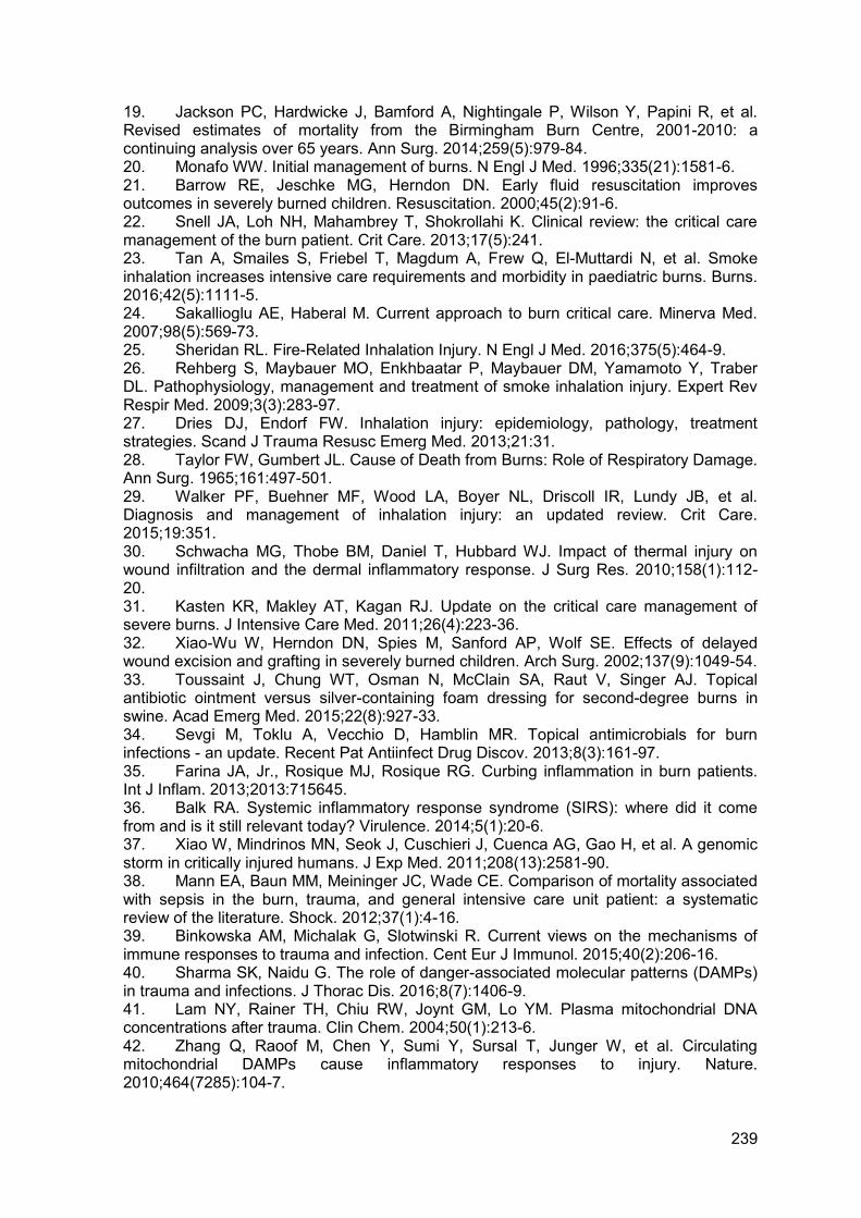
239
19. Jackson PC, Hardwicke J, Bamford A, Nightingale P, Wilson Y, Papini R, et al. Revised estimates of mortality from the Birmingham Burn Centre, 2001-2010: a continuing analysis over 65 years. Ann Surg. 2014;259(5):979-84. 20. Monafo WW. Initial management of burns. N Engl J Med. 1996;335(21):1581-6. 21. Barrow RE, Jeschke MG, Herndon DN. Early fluid resuscitation improves outcomes in severely burned children. Resuscitation. 2000;45(2):91-6. 22. Snell JA, Loh NH, Mahambrey T, Shokrollahi K. Clinical review: the critical care management of the burn patient. Crit Care. 2013;17(5):241. 23. Tan A, Smailes S, Friebel T, Magdum A, Frew Q, El-Muttardi N, et al. Smoke inhalation increases intensive care requirements and morbidity in paediatric burns. Burns. 2016;42(5):1111-5. 24. Sakallioglu AE, Haberal M. Current approach to burn critical care. Minerva Med. 2007;98(5):569-73. 25. Sheridan RL. Fire-Related Inhalation Injury. N Engl J Med. 2016;375(5):464-9. 26. Rehberg S, Maybauer MO, Enkhbaatar P, Maybauer DM, Yamamoto Y, Traber DL. Pathophysiology, management and treatment of smoke inhalation injury. Expert Rev Respir Med. 2009;3(3):283-97. 27. Dries DJ, Endorf FW. Inhalation injury: epidemiology, pathology, treatment strategies. Scand J Trauma Resusc Emerg Med. 2013;21:31. 28. Taylor FW, Gumbert JL. Cause of Death from Burns: Role of Respiratory Damage. Ann Surg. 1965;161:497-501. 29. Walker PF, Buehner MF, Wood LA, Boyer NL, Driscoll IR, Lundy JB, et al. Diagnosis and management of inhalation injury: an updated review. Crit Care. 2015;19:351. 30. Schwacha MG, Thobe BM, Daniel T, Hubbard WJ. Impact of thermal injury on wound infiltration and the dermal inflammatory response. J Surg Res. 2010;158(1):112-20. 31. Kasten KR, Makley AT, Kagan RJ. Update on the critical care management of severe burns. J Intensive Care Med. 2011;26(4):223-36. 32. Xiao-Wu W, Herndon DN, Spies M, Sanford AP, Wolf SE. Effects of delayed wound excision and grafting in severely burned children. Arch Surg. 2002;137(9):1049-54. 33. Toussaint J, Chung WT, Osman N, McClain SA, Raut V, Singer AJ. Topical antibiotic ointment versus silver-containing foam dressing for second-degree burns in swine. Acad Emerg Med. 2015;22(8):927-33. 34. Sevgi M, Toklu A, Vecchio D, Hamblin MR. Topical antimicrobials for burn infections - an update. Recent Pat Antiinfect Drug Discov. 2013;8(3):161-97. 35. Farina JA, Jr., Rosique MJ, Rosique RG. Curbing inflammation in burn patients. Int J Inflam. 2013;2013:715645. 36. Balk RA. Systemic inflammatory response syndrome (SIRS): where did it come from and is it still relevant today? Virulence. 2014;5(1):20-6. 37. Xiao W, Mindrinos MN, Seok J, Cuschieri J, Cuenca AG, Gao H, et al. A genomic storm in critically injured humans. J Exp Med. 2011;208(13):2581-90. 38. Mann EA, Baun MM, Meininger JC, Wade CE. Comparison of mortality associated with sepsis in the burn, trauma, and general intensive care unit patient: a systematic review of the literature. Shock. 2012;37(1):4-16. 39. Binkowska AM, Michalak G, Slotwinski R. Current views on the mechanisms of immune responses to trauma and infection. Cent Eur J Immunol. 2015;40(2):206-16. 40. Sharma SK, Naidu G. The role of danger-associated molecular patterns (DAMPs) in trauma and infections. J Thorac Dis. 2016;8(7):1406-9. 41. Lam NY, Rainer TH, Chiu RW, Joynt GM, Lo YM. Plasma mitochondrial DNA concentrations after trauma. Clin Chem. 2004;50(1):213-6. 42. Zhang Q, Raoof M, Chen Y, Sumi Y, Sursal T, Junger W, et al. Circulating mitochondrial DAMPs cause inflammatory responses to injury. Nature. 2010;464(7285):104-7.
240
43. Hazeldine J, Hampson P, Opoku FA, Foster M, Lord JM. N-Formyl peptides drive mitochondrial damage associated molecular pattern induced neutrophil activation through ERK1/2 and P38 MAP kinase signalling pathways. Injury. 2015;46(6):975-84. 44. Simmons JD, Lee YL, Mulekar S, Kuck JL, Brevard SB, Gonzalez RP, et al. Elevated levels of plasma mitochondrial DNA DAMPs are linked to clinical outcome in severely injured human subjects. Ann Surg. 2013;258(4):591-6; discussion 6-8. 45. Fernandez-Ruiz I, Arnalich F, Cubillos-Zapata C, Hernandez-Jimenez E, Moreno-Gonzalez R, Toledano V, et al. Mitochondrial DAMPs induce endotoxin tolerance in human monocytes: an observation in patients with myocardial infarction. PLoS One. 2014;9(5):e95073. 46. Angus DC, van der Poll T. Severe sepsis and septic shock. N Engl J Med. 2013;369(21):2063. 47. Paratz JD, Lipman J, Boots RJ, Muller MJ, Paterson DL. A new marker of sepsis post burn injury?*. Crit Care Med. 2014;42(9):2029-36. 48. Baue AE. Multiple, progressive, or sequential systems failure. A syndrome of the 1970s. Arch Surg. 1975;110(7):779-81. 49. Penner JA. Disseminated intravascular coagulation in patients with multiple organ failure of non-septic origin. Semin Thromb Hemost. 1998;24(1):45-52. 50. Kidokoro A, Iba T, Hong J. Role of DIC in multiple organ failure. Int J Surg Investig. 2000;2(1):73-80. 51. Vincent JL, De Backer D. Does disseminated intravascular coagulation lead to multiple organ failure? Crit Care Clin. 2005;21(3):469-77. 52. Toh CH, Alhamdi Y, Abrams ST. Current Pathological and Laboratory Considerations in the Diagnosis of Disseminated Intravascular Coagulation. Ann Lab Med. 2016;36(6):505-12. 53. Levi M, Toh CH, Thachil J, Watson HG. Guidelines for the diagnosis and management of disseminated intravascular coagulation. British Committee for Standards in Haematology. Br J Haematol. 2009;145(1):24-33. 54. Bick RL. Disseminated intravascular coagulation: a review of etiology, pathophysiology, diagnosis, and management: guidelines for care. Clin Appl Thromb Hemost. 2002;8(1):1-31. 55. Wada H, Hasegawa K, Watanabe M. DIC: an update on diagnosis and treatment. Rinsho Ketsueki. 2017;58(5):523-9. 56. Yanagida Y, Gando S, Sawamura A, Hayakawa M, Uegaki S, Kubota N, et al. Normal prothrombinase activity, increased systemic thrombin activity, and lower antithrombin levels in patients with disseminated intravascular coagulation at an early phase of trauma: comparison with acute coagulopathy of trauma-shock. Surgery. 2013;154(1):48-57. 57. Dobson GP, Letson HL, Sharma R, Sheppard FR, Cap AP. Mechanisms of early trauma-induced coagulopathy: The clot thickens or not? J Trauma Acute Care Surg. 2015;79(2):301-9. 58. Kono M, Saigo K, Takagi Y, Takahashi T, Kawauchi S, Wada A, et al. Heme-related molecules induce rapid production of neutrophil extracellular traps. Transfusion. 2014;54(11):2811-9. 59. Nguyen LN, Nguyen TG. Characteristics and outcomes of multiple organ dysfunction syndrome among severe-burn patients. Burns. 2009;35(7):937-41. 60. Dellinger RP, Levy MM, Rhodes A, Annane D, Gerlach H, Opal SM, et al. Surviving sepsis campaign: international guidelines for management of severe sepsis and septic shock: 2012. Crit Care Med. 2013;41(2):580-637. 61. Castellanos-Ortega A, Suberviola B, Garcia-Astudillo LA, Holanda MS, Ortiz F, Llorca J, et al. Impact of the Surviving Sepsis Campaign protocols on hospital length of stay and mortality in septic shock patients: results of a three-year follow-up quasi-experimental study. Crit Care Med. 2010;38(4):1036-43. 62. Levy MM, Dellinger RP, Townsend SR, Linde-Zwirble WT, Marshall JC, Bion J, et al. The Surviving Sepsis Campaign: results of an international guideline-based
241
performance improvement program targeting severe sepsis. Crit Care Med. 2010;38(2):367-74. 63. Ferrer R, Artigas A, Suarez D, Palencia E, Levy MM, Arenzana A, et al. Effectiveness of treatments for severe sepsis: a prospective, multicenter, observational study. Am J Respir Crit Care Med. 2009;180(9):861-6. 64. Orban C. Diagnostic criteria for sepsis in burn patients. Chirurgia (Bucur). 2012;107(6):697-700. 65. Greenhalgh DG, Saffle JR, Holmes JHt, Gamelli RL, Palmieri TL, Horton JW, et al. American Burn Association consensus conference to define sepsis and infection in burns. J Burn Care Res. 2007;28(6):776-90. 66. Singer M, Deutschman CS, Seymour CW, Shankar-Hari M, Annane D, Bauer M, et al. The Third International Consensus Definitions for Sepsis and Septic Shock (Sepsis-3). JAMA. 2016;315(8):801-10. 67. Blair JM, Webber MA, Baylay AJ, Ogbolu DO, Piddock LJ. Molecular mechanisms of antibiotic resistance. Nat Rev Microbiol. 2015;13(1):42-51. 68. van Langeveld I, Gagnon RC, Conrad PF, Gamelli RL, Martin B, Choudhry MA, et al. Multiple-Drug Resistance in Burn Patients: A Retrospective Study on the Impact of Antibiotic Resistance on Survival and Length of Stay. J Burn Care Res. 2017;38(2):99-105. 69. Assicot M, Gendrel D, Carsin H, Raymond J, Guilbaud J, Bohuon C. High serum procalcitonin concentrations in patients with sepsis and infection. Lancet. 1993;341(8844):515-8. 70. Mehanic S, Baljic R. The importance of serum procalcitonin in diagnosis and treatment of serious bacterial infections and sepsis. Mater Sociomed. 2013;25(4):277-81. 71. von Heimburg D, Stieghorst W, Khorram-Sefat R, Pallua N. Procalcitonin--a sepsis parameter in severe burn injuries. Burns. 1998;24(8):745-50. 72. Sachse C, Machens HG, Felmerer G, Berger A, Henkel E. Procalcitonin as a marker for the early diagnosis of severe infection after thermal injury. J Burn Care Rehabil. 1999;20(5):354-60. 73. Uzzan B, Cohen R, Nicolas P, Cucherat M, Perret GY. Procalcitonin as a diagnostic test for sepsis in critically ill adults and after surgery or trauma: a systematic review and meta-analysis. Crit Care Med. 2006;34(7):1996-2003. 74. Schuetz P, Raad I, Amin DN. Using procalcitonin-guided algorithms to improve antimicrobial therapy in ICU patients with respiratory infections and sepsis. Curr Opin Crit Care. 2013;19(5):453-60. 75. Cabral L, Afreixo V, Almeida L, Paiva JA. The Use of Procalcitonin (PCT) for Diagnosis of Sepsis in Burn Patients: A Meta-Analysis. PLoS One. 2016;11(12):e0168475. 76. Seoane L, Pertega S, Galeiras R, Astola I, Bouza T. Procalcitonin in the burn unit and the diagnosis of infection. Burns. 2014;40(2):223-9. 77. Lavrentieva A, Kontakiotis T, Lazaridis L, Tsotsolis N, Koumis J, Kyriazis G, et al. Inflammatory markers in patients with severe burn injury. What is the best indicator of sepsis? Burns. 2007;33(2):189-94. 78. Minami E, Ito S, Sugiura T, Fujita Y, Sasano H, Sobue K. Markedly elevated procalcitonin in early postoperative period in pediatric open heart surgery: a prospective cohort study. J Intensive Care. 2014;2(1):38. 79. Pepys MB, Hirschfield GM. C-reactive protein: a critical update. J Clin Invest. 2003;111(12):1805-12. 80. Kim H, Hur M, Moon HW, Yun YM, Di Somma S, Network G. Multi-marker approach using procalcitonin, presepsin, galectin-3, and soluble suppression of tumorigenicity 2 for the prediction of mortality in sepsis. Ann Intensive Care. 2017;7(1):27. 81. Sapin F, Biston P, Piagnerelli M. Predictive value of C-reactive protein in critically ill patients after abdominal surgery. Clinics (Sao Paulo). 2017;72(1):23-9.
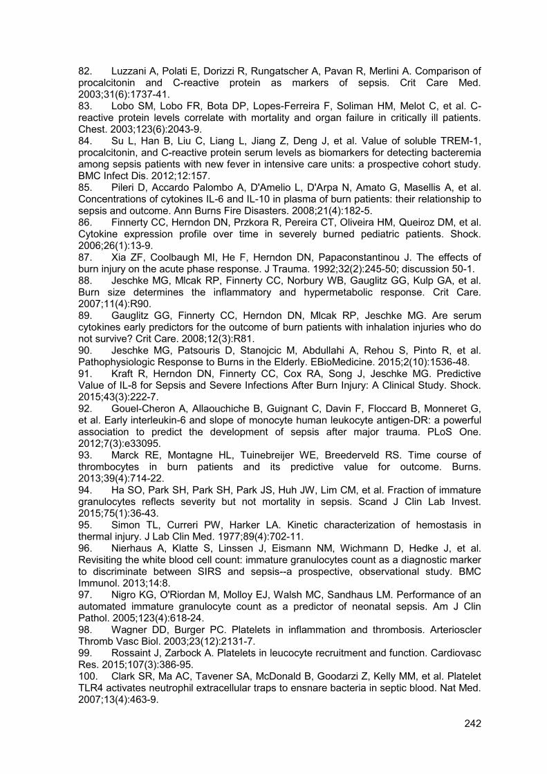
242
82. Luzzani A, Polati E, Dorizzi R, Rungatscher A, Pavan R, Merlini A. Comparison of procalcitonin and C-reactive protein as markers of sepsis. Crit Care Med. 2003;31(6):1737-41. 83. Lobo SM, Lobo FR, Bota DP, Lopes-Ferreira F, Soliman HM, Melot C, et al. C-reactive protein levels correlate with mortality and organ failure in critically ill patients. Chest. 2003;123(6):2043-9. 84. Su L, Han B, Liu C, Liang L, Jiang Z, Deng J, et al. Value of soluble TREM-1, procalcitonin, and C-reactive protein serum levels as biomarkers for detecting bacteremia among sepsis patients with new fever in intensive care units: a prospective cohort study. BMC Infect Dis. 2012;12:157. 85. Pileri D, Accardo Palombo A, D'Amelio L, D'Arpa N, Amato G, Masellis A, et al. Concentrations of cytokines IL-6 and IL-10 in plasma of burn patients: their relationship to sepsis and outcome. Ann Burns Fire Disasters. 2008;21(4):182-5. 86. Finnerty CC, Herndon DN, Przkora R, Pereira CT, Oliveira HM, Queiroz DM, et al. Cytokine expression profile over time in severely burned pediatric patients. Shock. 2006;26(1):13-9. 87. Xia ZF, Coolbaugh MI, He F, Herndon DN, Papaconstantinou J. The effects of burn injury on the acute phase response. J Trauma. 1992;32(2):245-50; discussion 50-1. 88. Jeschke MG, Mlcak RP, Finnerty CC, Norbury WB, Gauglitz GG, Kulp GA, et al. Burn size determines the inflammatory and hypermetabolic response. Crit Care. 2007;11(4):R90. 89. Gauglitz GG, Finnerty CC, Herndon DN, Mlcak RP, Jeschke MG. Are serum cytokines early predictors for the outcome of burn patients with inhalation injuries who do not survive? Crit Care. 2008;12(3):R81. 90. Jeschke MG, Patsouris D, Stanojcic M, Abdullahi A, Rehou S, Pinto R, et al. Pathophysiologic Response to Burns in the Elderly. EBioMedicine. 2015;2(10):1536-48. 91. Kraft R, Herndon DN, Finnerty CC, Cox RA, Song J, Jeschke MG. Predictive Value of IL-8 for Sepsis and Severe Infections After Burn Injury: A Clinical Study. Shock. 2015;43(3):222-7. 92. Gouel-Cheron A, Allaouchiche B, Guignant C, Davin F, Floccard B, Monneret G, et al. Early interleukin-6 and slope of monocyte human leukocyte antigen-DR: a powerful association to predict the development of sepsis after major trauma. PLoS One. 2012;7(3):e33095. 93. Marck RE, Montagne HL, Tuinebreijer WE, Breederveld RS. Time course of thrombocytes in burn patients and its predictive value for outcome. Burns. 2013;39(4):714-22. 94. Ha SO, Park SH, Park SH, Park JS, Huh JW, Lim CM, et al. Fraction of immature granulocytes reflects severity but not mortality in sepsis. Scand J Clin Lab Invest. 2015;75(1):36-43. 95. Simon TL, Curreri PW, Harker LA. Kinetic characterization of hemostasis in thermal injury. J Lab Clin Med. 1977;89(4):702-11. 96. Nierhaus A, Klatte S, Linssen J, Eismann NM, Wichmann D, Hedke J, et al. Revisiting the white blood cell count: immature granulocytes count as a diagnostic marker to discriminate between SIRS and sepsis--a prospective, observational study. BMC Immunol. 2013;14:8. 97. Nigro KG, O'Riordan M, Molloy EJ, Walsh MC, Sandhaus LM. Performance of an automated immature granulocyte count as a predictor of neonatal sepsis. Am J Clin Pathol. 2005;123(4):618-24. 98. Wagner DD, Burger PC. Platelets in inflammation and thrombosis. Arterioscler Thromb Vasc Biol. 2003;23(12):2131-7. 99. Rossaint J, Zarbock A. Platelets in leucocyte recruitment and function. Cardiovasc Res. 2015;107(3):386-95. 100. Clark SR, Ma AC, Tavener SA, McDonald B, Goodarzi Z, Kelly MM, et al. Platelet TLR4 activates neutrophil extracellular traps to ensnare bacteria in septic blood. Nat Med. 2007;13(4):463-9.
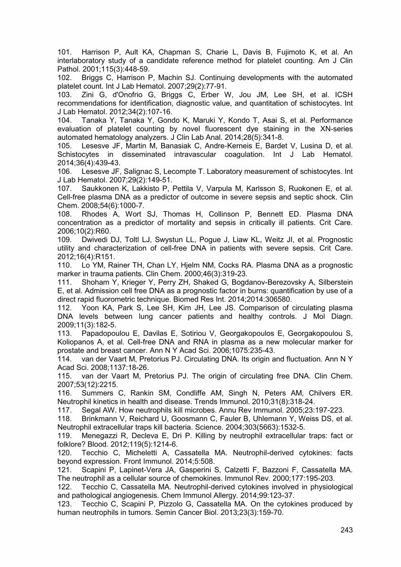
243
101. Harrison P, Ault KA, Chapman S, Charie L, Davis B, Fujimoto K, et al. An interlaboratory study of a candidate reference method for platelet counting. Am J Clin Pathol. 2001;115(3):448-59. 102. Briggs C, Harrison P, Machin SJ. Continuing developments with the automated platelet count. Int J Lab Hematol. 2007;29(2):77-91. 103. Zini G, d'Onofrio G, Briggs C, Erber W, Jou JM, Lee SH, et al. ICSH recommendations for identification, diagnostic value, and quantitation of schistocytes. Int J Lab Hematol. 2012;34(2):107-16. 104. Tanaka Y, Tanaka Y, Gondo K, Maruki Y, Kondo T, Asai S, et al. Performance evaluation of platelet counting by novel fluorescent dye staining in the XN-series automated hematology analyzers. J Clin Lab Anal. 2014;28(5):341-8. 105. Lesesve JF, Martin M, Banasiak C, Andre-Kerneis E, Bardet V, Lusina D, et al. Schistocytes in disseminated intravascular coagulation. Int J Lab Hematol. 2014;36(4):439-43. 106. Lesesve JF, Salignac S, Lecompte T. Laboratory measurement of schistocytes. Int J Lab Hematol. 2007;29(2):149-51. 107. Saukkonen K, Lakkisto P, Pettila V, Varpula M, Karlsson S, Ruokonen E, et al. Cell-free plasma DNA as a predictor of outcome in severe sepsis and septic shock. Clin Chem. 2008;54(6):1000-7. 108. Rhodes A, Wort SJ, Thomas H, Collinson P, Bennett ED. Plasma DNA concentration as a predictor of mortality and sepsis in critically ill patients. Crit Care. 2006;10(2):R60. 109. Dwivedi DJ, Toltl LJ, Swystun LL, Pogue J, Liaw KL, Weitz JI, et al. Prognostic utility and characterization of cell-free DNA in patients with severe sepsis. Crit Care. 2012;16(4):R151. 110. Lo YM, Rainer TH, Chan LY, Hjelm NM, Cocks RA. Plasma DNA as a prognostic marker in trauma patients. Clin Chem. 2000;46(3):319-23. 111. Shoham Y, Krieger Y, Perry ZH, Shaked G, Bogdanov-Berezovsky A, Silberstein E, et al. Admission cell free DNA as a prognostic factor in burns: quantification by use of a direct rapid fluorometric technique. Biomed Res Int. 2014;2014:306580. 112. Yoon KA, Park S, Lee SH, Kim JH, Lee JS. Comparison of circulating plasma DNA levels between lung cancer patients and healthy controls. J Mol Diagn. 2009;11(3):182-5. 113. Papadopoulou E, Davilas E, Sotiriou V, Georgakopoulos E, Georgakopoulou S, Koliopanos A, et al. Cell-free DNA and RNA in plasma as a new molecular marker for prostate and breast cancer. Ann N Y Acad Sci. 2006;1075:235-43. 114. van der Vaart M, Pretorius PJ. Circulating DNA. Its origin and fluctuation. Ann N Y Acad Sci. 2008;1137:18-26. 115. van der Vaart M, Pretorius PJ. The origin of circulating free DNA. Clin Chem. 2007;53(12):2215. 116. Summers C, Rankin SM, Condliffe AM, Singh N, Peters AM, Chilvers ER. Neutrophil kinetics in health and disease. Trends Immunol. 2010;31(8):318-24. 117. Segal AW. How neutrophils kill microbes. Annu Rev Immunol. 2005;23:197-223. 118. Brinkmann V, Reichard U, Goosmann C, Fauler B, Uhlemann Y, Weiss DS, et al. Neutrophil extracellular traps kill bacteria. Science. 2004;303(5663):1532-5. 119. Menegazzi R, Decleva E, Dri P. Killing by neutrophil extracellular traps: fact or folklore? Blood. 2012;119(5):1214-6. 120. Tecchio C, Micheletti A, Cassatella MA. Neutrophil-derived cytokines: facts beyond expression. Front Immunol. 2014;5:508. 121. Scapini P, Lapinet-Vera JA, Gasperini S, Calzetti F, Bazzoni F, Cassatella MA. The neutrophil as a cellular source of chemokines. Immunol Rev. 2000;177:195-203. 122. Tecchio C, Cassatella MA. Neutrophil-derived cytokines involved in physiological and pathological angiogenesis. Chem Immunol Allergy. 2014;99:123-37. 123. Tecchio C, Scapini P, Pizzolo G, Cassatella MA. On the cytokines produced by human neutrophils in tumors. Semin Cancer Biol. 2013;23(3):159-70.
244
124. Dinauer MC, Lekstrom-Himes JA, Dale DC. Inherited Neutrophil Disorders: Molecular Basis and New Therapies. Hematology Am Soc Hematol Educ Program. 2000:303-18. 125. Mocsai A. Diverse novel functions of neutrophils in immunity, inflammation, and beyond. J Exp Med. 2013;210(7):1283-99. 126. Jones DA, Abbassi O, McIntire LV, McEver RP, Smith CW. P-selectin mediates neutrophil rolling on histamine-stimulated endothelial cells. Biophys J. 1993;65(4):1560-9. 127. Ley K, Laudanna C, Cybulsky MI, Nourshargh S. Getting to the site of inflammation: the leukocyte adhesion cascade updated. Nat Rev Immunol. 2007;7(9):678-89. 128. Muller WA. Leukocyte-endothelial-cell interactions in leukocyte transmigration and the inflammatory response. Trends Immunol. 2003;24(6):327-34. 129. Anderson DC, Springer TA. Leukocyte adhesion deficiency: an inherited defect in the Mac-1, LFA-1, and p150,95 glycoproteins. Annu Rev Med. 1987;38:175-94. 130. Nuzzi PA, Lokuta MA, Huttenlocher A. Analysis of neutrophil chemotaxis. Methods Mol Biol. 2007;370:23-36. 131. Lin F, Nguyen CM, Wang SJ, Saadi W, Gross SP, Jeon NL. Neutrophil migration in opposing chemoattractant gradients using microfluidic chemotaxis devices. Ann Biomed Eng. 2005;33(4):475-82. 132. Boner A, Zeligs BJ, Bellanti JA. Chemotactic responses of various differentiational stages of neutrophils from human cord and adult blood. Infect Immun. 1982;35(3):921-8. 133. Hatano Y, Taniuchi S, Masuda M, Tsuji S, Ito T, Hasui M, et al. Phagocytosis of heat-killed Staphylococcus aureus by eosinophils: comparison with neutrophils. APMIS. 2009;117(2):115-23. 134. Leliefeld PH, Wessels CM, Leenen LP, Koenderman L, Pillay J. The role of neutrophils in immune dysfunction during severe inflammation. Crit Care. 2016;20:73. 135. Babior BM. NADPH oxidase: an update. Blood. 1999;93(5):1464-76. 136. Bedard K, Krause KH. The NOX family of ROS-generating NADPH oxidases: physiology and pathophysiology. Physiol Rev. 2007;87(1):245-313. 137. Liao Y, Liu P, Guo F, Zhang ZY, Zhang Z. Oxidative burst of circulating neutrophils following traumatic brain injury in human. PLoS One. 2013;8(7):e68963. 138. Dong YL, Abdullah K, Yan TZ, Rutan T, Broemeling L, Robson M, et al. Effect of thermal injury and sepsis on neutrophil function. J Trauma. 1993;34(3):417-21. 139. Spicer SS, Hardin JH. Ultrastructure, cytochemistry, and function of neutrophil leukocyte granules. A review. Lab Invest. 1969;20(5):488-97. 140. Belaaouaj A, Kim KS, Shapiro SD. Degradation of outer membrane protein A in Escherichia coli killing by neutrophil elastase. Science. 2000;289(5482):1185-8. 141. Fox S, Leitch AE, Duffin R, Haslett C, Rossi AG. Neutrophil apoptosis: relevance to the innate immune response and inflammatory disease. J Innate Immun. 2010;2(3):216-27. 142. Luo HR, Loison F. Constitutive neutrophil apoptosis: mechanisms and regulation. Am J Hematol. 2008;83(4):288-95. 143. Edwards SW, Derouet M, Howse M, Moots RJ. Regulation of neutrophil apoptosis by Mcl-1. Biochem Soc Trans. 2004;32(Pt3):489-92. 144. Akgul C, Moulding DA, White MR, Edwards SW. In vivo localisation and stability of human Mcl-1 using green fluorescent protein (GFP) fusion proteins. FEBS Lett. 2000;478(1-2):72-6. 145. Lovell JF, Billen LP, Bindner S, Shamas-Din A, Fradin C, Leber B, et al. Membrane binding by tBid initiates an ordered series of events culminating in membrane permeabilization by Bax. Cell. 2008;135(6):1074-84. 146. Scatizzi JC, Hutcheson J, Bickel E, Haines GK, 3rd, Perlman H. Pro-apoptotic Bid is required for the resolution of the effector phase of inflammatory arthritis. Arthritis Res Ther. 2007;9(3):R49.
245
147. Andina N, Conus S, Schneider EM, Fey MF, Simon HU. Induction of Bim limits cytokine-mediated prolonged survival of neutrophils. Cell Death Differ. 2009;16(9):1248-55. 148. Nolan B, Collette H, Baker S, Duffy A, De M, Miller C, et al. Inhibition of neutrophil apoptosis after severe trauma is NFkappabeta dependent. J Trauma. 2000;48(4):599-604; discussion -5. 149. Turina M, Hoth JJ, Turpen RM, Scott MJ, Cheadle WG. Alveolar interleukin-10 regulates neutrophil apoptosis in severely traumatized patients. J Trauma. 2007;63(4):733-9. 150. Paunel-Gorgulu A, Kirichevska T, Logters T, Windolf J, Flohe S. Molecular mechanisms underlying delayed apoptosis in neutrophils from multiple trauma patients with and without sepsis. Mol Med. 2012;18:325-35. 151. Matsuda T, Saito H, Fukatsu K, Han I, Inoue T, Furukawa S, et al. Cytokine-modulated inhibition of neutrophil apoptosis at local site augments exudative neutrophil functions and reflects inflammatory response after surgery. Surgery. 2001;129(1):76-85. 152. Scheel-Toellner D, Wang K, Assi LK, Webb PR, Craddock RM, Salmon M, et al. Clustering of death receptors in lipid rafts initiates neutrophil spontaneous apoptosis. Biochem Soc Trans. 2004;32(Pt 5):679-81. 153. Dunogue B, Pilmis B, Mahlaoui N, Elie C, Coignard-Biehler H, Amazzough K, et al. Chronic Granulomatous Disease in Patients Reaching Adulthood: A Nationwide Study in France. Clin Infect Dis. 2017;64(6):767-75. 154. Introne WJ, Westbroek W, Golas GA, Adams D. Chediak-Higashi Syndrome. In: Pagon RA, Adam MP, Ardinger HH, Wallace SE, Amemiya A, Bean LJH, et al., editors. GeneReviews(R). Seattle (WA)1993. 155. Robertson CM, Perrone EE, McConnell KW, Dunne WM, Boody B, Brahmbhatt T, et al. Neutrophil depletion causes a fatal defect in murine pulmonary Staphylococcus aureus clearance. J Surg Res. 2008;150(2):278-85. 156. Hazeldine J, Hampson P, Lord JM. The impact of trauma on neutrophil function. Injury. 2014;45(12):1824-33. 157. Cook KM, Sifri ZC, Baranski GM, Mohr AM, Livingston DH. The role of plasma granulocyte colony stimulating factor and bone marrow dysfunction after severe trauma. J Am Coll Surg. 2013;216(1):57-64. 158. Pillay J, Kamp VM, van Hoffen E, Visser T, Tak T, Lammers JW, et al. A subset of neutrophils in human systemic inflammation inhibits T cell responses through Mac-1. J Clin Invest. 2012;122(1):327-36. 159. Warden GD, Mason AD, Jr., Pruitt BA, Jr. Suppression of leukocyte chemotaxis in vitro by chemotherapeutic agents used in the management of thermal injuries. Ann Surg. 1975;181(3):363-9. 160. Kim Y, Goldstein E, Lippert W, Brofeldt T, Donovan R. Polymorphonuclear leucocyte motility in patients with severe burns. Burns. 1989;15(2):93-7. 161. Warden GD, Mason AD, Jr., Pruitt BA, Jr. Evaluation of leukocyte chemotaxis in vitro in thermally injured patients. J Clin Invest. 1974;54(4):1001-4. 162. Richards MK, Liu F, Iwasaki H, Akashi K, Link DC. Pivotal role of granulocyte colony-stimulating factor in the development of progenitors in the common myeloid pathway. Blood. 2003;102(10):3562-8. 163. Lord BI, Bronchud MH, Owens S, Chang J, Howell A, Souza L, et al. The kinetics of human granulopoiesis following treatment with granulocyte colony-stimulating factor in vivo. Proc Natl Acad Sci U S A. 1989;86(23):9499-503. 164. Liu F, Poursine-Laurent J, Wu HY, Link DC. Interleukin-6 and the granulocyte colony-stimulating factor receptor are major independent regulators of granulopoiesis in vivo but are not required for lineage commitment or terminal differentiation. Blood. 1997;90(7):2583-90. 165. Sugimoto K, Mori KJ. Cascade regulation of cytokine production in granulopoiesis. Leukemia. 1997;11 Suppl 3:464-7.
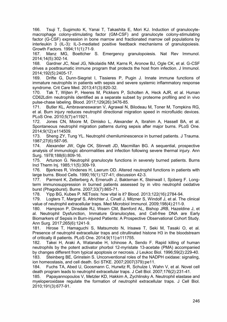
246
166. Tsuji T, Sugimoto K, Yanai T, Takashita E, Mori KJ. Induction of granulocyte-macrophage colony-stimulating factor (GM-CSF) and granulocyte colony-stimulating factor (G-CSF) expression in bone marrow and fractionated marrow cell populations by interleukin 3 (IL-3): IL-3-mediated positive feedback mechanisms of granulopoiesis. Growth Factors. 1994;11(1):71-9. 167. Manz MG, Boettcher S. Emergency granulopoiesis. Nat Rev Immunol. 2014;14(5):302-14. 168. Gardner JC, Noel JG, Nikolaidis NM, Karns R, Aronow BJ, Ogle CK, et al. G-CSF drives a posttraumatic immune program that protects the host from infection. J Immunol. 2014;192(5):2405-17. 169. Drifte G, Dunn-Siegrist I, Tissieres P, Pugin J. Innate immune functions of immature neutrophils in patients with sepsis and severe systemic inflammatory response syndrome. Crit Care Med. 2013;41(3):820-32. 170. Tak T, Wijten P, Heeres M, Pickkers P, Scholten A, Heck AJR, et al. Human CD62Ldim neutrophils identified as a separate subset by proteome profiling and in vivo pulse-chase labeling. Blood. 2017;129(26):3476-85. 171. Butler KL, Ambravaneswaran V, Agrawal N, Bilodeau M, Toner M, Tompkins RG, et al. Burn injury reduces neutrophil directional migration speed in microfluidic devices. PLoS One. 2010;5(7):e11921. 172. Jones CN, Moore M, Dimisko L, Alexander A, Ibrahim A, Hassell BA, et al. Spontaneous neutrophil migration patterns during sepsis after major burns. PLoS One. 2014;9(12):e114509. 173. Sheng ZY, Tung YL. Neutrophil chemiluminescence in burned patients. J Trauma. 1987;27(6):587-95. 174. Alexander JW, Ogle CK, Stinnett JD, Macmillan BG. A sequential, prospective analysis of immunologic abnormalities and infection following severe thermal injury. Ann Surg. 1978;188(6):809-16. 175. Arturson G. Neutrophil granulocyte functions in severely burned patients. Burns Incl Therm Inj. 1985;11(5):309-19. 176. Bjerknes R, Vindenes H, Laerum OD. Altered neutrophil functions in patients with large burns. Blood Cells. 1990;16(1):127-41; discussion 42-3. 177. Parment K, Zetterberg A, Ernerudh J, Bakteman K, Steinwall I, Sjoberg F. Long-term immunosuppression in burned patients assessed by in vitro neutrophil oxidative burst (Phagoburst). Burns. 2007;33(7):865-71. 178. Yipp BG, Kubes P. NETosis: how vital is it? Blood. 2013;122(16):2784-94. 179. Logters T, Margraf S, Altrichter J, Cinatl J, Mitzner S, Windolf J, et al. The clinical value of neutrophil extracellular traps. Med Microbiol Immunol. 2009;198(4):211-9. 180. Hampson P, Dinsdale RJ, Wearn CM, Bamford AL, Bishop JRB, Hazeldine J, et al. Neutrophil Dysfunction, Immature Granulocytes, and Cell-free DNA are Early Biomarkers of Sepsis in Burn-injured Patients: A Prospective Observational Cohort Study. Ann Surg. 2017;265(6):1241-9. 181. Hirose T, Hamaguchi S, Matsumoto N, Irisawa T, Seki M, Tasaki O, et al. Presence of neutrophil extracellular traps and citrullinated histone H3 in the bloodstream of critically ill patients. PLoS One. 2014;9(11):e111755. 182. Takei H, Araki A, Watanabe H, Ichinose A, Sendo F. Rapid killing of human neutrophils by the potent activator phorbol 12-myristate 13-acetate (PMA) accompanied by changes different from typical apoptosis or necrosis. J Leukoc Biol. 1996;59(2):229-40. 183. Steinberg BE, Grinstein S. Unconventional roles of the NADPH oxidase: signaling, ion homeostasis, and cell death. Sci STKE. 2007;2007(379):pe11. 184. Fuchs TA, Abed U, Goosmann C, Hurwitz R, Schulze I, Wahn V, et al. Novel cell death program leads to neutrophil extracellular traps. J Cell Biol. 2007;176(2):231-41. 185. Papayannopoulos V, Metzler KD, Hakkim A, Zychlinsky A. Neutrophil elastase and myeloperoxidase regulate the formation of neutrophil extracellular traps. J Cell Biol. 2010;191(3):677-91.
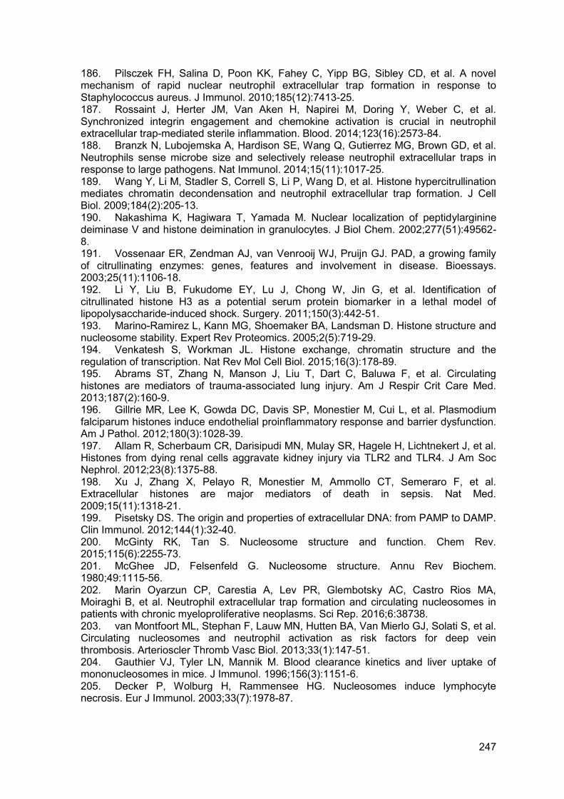
247
186. Pilsczek FH, Salina D, Poon KK, Fahey C, Yipp BG, Sibley CD, et al. A novel mechanism of rapid nuclear neutrophil extracellular trap formation in response to Staphylococcus aureus. J Immunol. 2010;185(12):7413-25. 187. Rossaint J, Herter JM, Van Aken H, Napirei M, Doring Y, Weber C, et al. Synchronized integrin engagement and chemokine activation is crucial in neutrophil extracellular trap-mediated sterile inflammation. Blood. 2014;123(16):2573-84. 188. Branzk N, Lubojemska A, Hardison SE, Wang Q, Gutierrez MG, Brown GD, et al. Neutrophils sense microbe size and selectively release neutrophil extracellular traps in response to large pathogens. Nat Immunol. 2014;15(11):1017-25. 189. Wang Y, Li M, Stadler S, Correll S, Li P, Wang D, et al. Histone hypercitrullination mediates chromatin decondensation and neutrophil extracellular trap formation. J Cell Biol. 2009;184(2):205-13. 190. Nakashima K, Hagiwara T, Yamada M. Nuclear localization of peptidylarginine deiminase V and histone deimination in granulocytes. J Biol Chem. 2002;277(51):49562-8. 191. Vossenaar ER, Zendman AJ, van Venrooij WJ, Pruijn GJ. PAD, a growing family of citrullinating enzymes: genes, features and involvement in disease. Bioessays. 2003;25(11):1106-18. 192. Li Y, Liu B, Fukudome EY, Lu J, Chong W, Jin G, et al. Identification of citrullinated histone H3 as a potential serum protein biomarker in a lethal model of lipopolysaccharide-induced shock. Surgery. 2011;150(3):442-51. 193. Marino-Ramirez L, Kann MG, Shoemaker BA, Landsman D. Histone structure and nucleosome stability. Expert Rev Proteomics. 2005;2(5):719-29. 194. Venkatesh S, Workman JL. Histone exchange, chromatin structure and the regulation of transcription. Nat Rev Mol Cell Biol. 2015;16(3):178-89. 195. Abrams ST, Zhang N, Manson J, Liu T, Dart C, Baluwa F, et al. Circulating histones are mediators of trauma-associated lung injury. Am J Respir Crit Care Med. 2013;187(2):160-9. 196. Gillrie MR, Lee K, Gowda DC, Davis SP, Monestier M, Cui L, et al. Plasmodium falciparum histones induce endothelial proinflammatory response and barrier dysfunction. Am J Pathol. 2012;180(3):1028-39. 197. Allam R, Scherbaum CR, Darisipudi MN, Mulay SR, Hagele H, Lichtnekert J, et al. Histones from dying renal cells aggravate kidney injury via TLR2 and TLR4. J Am Soc Nephrol. 2012;23(8):1375-88. 198. Xu J, Zhang X, Pelayo R, Monestier M, Ammollo CT, Semeraro F, et al. Extracellular histones are major mediators of death in sepsis. Nat Med. 2009;15(11):1318-21. 199. Pisetsky DS. The origin and properties of extracellular DNA: from PAMP to DAMP. Clin Immunol. 2012;144(1):32-40. 200. McGinty RK, Tan S. Nucleosome structure and function. Chem Rev. 2015;115(6):2255-73. 201. McGhee JD, Felsenfeld G. Nucleosome structure. Annu Rev Biochem. 1980;49:1115-56. 202. Marin Oyarzun CP, Carestia A, Lev PR, Glembotsky AC, Castro Rios MA, Moiraghi B, et al. Neutrophil extracellular trap formation and circulating nucleosomes in patients with chronic myeloproliferative neoplasms. Sci Rep. 2016;6:38738. 203. van Montfoort ML, Stephan F, Lauw MN, Hutten BA, Van Mierlo GJ, Solati S, et al. Circulating nucleosomes and neutrophil activation as risk factors for deep vein thrombosis. Arterioscler Thromb Vasc Biol. 2013;33(1):147-51. 204. Gauthier VJ, Tyler LN, Mannik M. Blood clearance kinetics and liver uptake of mononucleosomes in mice. J Immunol. 1996;156(3):1151-6. 205. Decker P, Wolburg H, Rammensee HG. Nucleosomes induce lymphocyte necrosis. Eur J Immunol. 2003;33(7):1978-87.
248
206. Bhagirath VC, Dwivedi DJ, Liaw PC. Comparison of the Proinflammatory and Procoagulant Properties of Nuclear, Mitochondrial, and Bacterial DNA. Shock. 2015;44(3):265-71. 207. Levine B, Kroemer G. Autophagy in the pathogenesis of disease. Cell. 2008;132(1):27-42. 208. Remijsen Q, Vanden Berghe T, Wirawan E, Asselbergh B, Parthoens E, De Rycke R, et al. Neutrophil extracellular trap cell death requires both autophagy and superoxide generation. Cell Res. 2011;21(2):290-304. 209. Nakagawa I, Amano A, Mizushima N, Yamamoto A, Yamaguchi H, Kamimoto T, et al. Autophagy defends cells against invading group A Streptococcus. Science. 2004;306(5698):1037-40. 210. Gutierrez MG, Master SS, Singh SB, Taylor GA, Colombo MI, Deretic V. Autophagy is a defense mechanism inhibiting BCG and Mycobacterium tuberculosis survival in infected macrophages. Cell. 2004;119(6):753-66. 211. Birmingham CL, Brumell JH. Autophagy recognizes intracellular Salmonella enterica serovar Typhimurium in damaged vacuoles. Autophagy. 2006;2(3):156-8. 212. Kenno S, Perito S, Mosci P, Vecchiarelli A, Monari C. Autophagy and Reactive Oxygen Species Are Involved in Neutrophil Extracellular Traps Release Induced by C. albicans Morphotypes. Front Microbiol. 2016;7:879. 213. Hashiba M, Huq A, Tomino A, Hirakawa A, Hattori T, Miyabe H, et al. Neutrophil extracellular traps in patients with sepsis. J Surg Res. 2015;194(1):248-54. 214. Czaikoski PG, Mota JM, Nascimento DC, Sonego F, Castanheira FV, Melo PH, et al. Neutrophil Extracellular Traps Induce Organ Damage during Experimental and Clinical Sepsis. PLoS One. 2016;11(2):e0148142. 215. Margraf S, Logters T, Reipen J, Altrichter J, Scholz M, Windolf J. Neutrophil-derived circulating free DNA (cf-DNA/NETs): a potential prognostic marker for posttraumatic development of inflammatory second hit and sepsis. Shock. 2008;30(4):352-8. 216. Yang J, Selvaganapathy PR, Gould TJ, Dwivedi DJ, Liu D, Fox-Robichaud AE, et al. A microfluidic device for rapid quantification of cell-free DNA in patients with severe sepsis. Lab Chip. 2015;15(19):3925-33. 217. Gupta S, Kaplan MJ. The role of neutrophils and NETosis in autoimmune and renal diseases. Nat Rev Nephrol. 2016;12(7):402-13. 218. Demers M, Wagner DD. NETosis: a new factor in tumor progression and cancer-associated thrombosis. Semin Thromb Hemost. 2014;40(3):277-83. 219. Jorch SK, Kubes P. An emerging role for neutrophil extracellular traps in noninfectious disease. Nat Med. 2017;23(3):279-87. 220. Altrichter J, Zedler S, Kraft R, Faist E, Mitzner SR, Sauer M, et al. Neutrophil-derived circulating free DNA (cf-DNA/NETs), a potential prognostic marker for mortality in patients with severe burn injury. Eur J Trauma Emerg Surg. 2010;36(6):551-7. 221. Meng W, Paunel-Gorgulu A, Flohe S, Hoffmann A, Witte I, MacKenzie C, et al. Depletion of neutrophil extracellular traps in vivo results in hypersusceptibility to polymicrobial sepsis in mice. Crit Care. 2012;16(4):R137. 222. Tanaka K, Koike Y, Shimura T, Okigami M, Ide S, Toiyama Y, et al. In vivo characterization of neutrophil extracellular traps in various organs of a murine sepsis model. PLoS One. 2014;9(11):e111888. 223. Alhamdi Y, Abrams ST, Cheng Z, Jing S, Su D, Liu Z, et al. Circulating Histones Are Major Mediators of Cardiac Injury in Patients With Sepsis. Crit Care Med. 2015;43(10):2094-103. 224. Caudrillier A, Kessenbrock K, Gilliss BM, Nguyen JX, Marques MB, Monestier M, et al. Platelets induce neutrophil extracellular traps in transfusion-related acute lung injury. J Clin Invest. 2012;122(7):2661-71. 225. Kolaczkowska E, Jenne CN, Surewaard BG, Thanabalasuriar A, Lee WY, Sanz MJ, et al. Molecular mechanisms of NET formation and degradation revealed by intravital imaging in the liver vasculature. Nat Commun. 2015;6:6673.
249
226. Kaplan MJ, Radic M. Neutrophil extracellular traps: double-edged swords of innate immunity. J Immunol. 2012;189(6):2689-95. 227. Urban CF, Ermert D, Schmid M, Abu-Abed U, Goosmann C, Nacken W, et al. Neutrophil extracellular traps contain calprotectin, a cytosolic protein complex involved in host defense against Candida albicans. PLoS Pathog. 2009;5(10):e1000639. 228. Kawai C, Kotani H, Miyao M, Ishida T, Jemail L, Abiru H, et al. Circulating Extracellular Histones Are Clinically Relevant Mediators of Multiple Organ Injury. Am J Pathol. 2016;186(4):829-43. 229. Andrews RK, Arthur JF, Gardiner EE. Neutrophil extracellular traps (NETs) and the role of platelets in infection. Thromb Haemost. 2014;112(4):659-65. 230. Fuchs TA, Brill A, Wagner DD. Neutrophil extracellular trap (NET) impact on deep vein thrombosis. Arterioscler Thromb Vasc Biol. 2012;32(8):1777-83. 231. Levi M, Schultz M, van der Poll T. Sepsis and thrombosis. Semin Thromb Hemost. 2013;39(5):559-66. 232. Fuchs TA, Brill A, Duerschmied D, Schatzberg D, Monestier M, Myers DD, Jr., et al. Extracellular DNA traps promote thrombosis. Proc Natl Acad Sci U S A. 2010;107(36):15880-5. 233. Brill A, Fuchs TA, Savchenko AS, Thomas GM, Martinod K, De Meyer SF, et al. Neutrophil extracellular traps promote deep vein thrombosis in mice. J Thromb Haemost. 2012;10(1):136-44. 234. Gould TJ, Vu TT, Swystun LL, Dwivedi DJ, Mai SH, Weitz JI, et al. Neutrophil extracellular traps promote thrombin generation through platelet-dependent and platelet-independent mechanisms. Arterioscler Thromb Vasc Biol. 2014;34(9):1977-84. 235. Gould TJ, Vu TT, Stafford AR, Dwivedi DJ, Kim PY, Fox-Robichaud AE, et al. Cell-Free DNA Modulates Clot Structure and Impairs Fibrinolysis in Sepsis. Arterioscler Thromb Vasc Biol. 2015;35(12):2544-53. 236. Boneschansker L, Inoue Y, Oklu R, Irimia D. Capillary plexuses are vulnerable to neutrophil extracellular traps. Integr Biol (Camb). 2016;8(2):149-55. 237. Hakkim A, Furnrohr BG, Amann K, Laube B, Abed UA, Brinkmann V, et al. Impairment of neutrophil extracellular trap degradation is associated with lupus nephritis. Proc Natl Acad Sci U S A. 2010;107(21):9813-8. 238. Kotzin BL. Systemic lupus erythematosus. Cell. 1996;85(3):303-6. 239. Meng W, Paunel-Gorgulu A, Flohe S, Witte I, Schadel-Hopfner M, Windolf J, et al. Deoxyribonuclease is a potential counter regulator of aberrant neutrophil extracellular traps formation after major trauma. Mediators Inflamm. 2012;2012:149560. 240. Lee WM, Galbraith RM. The extracellular actin-scavenger system and actin toxicity. N Engl J Med. 1992;326(20):1335-41. 241. Ananthakrishnan R, Ehrlicher A. The forces behind cell movement. Int J Biol Sci. 2007;3(5):303-17. 242. Mogilner A, Oster G. Cell motility driven by actin polymerization. Biophys J. 1996;71(6):3030-45. 243. Haddad JG, Harper KD, Guoth M, Pietra GG, Sanger JW. Angiopathic consequences of saturating the plasma scavenger system for actin. Proc Natl Acad Sci U S A. 1990;87(4):1381-5. 244. Lind SE, Smith CJ. Actin stimulates plasmin generation by tissue and urokinase-type plasminogen activators. Arch Biochem Biophys. 1993;307(1):138-45. 245. Lazarides E, Lindberg U. Actin is the naturally occurring inhibitor of deoxyribonuclease I. Proc Natl Acad Sci U S A. 1974;71(12):4742-6. 246. Lee PS, Patel SR, Christiani DC, Bajwa E, Stossel TP, Waxman AB. Plasma gelsolin depletion and circulating actin in sepsis: a pilot study. PLoS One. 2008;3(11):e3712. 247. Smith DB, Janmey PA, Herbert TJ, Lind SE. Quantitative measurement of plasma gelsolin and its incorporation into fibrin clots. J Lab Clin Med. 1987;110(2):189-95. 248. Yin HL, Kwiatkowski DJ, Mole JE, Cole FS. Structure and biosynthesis of cytoplasmic and secreted variants of gelsolin. J Biol Chem. 1984;259(8):5271-6.
250
249. Kwiatkowski DJ, Janmey PA, Yin HL. Identification of critical functional and regulatory domains in gelsolin. J Cell Biol. 1989;108(5):1717-26. 250. Yin HL, Zaner KS, Stossel TP. Ca2+ control of actin gelation. Interaction of gelsolin with actin filaments and regulation of actin gelation. J Biol Chem. 1980;255(19):9494-500. 251. Yin HL, Iida K, Janmey PA. Identification of a polyphosphoinositide-modulated domain in gelsolin which binds to the sides of actin filaments. J Cell Biol. 1988;106(3):805-12. 252. Lind SE, Smith DB, Janmey PA, Stossel TP. Depression of gelsolin levels and detection of gelsolin-actin complexes in plasma of patients with acute lung injury. Am Rev Respir Dis. 1988;138(2):429-34. 253. Ito H, Kambe H, Kimura Y, Nakamura H, Hayashi E, Kishimoto T, et al. Depression of plasma gelsolin level during acute liver injury. Gastroenterology. 1992;102(5):1686-92. 254. Huang LF, Yao YM, Li JF, Dong N, Liu C, Yu Y, et al. Reduction of plasma gelsolin levels correlates with development of multiple organ dysfunction syndrome and fatal outcome in burn patients. PLoS One. 2011;6(11):e25748. 255. Suhler E, Lin W, Yin HL, Lee WM. Decreased plasma gelsolin concentrations in acute liver failure, myocardial infarction, septic shock, and myonecrosis. Crit Care Med. 1997;25(4):594-8. 256. Kulakowska A, Ciccarelli NJ, Wen Q, Mroczko B, Drozdowski W, Szmitkowski M, et al. Hypogelsolinemia, a disorder of the extracellular actin scavenger system, in patients with multiple sclerosis. BMC Neurol. 2010;10:107. 257. Dahl B, Schiodt FV, Ott P, Gvozdenovic R, Yin HL, Lee WM. Plasma gelsolin is reduced in trauma patients. Shock. 1999;12(2):102-4. 258. Osborn TM, Verdrengh M, Stossel TP, Tarkowski A, Bokarewa M. Decreased levels of the gelsolin plasma isoform in patients with rheumatoid arthritis. Arthritis Res Ther. 2008;10(5):R117. 259. Hu Y, Li H, Li WH, Meng HX, Fan YZ, Li WJ, et al. The value of decreased plasma gelsolin levels in patients with systemic lupus erythematosus and rheumatoid arthritis in diagnosis and disease activity evaluation. Lupus. 2013;22(14):1455-61. 260. Ordija CM, Chiou TT, Yang Z, Deloid GM, de Oliveira Valdo M, Wang Z, et al. Free actin impairs macrophage bacterial defenses via scavenger receptor MARCO interaction with reversal by plasma gelsolin. Am J Physiol Lung Cell Mol Physiol. 2017;312(6):L1018-L28. 261. Rothenbach PA, Dahl B, Schwartz JJ, O'Keefe GE, Yamamoto M, Lee WM, et al. Recombinant plasma gelsolin infusion attenuates burn-induced pulmonary microvascular dysfunction. J Appl Physiol (1985). 2004;96(1):25-31. 262. Bikle DD, Gee E, Halloran B, Kowalski MA, Ryzen E, Haddad JG. Assessment of the free fraction of 25-hydroxyvitamin D in serum and its regulation by albumin and the vitamin D-binding protein. J Clin Endocrinol Metab. 1986;63(4):954-9. 263. Cooke NE, Haddad JG. Vitamin D binding protein (Gc-globulin). Endocr Rev. 1989;10(3):294-307. 264. Van Baelen H, Bouillon R, De Moor P. Vitamin D-binding protein (Gc-globulin) binds actin. J Biol Chem. 1980;255(6):2270-2. 265. Janmey PA, Lind SE. Capacity of human serum to depolymerize actin filaments. Blood. 1987;70(2):524-30. 266. Dueland S, Nenseter MS, Drevon CA. Uptake and degradation of filamentous actin and vitamin D-binding protein in the rat. Biochem J. 1991;274 ( Pt 1):237-41. 267. Herrmannsdoerfer AJ, Heeb GT, Feustel PJ, Estes JE, Keenan CJ, Minnear FL, et al. Vascular clearance and organ uptake of G- and F-actin in the rat. Am J Physiol. 1993;265(6 Pt 1):G1071-81. 268. Dahl B, Schiodt FV, Nielsen M, Kiaer T, Williams JG, Ott P. Admission level of Gc-globulin predicts outcome after multiple trauma. Injury. 1999;30(4):275-81.
251
269. Dahl B, Schiodt FV, Ott P, Wians F, Lee WM, Balko J, et al. Plasma concentration of Gc-globulin is associated with organ dysfunction and sepsis after injury. Crit Care Med. 2003;31(1):152-6. 270. Dahl B, Schiodt FV, Kiaer T, Ott P, Bondesen S, Tygstrup N. Serum Gc-globulin in the early course of multiple trauma. Crit Care Med. 1998;26(2):285-9. 271. Dahl B, Schiodt FV, Rudolph S, Ott P, Kiaer T, Heslet L. Trauma stimulates the synthesis of Gc-globulin. Intensive Care Med. 2001;27(2):394-9. 272. Wang X, Qin W, Sun B. New strategy for sepsis: Targeting a key role of platelet-neutrophil interaction. Burns Trauma. 2014;2(3):114-20. 273. Kimball AS, Obi AT, Diaz JA, Henke PK. The Emerging Role of NETs in Venous Thrombosis and Immunothrombosis. Front Immunol. 2016;7:236. 274. Yang S, Qi H, Kan K, Chen J, Xie H, Guo X, et al. Neutrophil Extracellular Traps Promote Hypercoagulability in Patients With Sepsis. Shock. 2017;47(2):132-9. 275. Martinod K, Fuchs TA, Zitomersky NL, Wong SL, Demers M, Gallant M, et al. PAD4-deficiency does not affect bacteremia in polymicrobial sepsis and ameliorates endotoxemic shock. Blood. 2015;125(12):1948-56. 276. Li P, Li M, Lindberg MR, Kennett MJ, Xiong N, Wang Y. PAD4 is essential for antibacterial innate immunity mediated by neutrophil extracellular traps. J Exp Med. 2010;207(9):1853-62. 277. Kraft R, Herndon DN, Finnerty CC, Shahrokhi S, Jeschke MG. Occurrence of multiorgan dysfunction in pediatric burn patients: incidence and clinical outcome. Ann Surg. 2014;259(2):381-7. 278. Knaus WA, Draper EA, Wagner DP, Zimmerman JE. APACHE II: a severity of disease classification system. Crit Care Med. 1985;13(10):818-29. 279. Vincent JL, Moreno R, Takala J, Willatts S, De Mendonca A, Bruining H, et al. The SOFA (Sepsis-related Organ Failure Assessment) score to describe organ dysfunction/failure. On behalf of the Working Group on Sepsis-Related Problems of the European Society of Intensive Care Medicine. Intensive Care Med. 1996;22(7):707-10. 280. Tobiasen J, Hiebert JM, Edlich RF. The abbreviated burn severity index. Ann Emerg Med. 1982;11(5):260-2. 281. Osler T, Glance LG, Hosmer DW. Simplified estimates of the probability of death after burn injuries: extending and updating the baux score. J Trauma. 2010;68(3):690-7. 282. Dinsdale RJ, Devi A, Hampson P, Wearn CM, Bamford AL, Hazeldine J, et al. Changes in novel haematological parameters following thermal injury: A prospective observational cohort study. Sci Rep. 2017;7(1):3211. 283. Brinkmann V, Laube B, Abu Abed U, Goosmann C, Zychlinsky A. Neutrophil extracellular traps: how to generate and visualize them. J Vis Exp. 2010(36). 284. Baxter C. Fluid resuscitation, burn percentage, and physiologic age. J Trauma. 1979;19(11 Suppl):864-5. 285. Baxter CR, Shires T. Physiological response to crystalloid resuscitation of severe burns. Ann N Y Acad Sci. 1968;150(3):874-94. 286. Goldshtein H, Hausmann MJ, Douvdevani A. A rapid direct fluorescent assay for cell-free DNA quantification in biological fluids. Ann Clin Biochem. 2009;46(Pt 6):488-94. 287. Gogenur M, Burcharth J, Gogenur I. The role of total cell-free DNA in predicting outcomes among trauma patients in the intensive care unit: a systematic review. Crit Care. 2017;21(1):14. 288. Hoppenbrouwers T, Autar ASA, Sultan AR, Abraham TE, van Cappellen WA, Houtsmuller AB, et al. In vitro induction of NETosis: Comprehensive live imaging comparison and systematic review. PLoS One. 2017;12(5):e0176472. 289. Leshner M, Wang S, Lewis C, Zheng H, Chen XA, Santy L, et al. PAD4 mediated histone hypercitrullination induces heterochromatin decondensation and chromatin unfolding to form neutrophil extracellular trap-like structures. Front Immunol. 2012;3:307. 290. Ahmed AI, Soliman RA, Samir S. Cell Free DNA and Procalcitonin as Early Markers of Complications in ICU Patients with Multiple Trauma and Major Surgery. Clin Lab. 2016;62(12):2395-404.
252
291. Hazeldine J, Naumann DN, Toman E, Davies D, Bishop JRB, Su Z, et al. Prehospital immune responses and development of multiple organ dysfunction syndrome following traumatic injury: A prospective cohort study. PLoS Med. 2017;14(7):e1002338. 292. Thalin C, Daleskog M, Goransson SP, Schatzberg D, Lasselin J, Laska AC, et al. Validation of an enzyme-linked immunosorbent assay for the quantification of citrullinated histone H3 as a marker for neutrophil extracellular traps in human plasma. Immunol Res. 2017;65(3):706-12. 293. Liu S, Su X, Pan P, Zhang L, Hu Y, Tan H, et al. Neutrophil extracellular traps are indirectly triggered by lipopolysaccharide and contribute to acute lung injury. Sci Rep. 2016;6:37252. 294. Nakazawa D, Kumar S, Desai J, Anders HJ. Neutrophil extracellular traps in tissue pathology. Histol Histopathol. 2017;32(3):203-13. 295. Slaba I, Wang J, Kolaczkowska E, McDonald B, Lee WY, Kubes P. Imaging the dynamic platelet-neutrophil response in sterile liver injury and repair in mice. Hepatology. 2015;62(5):1593-605. 296. Engelmann B, Massberg S. Thrombosis as an intravascular effector of innate immunity. Nat Rev Immunol. 2013;13(1):34-45. 297. de Stoppelaar SF, van 't Veer C, van der Poll T. The role of platelets in sepsis. Thromb Haemost. 2014;112(4):666-77. 298. Chen G, Zhang D, Fuchs TA, Manwani D, Wagner DD, Frenette PS. Heme-induced neutrophil extracellular traps contribute to the pathogenesis of sickle cell disease. Blood. 2014;123(24):3818-27. 299. Dellinger RP, Levy MM, Rhodes A, Annane D, Gerlach H, Opal SM, et al. Surviving Sepsis Campaign: international guidelines for management of severe sepsis and septic shock, 2012. Intensive Care Med. 2013;39(2):165-228. 300. Kumar A, Roberts D, Wood KE, Light B, Parrillo JE, Sharma S, et al. Duration of hypotension before initiation of effective antimicrobial therapy is the critical determinant of survival in human septic shock. Crit Care Med. 2006;34(6):1589-96. 301. Rhodes A, Evans LE, Alhazzani W, Levy MM, Antonelli M, Ferrer R, et al. Surviving Sepsis Campaign: International Guidelines for Management of Sepsis and Septic Shock: 2016. Intensive Care Med. 2017;43(3):304-77. 302. Washington JA, 2nd, Ilstrup DM. Blood cultures: issues and controversies. Rev Infect Dis. 1986;8(5):792-802. 303. Weinstein MP. Blood culture contamination: persisting problems and partial progress. J Clin Microbiol. 2003;41(6):2275-8. 304. Avriel A, Paryente Wiessman M, Almog Y, Perl Y, Novack V, Galante O, et al. Admission cell free DNA levels predict 28-day mortality in patients with severe sepsis in intensive care. PLoS One. 2014;9(6):e100514. 305. Garnacho-Montero J, Huici-Moreno MJ, Gutierrez-Pizarraya A, Lopez I, Marquez-Vacaro JA, Macher H, et al. Prognostic and diagnostic value of eosinopenia, C-reactive protein, procalcitonin, and circulating cell-free DNA in critically ill patients admitted with suspicion of sepsis. Crit Care. 2014;18(3):R116. 306. Rhodes A, Cecconi M. Cell-free DNA and outcome in sepsis. Crit Care. 2012;16(6):170. 307. Lo YM, Zhang J, Leung TN, Lau TK, Chang AM, Hjelm NM. Rapid clearance of fetal DNA from maternal plasma. Am J Hum Genet. 1999;64(1):218-24. 308. Kolialexi A, Tsangaris GT, Antsaklis A, Mavroua A. Rapid clearance of fetal cells from maternal circulation after delivery. Ann N Y Acad Sci. 2004;1022:113-8. 309. Velders M, Treff G, Machus K, Bosnyak E, Steinacker J, Schumann U. Exercise is a potent stimulus for enhancing circulating DNase activity. Clin Biochem. 2014;47(6):471-4. 310. Sur Chowdhury C, Giaglis S, Walker UA, Buser A, Hahn S, Hasler P. Enhanced neutrophil extracellular trap generation in rheumatoid arthritis: analysis of underlying signal transduction pathways and potential diagnostic utility. Arthritis Res Ther. 2014;16(3):R122.
253
311. Hamaguchi S, Akeda Y, Yamamoto N, Seki M, Yamamoto K, Oishi K, et al. Origin of Circulating Free DNA in Sepsis: Analysis of the CLP Mouse Model. Mediators Inflamm. 2015;2015:614518. 312. Bossola M, Sanguinetti M, Scribano D, Zuppi C, Giungi S, Luciani G, et al. Circulating bacterial-derived DNA fragments and markers of inflammation in chronic hemodialysis patients. Clin J Am Soc Nephrol. 2009;4(2):379-85. 313. Szeto CC, Kwan BC, Chow KM, Kwok JS, Lai KB, Cheng PM, et al. Circulating bacterial-derived DNA fragment level is a strong predictor of cardiovascular disease in peritoneal dialysis patients. PLoS One. 2015;10(5):e0125162. 314. Simmons JD, Lee YL, Pastukh VM, Capley G, Muscat CA, Muscat DC, et al. Potential contribution of mitochondrial DNA damage associated molecular patterns in transfusion products to the development of acute respiratory distress syndrome after multiple transfusions. J Trauma Acute Care Surg. 2017;82(6):1023-9. 315. Itagaki K, Kaczmarek E, Lee YT, Tang IT, Isal B, Adibnia Y, et al. Mitochondrial DNA released by trauma induces neutrophil extracellular traps. PLoS One. 2015;10(3):e0120549. 316. Magna M, Pisetsky DS. The Alarmin Properties of DNA and DNA-associated Nuclear Proteins. Clin Ther. 2016;38(5):1029-41. 317. Timmermans K, Kox M, Vaneker M, van den Berg M, John A, van Laarhoven A, et al. Plasma levels of danger-associated molecular patterns are associated with immune suppression in trauma patients. Intensive Care Med. 2016;42(4):551-61. 318. Gu X, Yao Y, Wu G, Lv T, Luo L, Song Y. The plasma mitochondrial DNA is an independent predictor for post-traumatic systemic inflammatory response syndrome. PLoS One. 2013;8(8):e72834. 319. Huang H, Tohme S, Al-Khafaji AB, Tai S, Loughran P, Chen L, et al. Damage-associated molecular pattern-activated neutrophil extracellular trap exacerbates sterile inflammatory liver injury. Hepatology. 2015;62(2):600-14. 320. Boudreau LH, Duchez AC, Cloutier N, Soulet D, Martin N, Bollinger J, et al. Platelets release mitochondria serving as substrate for bactericidal group IIA-secreted phospholipase A2 to promote inflammation. Blood. 2014;124(14):2173-83. 321. Demaret J, Venet F, Friggeri A, Cazalis MA, Plassais J, Jallades L, et al. Marked alterations of neutrophil functions during sepsis-induced immunosuppression. J Leukoc Biol. 2015;98(6):1081-90. 322. Bhatia R, Dent C, Topley N, Pallister I. Neutrophil priming for elastase release in adult blunt trauma patients. J Trauma. 2006;60(3):590-6. 323. Partrick DA, Moore FA, Moore EE, Barnett CC, Jr., Silliman CC. Neutrophil priming and activation in the pathogenesis of postinjury multiple organ failure. New Horiz. 1996;4(2):194-210. 324. Raoof M, Zhang Q, Itagaki K, Hauser CJ. Mitochondrial peptides are potent immune activators that activate human neutrophils via FPR-1. J Trauma. 2010;68(6):1328-32; discussion 32-4. 325. Chen Y, Corriden R, Inoue Y, Yip L, Hashiguchi N, Zinkernagel A, et al. ATP release guides neutrophil chemotaxis via P2Y2 and A3 receptors. Science. 2006;314(5806):1792-5. 326. Li H, Itagaki K, Sandler N, Gallo D, Galenkamp A, Kaczmarek E, et al. Mitochondrial damage-associated molecular patterns from fractures suppress pulmonary immune responses via formyl peptide receptors 1 and 2. J Trauma Acute Care Surg. 2015;78(2):272-9; discussion 9-81. 327. Laskay T, van Zandbergen G, Solbach W. Neutrophil granulocytes--Trojan horses for Leishmania major and other intracellular microbes? Trends Microbiol. 2003;11(5):210-4. 328. Cornet E, Boubaya M, Troussard X. Contribution of the new XN-1000 parameters NEUT-RI and NEUT-WY for managing patients with immature granulocytes. Int J Lab Hematol. 2015;37(5):e123-6.
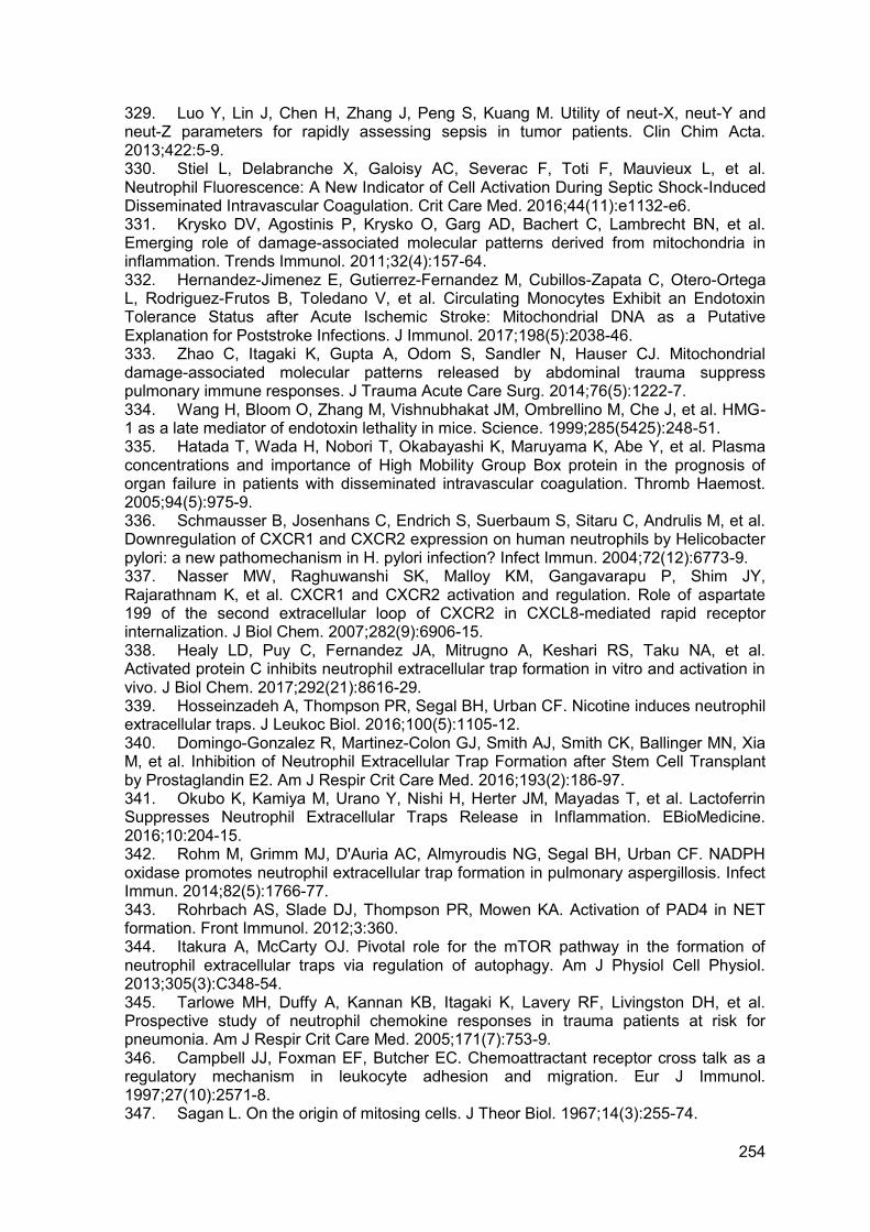
254
329. Luo Y, Lin J, Chen H, Zhang J, Peng S, Kuang M. Utility of neut-X, neut-Y and neut-Z parameters for rapidly assessing sepsis in tumor patients. Clin Chim Acta. 2013;422:5-9. 330. Stiel L, Delabranche X, Galoisy AC, Severac F, Toti F, Mauvieux L, et al. Neutrophil Fluorescence: A New Indicator of Cell Activation During Septic Shock-Induced Disseminated Intravascular Coagulation. Crit Care Med. 2016;44(11):e1132-e6. 331. Krysko DV, Agostinis P, Krysko O, Garg AD, Bachert C, Lambrecht BN, et al. Emerging role of damage-associated molecular patterns derived from mitochondria in inflammation. Trends Immunol. 2011;32(4):157-64. 332. Hernandez-Jimenez E, Gutierrez-Fernandez M, Cubillos-Zapata C, Otero-Ortega L, Rodriguez-Frutos B, Toledano V, et al. Circulating Monocytes Exhibit an Endotoxin Tolerance Status after Acute Ischemic Stroke: Mitochondrial DNA as a Putative Explanation for Poststroke Infections. J Immunol. 2017;198(5):2038-46. 333. Zhao C, Itagaki K, Gupta A, Odom S, Sandler N, Hauser CJ. Mitochondrial damage-associated molecular patterns released by abdominal trauma suppress pulmonary immune responses. J Trauma Acute Care Surg. 2014;76(5):1222-7. 334. Wang H, Bloom O, Zhang M, Vishnubhakat JM, Ombrellino M, Che J, et al. HMG-1 as a late mediator of endotoxin lethality in mice. Science. 1999;285(5425):248-51. 335. Hatada T, Wada H, Nobori T, Okabayashi K, Maruyama K, Abe Y, et al. Plasma concentrations and importance of High Mobility Group Box protein in the prognosis of organ failure in patients with disseminated intravascular coagulation. Thromb Haemost. 2005;94(5):975-9. 336. Schmausser B, Josenhans C, Endrich S, Suerbaum S, Sitaru C, Andrulis M, et al. Downregulation of CXCR1 and CXCR2 expression on human neutrophils by Helicobacter pylori: a new pathomechanism in H. pylori infection? Infect Immun. 2004;72(12):6773-9. 337. Nasser MW, Raghuwanshi SK, Malloy KM, Gangavarapu P, Shim JY, Rajarathnam K, et al. CXCR1 and CXCR2 activation and regulation. Role of aspartate 199 of the second extracellular loop of CXCR2 in CXCL8-mediated rapid receptor internalization. J Biol Chem. 2007;282(9):6906-15. 338. Healy LD, Puy C, Fernandez JA, Mitrugno A, Keshari RS, Taku NA, et al. Activated protein C inhibits neutrophil extracellular trap formation in vitro and activation in vivo. J Biol Chem. 2017;292(21):8616-29. 339. Hosseinzadeh A, Thompson PR, Segal BH, Urban CF. Nicotine induces neutrophil extracellular traps. J Leukoc Biol. 2016;100(5):1105-12. 340. Domingo-Gonzalez R, Martinez-Colon GJ, Smith AJ, Smith CK, Ballinger MN, Xia M, et al. Inhibition of Neutrophil Extracellular Trap Formation after Stem Cell Transplant by Prostaglandin E2. Am J Respir Crit Care Med. 2016;193(2):186-97. 341. Okubo K, Kamiya M, Urano Y, Nishi H, Herter JM, Mayadas T, et al. Lactoferrin Suppresses Neutrophil Extracellular Traps Release in Inflammation. EBioMedicine. 2016;10:204-15. 342. Rohm M, Grimm MJ, D'Auria AC, Almyroudis NG, Segal BH, Urban CF. NADPH oxidase promotes neutrophil extracellular trap formation in pulmonary aspergillosis. Infect Immun. 2014;82(5):1766-77. 343. Rohrbach AS, Slade DJ, Thompson PR, Mowen KA. Activation of PAD4 in NET formation. Front Immunol. 2012;3:360. 344. Itakura A, McCarty OJ. Pivotal role for the mTOR pathway in the formation of neutrophil extracellular traps via regulation of autophagy. Am J Physiol Cell Physiol. 2013;305(3):C348-54. 345. Tarlowe MH, Duffy A, Kannan KB, Itagaki K, Lavery RF, Livingston DH, et al. Prospective study of neutrophil chemokine responses in trauma patients at risk for pneumonia. Am J Respir Crit Care Med. 2005;171(7):753-9. 346. Campbell JJ, Foxman EF, Butcher EC. Chemoattractant receptor cross talk as a regulatory mechanism in leukocyte adhesion and migration. Eur J Immunol. 1997;27(10):2571-8. 347. Sagan L. On the origin of mitosing cells. J Theor Biol. 1967;14(3):255-74.
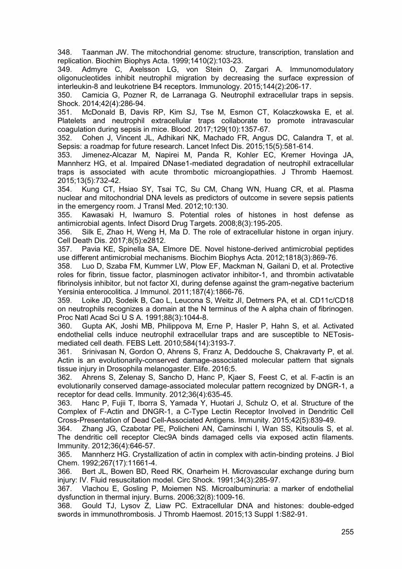
255
348. Taanman JW. The mitochondrial genome: structure, transcription, translation and replication. Biochim Biophys Acta. 1999;1410(2):103-23. 349. Admyre C, Axelsson LG, von Stein O, Zargari A. Immunomodulatory oligonucleotides inhibit neutrophil migration by decreasing the surface expression of interleukin-8 and leukotriene B4 receptors. Immunology. 2015;144(2):206-17. 350. Camicia G, Pozner R, de Larranaga G. Neutrophil extracellular traps in sepsis. Shock. 2014;42(4):286-94. 351. McDonald B, Davis RP, Kim SJ, Tse M, Esmon CT, Kolaczkowska E, et al. Platelets and neutrophil extracellular traps collaborate to promote intravascular coagulation during sepsis in mice. Blood. 2017;129(10):1357-67. 352. Cohen J, Vincent JL, Adhikari NK, Machado FR, Angus DC, Calandra T, et al. Sepsis: a roadmap for future research. Lancet Infect Dis. 2015;15(5):581-614. 353. Jimenez-Alcazar M, Napirei M, Panda R, Kohler EC, Kremer Hovinga JA, Mannherz HG, et al. Impaired DNase1-mediated degradation of neutrophil extracellular traps is associated with acute thrombotic microangiopathies. J Thromb Haemost. 2015;13(5):732-42. 354. Kung CT, Hsiao SY, Tsai TC, Su CM, Chang WN, Huang CR, et al. Plasma nuclear and mitochondrial DNA levels as predictors of outcome in severe sepsis patients in the emergency room. J Transl Med. 2012;10:130. 355. Kawasaki H, Iwamuro S. Potential roles of histones in host defense as antimicrobial agents. Infect Disord Drug Targets. 2008;8(3):195-205. 356. Silk E, Zhao H, Weng H, Ma D. The role of extracellular histone in organ injury. Cell Death Dis. 2017;8(5):e2812. 357. Pavia KE, Spinella SA, Elmore DE. Novel histone-derived antimicrobial peptides use different antimicrobial mechanisms. Biochim Biophys Acta. 2012;1818(3):869-76. 358. Luo D, Szaba FM, Kummer LW, Plow EF, Mackman N, Gailani D, et al. Protective roles for fibrin, tissue factor, plasminogen activator inhibitor-1, and thrombin activatable fibrinolysis inhibitor, but not factor XI, during defense against the gram-negative bacterium Yersinia enterocolitica. J Immunol. 2011;187(4):1866-76. 359. Loike JD, Sodeik B, Cao L, Leucona S, Weitz JI, Detmers PA, et al. CD11c/CD18 on neutrophils recognizes a domain at the N terminus of the A alpha chain of fibrinogen. Proc Natl Acad Sci U S A. 1991;88(3):1044-8. 360. Gupta AK, Joshi MB, Philippova M, Erne P, Hasler P, Hahn S, et al. Activated endothelial cells induce neutrophil extracellular traps and are susceptible to NETosis-mediated cell death. FEBS Lett. 2010;584(14):3193-7. 361. Srinivasan N, Gordon O, Ahrens S, Franz A, Deddouche S, Chakravarty P, et al. Actin is an evolutionarily-conserved damage-associated molecular pattern that signals tissue injury in Drosophila melanogaster. Elife. 2016;5. 362. Ahrens S, Zelenay S, Sancho D, Hanc P, Kjaer S, Feest C, et al. F-actin is an evolutionarily conserved damage-associated molecular pattern recognized by DNGR-1, a receptor for dead cells. Immunity. 2012;36(4):635-45. 363. Hanc P, Fujii T, Iborra S, Yamada Y, Huotari J, Schulz O, et al. Structure of the Complex of F-Actin and DNGR-1, a C-Type Lectin Receptor Involved in Dendritic Cell Cross-Presentation of Dead Cell-Associated Antigens. Immunity. 2015;42(5):839-49. 364. Zhang JG, Czabotar PE, Policheni AN, Caminschi I, Wan SS, Kitsoulis S, et al. The dendritic cell receptor Clec9A binds damaged cells via exposed actin filaments. Immunity. 2012;36(4):646-57. 365. Mannherz HG. Crystallization of actin in complex with actin-binding proteins. J Biol Chem. 1992;267(17):11661-4. 366. Bert JL, Bowen BD, Reed RK, Onarheim H. Microvascular exchange during burn injury: IV. Fluid resuscitation model. Circ Shock. 1991;34(3):285-97. 367. Vlachou E, Gosling P, Moiemen NS. Microalbuminuria: a marker of endothelial dysfunction in thermal injury. Burns. 2006;32(8):1009-16. 368. Gould TJ, Lysov Z, Liaw PC. Extracellular DNA and histones: double-edged swords in immunothrombosis. J Thromb Haemost. 2015;13 Suppl 1:S82-91.
256
369. Cohen TS, Bucki R, Byfield FJ, Ciccarelli NJ, Rosenberg B, DiNubile MJ, et al. Therapeutic potential of plasma gelsolin administration in a rat model of sepsis. Cytokine. 2011;54(3):235-8. 370. Mathews ZR, Koyfman A. Blast Injuries. J Emerg Med. 2015;49(4):573-87. 371. Mitra B, Mori A, Cameron PA, Fitzgerald M, Paul E, Street A. Fresh frozen plasma (FFP) use during massive blood transfusion in trauma resuscitation. Injury. 2010;41(1):35-9. 372. Chhabra D, Nosworthy NJ, dos Remedios CG. The N-terminal fragment of gelsolin inhibits the interaction of DNase I with isolated actin, but not with the cofilin-actin complex. Proteomics. 2005;5(12):3131-6. 373. Zhang LM, Li R, Zhao XC, Zhang Q, Luo XL. Increased Transfusion of Fresh Frozen Plasma is Associated with Mortality or Worse Functional Outcomes After Severe Traumatic Brain Injury: A Retrospective Study. World Neurosurg. 2017;104:381-9. 374. Mesar T, Larentzakis A, Dzik W, Chang Y, Velmahos G, Yeh DD. Association Between Ratio of Fresh Frozen Plasma to Red Blood Cells During Massive Transfusion and Survival Among Patients Without Traumatic Injury. JAMA Surg. 2017;152(6):574-80. 375. Hagiwara A, Kushimoto S, Kato H, Sasaki J, Ogura H, Matsuoka T, et al. Can Early Aggressive Administration of Fresh Frozen Plasma Improve Outcomes in Patients with Severe Blunt Trauma?--A Report by the Japanese Association for the Surgery of Trauma. Shock. 2016;45(5):495-501. 376. Chen Q, Ye L, Jin Y, Zhang N, Lou T, Qiu Z, et al. Circulating nucleosomes as a predictor of sepsis and organ dysfunction in critically ill patients. Int J Infect Dis. 2012;16(7):e558-64. 377. Marsman G, Zeerleder S, Luken BM. Extracellular histones, cell-free DNA, or nucleosomes: differences in immunostimulation. Cell Death Dis. 2016;7(12):e2518. 378. Wildhagen KC, Garcia de Frutos P, Reutelingsperger CP, Schrijver R, Areste C, Ortega-Gomez A, et al. Nonanticoagulant heparin prevents histone-mediated cytotoxicity in vitro and improves survival in sepsis. Blood. 2014;123(7):1098-101. 379. Demidenko O, Tsvetkova S. Action of heparin on protein fractions of isolated nuclei and on their DNA content. Histochemistry. 1978;57(3):265-71. 380. Ronnefarth VM, Erbacher AI, Lamkemeyer T, Madlung J, Nordheim A, Rammensee HG, et al. TLR2/TLR4-independent neutrophil activation and recruitment upon endocytosis of nucleosomes reveals a new pathway of innate immunity in systemic lupus erythematosus. J Immunol. 2006;177(11):7740-9. 381. Abrams ST, Zhang N, Dart C, Wang SS, Thachil J, Guan Y, et al. Human CRP defends against the toxicity of circulating histones. J Immunol. 2013;191(5):2495-502. 382. Du Clos TW, Zlock LT, Rubin RL. Analysis of the binding of C-reactive protein to histones and chromatin. J Immunol. 1988;141(12):4266-70. 383. Du Clos TW, Marnell L, Zlock LR, Burlingame RW. Analysis of the binding of C-reactive protein to chromatin subunits. J Immunol. 1991;146(4):1220-5. 384. Lehmann-Werman R, Neiman D, Zemmour H, Moss J, Magenheim J, Vaknin-Dembinsky A, et al. Identification of tissue-specific cell death using methylation patterns of circulating DNA. Proc Natl Acad Sci U S A. 2016;113(13):E1826-34. 385. Bergman Y, Cedar H. DNA methylation dynamics in health and disease. Nat Struct Mol Biol. 2013;20(3):274-81. 386. Rodriguez AM, Rodin D, Nomura H, Morton CC, Weremowicz S, Schneider MC. Identification, localization, and expression of two novel human genes similar to deoxyribonuclease I. Genomics. 1997;42(3):507-13. 387. Napirei M, Wulf S, Eulitz D, Mannherz HG, Kloeckl T. Comparative characterization of rat deoxyribonuclease 1 (Dnase1) and murine deoxyribonuclease 1-like 3 (Dnase1l3). Biochem J. 2005;389(Pt 2):355-64. 388. Koyama R, Arai T, Kijima M, Sato S, Miura S, Yuasa M, et al. DNase gamma, DNase I and caspase-activated DNase cooperate to degrade dead cells. Genes Cells. 2016;21(11):1150-63.
257
389. Xu XY, Yang WF, Zhang SG, Zhao Q, Linag LJ, Wang X, et al. [Correlation of DNase I in serum and synovial fluid with inflammatory activity in patients with rheumatoid arthritis]. Nan Fang Yi Ke Da Xue Xue Bao. 2016;36(9):1204-8. 390. Shiokawa D, Ohyama H, Yamada T, Takahashi K, Tanuma S. Identification of an endonuclease responsible for apoptosis in rat thymocytes. Eur J Biochem. 1994;226(1):23-30. 391. Shiokawa D, Kobayashi T, Tanuma S. Involvement of DNase gamma in apoptosis associated with myogenic differentiation of C2C12 cells. J Biol Chem. 2002;277(34):31031-7. 392. Nishimura K, Tanuma S. Presence of DNase gamma-like endonuclease in nuclei of neuronal differentiated PC12 cells. Apoptosis. 1998;3(2):97-103. 393. Baron WF, Pan CQ, Spencer SA, Ryan AM, Lazarus RA, Baker KP. Cloning and characterization of an actin-resistant DNase I-like endonuclease secreted by macrophages. Gene. 1998;215(2):291-301. 394. Wilber A, Lu M, Schneider MC. Deoxyribonuclease I-like III is an inducible macrophage barrier to liposomal transfection. Mol Ther. 2002;6(1):35-42. 395. Napirei M, Ludwig S, Mezrhab J, Klockl T, Mannherz HG. Murine serum nucleases--contrasting effects of plasmin and heparin on the activities of DNase1 and DNase1-like 3 (DNase1l3). FEBS J. 2009;276(4):1059-73. 396. Carbonella A, Mancano G, Gremese E, Alkuraya FS, Patel N, Gurrieri F, et al. An autosomal recessive DNASE1L3-related autoimmune disease with unusual clinical presentation mimicking systemic lupus erythematosus. Lupus. 2017;26(7):768-72. 397. Al-Mayouf SM, Sunker A, Abdwani R, Abrawi SA, Almurshedi F, Alhashmi N, et al. Loss-of-function variant in DNASE1L3 causes a familial form of systemic lupus erythematosus. Nat Genet. 2011;43(12):1186-8. 398. Zhao Q, Yang C, Wang J, Li Y, Yang P. Serum level of DNase1l3 in patients with dermatomyositis/polymyositis, systemic lupus erythematosus and rheumatoid arthritis, and its association with disease activity. Clin Exp Med. 2016. 399. Sisirak V, Sally B, D'Agati V, Martinez-Ortiz W, Ozcakar ZB, David J, et al. Digestion of Chromatin in Apoptotic Cell Microparticles Prevents Autoimmunity. Cell. 2016;166(1):88-101. 400. Russell RT, Christiaans SC, Nice T, Banks M, Mortellaro V, Morgan C, et al. Histone-Complexed DNA Fragments Levels are Associated with Coagulopathy, Endothelial Cell Damage, and Increased Mortality after Severe Pediatric Trauma. Shock. 2017. 401. Martinod K, Demers M, Fuchs TA, Wong SL, Brill A, Gallant M, et al. Neutrophil histone modification by peptidylarginine deiminase 4 is critical for deep vein thrombosis in mice. Proc Natl Acad Sci U S A. 2013;110(21):8674-9. 402. Mai SH, Khan M, Dwivedi DJ, Ross CA, Zhou J, Gould TJ, et al. Delayed but not Early Treatment with DNase Reduces Organ Damage and Improves Outcome in a Murine Model of Sepsis. Shock. 2015;44(2):166-72. 403. Jorgensen CS, Christiansen M, Laursen I, Krogsoe LB, Hojrup P, Blou L, et al. Large-scale purification and characterization of non-glycosylated Gc globulin (vitamin D-binding protein) from plasma fraction IV. Biotechnol Appl Biochem. 2006;44(Pt 1):35-44. 404. Lee PS, Waxman AB, Cotich KL, Chung SW, Perrella MA, Stossel TP. Plasma gelsolin is a marker and therapeutic agent in animal sepsis. Crit Care Med. 2007;35(3):849-55. 405. Lee PS, Drager LR, Stossel TP, Moore FD, Rogers SO. Relationship of plasma gelsolin levels to outcomes in critically ill surgical patients. Ann Surg. 2006;243(3):399-403. 406. Christofidou-Solomidou M, Scherpereel A, Solomides CC, Christie JD, Stossel TP, Goelz S, et al. Recombinant plasma gelsolin diminishes the acute inflammatory response to hyperoxia in mice. J Investig Med. 2002;50(1):54-60.
258
407. Goldschmidt-Clermont PJ, Van Baelen H, Bouillon R, Shook TE, Williams MH, Nel AE, et al. Role of group-specific component (vitamin D binding protein) in clearance of actin from the circulation in the rabbit. J Clin Invest. 1988;81(5):1519-27. 408. Lind SE, Smith DB, Janmey PA, Stossel TP. Role of plasma gelsolin and the vitamin D-binding protein in clearing actin from the circulation. J Clin Invest. 1986;78(3):736-42. 409. Livingston DH, Anjaria D, Wu J, Hauser CJ, Chang V, Deitch EA, et al. Bone marrow failure following severe injury in humans. Ann Surg. 2003;238(5):748-53. 410. Posluszny JA, Jr., Muthumalaiappan K, Kini AR, Szilagyi A, He LK, Li Y, et al. Burn injury dampens erythroid cell production through reprioritizing bone marrow hematopoietic response. J Trauma. 2011;71(5):1288-96. 411. Miranda MB, Xu H, Torchia JA, Johnson DE. Cytokine-induced myeloid differentiation is dependent on activation of the MEK/ERK pathway. Leuk Res. 2005;29(11):1293-306. 412. Chakraborty A, Tweardy DJ. Stat3 and G-CSF-induced myeloid differentiation. Leuk Lymphoma. 1998;30(5-6):433-42. 413. Ertel W, Keel M, Buergi U, Hartung T, Imhof HG, Trentz O. Granulocyte colony-stimulating factor inhibits neutrophil apoptosis at the local site after severe head and thoracic injury. J Trauma. 1999;46(5):784-92; discussion 92-3. 414. Sartorelli KH, Silver GM, Gamelli RL. The effect of granulocyte colony-stimulating factor (G-CSF) upon burn-induced defective neutrophil chemotaxis. J Trauma. 1991;31(4):523-9; discussion 9-30. 415. Nathan C. Points of control in inflammation. Nature. 2002;420(6917):846-52. 416. Serhan CN, Gotlinger K, Hong S, Arita M. Resolvins, docosatrienes, and neuroprotectins, novel omega-3-derived mediators, and their aspirin-triggered endogenous epimers: an overview of their protective roles in catabasis. Prostaglandins Other Lipid Mediat. 2004;73(3-4):155-72. 417. Serhan CN, Clish CB, Brannon J, Colgan SP, Chiang N, Gronert K. Novel functional sets of lipid-derived mediators with antiinflammatory actions generated from omega-3 fatty acids via cyclooxygenase 2-nonsteroidal antiinflammatory drugs and transcellular processing. J Exp Med. 2000;192(8):1197-204. 418. Levy BD, Clish CB, Schmidt B, Gronert K, Serhan CN. Lipid mediator class switching during acute inflammation: signals in resolution. Nat Immunol. 2001;2(7):612-9. 419. Serhan CN, Hong S, Gronert K, Colgan SP, Devchand PR, Mirick G, et al. Resolvins: a family of bioactive products of omega-3 fatty acid transformation circuits initiated by aspirin treatment that counter proinflammation signals. J Exp Med. 2002;196(8):1025-37. 420. Spite M, Norling LV, Summers L, Yang R, Cooper D, Petasis NA, et al. Resolvin D2 is a potent regulator of leukocytes and controls microbial sepsis. Nature. 2009;461(7268):1287-91. 421. Kurihara T, Jones CN, Yu YM, Fischman AJ, Watada S, Tompkins RG, et al. Resolvin D2 restores neutrophil directionality and improves survival after burns. FASEB J. 2013;27(6):2270-81. 422. Krishnamoorthy S, Recchiuti A, Chiang N, Fredman G, Serhan CN. Resolvin D1 receptor stereoselectivity and regulation of inflammation and proresolving microRNAs. Am J Pathol. 2012;180(5):2018-27. 423. Lee J, Jackman JG, Kwun J, Manook M, Moreno A, Elster EA, et al. Nucleic acid scavenging microfiber mesh inhibits trauma-induced inflammation and thrombosis. Biomaterials. 2017;120:94-102. 424. Kumagai T, Takeyama N, Yabuki T, Harada M, Miki Y, Kanou H, et al. Apheresis of activated leukocytes with an immobilized polymyxin B filter in patients with septic shock. Shock. 2010;34(5):461-6. 425. Ronco C, Klein DJ. Polymyxin B hemoperfusion: a mechanistic perspective. Crit Care. 2014;18(3):309.