Embed Size (px)
DESCRIPTION
panoramic dental radiographs intro
Citation preview
Dr. Aruna Ramesh
Panoramic radiography is based on body-section radiography
A special radiographic technique that blurs out the shadows of superimposed structures
Object of interest less blurred Does not improve the sharpness
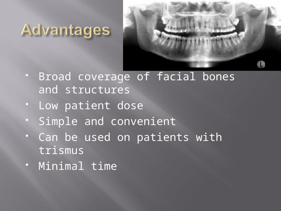
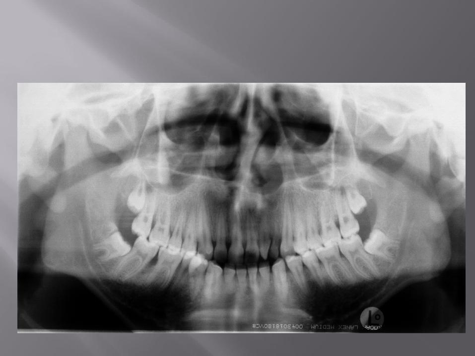
Broad coverage of facial bones and structures
Low patient dose Simple and convenient Can be used on patients with trismus Minimal time
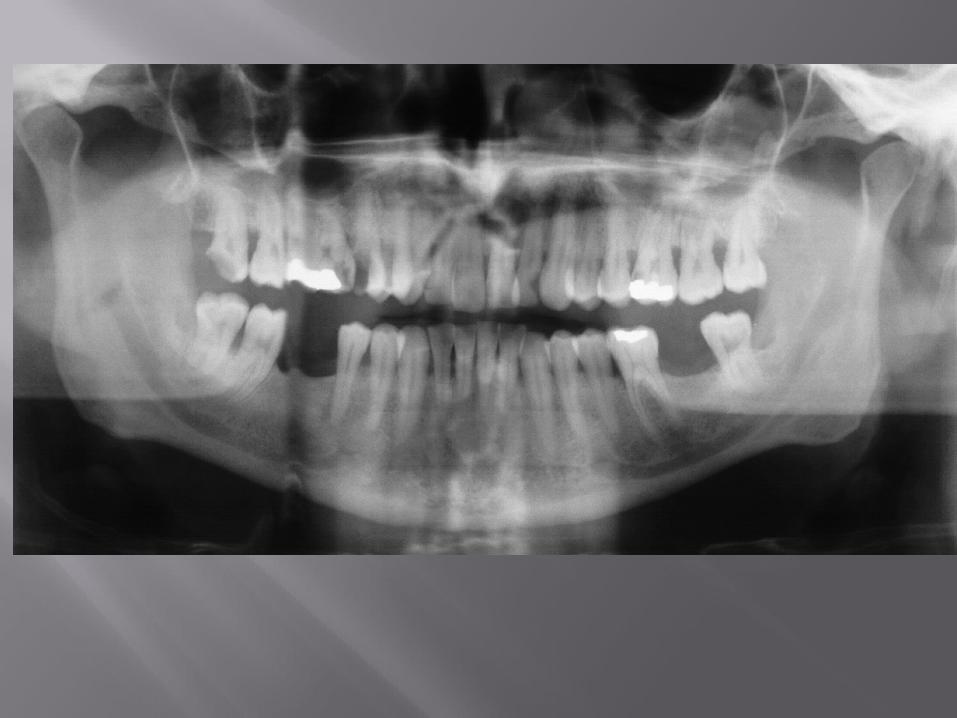
Critical patient positioning Inherent magnification Poor resolution Incorrect interpretation of ghost image Overlapped premolar region
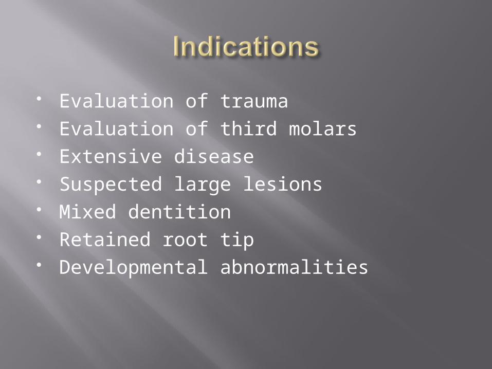
Evaluation of trauma Evaluation of third molars Extensive disease Suspected large lesions Mixed dentition Retained root tip Developmental abnormalities
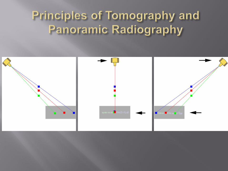

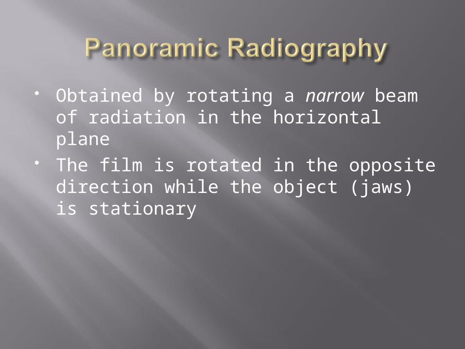
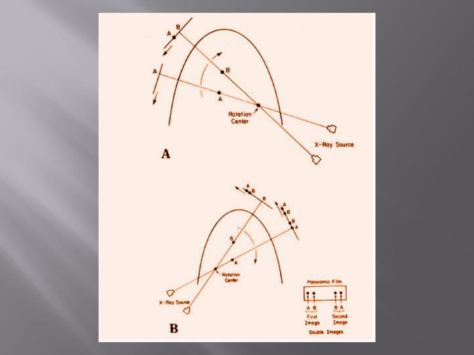
Obtained by rotating a narrow beam of radiation in the horizontal plane
The film is rotated in the opposite direction while the object (jaws) is stationary
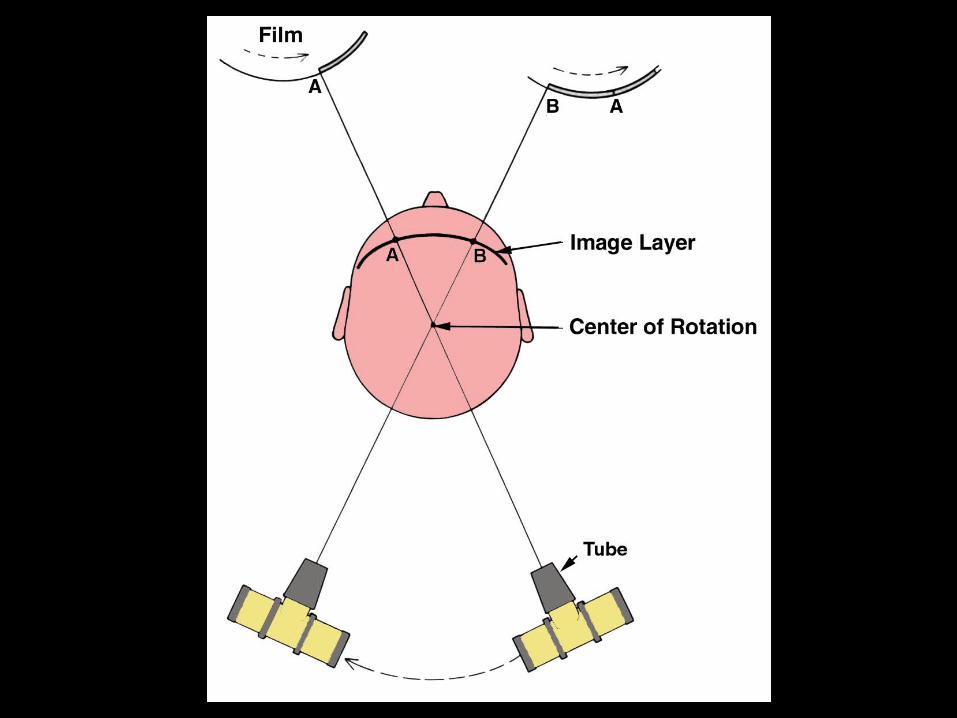
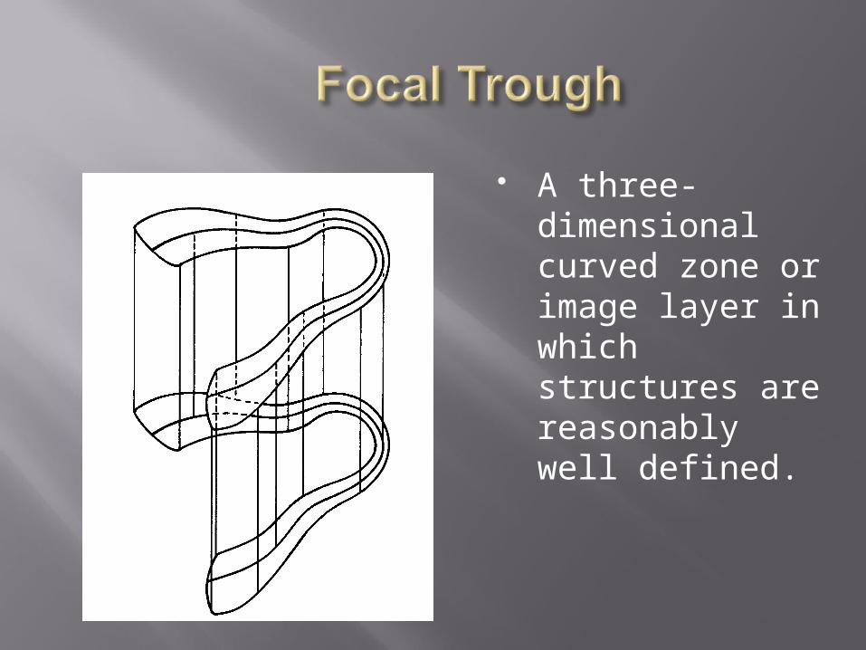
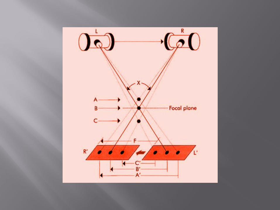
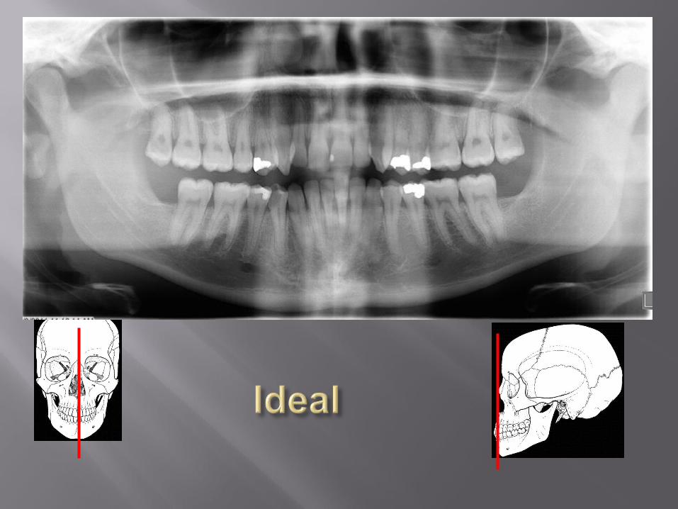
A three-dimensional curved zone or image layer in which structures are reasonably well defined.
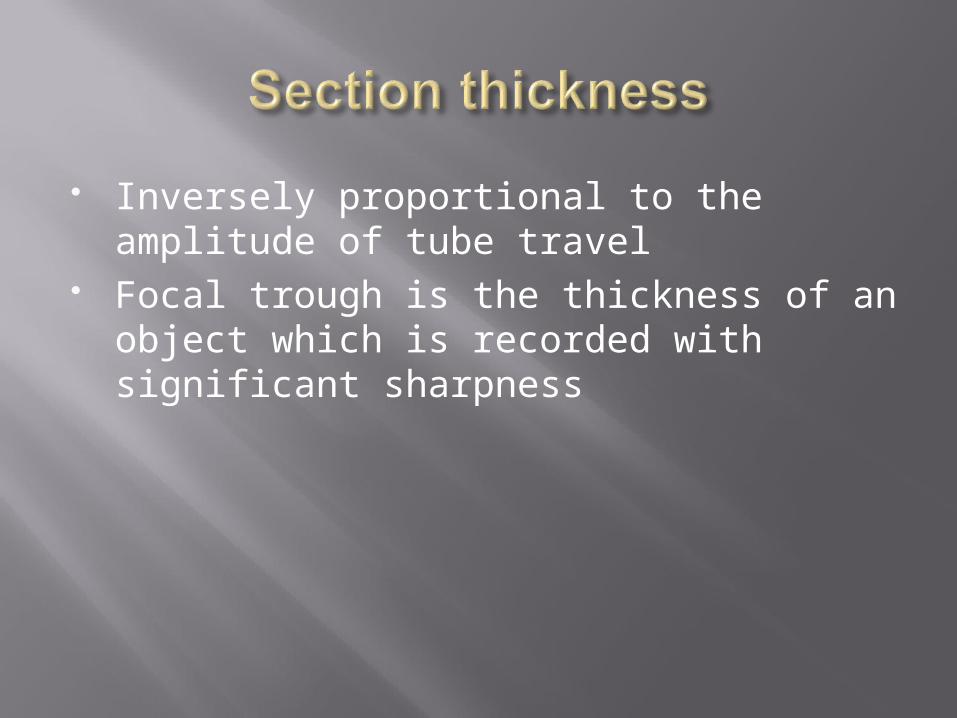
Inversely proportional to the amplitude of tube travel
Focal trough is the thickness of an object which is recorded with significant sharpness
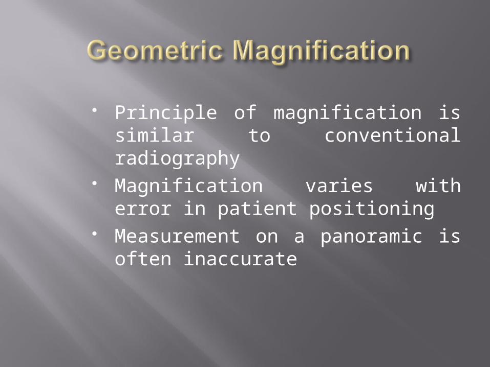
Principle of magnification is similar to conventional radiography
Magnification varies with error in patient positioning
Measurement on a panoramic is often inaccurate
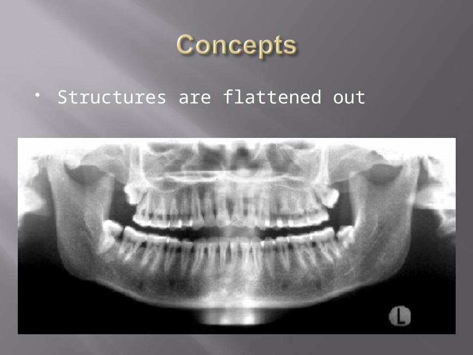
Structures are flattened out
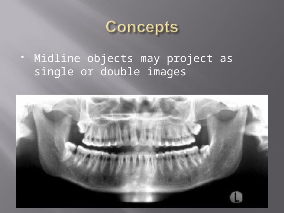
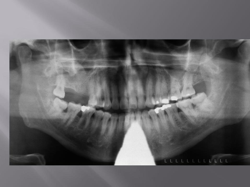
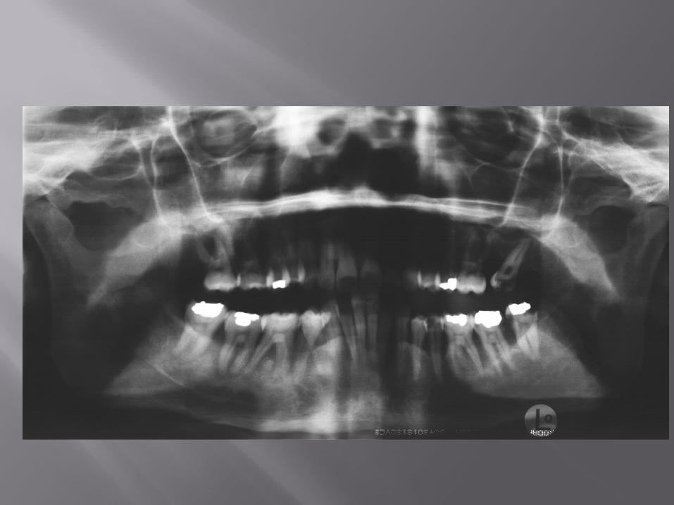
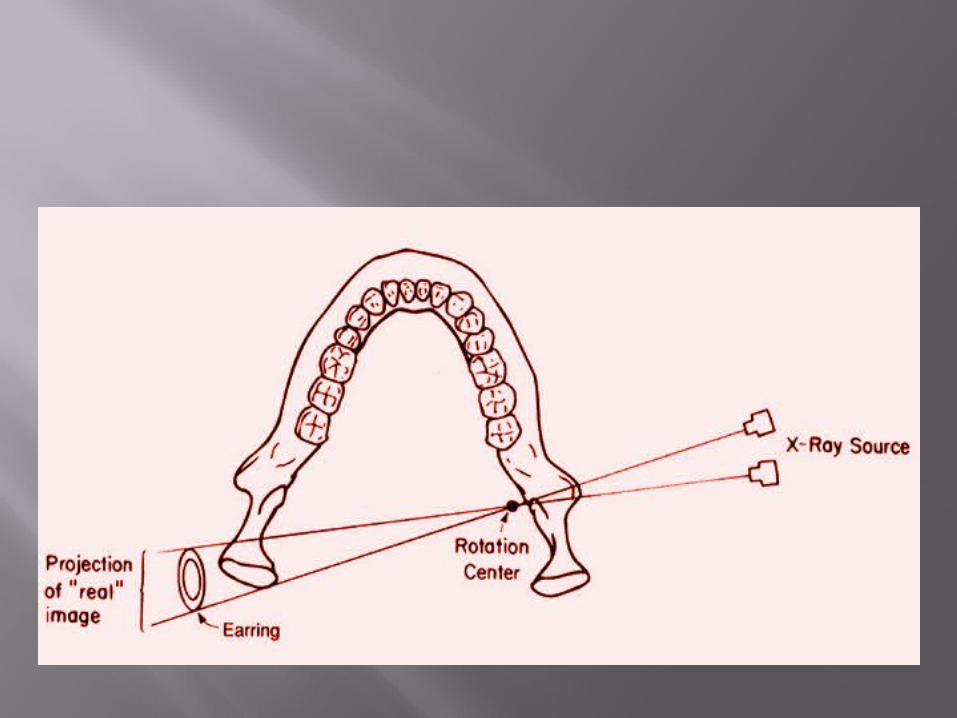
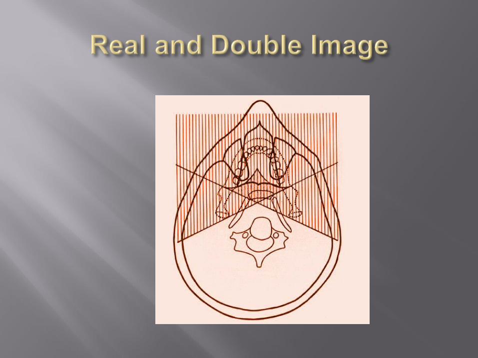
Midline objects may project as single or double images
Objects are intercepted twice by beam One image is the mirror image of the
other Occur with midline objects
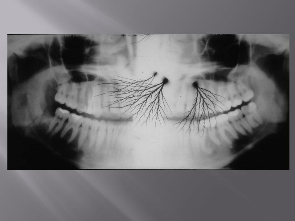
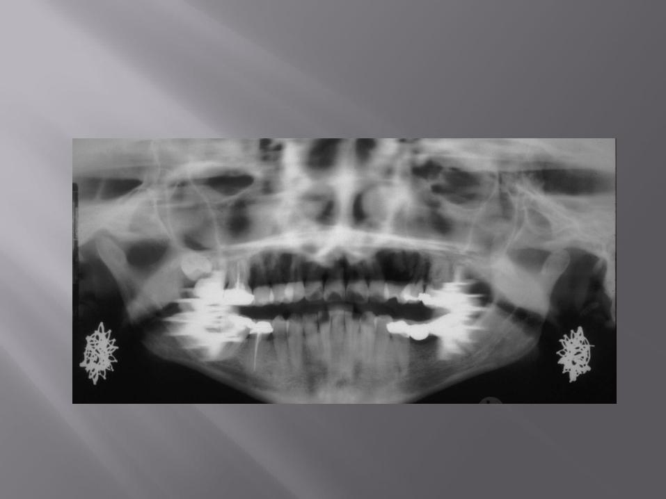
A real image is formed when the object is located between the rotation center and film
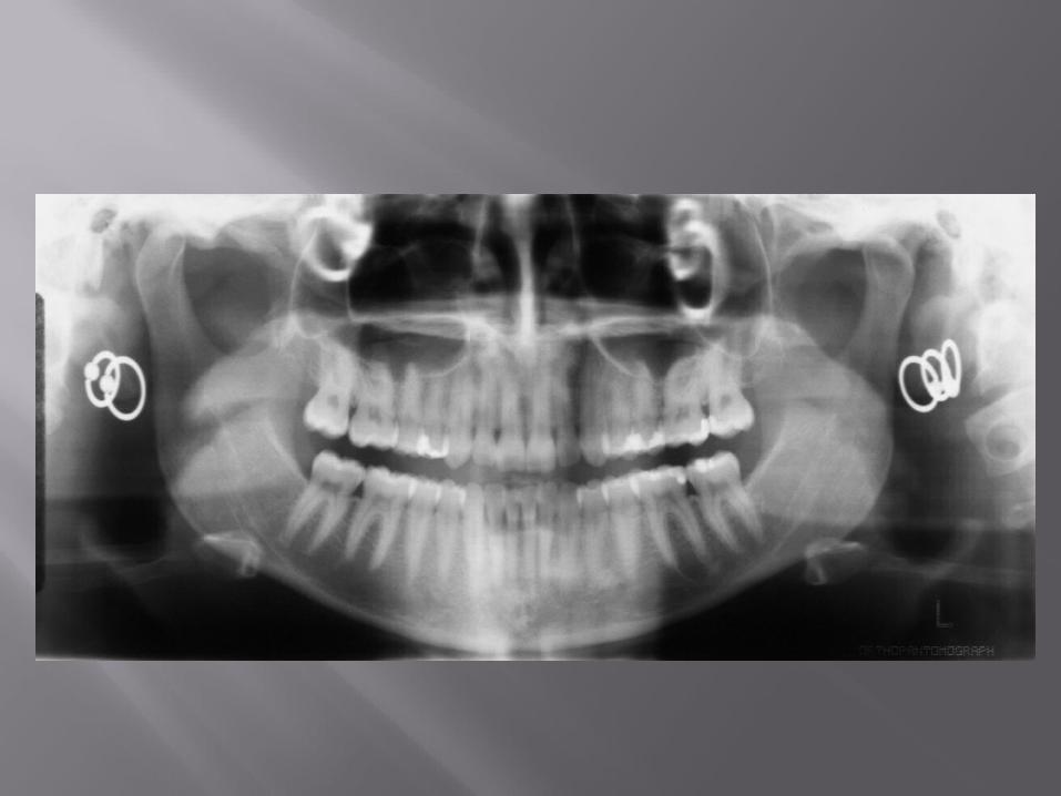
Arise from structures located on the opposite side of the center of rotation away from the image layer
Produced when the object is located between the x-ray source and the rotational center.
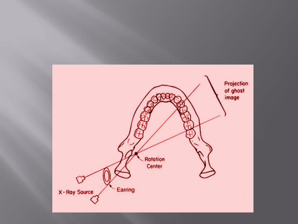
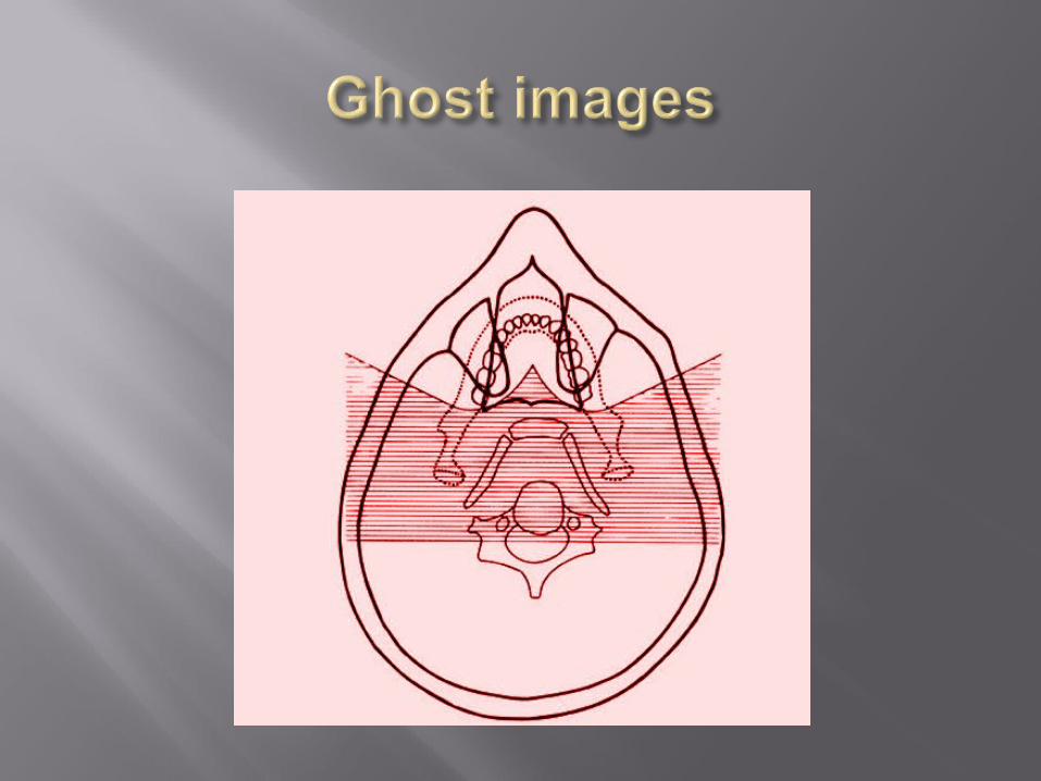
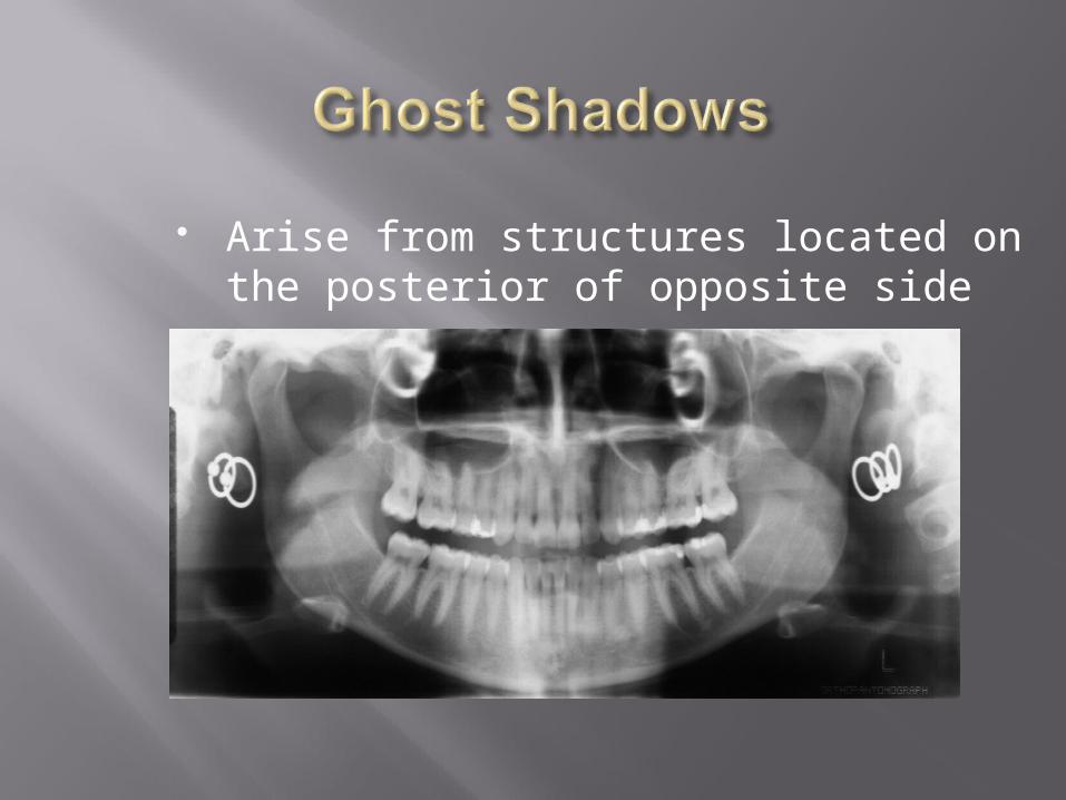
Arise from structures located on the posterior of opposite side
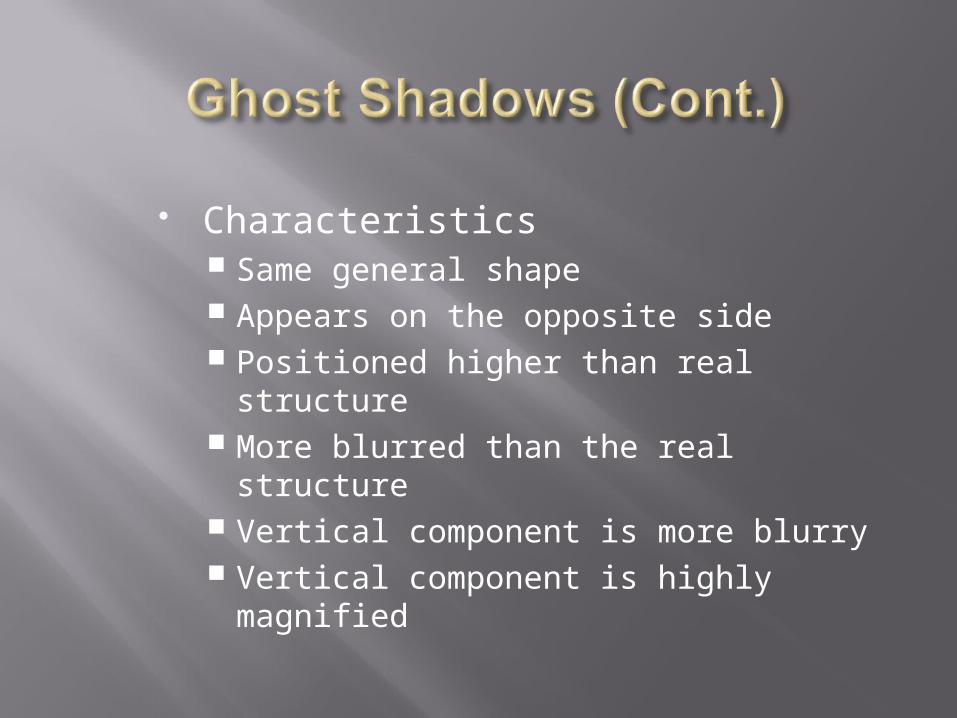
Characteristics Same general shape Appears on the opposite side Positioned higher than real structure More blurred than the real structure Vertical component is more blurry Vertical component is highly magnified
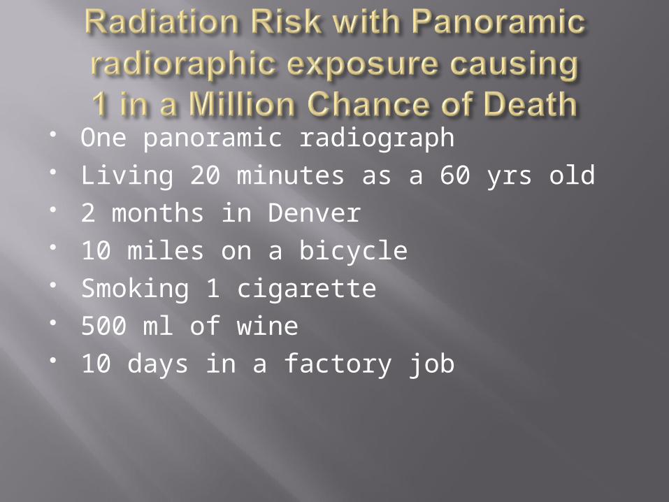
One panoramic radiograph Living 20 minutes as a 60 yrs old 2 months in Denver 10 miles on a bicycle Smoking 1 cigarette 500 ml of wine 10 days in a factory job
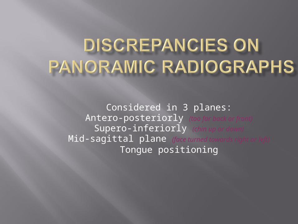
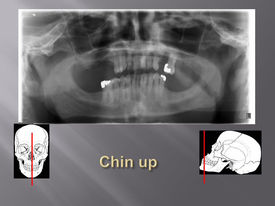
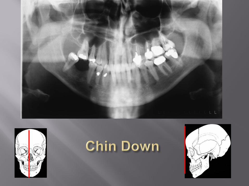
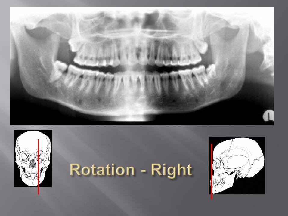
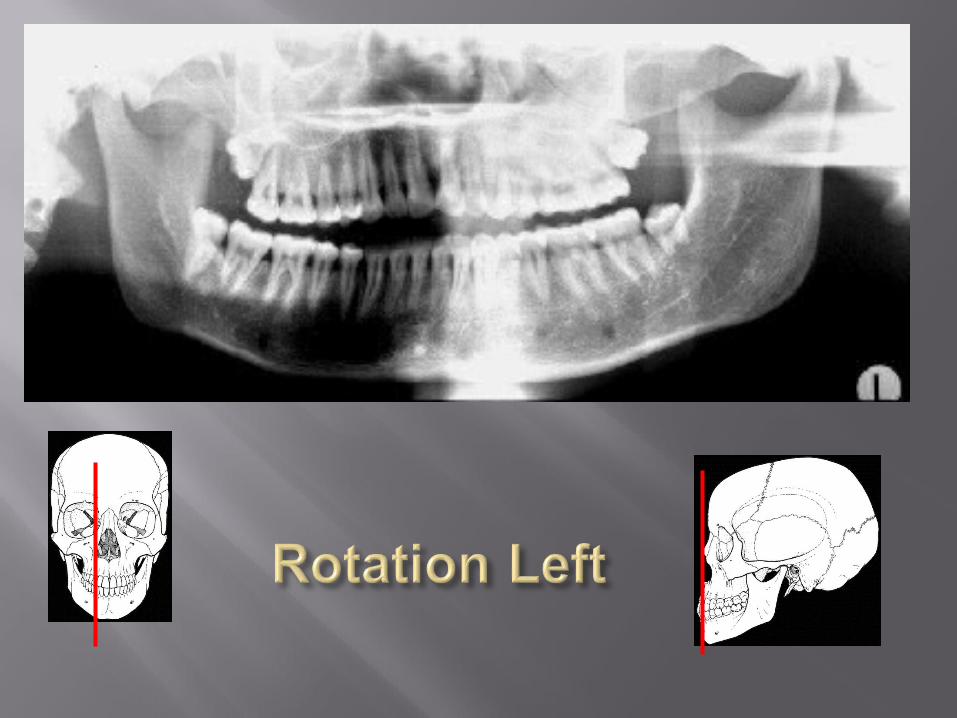
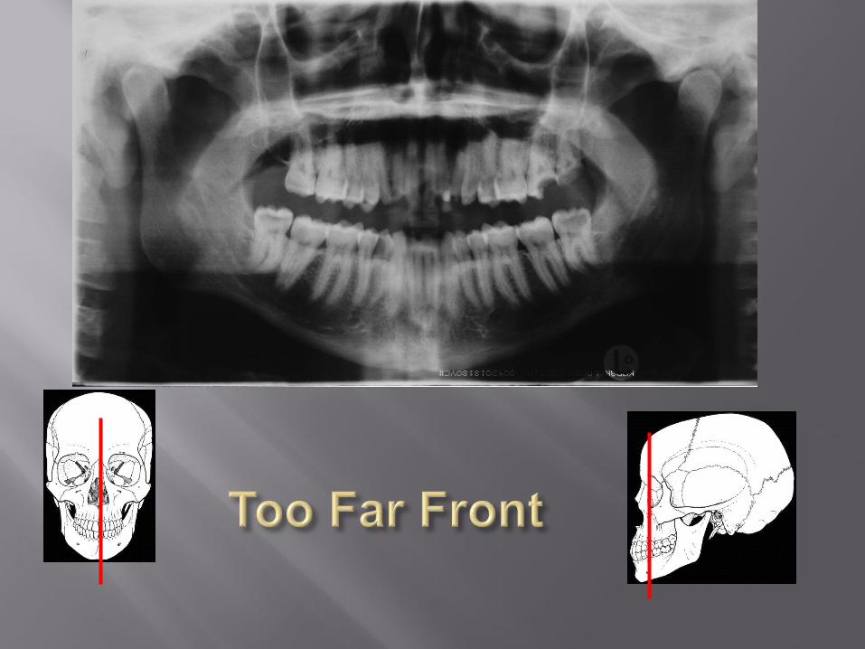
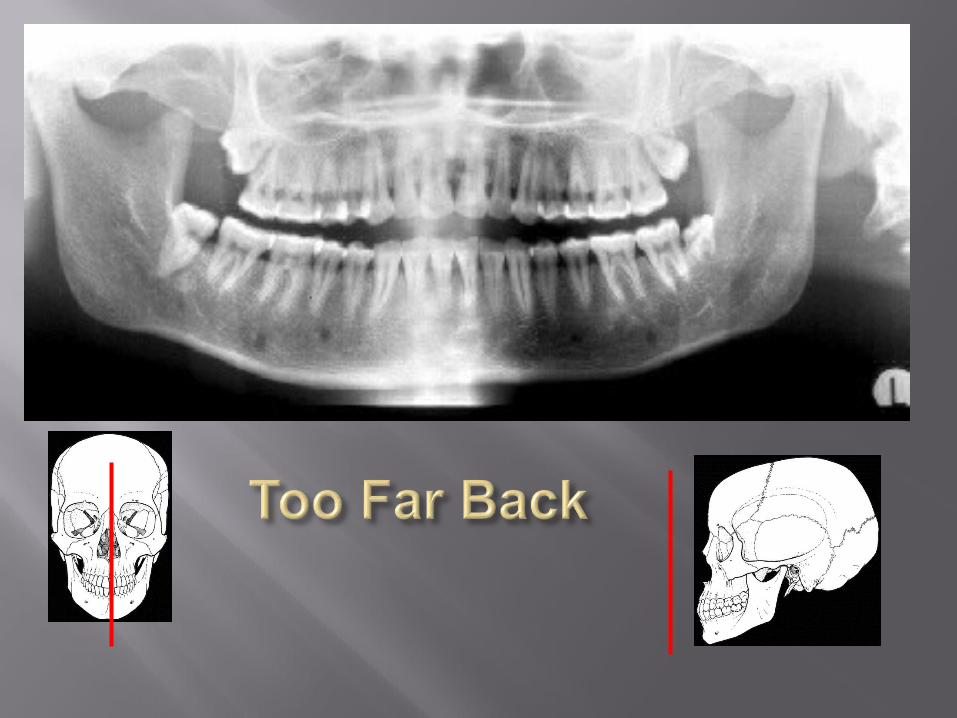
Considered in 3 planes:Antero-posteriorly (too far back or front)
Supero-inferiorly (chin up or down)
Mid-sagittal plane (face turned towards right or left)
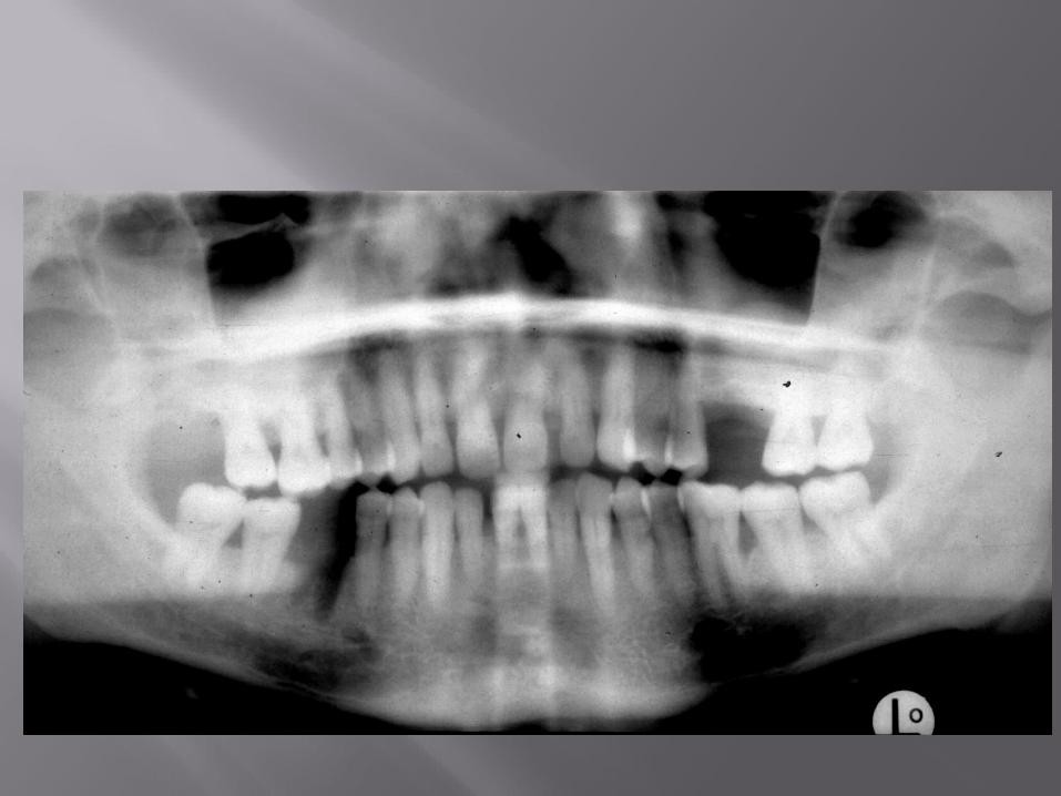
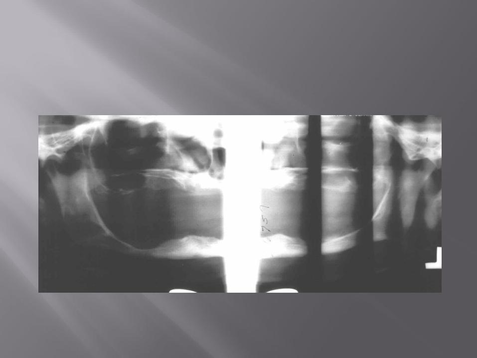
Tongue positioning
HELLO
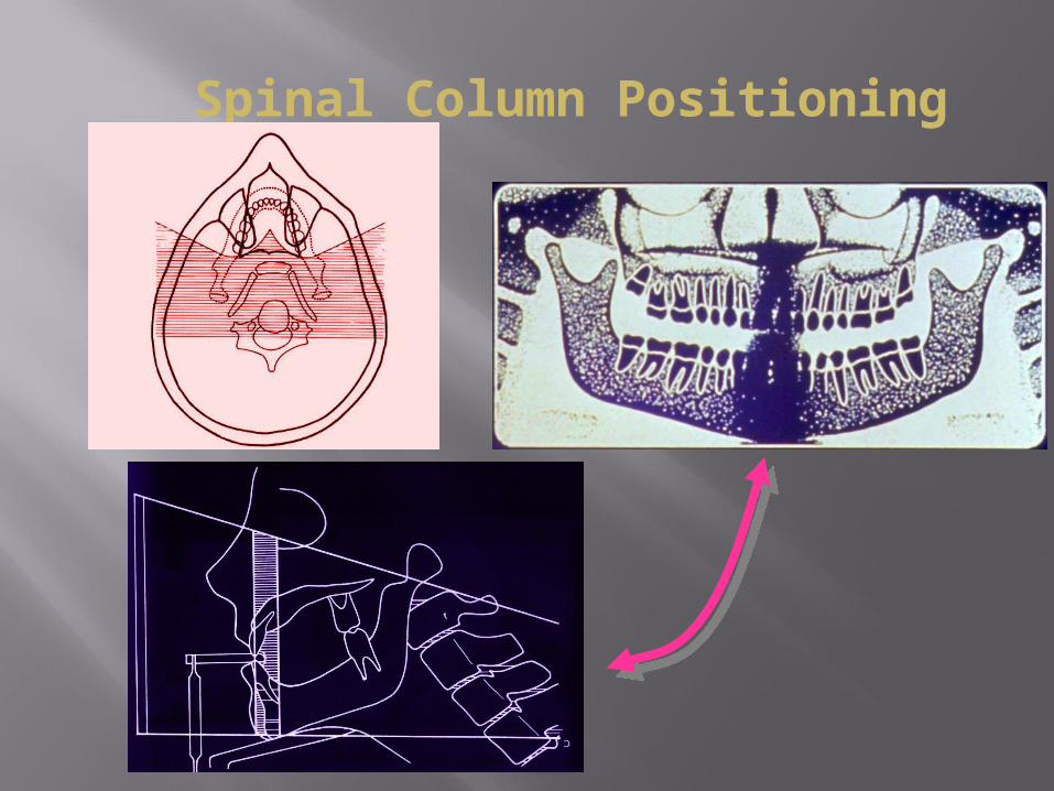
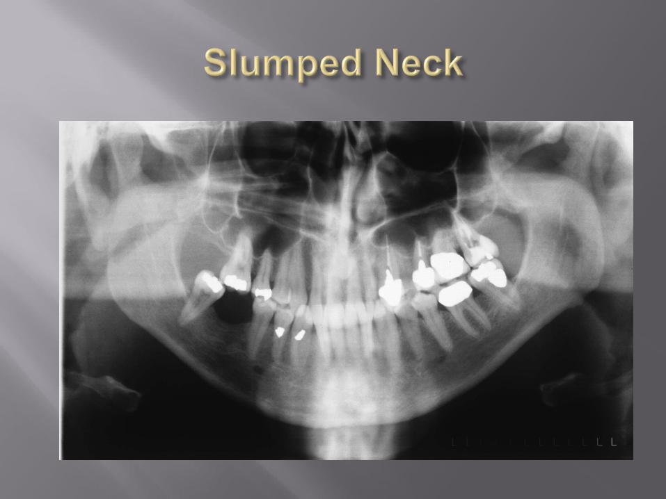
Spinal Column Positioning