Embed Size (px)
Citation preview
Introduction
When the human body is invaded by foreign organ-isms it defends itself either through the propagation of a cellular response (cell-mediated immunity) or by the production of soluble proteins. This produc-tion of soluble proteins is known as humoral immu-nity, so-called because it refers to substances found within the humours, or body fluids. While there are a number of components that have been identified as important in the humoral immune response, includ-ing complement proteins [1], the key molecules necessary for mediating specific humoral immunity are immunoglobulins (Ig), also known as antibodies (Ab) (see also chapter A3).
Ig molecules are complex proteins made up of two identical heavy chains and two identical light chains, linked by disulphide bonds and arranged in a ‘Y’-shape (Fig. 1A) [2]. They are expressed on the sur-face of B cells or secreted, recognising and binding to antigens (Ag), i.e. ‘foreign’ or ‘dangerous’ structures, such as bacterial or viral surface proteins and surface polysaccharides, as well as secreted bacterial toxins. Variable domains in the N-terminal regions of the heavy and light chains recognise and bind Ag, while the constant regions of the heavy chain C-terminal domains mediate effector functions and define the class or isotype of Ig (Box 1) (see also chapter A3). An Ag is recognised by shape. While this may be a
continuous peptide or surface polysaccharide, more often it is the three-dimensional shape made when the Ag is folded into its native structure. The shape recognised by the Ig is called an epitope. Each B cell can only produce one specific Ab, which defines its idiotype and the total number of idiotypes a body can produce is known as the immune repertoire [3]. The process by which antibody diversity is generated has been described in detail in chapter A3.
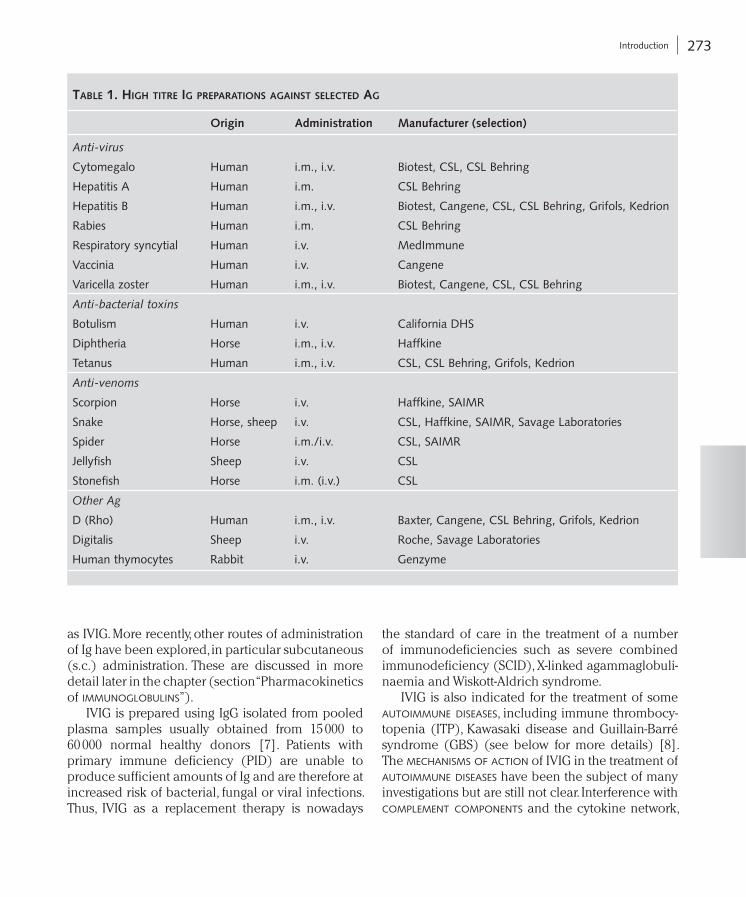
The therapeutic use of Ig has been known for many years. Hyperimmune sera collected from immunised animals, or now more often from immu-nised or naturally exposed donors, have long been used as a passive immunotherapy treatment to pro-vide specific Ab to an individual immediately, e.g. after snake envenomation, Clostridium tetani infec-tion or rabies infection (Tab. 1) [4, 5]. Similarly, anti-Rhesus D (RhD) Ig has been used successfully in the prevention of haemolytic disease in neonates due to Rhesus incompatibility between mother and child, or after mistransfusion of blood components con-taining RhD-positive erythrocytes to RhD-negative recipients. More details on hyperimmune Ig are pro-vided later in this chapter (section “Immunoglobulin preparations for medical use”).
For over half a century, Ig has also been used therapeutically in patients with immune deficien-cies [6]. Initially administered intramuscularly (i.m.), benefits in terms of efficacy and safety saw a switch to intravenous (i.v.) administration of Ig, referred to
Plasma-derived immunoglobulins
Adrian W. Zuercher, Lorenz Amsler, Hanspeter Amstutz, Irmgard Andresen, Reinhard Bolli, Wolfram Hummel, Fabian Käsermann, Christoph Kempf, Peter Lerch, Marius Lötscher, Alexander Schaub, Martin Spycher and Sylvia M. Miescher
C2
Box 1
There are five different classes of Ig: IgG, IgM, IgA, IgD and IgE as well as four subclasses of IgG and two subclasses of IgA; each class has a different function (see Fig. 2).
F.P. Nijkamp and M.J. Parnham (eds.), Principles of Immunopharmacology: 3rd revised and extended edition, DOI 10.1007/978-3-0346-0136-8_17, © Springer Basel AG 2011
271
272 Plasma-derived immunoglobulins
Hea
vy c
hain
N-linked carbohydrate
SS
SS
Hinge region
CH2
CH3
Ligh
t cha
in
CDRs
SS
CL
VH
VL
CH1
SS
Binding partner
Antigen
C3a, C3b, C5a
MBL
FcRn, SpA, SpG
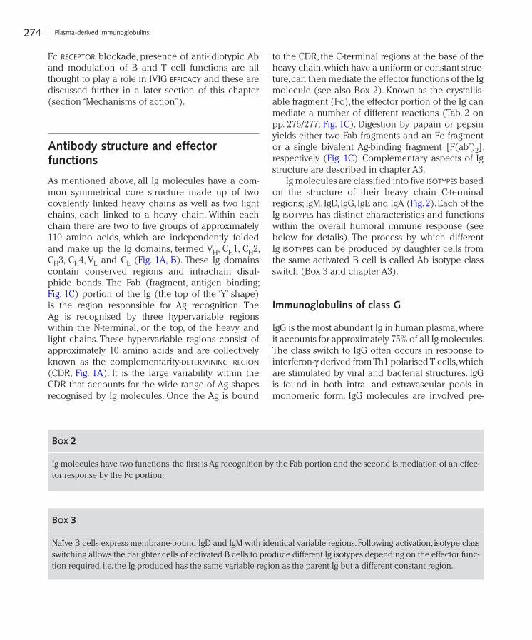
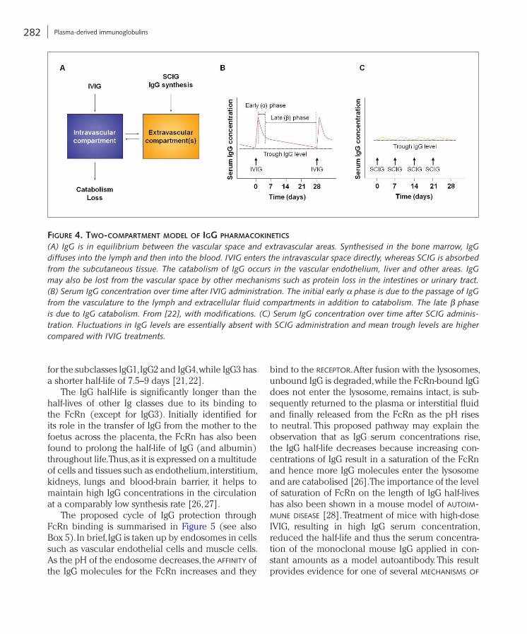
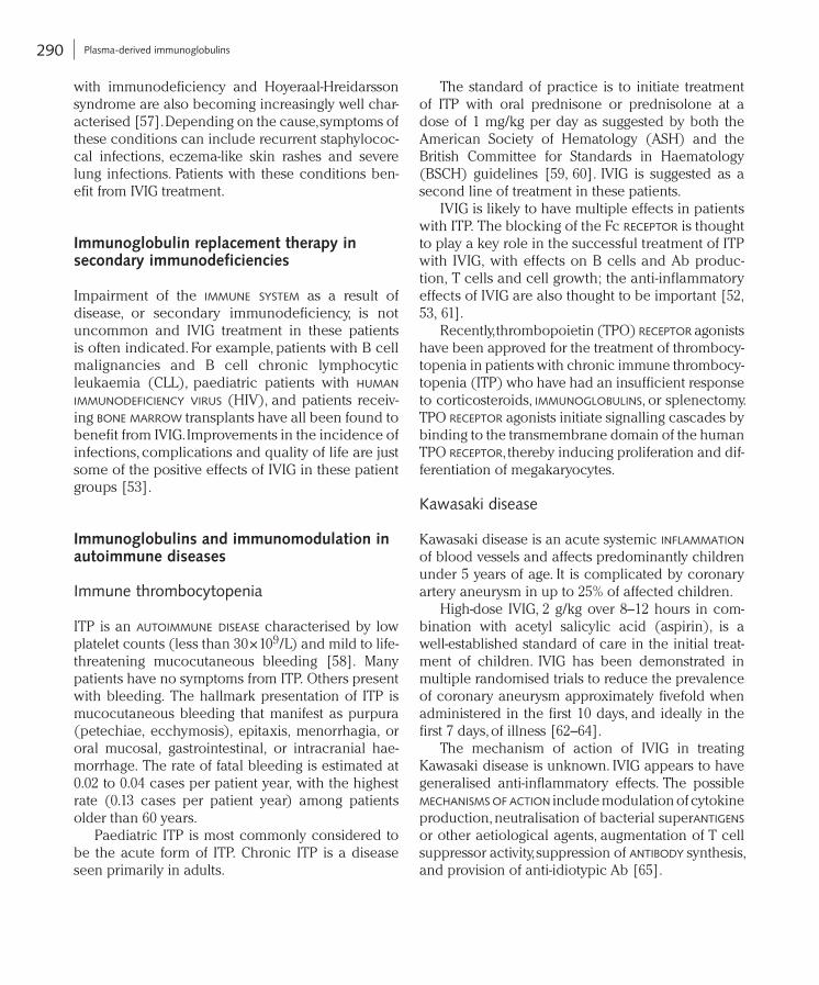
Figure 1. Structure oF igg(A) Detailed structure of an IgG1 molecule. CDR, Complementarity Determining Region; CH, constant, heavy; CL, constant, light; VH, variable, heavy; VL, variable, light; S-S, disulphide bonds; C3a, C3b, C5a, C1q, complement pro-teins; FcγR, Fcγ receptor; MBL, mannan binding lectin; FcRn, neonatal Fc receptor, SpA, Staphylococcus protein A; SpG, Streptococcus protein G. (B) Ribbon structure of an IgG2 antibody. This image has been released worldwide into the public domain by its author, Tim Vickers, at the Wikipedia project. Carbohydrates are not shown in this figure. (C) IgG fragments.
F(ab’)2 peptide fragments
pepsin
papainFab
Fc
a
B
c
273 Introduction
the standard of care in the treatment of a number of immunodeficiencies such as severe combined immunodeficiency (SCID), X-linked agammaglobuli-naemia and Wiskott-Aldrich syndrome.
IVIG is also indicated for the treatment of some autoimmune diseases, including immune thrombocy-topenia (ITP), Kawasaki disease and Guillain-Barré syndrome (GBS) (see below for more details) [8]. The mechanisms of action of IVIG in the treatment of autoimmune diseases have been the subject of many investigations but are still not clear. Interference with complement components and the cytokine network,
as IVIG. More recently, other routes of administration of Ig have been explored, in particular subcutaneous (s.c.) administration. These are discussed in more detail later in the chapter (section “Pharmacokinetics of immunoglobulins”).
IVIG is prepared using IgG isolated from pooled plasma samples usually obtained from 15 000 to 60 000 normal healthy donors [7]. Patients with primary immune deficiency (PID) are unable to produce sufficient amounts of Ig and are therefore at increased risk of bacterial, fungal or viral infections. Thus, IVIG as a replacement therapy is nowadays
taBle 1. high titre ig preparationS againSt Selected ag
Origin Administration Manufacturer (selection)
Anti-virus
Cytomegalo Human i.m., i.v. Biotest, CSL, CSL Behring
Hepatitis A Human i.m. CSL Behring
Hepatitis B Human i.m., i.v. Biotest, Cangene, CSL, CSL Behring, Grifols, Kedrion
Rabies Human i.m. CSL Behring
Respiratory syncytial Human i.v. MedImmune
Vaccinia Human i.v. Cangene
Varicella zoster Human i.m., i.v. Biotest, Cangene, CSL, CSL Behring
Anti-bacterial toxins
Botulism Human i.v. California DHS
Diphtheria Horse i.m., i.v. Haffkine
Tetanus Human i.m., i.v. CSL, CSL Behring, Grifols, Kedrion
Anti-venoms
Scorpion Horse i.v. Haffkine, SAIMR
Snake Horse, sheep i.v. CSL, Haffkine, SAIMR, Savage Laboratories
Spider Horse i.m./i.v. CSL, SAIMR
Jellyfish Sheep i.v. CSL
Stonefish Horse i.m. (i.v.) CSL
Other Ag
D (Rho) Human i.m., i.v. Baxter, Cangene, CSL Behring, Grifols, Kedrion
Digitalis Sheep i.v. Roche, Savage Laboratories
Human thymocytes Rabbit i.v. Genzyme
274 Plasma-derived immunoglobulins
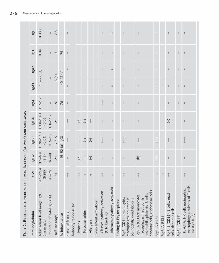
to the CDR, the C-terminal regions at the base of the heavy chain, which have a uniform or constant struc-ture, can then mediate the effector functions of the Ig molecule (see also Box 2). Known as the crystallis-able fragment (Fc), the effector portion of the Ig can mediate a number of different reactions (Tab. 2 on pp. 276/277; Fig. 1C). Digestion by papain or pepsin yields either two Fab fragments and an Fc fragment or a single bivalent Ag-binding fragment [F(ab’)2], respectively (Fig. 1C). Complementary aspects of Ig structure are described in chapter A3.
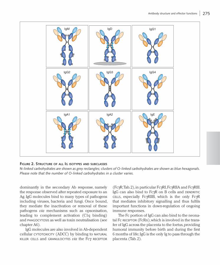
Ig molecules are classified into five isotypes based on the structure of their heavy chain C-terminal regions; IgM, IgD, IgG, IgE and IgA (Fig. 2). Each of the Ig isotypes has distinct characteristics and functions within the overall humoral immune response (see below for details). The process by which different Ig isotypes can be produced by daughter cells from the same activated B cell is called Ab isotype class switch (Box 3 and chapter A3).
Immunoglobulins of class G
IgG is the most abundant Ig in human plasma, where it accounts for approximately 75% of all Ig molecules. The class switch to IgG often occurs in response to interferon-γ derived from Th1 polarised T cells, which are stimulated by viral and bacterial structures. IgG is found in both intra- and extravascular pools in monomeric form. IgG molecules are involved pre-
Fc receptor blockade, presence of anti-idiotypic Ab and modulation of B and T cell functions are all thought to play a role in IVIG efficacy and these are discussed further in a later section of this chapter (section “Mechanisms of action”).
Antibody structure and effector functions
As mentioned above, all Ig molecules have a com-mon symmetrical core structure made up of two covalently linked heavy chains as well as two light chains, each linked to a heavy chain. Within each chain there are two to five groups of approximately 110 amino acids, which are independently folded and make up the Ig domains, termed VH, CH1, CH2, CH3, CH4, VL and CL (Fig. 1A, B). These Ig domains contain conserved regions and intrachain disul-phide bonds. The Fab (fragment, antigen binding; Fig. 1C) portion of the Ig (the top of the ‘Y’ shape) is the region responsible for Ag recognition. The Ag is recognised by three hypervariable regions within the N-terminal, or the top, of the heavy and light chains. These hypervariable regions consist of approximately 10 amino acids and are collectively known as the complementarity-determining region (CDR; Fig. 1A). It is the large variability within the CDR that accounts for the wide range of Ag shapes recognised by Ig molecules. Once the Ag is bound
Box 2
Ig molecules have two functions; the first is Ag recognition by the Fab portion and the second is mediation of an effec-tor response by the Fc portion.
Box 3
Naïve B cells express membrane-bound IgD and IgM with identical variable regions. Following activation, isotype class switching allows the daughter cells of activated B cells to produce different Ig isotypes depending on the effector func-tion required, i.e. the Ig produced has the same variable region as the parent Ig but a different constant region.
275 Antibody structure and effector functions
(FcγR; Tab. 2), in particular FcγRI, FcγRIIA and FcγRIII. IgG can also bind to FcγR on B cells and dendritic cells, especially FcγRIIB, which is the only FcγR that mediates inhibitory signalling and thus fulfils important functions in down-regulation of ongoing immune responses.
The Fc portion of IgG can also bind to the neona-tal Fc receptor (FcRn), which is involved in the trans-fer of IgG across the placenta to the foetus, providing humoral immunity before birth and during the first 6 months of life; IgG is the only Ig to pass through the placenta (Tab. 2).
dominantly in the secondary Ab response, namely the response observed after repeated exposure to an Ag. IgG molecules bind to many types of pathogens including viruses, bacteria and fungi. Once bound, they mediate the inactivation or removal of these pathogens via mechanisms such as opsonisation, leading to complement activation (C1q binding) and phagocytosis as well as toxin neutralisation (see chapter A6).
IgG molecules are also involved in Ab-dependent cellular cytotoxicity (ADCC) by binding to natural killer cells and granulocytes via the Fcγ receptor
IgE
IgM
IgA2
IgG1
IgG4IgG3IgG2
IgA1
IgD
Figure 2. Structure oF all ig iSotypeS and SuBclaSSeS
N-linked carbohydrates are shown as grey rectangles; clusters of O-linked carbohydrates are shown as blue hexagonals. Please note that the number of O-linked carbohydrates in a cluster varies.
276 Plasma-derived immunoglobulins
taB
le 2
. B
iolo
gic
al
Fun
cti
on
S o
F h
um
an i
g c
laSS
eS (
iSo
type
S) a
nd S
uB
cla
SSeS
Imm
unog
lobu
linIg
G1
IgG
2Ig
G3
IgG
4Ig
MIg
A1
IgA
2Ig
DIg
E
Adu
lt se
rum
leve
l ran
ge, g
/L
(mea
n g/
L)4.
9–11
.4
(6.9
8)1.
5–6.
4 (3
.8)
0.20
–1.1
0 (0
.51)
0.08
–1.4
0 (0
.56)
0.7–
1.7
1.5–
2.6
(a)
0.04
0.00
03
Prop
ortio
n of
tot
al Ig
G (
%)
43–7
516
–48
1.7–
7.5
0.8–
11.7
––
––
Hal
f–lif
e (d
ays)
2121
7.5–
921
56
(a)
32.
5
% in
trav
ascu
lar
45–5
2 (a
ll Ig
G)
7640
–42
(a)
75–
Plac
enta
l tra
nsfe
r+
++
++
++
––
––
–
Ant
ibod
y re
spon
se t
o:
P
rote
ins
++
+/–
++
+/–
P
olys
acch
arid
es+
++
(–)
(–)
A
llerg
ens
+(–
)(–
)+
+
Com
plem
ent
activ
atio
n:
C
lass
ical
pat
hway
act
ivat
ion
(C
1q b
indi
ng)
++
++
++
–+
++
––
––
A
ltern
ativ
e pa
thw
ay a
ctiv
atio
n–
––
––
++
––
Bind
ing
to F
cγ r
ecep
tors
:
FcγR
I (C
D64
): m
onoc
ytes
, m
acro
phag
es, n
eutr
ophi
ls,
eosi
noph
ils, d
endr
itic
cells
++
–+
++
+–
––
––
FcγR
IIA (
CD
32):
mon
ocyt
es,
mac
ro ph
ages
, neu
trop
hils
, eo
sino
phils
, pla
tele
ts, B
cel
ls,
dend
ritic
cel
ls, e
ndot
helia
l cel
ls
++
(b)
++
––
––
––
FcγR
IIA-H
131
++
++
++
++
––
––
––
FcγR
IIA-R
131
++
–+
+–
––
––
–
FcγR
IIB (
CD
32):
B c
ells
, mas
t ce
lls, d
endr
itic
cells
++
–+
+(+
)–
––
––
FcγR
III (
CD
16)
––
––
–
F
cγR
IIIA
: NK
cel
ls e
osin
ophi
ls,
mac
roph
ages
, sub
sets
of
T ce
lls,
mas
t ce
lls (
c)
++
–+
++
––
––
––
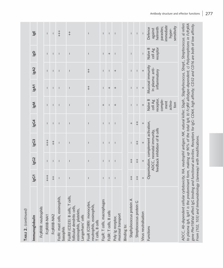
277 Antibody structure and effector functions
taB
le 2
. (c
onti
nued
)
Imm
unog
lobu
linIg
G1
IgG
2Ig
G3
IgG
4Ig
MIg
A1
IgA
2Ig
DIg
E
F
cγR
IIIB:
neu
trop
hils
Fc
γRIII
B-N
A1
++
+–
++
+–
––
––
–
Fc
γRIII
B-N
A2
++
–+
+–
––
––
–
FcεR
I: m
ast
cells
, eos
inop
hils
, ba
soph
ils–
––
––
––
–+
++
FcεR
II (C
D23
): B
cel
ls, T
cel
ls,
folli
cula
r de
ndrit
ic c
ells
, eo
sino
phils
, pla
tele
ts,
Lang
erha
ns’ c
ells
––
––
––
––
++
FcαR
(C
D89
): m
onoc
ytes
, ne
utro
phils
, eos
inop
hils
, T
cells
, B c
ells
––
––
–+
++
+–
–
FcµR
: T c
ells
, mac
roph
ages
––
––
+–
––
–
FcδR
: T c
ells
, B c
ells
–
––
––
+–
+–
Poly
Ig r
ecep
tor,
m
ucos
al t
rans
port
––
––
++
+–
–
Bind
ing
to:
S
taph
yloc
occu
s pr
otei
n A
++
++
–+
––
––
–
S
trep
toco
ccus
pro
tein
G+
++
++
++
+–
––
–
Viru
s ne
utra
lisat
ion
++
++
++
+–
–
Func
tions
Ops
onis
atio
n, c
ompl
emen
t ac
tivat
ion,
A
DC
C, n
eona
tal i
mm
unity
, fe
edba
ck in
hibi
tion
of B
cel
ls
Naï
ve B
ce
ll A
g re
cept
or,
com
ple-
men
t ac
tiva-
tion
Muc
osal
imm
unity
In
pla
sma:
ant
i-in
flam
mat
ory
Naï
ve B
ce
ll A
g re
cept
or
Def
ence
ag
ains
t he
lmin
th
para
site
s;
imm
edia
te
hype
r-se
nsiti
vity
AD
CC
, Ab-
depe
nden
t ce
llula
r cy
toto
xici
ty; N
A, n
eutr
ophi
l ant
igen
; NK
, nat
ural
kill
er; S
taph
., St
aphy
loco
ccus
, Str
ept.
, Str
epto
cocc
us. a
) re
fers
to
pla
sma
IgA
, Ig
A1
is t
he p
redo
min
ant
form
, m
akin
g up
90%
of
the
tota
l Ig
A;
b) F
c γR
II al
loty
pe-d
epen
dent
; c)
Pol
ymor
phis
ms
in F
cRγI
IIA
gene
Phe
158V
al a
ffec
t Ig
G b
indi
ng a
nd f
unct
iona
l act
ivit
ies.
Rec
epto
rs f
or Ig
G: C
D64
, hig
h af
fini
ty; C
D32
and
CD
16 a
re b
oth
of lo
w a
ffin
ity.
Fr
om [
102,
103
] an
d Im
mun
obio
logy
(Ja
new
ay)
wit
h m
odif
icat
ions
.
278 Plasma-derived immunoglobulins
of apoptotic cells, clearance of intracellular proteins released from necrotic cells) and tumour surveil-lance (recognition of newly emerging carbohydrate, glycolipid or glycoprotein pattern on malignant cells). Other actions of IgM NAb molecules include proteolysis and modulation of B and T cell immune responses, as well as promotion of diseases arising from a sudden impact, such as in infarction or sys-temic inflammatory response syndrome [11].
Immunoglobulin of class A
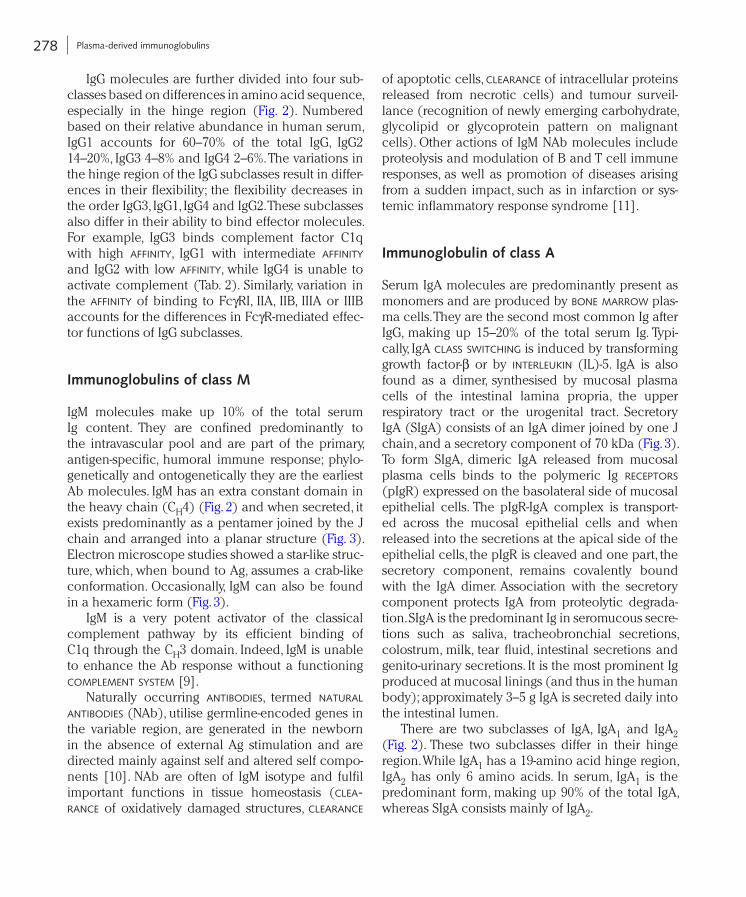
Serum IgA molecules are predominantly present as monomers and are produced by bone marrow plas-ma cells. They are the second most common Ig after IgG, making up 15–20% of the total serum Ig. Typi-cally, IgA class switching is induced by transforming growth factor-β or by interleukin (IL)-5. IgA is also found as a dimer, synthesised by mucosal plasma cells of the intestinal lamina propria, the upper respiratory tract or the urogenital tract. Secretory IgA (SIgA) consists of an IgA dimer joined by one J chain, and a secretory component of 70 kDa (Fig. 3). To form SIgA, dimeric IgA released from mucosal plasma cells binds to the polymeric Ig receptors (pIgR) expressed on the basolateral side of mucosal epithelial cells. The pIgR-IgA complex is transport-ed across the mucosal epithelial cells and when released into the secretions at the apical side of the epithelial cells, the pIgR is cleaved and one part, the secretory component, remains covalently bound with the IgA dimer. Association with the secretory component protects IgA from proteolytic degrada-tion. SIgA is the predominant Ig in seromucous secre-tions such as saliva, tracheobronchial secretions, colostrum, milk, tear fluid, intestinal secretions and genito-urinary secretions. It is the most prominent Ig produced at mucosal linings (and thus in the human body); approximately 3–5 g IgA is secreted daily into the intestinal lumen.
There are two subclasses of IgA, IgA1 and IgA2 (Fig. 2). These two subclasses differ in their hinge region. While IgA1 has a 19-amino acid hinge region, IgA2 has only 6 amino acids. In serum, IgA1 is the predominant form, making up 90% of the total IgA, whereas SIgA consists mainly of IgA2.
IgG molecules are further divided into four sub-classes based on differences in amino acid sequence, especially in the hinge region (Fig. 2). Numbered based on their relative abundance in human serum, IgG1 accounts for 60–70% of the total IgG, IgG2 14–20%, IgG3 4–8% and IgG4 2–6%. The variations in the hinge region of the IgG subclasses result in differ-ences in their flexibility; the flexibility decreases in the order IgG3, IgG1, IgG4 and IgG2. These subclasses also differ in their ability to bind effector molecules. For example, IgG3 binds complement factor C1q with high affinity, IgG1 with intermediate affinity and IgG2 with low affinity, while IgG4 is unable to activate complement (Tab. 2). Similarly, variation in the affinity of binding to FcγRI, IIA, IIB, IIIA or IIIB accounts for the differences in FcγR-mediated effec-tor functions of IgG subclasses.
Immunoglobulins of class M
IgM molecules make up 10% of the total serum Ig content. They are confined predominantly to the intravascular pool and are part of the primary, antigen-specific, humoral immune response; phylo-genetically and ontogenetically they are the earliest Ab molecules. IgM has an extra constant domain in the heavy chain (CH4) (Fig. 2) and when secreted, it exists predominantly as a pentamer joined by the J chain and arranged into a planar structure (Fig. 3). Electron microscope studies showed a star-like struc-ture, which, when bound to Ag, assumes a crab-like conformation. Occasionally, IgM can also be found in a hexameric form (Fig. 3).
IgM is a very potent activator of the classical complement pathway by its efficient binding of C1q through the CH3 domain. Indeed, IgM is unable to enhance the Ab response without a functioning complement system [9].
Naturally occurring antibodies, termed natural antibodies (NAb), utilise germline-encoded genes in the variable region, are generated in the newborn in the absence of external Ag stimulation and are directed mainly against self and altered self compo-nents [10]. NAb are often of IgM isotype and fulfil important functions in tissue homeostasis (clea-rance of oxidatively damaged structures, clearance
279 Antibody structure and effector functions
biota in the intestinal lumen by ‘immune exclusion’ [12]. SIgA can neither bind nor activate complement.
Immunoglobulin of class D
IgD is present at very low concentrations in serum; it accounts for less than 1% of the total serum Ig (Fig. 2). It is mostly found as a membrane-bound monomer, co-expressed with IgM, on naïve B cells. Upon activa-tion of naïve B cells with specific Ag, class switch recombination occurs and the expression of IgD is lost. Recently, a potential function of secreted IgD in anti-bacterial immunity of the respiratory tract has been described [14, 15].
Binding of monomeric serum IgA to the FcαRI present on monocytes, macrophages, neutrophils and eosinophils, induces inhibitory signalling path-ways [12]. Thus serum IgA serves an important anti-inflammatory function in the systemic circulation. In aggregated form, serum IgA can activate the lectin pathway of the complement system as a result of binding to the carbohydrate recognition domain of mannan-binding lectin (MBL) [13].
SIgA is present as high- and low-affinity Ab; high-affinity SIgA helps defend mucous membranes of the intestine and nose against viral or bacterial infections and takes part in the neutralisation of bacterial toxins, while low-affinity SIgA has a more ‘homeostatic’ func-tion in shaping and controlling the commensal micro-
Hexameric IgM
Dimeric IgA
Poly-Ig Receptor
Secretory IgA
Pentameric IgM
J chain
J chainJ chain
Figure 3. StructureS oF polymeric igm and iga
280 Plasma-derived immunoglobulins
Ab repertoire, and cross-talk between the innate and adaptive immune pathways. The five classes of Ig are highly diverse in terms of the location and number of conserved N-linked glycosylation sites situated on the Fc and Fab portions (Figs. 1A and 2) (see Box 4) [16]. Abnormalities in the glycosylation profiles of Ig molecules have been linked to certain diseases. For example, increases in agalactosyl glycoforms of IgG have been isolated from patients with rheumatoid arthritis; the pathogenesis of IgA nephropathy is influenced by abnormal IgA1 O-glycosylation and hence reduced IgA clearance; the glycosylation of all glycoproteins is also affected in congenital disorders of glycosylation and abnormal IgG glycoforms are used in the diagnosis of these disorders [16].
IgG glycosylation
All IgG molecules have a single conserved N-linked glycosylation site, Asn297, in the Fc region, which is important in maintaining Fc effector function (Figs. 1A and 2). The oligosaccharides present in this region are of the complex di-antennary type. Com-prised of a core heptasaccharide, these oligosaccha-rides have the variable addition of fucose, galactose, bisecting N-acetylglucosamine and sialic acid. Sia-lylation tends to be present in less than 10% of IgG. Glycosylation is critical for effector functions medi-ated via the FcγR, the C1q component of comple-ment and the MBL. For example, the absence of core fucose on the Fc glycans leads to increased binding to FcγRIIIA and enhanced ADCC [17]. Furthermore, terminal sialylation of the Fc glycans has been reported to be responsible for the anti-inflammatory properties of human IgG in mice [18]. In contrast, Fc glycosylation does not appear to play a role in FcRn interactions [17].
Immunoglobulin of class E
IgE is found only in trace amounts in human serum. Class switch to IgE production is classically induced by IL-4 derived from Th2 polarised T cells, but alter-native pathways have also been described. Similar to IgM, IgE has an extra constant domain in the heavy chain (CH4) (Fig. 2). It is found predominantly bound to the high-affinity Fcε receptor (FcεRI) on basophils and mast cells even prior to the interaction with its cognate Ag. When Ag/allergen binds to the IgE on mast cells and basophils it causes the aggregation of the Fcε receptors and subsequent degranulation of the cell, with the release of vasoactive and chemot-actic mediators. This results in allergic reactions such as hay fever, extrinsic asthma and the Prausnitz-Küstner skin reaction; in severe cases anaphylaxis can even be life-threatening. As well as its role in atopic allergy, IgE is important in protection against helminths and parasites. Indeed, elevation of IgE levels can be used as a diagnostic tool for parasitic infections. Circulating IgE cannot activate comple-ment via the classical pathway (Tab. 2).
IgE can bind to two Fc receptors, namely FcεRI and FcεRII. IgE is mostly bound to the FcεRI; the FcεRI binding site on IgE is located at the interface between CH2 and CH3. The IgE binding affinity for the FcεRII, which is an Fc receptor found on mono-cytes, B cells and platelets, is lower than for FcεRI.
Glycosylation of immunoglobulins
Glycosylation of Ig has a number of important roles including the maintenance of structure and stability, receptor binding, Fc effector function, intracellular transport, secretion and clearance, expansion of the
Box 4
Only 2–3% of the molecular weight of IgG is made up of glycosylation in the heavy chain, whereas IgM, IgD and IgE molecules are more highly glycosylated; 12–14% of the molecular weight is accounted for by glycosylation of the heavy chain.
281 Pharmacokinetics of immunoglobulins
Absence of this site results in incomplete assembly and lack of secretion [16].
Pharmacokinetics of immunoglobulins
Catabolism provides the main route of Ig elimination [21]. Due to its large size, very little Ig is excreted in the urine and only small amounts have been identi-fied in the bile. It is assumed that approximately one-third of Ig is broken down in the liver, with another third in the intestines. The cellular compartments in close proximity to the blood and lymph such as the endothelial cells constitute other sites important in Ig catabolism. There are differences in sites of catab-olism between Ig classes with IgA, particularly SIgA, being different from other classes as it is present in high amounts in body excretions such as intestinal fluid and saliva with a resulting high turnover.
Pharmacokinetics of IgG
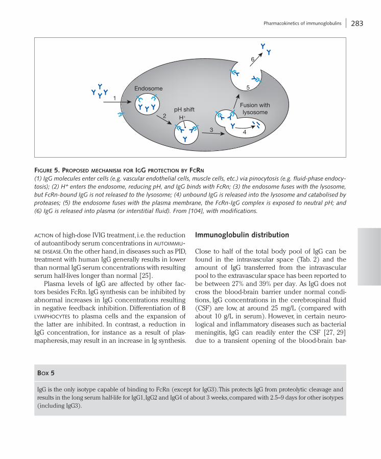
In pharmacokinetic studies with IVIG administra-tion, a two-compartment model has been proposed to describe the time course of IgG serum concen-trations (Fig. 4A) [22, 23]. After the initial rapid rise in serum IgG concentration a comparably rapid decrease follows (Fig. 4B). This early (or α) phase, which in humans roughly occurs during the first 5 days after infusion, is the result of both catabolism of IgG and distribution between the intravascular and extravascular spaces. The following β phase shows a slower decrease in IgG serum concentra-tion, which is caused by the catabolism of IgG and thus determines the serum half-life of IgG. This gen-eral model has been confirmed using various IVIG preparations [23, 24]. A high inter-individual vari-ability of pharmacokinetic parameters was found, which is not unexpected considering the complex mechanisms of catabolism and distribution of IgG [24, 25].
The use of radioiodinated IgG molecules has shown that IgG, except for IgG3, can survive longer in the blood than any other serum proteins, with a mean half-time of survival of about 3 weeks (Tab. 2)
In addition to Fc glycosylation, approximately 20–30% of normal plasma IgG is glycosylated in the fab region. In contrast to the Fc oligosaccharides, Fab oligosaccharides are bisected, extensively galac-tosylated and substantially sialylated. This difference may be due to a lack of accessibility of the Fc por-tion to specific transferases [19].
Various factors influence the glycosylation of IgG, including age and sex. Age dependency is particularly clear; galactosylation and sialylation increases up to the age of 25 years and then decreases again throughout life. Moreover, during pregnancy there is a temporary increase in IgG galactosylation.
Glycosylation of other Ig molecules
IgA molecules possess two conserved N-glycosylati-on sites in the heavy chains, one in the CH2 region and one in the tailpiece of the Fc region (Fig. 2). In addition, there are two to three further N-glycosylati-on sites in IgA2, up to five O-glycosylation sites in the hinge region of IgA1, seven N-glycosylation sites in the secretory component and a further one in the J chain. In contrast to IgG, over 90% of IgA molecules are sialylated [20].
The µ-chain of IgM molecules has five N-linked glycosylation sites; three are of the complex type and two of the oligomannose type. These contain pre-dominantly monosialylated, bi-antennary structures. In addition, there is one N-linked glycosylation site in the J chain containing mainly sialylated bi-antennary glycans. Circulating predominantly as pentamers, the glycan epitopes are thought to allow IgM to aggluti-nate microorganisms in the serum [16].
IgE is the most heavily glycosylated Ig. It contains seven N-linked glycosylation sites in the ε-chain. These N-linked glycans result in a reduction in the flexibility of the IgE molecules, which is thought to control Ag binding and prevent unwanted and potentially fatal immune responses through inappro-priate Ag binding [16].
IgD has three N-linked glycosylation sites in the δ-chain and site-directed mutagenesis has shown that the presence of oligomannose glycans at Asn354 is of vital importance for the production of IgD.
282 Plasma-derived immunoglobulins
bind to the receptor. After fusion with the lysosomes, unbound IgG is degraded, while the FcRn-bound IgG does not enter the lysosome, remains intact, is sub-sequently returned to the plasma or interstitial fluid and finally released from the FcRn as the pH rises to neutral. This proposed pathway may explain the observation that as IgG serum concentrations rise, the IgG half-life decreases because increasing con-centrations of IgG result in a saturation of the FcRn and hence more IgG molecules enter the lysosome and are catabolised [26]. The importance of the level of saturation of FcRn on the length of IgG half-lives has also been shown in a mouse model of autoim-mune disease [28]. Treatment of mice with high-dose IVIG, resulting in high IgG serum concentration, reduced the half-life and thus the serum concentra-tion of the monoclonal mouse IgG applied in con-stant amounts as a model autoantibody. This result provides evidence for one of several mechanisms of
for the subclasses IgG1, IgG2 and IgG4, while IgG3 has a shorter half-life of 7.5–9 days [21, 22].
The IgG half-life is significantly longer than the half-lives of other Ig classes due to its binding to the FcRn (except for IgG3). Initially identified for its role in the transfer of IgG from the mother to the foetus across the placenta, the FcRn has also been found to prolong the half-life of IgG (and albumin) throughout life. Thus, as it is expressed on a multitude of cells and tissues such as endothelium, interstitium, kidneys, lungs and blood-brain barrier, it helps to maintain high IgG concentrations in the circulation at a comparably low synthesis rate [26, 27].
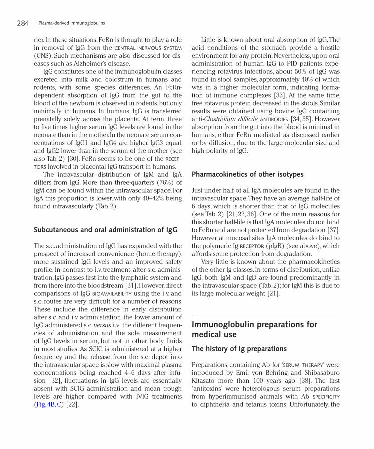
The proposed cycle of IgG protection through FcRn binding is summarised in Figure 5 (see also Box 5). In brief, IgG is taken up by endosomes in cells such as vascular endothelial cells and muscle cells. As the pH of the endosome decreases, the affinity of the IgG molecules for the FcRn increases and they
Figure 4. two-compartment model oF igg pharmacokineticS
(A) IgG is in equilibrium between the vascular space and extravascular areas. Synthesised in the bone marrow, IgG diffuses into the lymph and then into the blood. IVIG enters the intravascular space directly, whereas SCIG is absorbed from the subcutaneous tissue. The catabolism of IgG occurs in the vascular endothelium, liver and other areas. IgG may also be lost from the vascular space by other mechanisms such as protein loss in the intestines or urinary tract. (B) Serum IgG concentration over time after IVIG administration. The initial early α phase is due to the passage of IgG from the vasculature to the lymph and extracellular fluid compartments in addition to catabolism. The late β phase is due to IgG catabolism. From [22], with modifications. (C) Serum IgG concentration over time after SCIG adminis-tration. Fluctuations in IgG levels are essentially absent with SCIG administration and mean trough levels are higher compared with IVIG treatments.
283 Pharmacokinetics of immunoglobulins
Immunoglobulin distribution
Close to half of the total body pool of IgG can be found in the intravascular space (Tab. 2) and the amount of IgG transferred from the intravascular pool to the extravascular space has been reported to be between 27% and 39% per day. As IgG does not cross the blood-brain barrier under normal condi-tions, IgG concentrations in the cerebrospinal fluid (CSF) are low, at around 25 mg/L (compared with about 10 g/L in serum). However, in certain neuro-logical and inflammatory diseases such as bacterial meningitis, IgG can readily enter the CSF [27, 29] due to a transient opening of the blood-brain bar-
action of high-dose IVIG treatment, i.e. the reduction of autoantibody serum concentrations in autoimmu-ne disease. On the other hand, in diseases such as PID, treatment with human IgG generally results in lower than normal IgG serum concentrations with resulting serum half-lives longer than normal [25].
Plasma levels of IgG are affected by other fac-tors besides FcRn. IgG synthesis can be inhibited by abnormal increases in IgG concentrations resulting in negative feedback inhibition. Differentiation of b lymphocytes to plasma cells and the expansion of the latter are inhibited. In contrast, a reduction in IgG concentration, for instance as a result of plas-mapheresis, may result in an increase in Ig synthesis.
1
2
3 4
5
6
H+
Endosome
pH shiftFusion with lysosome
Figure 5. propoSed mechaniSm For igg protection By Fcrn
(1) IgG molecules enter cells (e.g. vascular endothelial cells, muscle cells, etc.) via pinocytosis (e.g. fluid-phase endocy-tosis); (2) H+ enters the endosome, reducing pH, and IgG binds with FcRn; (3) the endosome fuses with the lysosome, but FcRn-bound IgG is not released to the lysosome; (4) unbound IgG is released into the lysosome and catabolised by proteases; (5) the endosome fuses with the plasma membrane, the FcRn-IgG complex is exposed to neutral pH; and (6) IgG is released into plasma (or interstitial fluid). From [104], with modifications.
Box 5
IgG is the only isotype capable of binding to FcRn (except for IgG3). This protects IgG from proteolytic cleavage and results in the long serum half-life for IgG1, IgG2 and IgG4 of about 3 weeks, compared with 2.5–9 days for other isotypes (including IgG3).
284 Plasma-derived immunoglobulins
Little is known about oral absorption of IgG. The acid conditions of the stomach provide a hostile environment for any protein. Nevertheless, upon oral administration of human IgG to PID patients expe-riencing rotavirus infections, about 50% of IgG was found in stool samples, approximately 40% of which was in a higher molecular form, indicating forma-tion of immune complexes [33]. At the same time, free rotavirus protein decreased in the stools. Similar results were obtained using bovine IgG containing anti-Clostridium difficile antibodies [34, 35]. However, absorption from the gut into the blood is minimal in humans, either FcRn mediated as discussed earlier or by diffusion, due to the large molecular size and high polarity of IgG.
Pharmacokinetics of other isotypes
Just under half of all IgA molecules are found in the intravascular space. They have an average half-life of 6 days, which is shorter than that of IgG molecules (see Tab. 2) [21, 22, 36]. One of the main reasons for this shorter half-life is that IgA molecules do not bind to FcRn and are not protected from degradation [37]. However, at mucosal sites IgA molecules do bind to the polymeric Ig receptor (pIgR) (see above), which affords some protection from degradation.
Very little is known about the pharmacokinetics of the other Ig classes. In terms of distribution, unlike IgG, both IgM and IgD are found predominantly in the intravascular space (Tab. 2); for IgM this is due to its large molecular weight [21].
Immunoglobulin preparations for medical use
The history of Ig preparations
Preparations containing Ab for ‘serum therapy’ were introduced by Emil von Behring and Shibasaburo Kitasato more than 100 years ago [38]. The first ‘antitoxins’ were heterologous serum preparations from hyperimmunised animals with Ab specificity to diphtheria and tetanus toxins. Unfortunately, the
rier. In these situations, FcRn is thought to play a role in removal of IgG from the central nervous system (CNS). Such mechanisms are also discussed for dis-eases such as Alzheimer’s disease.
IgG constitutes one of the immunoglobulin classes excreted into milk and colostrum in humans and rodents, with some species differences. An FcRn-dependent absorption of IgG from the gut to the blood of the newborn is observed in rodents, but only minimally in humans. In humans, IgG is transferred prenatally solely across the placenta. At term, three to five times higher serum IgG levels are found in the neonate than in the mother. In the neonate, serum con-centrations of IgG1 and IgG4 are higher, IgG3 equal, and IgG2 lower than in the serum of the mother (see also Tab. 2) [30]. FcRn seems to be one of the recep-tors involved in placental IgG transport in humans.
The intravascular distribution of IgM and IgA differs from IgG. More than three-quarters (76%) of IgM can be found within the intravascular space. For IgA this proportion is lower, with only 40–42% being found intravascularly (Tab. 2).
Subcutaneous and oral administration of IgG
The s.c. administration of IgG has expanded with the prospect of increased convenience (home therapy), more sustained IgG levels and an improved safety profile. In contrast to i.v. treatment, after s.c. adminis-tration, IgG passes first into the lymphatic system and from there into the bloodstream [31]. However, direct comparisons of IgG bioavailability using the i.v. and s.c. routes are very difficult for a number of reasons. These include the difference in early distribution after s.c. and i.v. administration, the lower amount of IgG administered s.c. versus i.v., the different frequen-cies of administration and the sole measurement of IgG levels in serum, but not in other body fluids in most studies. As SCIG is administered at a higher frequency and the release from the s.c. depot into the intravascular space is slow with maximal plasma concentrations being reached 4–6 days after infu-sion [32], fluctuations in IgG levels are essentially absent with SCIG administration and mean trough levels are higher compared with IVIG treatments (Fig. 4B, C) [22].
285 Immunoglobulin preparations for medical use
Subsequent IVIG preparations were produced by cold ethanol precipitation followed by chemical modification, which led to a marked reduction in inherent complement binding [39]. Unfortunately, such chemical treatments led to reversible or irre-versible modifications of the Ig molecule, which did not demonstrate fully active Fab and Fc functions. Although effective in the treatment of some diseases, they were not as functional as native, intact Ig. As a result, they became obsolete for all major markets.
Immunoglobulin formulations today
More recent developments in the production of IVIG rely on more gentle isolation and purification pro-cedures [40]. Cold ethanol fractionation is followed by a series of individual purification procedures that may also be used in combination. Polymers such as polyethylene glycol, which support selective precipi-tation and stabilisation may also be used. State of the art production processes use cation and/or anion exchange chromatography for polishing the IgG, especially for the removal of Ig classes other than G (i.e. IgA, IgM, IgE). These techniques yield unaltered and functionally intact IgG with at least 96% IgG, a subclass distribution characteristic of human serum and a normal half-life of IgG (Box 6).
Virus inactivation and removal steps are integrat-ed within the IVIG production process. More detail on this topic is given later in the chapter (section “Viral safety of immunoglobulins”).
Lyophilised IVIG preparations offer excellent sta-bility and have a shelf life of up to several years at room temperature. A disadvantage of lyophilised products is that they are less convenient because they require reconstitution prior to administration.
With this in mind, the latest preparations of IVIG have been formulated as a ready-to-use liquid. While
initial success, particularly with the diphtheria anti-sera, was marred by the increasing incidence of side effects from the heterologous animal serum proteins, known as serum sickness. As a result, purer and there-fore more compatible preparations were developed.
The initial Ig preparations derived from human plasma were for i.m. injection only. Disadvantages of the i.m. administration included pain at the injection site, limits to the volumes that could be adminis-tered, reduced bioavailability, and delays of at least 24 hours and up to several days before maximum serum levels were achieved. The i.m. preparations of Ig (IMIG) were not suitable for i.v. administration due to adverse reactions, which were often severe and even fatal. Such adverse reactions occurred in at least 15% of administrations and were attributed to the presence of aggregates in these preparations [6]. Ig aggregates can interact with the first component of the complement system, C1q, thereby triggering the classical pathway of complement activation and causing the in vivo generation of anaphylatoxins (C3a, C5a). The presence of endogenous prekallikrein activator (PKA) activity, known to induce hypoten-sion, further contributed to the adverse event profile of IMIG after i.v. administration. Together with the lack of appropriate stability, these factors made the initial IMIG preparations unsuitable for i.v. administration.
Initial IVIG preparations developed in the 1970s underwent plasma fractionation by cold ethanol precipitation followed by protease digestion, includ-ing trypsin, plasmin and pepsin. The resulting Ig preparation contained predominantly fragment-ed Ig including the bivalent Ab molecule, F(ab’)2 (Fig. 1C). However, these preparations contained no Fc-mediated activity, which is essential for the full spectrum of Ig activities. Moreover, although well tolerated, these preparations had a very short half-life of 24 hours or less, making them generally unsuitable for Ab replacement therapy.
Box 6
Current production methods for IVIG generate non-cleaved, native and functionally intact IgG with an almost normal distribution of IgG subclasses and a physiological half-life.
286 Plasma-derived immunoglobulins
‘source’ plasma. Plasma from whole blood donations, termed ‘recovered’ plasma, is also used [49]. Despite the use of a large number of donors, Ig preparations still demonstrate a high degree of variability in speci-ficity and titre levels. Each Ig preparation is different; it represents the cumulative Ag experience of the donors and therefore depends on the donor popula-tion and also the period of time when the plasma was collected. For example, vaccination programmes have significantly affected titres of the pooled Ig preparations. As the measles disease triggers a higher Ab titre than the vaccine, mean measles titres have been declining since the introduction of a widely accepted vaccination programme, whereas mean titres to hepatitis b virus have increased significantly since the mid nineties (after the introduction of the vaccine) [50].
Hyperimmune plasma from volunteers immu-nised with approved vaccines against diseases such as hepatitis B, varicella zoster, tetanus toxoid or rabies provides a source of pathogen-specific, high-titre therapeutic Ig [48]. This passive prophylaxis against certain infectious diseases is receiving increasing interest. While the use of animal sera and antitoxins often results in unwanted side effects due to the non-human origin [4], hyperimmune Ig from human sources are similar to regular IVIG but contain a high level of Ab against a specific Ag and are often useful in certain conditions where normal Ig would be of little benefit [48]. A good example is anti-RhD IgG prepared from the plasma of RhD-negative human donors who agree to be hyperimmunised with RhD-positive red blood cells. This preparation has been successfully used for decades to prevent RhD incompatibility between mother and child in the course of pregnancy or during delivery, or after mis-transfusion of RhD-positive blood to RhD-negative recipients. Recently, a new production process has been developed that selectively concentrates anti-D IgG and provides higher yields of this hyperimmune Ig than conventional manufacturing processes for plasma-derived IgG [51].
Table 1 provides details of a selection of currently available hyperIg preparations.
the first liquid preparations of IVIG required refriger-ation and had a limited shelf life, more recent formu-lations are stable at room temperature for extended periods. Studies have shown that by reducing the pH to moderately acidic and adding appropriate stabilis-ers to the formulation, the stability of the liquid IVIG can be maintained for up to 3 years [41, 42].
In liquid IVIG, isolated from pooled plasma of numerous donors, idiotype/anti-idiotype Ab dimers are formed [43, 44]. idiotype/anti-idiotype dimers form when the Ag-binding region of one IgG mole-cule binds to the variable region of another. High lev-els of dimeric IgG in IVIG have been described to be associated with adverse reactions such as headache, fever and flushing observed during i.v. infusion [45, 46]. Therefore, in liquid IVIG preparations the forma-tion of IgG dimers has to be controlled to maintain good clinical tolerability.
Table 3 (see pp. 288/289) provides characteristics of a selection of currently available IVIG prepara-tions in major markets.
For certain patients, such as children, pregnant women, and individuals with poor venous access, s.c. administration of Ig is more convenient [32]. Moreover, a number of validated health-related qual-ity of life questionnaires have found that many patients prefer SCIG to IVIG [32, 47]. Although some of the current preparations of IVIG are suitable for s.c. administration, specific formulations of Ig for s.c. administration are being developed. So far, the FDA has approved the use of a few SCIG preparations. In Europe SCIG is more extensively used [48]. The new-est SCIG preparation contains Ig concentrations as high as 20% (Tab. 3) [49].
The basic material for the production of poly-valent human Ig, polyvalent homologous Ig (or, according to pharmacopoeia terminology, ‘normal’ Ig), is a pool of plasma from thousands of healthy human donors. Special Ig may be isolated from the plasma of specifically immunised donors, from the plasma of convalescents or from screened plasma containing high titres of a desired specificity. Most of the plasma obtained for the production of therapeu-tic Ig is obtained by plasmapheresis and is called
287 Indications for immunoglobulins
for SCID are often agammaglobulinaemic and also benefit from IVIG.
Hypogammaglobulinaemia
In hypogammaglobulinaemia, patients present with recurrent and often severe infections such as sinus-itis, otitis media, pneumonia and meningitis. The infections may also involve the bones and joints and complications such as chronic asymmetric polyar-thritis are not uncommon. Hypogammaglobulinae-mia is a characteristic of CVID and studies in these patients have demonstrated the benefits of IVIG, with a reduction in the incidence of infections compared with the pre-treatment rates. CVID patients are par-ticularly at risk of chronic, subclinical lung and pul-monary infections. IVIG can reduce the incidence of pneumonia and also prevent the progression of lung disease in these patients [53].
Regular treatment with IVIG is also of benefit to patients with hyper-IgM syndromes, characterised by hypogammaglobulinaemia and severe impair-ment of specific Ab production. Studies found that meningitis was eradicated in the patients tested and the incidence of pneumonia was markedly reduced [53].
Other primary immune deficiencies
Other well-defined immunodeficiency syndromes include hyper-IgE syndrome, in which patients dem-onstrate normal levels of IgG, IgM and IgA but have poor Ab-mediated responses, and Wiskott-Aldrich syndrome, which is characterised by normal IgG levels but abnormal Ab responses [53]. In addition, conditions caused by DNA repair defects, such as ataxia-telangiectasia, and thymic defects, as well as immune-osseous dysplasias, chronic mucocuta-neous candidiasis, hepatic veno-occlusive disease
Indications for immunoglobulins
When first introduced, IVIG was used as a replace-ment therapy for immunodeficiency diseases. Cur-rently, IVIG is also used as a treatment for a number of autoimmune and inflammatory diseases (Box 7). IVIG is used at low doses when administered as a ‘replacement’ therapy (200–500 mg/kg) and at high doses when used as an ‘immunomodulatory/anti-inflammatory’ therapy (up to 2 g/kg).
Immunoglobulin replacement therapy in primary immunodeficiencies
IVIG is indicated as a replacement therapy in patients with primary immunodeficiency (PID) characterised by reduced or absent Ab production and recurrent infections [52–54]. More than 140 distinct, inherited PID disorders have been identified [55]. The most common antibody deficiency is common variable immunodeficiency (CVID) [56].
X-linked agammaglobulinaemia
In a rare X-linked genetic disorder, patients with X-linked agammaglobulinaemia experience repeat-ed infections, particularly of the respiratory tract, paranasal sinuses, ears and meninges. Pneumococci or encapsulated Haemophilus influenzae are often the cause. Intestinal infections caused by bacteria such as Campylobacter jejuni are also common. In these patients, who have a deficit in Ab production, IVIG is of clear benefit, reducing both acute and chronic infections. Analyses of data in agammaglob-ulinaemic children have found that the number and severity of infections is inversely proportional to the dose of IVIG [53]. Recipients of stem cell transplants
Box 7
IVIG is indicated in the treatment of PID as well as some secondary immunodeficiencies. In addition, IVIG has been used to successfully treat a number of autoimmune diseases including ITP, Kawasaki disease, GBS and CIDP. Experimen-tal indications for IVIG include Alzheimer’s disease.
288 Plasma-derived immunoglobulins
taB
le 3
. v
ar
iou
S i.v
. a
nd S
.c.
imm
un
og
loB
uli
n p
rep
ar
ati
on
S
Prod
uct
Man
ufac
turi
ng
proc
ess
Vir
al s
afet
yEx
cipi
ents
Prot
ein
Stor
age,
ph
ysic
al s
tate
Rou
te o
f ad
min
istr
atio
nM
anuf
actu
rer
Fleb
ogam
ma
DIF
Etha
nol,
PEG
, IE
XS/
D, p
ast,
viru
s fil
trat
ion
Sorb
itol,
PEG
, N
aCl
5%2–
25°C
, liq
uid
i.v.
Inst
ituto
G
rifol
s
Gam
mag
ard
Liqu
id (
US)
K
iovi
g (E
U)
Etha
nol,
IEX
S/D
, viru
s fil
trat
ion,
lo
w p
HG
lyci
ne10
%2–
8°C
, liq
uid
i.v.
Baxt
er
Gam
mag
ard
SDEt
hano
l, A
IXEt
hano
l-pr
ec, S
/DA
lbum
in, g
lyci
ne,
gluc
ose,
PEG
, P8
0
5%, 1
0%0–
25°C
, lyo
i.v.
Baxt
er
Gam
man
orm
Etha
nol
S/D
Gly
cine
, NaC
l, N
aAc
16.5
%2–
8°C
, liq
uid
s.c.
Oct
apha
rma
Gam
map
lex
Etha
nol,
IEX
S/D
, low
pH
, viru
s fil
ter
Sorb
itol,
glyc
ine,
N
aAc,
NaC
l, P8
05%
2–25
°C, l
iqui
di.v
.Bi
o Pr
oduc
ts
Labo
rato
ry
Gam
unex
Etha
nol,
capr
ylat
e, A
IXC
apry
late
, low
pH
, ch
rom
atog
raph
yG
lyci
ne10
%2–
8°C
, liq
uid
i.v.
Tale
cris
Hiz
entr
aEt
hano
l, A
IXLo
w p
H, c
apry
late
fr
actio
natio
n/de
pth
fil-
trat
ion,
viru
s fil
trat
ion
Prol
ine
20%
2–25
°C, l
iqui
ds.
c.C
SL B
ehrin
g
Intr
agam
PIE
XPa
st, l
ow p
HM
alto
se6%
2–8°
C, l
iqui
di.v
.C
SL
Intr
atec
tC
IXS/
DG
lyci
ne5%
2–25
°C, l
iqui
di.v
.Bi
otes
t
Oct
agam
Etha
nol,
chro
mat
ogra
phy
S/D
, low
pH
Mal
tose
5%, 1
0%2–
25°C
, liq
uid
i.v.
Oct
apha
rma
Om
rigam
Etha
nol
S/D
, viru
s fil
trat
ion
Mal
tose
5%2–
25°C
, liq
uid
i.v.
Om
rix
Priv
igen
Etha
nol,
AIX
Cap
ryla
te f
ract
ion-
atio
n/de
pth
filtr
atio
n,
low
pH
, viru
s fil
trat
ion
Prol
ine
10%
2–25
°Ci.v
.C
SL B
ehrin
g
Sand
oglo
bulin
, C
arim
une
NF
Etha
nol
Low
pH
, pep
sin,
viru
s fil
trat
ion
Sucr
ose
6%2–
30°C
, lyo
i.v.
CSL
Beh
ring
289 Indications for immunoglobulins
taB
le 3
. (c
onti
nued
)
Prod
uct
Man
ufac
turi
ng
proc
ess
Vir
al s
afet
yEx
cipi
ents
Prot
ein
Stor
age,
ph
ysic
al s
tate
Rou
te o
f ad
min
istr
atio
nM
anuf
actu
rer
Subc
uvia
N
/AS/
DG
lyci
ne, N
aCl
16%
2–8°
, liq
uid
s.c.
, i.m
.Ba
xter
Subg
amN
/AN
/AG
lyci
ne, N
aAc,
N
aCl,
P80
16%
2–8°
Cs.
c.Bi
oPro
duct
s La
bora
tory
Viv
aglo
bin
Etha
nol
Past
, Eth
anol
-pre
cG
lyci
ne, N
aCl
16%
2–8°
C, l
iqui
ds.
c.C
SL B
ehrin
g
AIX
, an
ion-
exch
ange
chr
omat
ogra
phy;
CIX
, ca
tion
-exc
hang
e ch
rom
atog
raph
y; I
EX,
ion-
exch
ange
chr
omat
ogra
phy;
N/A
, no
inf
orm
atio
n av
aila
ble;
PEG
, pol
yeth
ylen
e gl
ycol
; P80
, pol
ysor
bate
80;
S/D
, sol
vent
/det
erge
nt t
reat
men
t; N
aAc,
sod
ium
ace
tate
; pas
t, p
aste
uriz
atio
n; p
rec,
pr
ecip
itat
ion
290 Plasma-derived immunoglobulins
The standard of practice is to initiate treatment of ITP with oral prednisone or prednisolone at a dose of 1 mg/kg per day as suggested by both the American Society of Hematology (ASH) and the British Committee for Standards in Haematology (BSCH) guidelines [59, 60]. IVIG is suggested as a second line of treatment in these patients.
IVIG is likely to have multiple effects in patients with ITP. The blocking of the Fc receptor is thought to play a key role in the successful treatment of ITP with IVIG, with effects on B cells and Ab produc-tion, T cells and cell growth; the anti-inflammatory effects of IVIG are also thought to be important [52, 53, 61].
Recently, thrombopoietin (TPO) receptor agonists have been approved for the treatment of thrombocy-topenia in patients with chronic immune thrombocy-topenia (ITP) who have had an insufficient response to corticosteroids, immunoglobulins, or splenectomy. TPO receptor agonists initiate signalling cascades by binding to the transmembrane domain of the human TPO receptor, thereby inducing proliferation and dif-ferentiation of megakaryocytes.
Kawasaki disease
Kawasaki disease is an acute systemic inflammation of blood vessels and affects predominantly children under 5 years of age. It is complicated by coronary artery aneurysm in up to 25% of affected children.
High-dose IVIG, 2 g/kg over 8–12 hours in com-bination with acetyl salicylic acid (aspirin), is a well-established standard of care in the initial treat-ment of children. IVIG has been demonstrated in multiple randomised trials to reduce the prevalence of coronary aneurysm approximately fivefold when administered in the first 10 days, and ideally in the first 7 days, of illness [62–64].
The mechanism of action of IVIG in treating Kawasaki disease is unknown. IVIG appears to have generalised anti-inflammatory effects. The possible mechanisms of action include modulation of cytokine production, neutralisation of bacterial superantigens or other aetiological agents, augmentation of T cell suppressor activity, suppression of antibody synthesis, and provision of anti-idiotypic Ab [65].
with immunodeficiency and Hoyeraal-Hreidarsson syndrome are also becoming increasingly well char-acterised [57]. Depending on the cause, symptoms of these conditions can include recurrent staphylococ-cal infections, eczema-like skin rashes and severe lung infections. Patients with these conditions ben-efit from IVIG treatment.
Immunoglobulin replacement therapy in secondary immunodeficiencies
Impairment of the immune system as a result of disease, or secondary immunodeficiency, is not uncommon and IVIG treatment in these patients is often indicated. For example, patients with B cell malignancies and B cell chronic lymphocytic leukaemia (CLL), paediatric patients with human immunodeficiency virus (HIV), and patients receiv-ing bone marrow transplants have all been found to benefit from IVIG. Improvements in the incidence of infections, complications and quality of life are just some of the positive effects of IVIG in these patient groups [53].
Immunoglobulins and immunomodulation in autoimmune diseases
Immune thrombocytopenia
ITP is an autoimmune disease characterised by low platelet counts (less than 30 × 109/L) and mild to life-threatening mucocutaneous bleeding [58]. Many patients have no symptoms from ITP. Others present with bleeding. The hallmark presentation of ITP is mucocutaneous bleeding that manifest as purpura (petechiae, ecchymosis), epitaxis, menorrhagia, or oral mucosal, gastrointestinal, or intracranial hae-morrhage. The rate of fatal bleeding is estimated at 0.02 to 0.04 cases per patient year, with the highest rate (0.13 cases per patient year) among patients older than 60 years.
Paediatric ITP is most commonly considered to be the acute form of ITP. Chronic ITP is a disease seen primarily in adults.
291 Indications for immunoglobulins
of T cell infiltrates but many activated macrophages. Complement-fixing IgG and IgM are deposited on the patient’s myelin sheath, indicative of myelin Ab. Ab to glycolipids LM1, GM1, or GD1b are also seen in some CIDP patients, but less frequently than in GBS patients.
In several clinical studies, IVIG has been effec-tive in the majority of patients with CIDP. IVIG can be used effectively as a first-line therapy to avoid steroid-related side effects. A new randomised trial, the largest ever conducted versus placebo, demon-strated that IVIG is not only effective compared with placebo, but it had a long-term benefit. When given at 1 g/kg every 3 weeks for up to 1 year as a mainte-nance therapy, IVIG prevented relapses and axonal loss [67]. In addition, CIDP is classically a steroid-responsive polyneuropathy. The efficacy of steroids has been proven in a controlled study [68].
Multifocal motor neuropathy
Multifocal motor neuropathy (MMN) is a rare disease of the peripheral nervous system. MMN presents with progressive asymmetrical muscle weakness in one or more limbs. Although the pathophysiology of MMN is far from being understood, the presence of Ab directed against the ganglioside GM1 in nearly 50% of patients and the good response to immuno-modulatory treatment, point to an immune-mediated mechanism.
MMN responds very well and essentially only to high-dose IVIG, which is the treatment of choice based on controlled clinical trials.
In a Cochrane review of four randomised studies including 34 patients, the authors concluded that IVIG does improve muscle strength with a possible improvement in disability [69]. Improvements in conduction block have also been reported with IVIG [70]. A recent retrospective study found obvious short-term benefits for MMN patients following IVIG treatment, although longer-term benefits were less well-defined in the 40 patients included in the study [71]. Recent studies have looked at the feasibility and safety of using SCIG in place of IVIG in MMN patients. SCIG was found to be as safe and effective as IVIG and may offer an alternative maintenance therapy in some patients [72, 73].
Guillain-Barré syndrome and chronic inflammatory demyelinating polyneuropathy
GBS is an acute demyelinating polyneuropathy char-acterised by acute (within 1 week) or subacute (within 4 weeks) ascending motor weakness, areflex-ia, and mild or moderate sensory abnormalities. It is a disease of all ages and occurs sporadically, although occasionally outbreaks have been noted. GBS is not one syndrome, but several syndromes, reflecting the varying degree of involvement of the motor or sen-sory nerve fibres and the myelin sheath and axon. These include acute inflammatory demyelinating polyneuropathy (AIDP), acute motor neuropathy (AMAN) and Miller-Fischer syndrome.
On the basis of two controlled clinical studies using IVIG versus plasmapheresis, IVIG, given at 2 g/kg over 2–5 days, has been shown to be equally effective as plasmapheresis, but no benefit was found when the two procedures were combined [66].
A Cochrane review of IVIG treatment in GBS, which included a meta-analysis of five trials involv-ing 537 participants, concluded that IVIG can hasten the recovery of this syndrome to the same extent as plasma exchange [66]. The decision as to which treatment to use first, IVIG or plasmapheresis, is governed by circumstances, availability of the treat-ment modality, experience, age of the patients, and other associated conditions. Because IVIG is easy to administer and more readily available, and because time to initiate treatment is essential, IVIG has become the therapeutic choice worldwide.
Chronic inflammatory demyelinating polyneu-ropathy (CIDP) is the most common form of the autoimmune peripheral neuropathies. It can be con-sidered as the chronic counterpart of GBS because of their various clinical, electrophysiological, his-tological, and laboratory similarities. CIDP differs from GBS predominantly by its speed of progression, mode of evolution, prognosis and the responsiveness to steroids.
CIDP presents with a slowly progressive weakness and paraesthesias that evolve over weeks or months. The weakness affects both distal and proximal mus-cles, and it follows a progressive or a relapsing-remit-ting course. Although CIDP is defined as an inflam-matory polyneuropathy, there are only minimal signs
292 Plasma-derived immunoglobulins
to produce antibodies in sufficient amounts to pro-tect themselves from microbial infection. In those patients, protection is mediated by the neutralisation and opsono-phagocytic effects of the four differ-ent IgG subtypes described above. If, however, IVIG is used in autoimmune or chronic inflammatory diseases, the beneficial effects are due to the interac-tion of specific Ab binding to, and thus modulating, humoral and cellular constituents of the dysregu-lated immune system in the patient.
Effects on soluble factors in the patient’s circula-tion include:
• Thedirectbindingofpathologicalautoantibodies by their anti-idiotypes in IVIG, leading to a reduc-tion in autoantibody-mediated phagocytosis and damage to self structures.
• The binding of potentially detrimental comple-ment components like C3b, C4b, C3a and C5a. This is a mechanism termed complement scavenging. This binding is believed to attenuate complement deposition and the ensuing damage inflicted by complement-mediated cytotoxicity and concur-rent inflammation [16]. Improvement of disease symptoms in patients with dermatomyositis was at least partially attributed to the inhibition of C3 uptake by cells [17].
Mechanisms directed towards immune cells include:
• CompetitiveFcRblockadeonneutrophils and a shift in expression from activating to inhibitory FcR expression on dendritic cells and monocy-tes/macrophages.
• Interaction of NAb in IVIG with immunoregu-latory molecules, such as B/t cell receptors, cytokine receptors, human leukocyte antigen (HLA) molecules and sialic acid-binding Ig-like lectins (Siglecs) [81], thereby reducing proin-flammatory cytokine production, the expansion of autoreactive B cells, and the number of poten-tially harmful effector T cells.
• The modification of dendritic cell function infavour of an anti-inflammatory state, including the production of IL-10, the prevention of den-dritic cell-mediated self-reactive T cell activation and general T cell priming.
Myasthenia gravis
In most cases, the first symptom of myasthenia gravis is a weakness of the eye muscles. Other cases may present with difficulty swallowing or slurred speech. The degree of muscle weakness can vary significant-ly from case to case. Some may only involve the eye muscles, while others may result in more widespread muscle weakness.
Thymectomy is the most common treatment in most cases. In patients unsuitable for surgery or who have remained symptomatic following surgery, ste-roids are the most widely used immunosuppressive treatment. Other treatment options include cholin-esterase inhibitors and plasma exchange. IVIG use in myasthenia gravis patients has been found to be comparable to plasma exchange, decreasing myas-thenia gravis clinical scores [74, 75]. IVIG was gener-ally better tolerated than plasma exchange [74]. A further study found no significant effect of IVIG com-pared with placebo after a 6-week treatment period, although a subsequent 6-week open-label study with IVIG showed positive trends [76]. IVIG is used mainly in myasthenia gravis exacerbation and crisis.
IVIG use in treatment of other conditions
Research into the use of IVIG in other conditions is ongoing [77]. These include relapsing remitting multiple sclerosis, dermatomyositis, polymyositis, anti-neutrophil cytoplasmic Ab-positive systemic vasculitis refractory to standard immunosuppressive therapy, systemic lupus erythematosus, Sjögren’s syndrome, anti-phospholipid syndrome, fibrosis-associated disorders, Steven Johnson syndrome and toxic epidermal necrolysis. In addition, IVIG may be of benefit in the treatment of graft-versus-host disease and Ab-mediated transplant rejection, post-polio syndrome, narcolepsy and even stroke. The use of IVIG in Alzheimer’s disease is also receiving particular attention and is the focus of recent studies [78–80].
Mechanisms of action of IVIG
Historically, the primary use of IVIG was as a recon-stitution therapy for people lacking the capacity
293 Adverse reactions to IgG therapy
patients who had received at least six IVIG infusions were entered into the study. The same publication clearly confirmed that the occurrence of gen-eralised reactions in immunodeficiency patients is frequently associated with intercurrent infections [89].
Hypersensitivity and anaphylactoid reactions
True allergic/anaphylactic reactions (i.e. IgE-mediat-ed) are not expected with homologous IgG prepara-tions. However, ADR do occur, which are clinically indistinguishable from anaphylactic reactions. They may be called anaphylactoid reactions and they respond to the same therapeutic approach: the infu-sion is to be stopped and usually, antihistamines and steroids are administered. The need for adrenaline (epinephrine) is very rarely reported.
IgA deficiency is one of the best known causes for such anaphylactoid reactions [90]. About 40% of patients with absolute IgA deficiency have devel-oped Ab against IgA. For these patients, it is advis-able to select an IVIG product containing only trace amounts of IgA in an attempt to minimise the risk for such reactions.
For patients who experience moderate to severe systemic reactions after IVIG products, s.c. adminis-tration of IgG may be a good alternative [87, 90].
Aseptic meningitis
Headache is the most common reaction to IVIG treatment. Severe headache, in combination with rigidity of the neck, vomiting and photophobia may occur as an infrequent complication of IVIG. This phenomenon, which is called acute aseptic meningitis occurs usually with a latency of several hours to 2 days following IVIG treatment. Analysis of the CSF is negative for pathogens, although invasion of white blood cells, predominantly granulocytes, is frequently observed in patients suffering from aseptic meningitis. The aetiology of this adverse reaction is unclear. However, patients with a history of migraine seem to be at higher risk for aseptic meningitis [91].
Indeed, many more mechanisms of action are described in the scientific literature and have recent-ly been reviewed [82–85].
Adverse reactions to IgG therapy
Infusion of IVIG is considered safe and normally well tolerated. Occasionally, however, it can be asso-ciated with side effects (adverse drug reactions, ADR), which are most often transient and mild in nature. ADR occur more frequently at the initiation or resumption of therapy and are often related to the infusion rate [86, 87].
Infusion-related systemic adverse reactions
Systemic or generalised ADR include ‘flu-like’ symp-toms such as headache, fatigue, chills, fever, nausea, vomiting, flushing, dizziness, back pain or lower abdominal pain, myalgia, arthralgia and changes in blood pressure. The molecular mechanisms lead-ing to these symptoms are not really clear, but there is evidence to suggest that the reactions could be caused by inflammatory cytokines such as TNF-α, IL-6 and IL-8, whose production may be induced by the IVIG itself [88] and/or by immune complexes, which may be formed by the infused IgG molecules. Generalised reactions to IVIG vary in severity. Lower-ing the infusion rate or temporarily interrupting the infusion may be important therapeutic measures for mild-to-moderate ADR.
Pre-medication (e.g. paracetamol, non-steroidal anti-inflammatory drugs or anti-histamine) is often used to prevent generalised reactions.
Experienced clinicians point out that the maxi-mally tolerated infusion rate differs widely between individuals and may depend on the medical his-tory of the patient. In clinical studies, the above described reactions occur in 5–30% of the infusions overall, as stated in the manufacturers’ prescribing information. This number contrasts strongly with the results of a study, which followed ‘experienced’ immunodeficiency patients over 2 years and found an overall ADR rate of 0.8% of the infusions. Only
294 Plasma-derived immunoglobulins
in some IVIG products [96]. Products containing sucrose account for a disproportionate share of the total number of such cases, but products contain-ing maltose or glucose have also been associated with renal failure [97]. Products not containing any sugar excipients do not appear in scientific literature reviews of case reports with renal failure [98, 99].
Viral safety of immunoglobulins
The importance of viral safety has long been rea-lised with all products originating from human plasma. Pathogen safety is of particular importance with IVIG preparations. Patients receiving these products have impaired immune defences and often receive these preparations for many years and sometimes at high or very high doses. The increase in reported cases of acute hepatitis c virus in patients receiving IVIG therapy in the early 1980s led to the development of more rigorous screening and manufacturing processes for IVIG production.
Viral safety is generally based on three measures: donor selection, donation testing and manufactur-ing. Of critical importance is the manufacturing step (see Box 8).
Donor selection
Prospective donors are very carefully screened prior to donation. This includes the completion of detailed questionnaires, a medical examination, and for repeat donors, regular physical examination. In particular, recent concerns regarding the spread of variant Creutzfeldt-Jakob disease or spongiform encephal-opathies has meant that the questionnaires include sections aimed at eliminating those donors that may have been exposed to either of these diseases. In the US, even careful selection of the positioning of the plasmapheresis centres has been employed, avoid-ing areas where there may be a higher prevalence of infected donors [86].
Haemolytic reactions
IVIG products can contain blood group antibodies, which may act as haemolysins and induce in vivo coating of red blood cells (RBC) with IgG. Haemo-lytic anaemia can develop subsequent to high-dose IVIG therapy in patients with blood group A, B, and AB due to enhanced RBC sequestration. The major-ity of reports on haemolytic reactions are associated with the more “modern” IVIG products after high cumulative total doses [92, 93].
Thromboembolic events
There is evidence of an association between IVIG administration and rare thromboembolic events such as myocardial infarction, stroke, pulmonary embolism and deep vein thromboses in at-risk patients. While a common hypothesis relates to a relative increase in blood viscosity through the high influx of immunoglobulin, the significant difference between products [94] rather points to another, yet unknown mechanism.
Skin reactions
There are different types of skin reactions, which might be sub-classified into immediate and delayed reactions. Immediate skin reactions (reported as rash or urticaria) are characteristic of acute hypersensiti-vity reactions and are often their only symptom.
Rare, delayed skin reactions present as an eczem-atous reaction with pruriginous maculopapular lesions involving the whole body. They typically begin at the palms and soles with dyshidrotic lesions about 5–10 days after the start of high-dose IVIG administration. This type of reaction may reoccur at rechallenge, even more intensively [95].
Renal failure
Osmotic nephrosis leading to renal failure may develop after IVIG therapy due to sugar excipients
295 Viral safety of immunoglobulins
balance or to explain and substantiate the mecha-nism of virus reduction. For example, in a virus frac-tionation step, the sum of virus titres recovered from the supernatant and the precipitate should equal the titre of the spiked virus. For virus inactivation processes, the kinetics of decline in infectivity must be shown. Virus reduction steps may include virus elimination, virus inactivation or even a combination of both. Either is acceptable as long as a certified validation procedure takes place.
As highlighted in Box 8, the three mechanisms applicable within a manufacturing process are parti-tioning, inactivation and virus filtration (see Tab. 4).
Partitioning
Protein precipitation steps followed by filtration or centrifugation are widely used during the purifica-tion of plasma proteins and can be very successful at removing the viruses to the waste fractions. This includes the removal of small, non-enveloped viruses that may be resistant to inactivation steps such as pasteurisation or solvent/detergent treatment. It is also one of the key methods for reducing theoretical contamination of IVIG by mis-folded prion proteins (PrPSc). One commonly used partitioning process is depth filtration in the presence of filter aids, which is usually applied after precipitation of plasma pro-teins. The use of column chromatography also relies on partitioning as the principal mechanism. When using chromatography, a careful validation of the col-umns, taking into account the resin’s expected life span in production, is vital to ensure no significant loss of partitioning efficacy over time.
Donation testing
Three decades ago, higher rates of markers for trans-fusion-transmitted infectious diseases were repeat-edly found among remunerated donors compared with non-remunerated donors. As a consequence, a safety gap between source and recovered plasma was claimed. This is no longer the case due to the voluntary introduction by the source plasma collect-ing and manufacturing industry of the International Quality Plasma Program (IQPP) and the Quality Standards of Excellence, Assurance and Leadership (QSEAL).
Donor sera are tested not only with Ab against certain viral pathogens but also by nucleic acid pro-cedures such as polymerase chain reaction; the latter picking up viral genomes before an Ab response has been raised.
Immunoglobulin manufacture
Manufacturers of Ig are required to show that their manufacturing processes inactivate and remove both known blood-borne pathogens as well as novel virus contaminants. By developing scaled-down ver-sions of the production processes, manufacturers can spike samples with viruses and ensure their inac-tivation and/or removal. Virus removal or inactivation is expressed in the form of logarithmic reduction fac-tors, calculated as the decimal logarithm of the ratio of virus titre spiked to the virus titre recovered after the manufacturing step. Validation of virus partition-ing relies on the ability to establish acceptable virus
Box 8
The safety strategy for plasma products is based on three measures, namely donor selection, donation and pool screen-ing (serological and nucleic acid testing), and the reduction throughout the manufacturing process of viruses poten-tially present in the starting material. A manufacturing process encompasses multiple steps that reduce pathogens that are potentially present. These steps complement each other by representing different mechanisms of virus reduction, which should ideally be used in succession. The three mechanisms are partitioning, inactivation and elimination based on size.
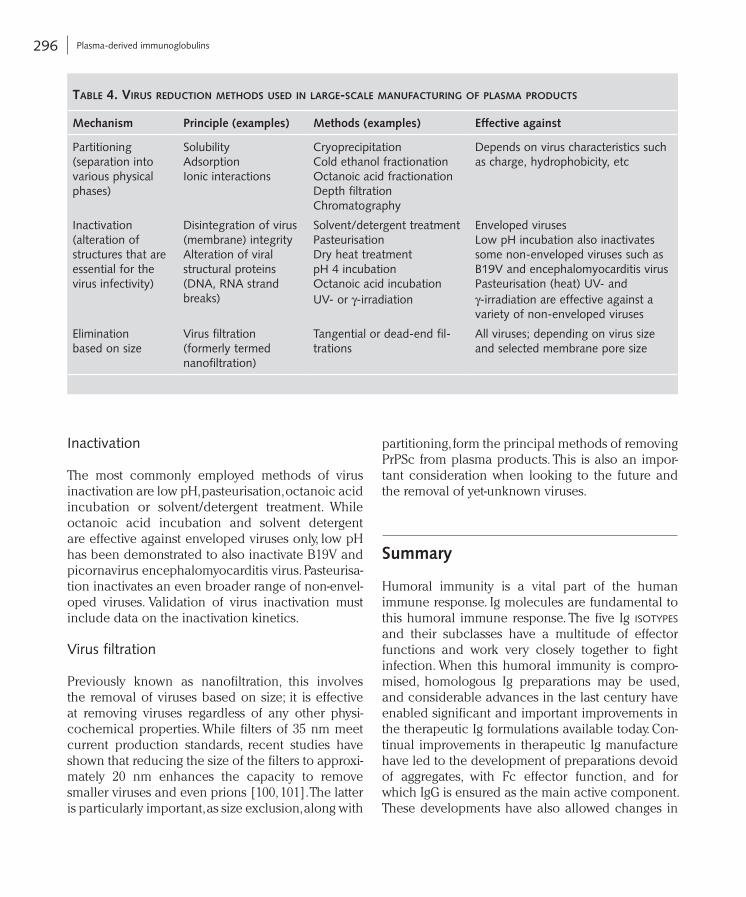
296 Plasma-derived immunoglobulins
partitioning, form the principal methods of removing PrPSc from plasma products. This is also an impor-tant consideration when looking to the future and the removal of yet-unknown viruses.
Summary
Humoral immunity is a vital part of the human immune response. Ig molecules are fundamental to this humoral immune response. The five Ig isotypes and their subclasses have a multitude of effector functions and work very closely together to fight infection. When this humoral immunity is compro-mised, homologous Ig preparations may be used, and considerable advances in the last century have enabled significant and important improvements in the therapeutic Ig formulations available today. Con-tinual improvements in therapeutic Ig manufacture have led to the development of preparations devoid of aggregates, with Fc effector function, and for which IgG is ensured as the main active component. These developments have also allowed changes in
Inactivation
The most commonly employed methods of virus inactivation are low pH, pasteurisation, octanoic acid incubation or solvent/detergent treatment. While octanoic acid incubation and solvent detergent are effective against enveloped viruses only, low pH has been demonstrated to also inactivate B19V and picornavirus encephalomyocarditis virus. Pasteurisa-tion inactivates an even broader range of non-envel-oped viruses. Validation of virus inactivation must include data on the inactivation kinetics.
Virus filtration
Previously known as nanofiltration, this involves the removal of viruses based on size; it is effective at removing viruses regardless of any other physi-cochemical properties. While filters of 35 nm meet current production standards, recent studies have shown that reducing the size of the filters to approxi-mately 20 nm enhances the capacity to remove smaller viruses and even prions [100, 101]. The latter is particularly important, as size exclusion, along with
taBle 4. viruS reduction methodS uSed in large-Scale manuFacturing oF plaSma productS
Mechanism Principle (examples) Methods (examples) Effective against
Partitioning (separation into various physical phases)
Solubility Adsorption Ionic interactions
Cryoprecipitation Cold ethanol fractionation Octanoic acid fractionation Depth filtration Chromatography
Depends on virus characteristics such as charge, hydrophobicity, etc
Inactivation (alteration of structures that are essential for the virus infectivity)
Disintegration of virus (membrane) integrity Alteration of viral structural proteins (DNA, RNA strand breaks)
Solvent/detergent treatment Pasteurisation Dry heat treatment pH 4 incubation Octanoic acid incubation UV- or γ-irradiation
Enveloped viruses Low pH incubation also inactivates some non-enveloped viruses such as B19V and encephalomyocarditis virus Pasteurisation (heat) UV- and γ-irradiation are effective against a variety of non-enveloped viruses
Elimination based on size
Virus filtration (formerly termed nanofiltration)
Tangential or dead-end fil-trations
All viruses; depending on virus size and selected membrane pore size
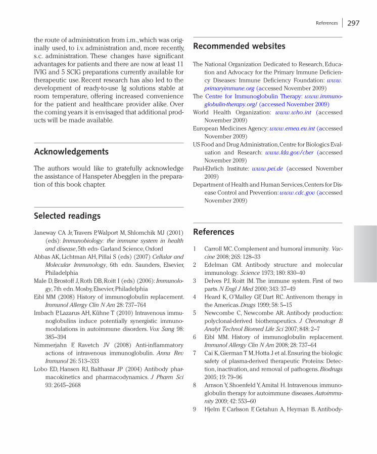
297 References
Recommended websites
The National Organization Dedicated to Research, Educa-tion and Advocacy for the Primary Immune Deficien-cy Diseases: Immune Deficiency Foundation: www.primaryimmune.org (accessed November 2009)
The Centre for Immunoglobulin Therapy: www.immunoglobulintherapy.org/ (accessed November 2009)
World Health Organization: www.who.int (accessed November 2009)
European Medicines Agency: www.emea.eu.int (accessed November 2009)
US Food and Drug Administration, Centre for Biologics Eval-uation and Research: www.fda.gov/cber (accessed November 2009)
Paul-Ehrlich Institute: www.pei.de (accessed November 2009)
Department of Health and Human Services, Centers for Dis-ease Control and Prevention: www.cdc.gov (accessed November 2009)
References
1 Carroll MC. Complement and humoral immunity. Vaccine 2008; 26S: 128–33
2 Edelman GM. Antibody structure and molecular immunology. Science 1973; 180: 830–40
3 Delves PJ, Roitt IM. The immune system. First of two parts. N Engl J Med 2000; 343: 37–49
4 Heard K, O’Malley GF, Dart RC. Antivenom therapy in the Americas. Drugs 1999; 58: 5–15
5 Newcombe C, Newcombe AR. Antibody production: polyclonal-derived biotherapeutics. J Chromatogr B Analyt Technol Biomed Life Sci 2007; 848: 2–7
6 Eibl MM. History of immunoglobulin replacement. Immunol Allergy Clin N Am 2008; 28: 737–64
7 Cai K, Gierman T M, Hotta J et al. Ensuring the biologic safety of plasma-derived therapeutic Proteins: Detec-tion, inactivation, and removal of pathogens. Biodrugs 2005; 19: 79–96
8 Arnson Y, Shoenfeld Y, Amital H. Intravenous immuno-globulin therapy for autoimmune diseases. Autoimmunity 2009; 42: 553–60
9 Hjelm F, Carlsson F, Getahun A, Heyman B. Antibody-
the route of administration from i.m., which was orig-inally used, to i.v. administration and, more recently, s.c. administration. These changes have significant advantages for patients and there are now at least 11 IVIG and 5 SCIG preparations currently available for therapeutic use. Recent research has also led to the development of ready-to-use Ig solutions stable at room temperature, offering increased convenience for the patient and healthcare provider alike. Over the coming years it is envisaged that additional prod-ucts will be made available.
Acknowledgements
The authors would like to gratefully acknowledge the assistance of Hanspeter Abegglen in the prepara-tion of this book chapter.
Selected readings
Janeway CA Jr, Travers P, Walport M, Shlomchik MJ (2001) (eds): Immunobiology: the immune system in health and disease, 5th edn- Garland Science, Oxford
Abbas AK, Lichtman AH, Pillai S (eds) (2007) Cellular and Molecular Immunology, 6th edn. Saunders, Elsevier, Philadelphia
Male D, Brostoff J, Roth DB, Roitt I (eds) (2006): Immunology, 7th edn. Mosby, Elsevier, Philadelphia
Eibl MM (2008) History of immunoglobulin replacement. Immunol Allergy Clin N Am 28: 737–764
Imbach P, Lazarus AH, Kühne T (2010) Intravenous immu-noglobulins induce potentially synergistic immuno-modulations in autoimmune disorders. Vox Sang 98: 385–394
Nimmerjahn F, Ravetch JV (2008) Anti-inflammatory actions of intravenous immunoglobulin. Annu Rev Immunol 26: 513–333
Lobo ED, Hansen RJ, Balthasar JP (2004) Antibody phar-macokinetics and pharmacodynamics. J Pharm Sci 93: 2645–2668
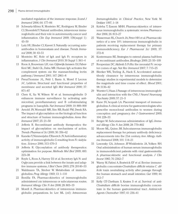
298 Plasma-derived immunoglobulins
Immunoglobulins in Clinical Practice, New York: M. Dekker 1997, 1–18
24 Koleba T, Ensom MHH. Pharmacokinetics of intrave-nous immunoglobulin: a systematic review. Pharmacother 2006; 26: 813–27
25 Wasserman RL, Church JA, Peter HH et al. Pharmacoki-netics of a new 10% intravenous immunoglobulin in patients receiving replacement therapy for primary immunodeficiency. Eur J Pharmaceut Sci 2009; 37: 272–8
26 Kontermann RE. Strategies to extend plasma half-lives of recombinant antibodies. Biodrugs 2009; 23: 93–109
27 Roopenian DC, Akilesh S. FcRn: the neonatal Fc recep-tor comes of age. Nat Rev Immunol 2007; 7: 715–25
28 Bleeker WK, Teeling JL, Hack E. Accelerated autoan-tibody clearance by intravenous immunoglobulin therapy: studies in experimental models to determine the magnitude and time course of effect. Blood 2001; 98: 3136–42
29 Wurster U, Haas J. Passage of intravenous immunoglob-ulin and interaction with the CNS. J Neurol Neurosurg Psychiatry 2009; 57: 21–5
30 Kane SV, Acquah LA. Placental transport of immuno-globulins: A clinical review for gastroenterologists who prescribe monoclonal antibodies to women during conception and pregnancy. Am J Gastroenterol 2009; 104: 228–33
31 Berger M. Subcutaneous administration of IgG. Immunol Allergy Clin N Am 2008; 28: 779–802
32 Moore ML, Quinn JM. Subcutaneous immunoglobulin replacement therapy for primary antibody deficiency: advancements into the 21st century. Ann Allergy Asthma Immunol 2008; 101: 114–21
33 Losonsky GA, Johnson JP, Winklestein JA, Yolken RH. Oral administration of human serum immunoglobulin in immunodeficient patients with viral gastroenteritis. A pharmacokinetic and functional analysis. J Clin Invest 1985; 76: 2362–7
34 Warny M, Fatimi A, Bostwick EF et al. Bovine immuno-globulin concentrate-Clostridium difficile retains C. difficile toxin neutralising activity after passage through the human stomach and small intestine. Gut 1999; 44: 212–7
35 Kelly CP, Chetham S, Keates S et al. Survival of anti-Clostridium difficile bovine immunoglobulin concen-trate in the human gastrointestinal tract. Antimicrob Agents Chemother 1997; 41: 236–41
mediated regulation of the immune response. Scand J Immunol 2006; 64: 177–84
10 Schwartz-Albiez R, Monteiro RC, Rodriguez M, Binder CJ, Shoenfeld Y. Natural antibodies, intravenous immu-noglobulin and their role in autoimmunity, cancer and inflammation. Clin Exp Immunol 2009; 158(suppl 1): 43–50
11 Lutz HU, Binder CJ, Kaveri S. Naturally occurring auto-antibodies in homeostasis and disease. Trends Immunol 2008; 30: 43–51
12 Monteiro RC. Role of IgA and IgA Fc receptors in inflammation. J Clin Immunol 2010; 30 Suppl 1: S61–4
13 Roos A, Bouwman LH, van Gijlswijk-Janssen DJ, Faber-Krol MC, Stahl GL, Daha MR. Human IgA activates the complement system via the mannan-binding lectin pathway. J Immunol 2001; 167: 2861–8
14 Preud’homme J-L, Petit I, Barra A, Morel F, Lecron J-C, Lelièvre. Structural and functional properties of membrane and secreted IgD. Mol Immunol 2000; 37: 871–87
15 Chen K, Xu W, Wilson M et al. Immunoglobulin D enhances immune surveillance by activating anti-microbial, proinflammatory and B cell-stimulating programs in basophils. Nat Immunol 2009; 10: 889–900
16 Arnold JN, Wormald MR, Sim RB, Rudd PM, Dwek RA. The impact of glycosylation on the biological function and structure of human immunoglobulins. Annu Rev Immunol 2007; 25: 21–50
17 Jefferis R. Recombinant antibody therapeutics: the impact of glycosylation on mechanisms of action. Trends Pharmacol Sci 2009; 30: 356–62
18 Kaneko Y, Nimmerjahn F, Ravetch JV. Anti-inflammatory activity of immunoglobulin G resulting from Fc sialyla-tion. Science 2006; 313: 670–3
19 Jefferis R. Glycosylation of antibody therapeutics: optimisation for purpose. Methods Mol Biol 2009; 483: 223–38
20 Royle L, Roos A, Harvey DJ et al. Secretory IgA N- and O-glycans provide a link between the innate and adap-tive immune systems. J Biol Chem 2003; 278: 20140–53
21 Waldmann TA, Strober W. Metabolism of immuno-globulins. Prog Allergy 1969; 13: 1–110
22 Bonilla FA. Pharmacokinetics of immunoglobulin administered via intravenous or subcutaneous routes. Immunol Allergy Clin N Am 2008; 28: 803–19
23 Morell A. Pharmacokinetics of intravenous immuno-globulin preparations. In: Lee ML (ed): Intravenous
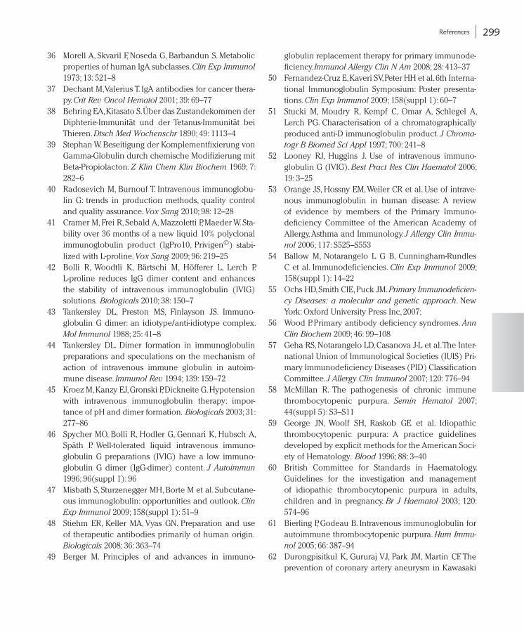
299 References
globulin replacement therapy for primary immunode-ficiency. Immunol Allergy Clin N Am 2008; 28: 413–37
50 Fernandez-Cruz E, Kaveri SV, Peter HH et al. 6th Interna-tional Immunoglobulin Symposium: Poster presenta-tions. Clin Exp Immunol 2009; 158(suppl 1): 60–7
51 Stucki M, Moudry R, Kempf C, Omar A, Schlegel A, Lerch PG. Characterisation of a chromatographically produced anti-D immunoglobulin product. J Chromatogr B Biomed Sci Appl 1997; 700: 241–8
52 Looney RJ, Huggins J. Use of intravenous immuno-globulin G (IVIG). Best Pract Res Clin Haematol 2006; 19: 3–25
53 Orange JS, Hossny EM, Weiler CR et al. Use of intrave-nous immunoglobulin in human disease: A review of evidence by members of the Primary Immuno-deficiency Committee of the American Academy of Allergy, Asthma and Immunology. J Allergy Clin Immunol 2006; 117: S525–S553
54 Ballow M, Notarangelo L G B, Cunningham-Rundles C et al. Immunodeficiencies. Clin Exp Immunol 2009; 158(suppl 1): 14–22
55 Ochs HD, Smith CIE, Puck JM. Primary Immunodeficiency Diseases: a molecular and genetic approach. New York: Oxford University Press Inc, 2007;
56 Wood P. Primary antibody deficiency syndromes. Ann Clin Biochem 2009; 46: 99–108
57 Geha RS, Notarangelo LD, Casanova J-L et al. The Inter-national Union of Immunological Societies (IUIS) Pri-mary Immunodeficiency Diseases (PID) Classification Committee. J Allergy Clin Immunol 2007; 120: 776–94
58 McMillan R. The pathogenesis of chronic immune thrombocytopenic purpura. Semin Hematol 2007; 44(suppl 5): S3–S11
59 George JN, Woolf SH, Raskob GE et al. Idiopathic thrombocytopenic purpura: A practice guidelines developed by explicit methods for the American Soci-ety of Hematology. Blood 1996; 88: 3–40
60 British Committee for Standards in Haematology. Guidelines for the investigation and management of idiopathic thrombocytopenic purpura in adults, children and in pregnancy. Br J Haematol 2003; 120: 574–96
61 Bierling P, Godeau B. Intravenous immunoglobulin for autoimmune thrombocytopenic purpura. Hum Immunol 2005; 66: 387–94
62 Durongpisitkul K, Gururaj VJ, Park JM, Martin CF. The prevention of coronary artery aneurysm in Kawasaki
36 Morell A, Skvaril F, Noseda G, Barbandun S. Metabolic properties of human IgA subclasses. Clin Exp Immunol 1973; 13: 521–8
37 Dechant M, Valerius T. IgA antibodies for cancer thera-py. Crit Rev Oncol Hematol 2001; 39: 69–77
38 Behring EA, Kitasato S. Über das Zustandekommen der Diphterie-Immunität und der Tetanus-Immunität bei Thieren. Dtsch Med Wochenschr 1890; 49: 1113–4
39 Stephan W. Beseitigung der Komplementfixierung von Gamma-Globulin durch chemische Modifizierung mit Beta-Propiolacton. Z Klin Chem Klin Biochem 1969; 7: 282–6
40 Radosevich M, Burnouf T. Intravenous immunoglobu-lin G: trends in production methods, quality control and quality assurance. Vox Sang 2010; 98: 12–28
41 Cramer M, Frei R, Sebald A, Mazzoletti P, Maeder W. Sta-bility over 36 months of a new liquid 10% polyclonal immunoglobulin product (IgPro10, Privigen©) stabi-lized with L-proline. Vox Sang 2009; 96: 219–25
42 Bolli R, Woodtli K, Bärtschi M, Höfferer L, Lerch P. L-proline reduces IgG dimer content and enhances the stability of intravenous immunoglobulin (IVIG) solutions. Biologicals 2010; 38: 150–7
43 Tankersley DL, Preston MS, Finlayson JS. Immuno-globulin G dimer: an idiotype/anti-idiotype complex. Mol Immunol 1988; 25: 41–8
44 Tankersley DL. Dimer formation in immunoglobulin preparations and speculations on the mechanism of action of intravenous immune globulin in autoim-mune disease. Immunol Rev 1994; 139: 159–72
45 Kroez M, Kanzy EJ, Gronski P, Dickneite G. Hypotension with intravenous immunoglobulin therapy: impor-tance of pH and dimer formation. Biologicals 2003; 31: 277–86
46 Spycher MO, Bolli R, Hodler G, Gennari K, Hubsch A, Späth P. Well-tolerated liquid intravenous immuno-globulin G preparations (IVIG) have a low immuno-globulin G dimer (IgG-dimer) content. J Autoimmun 1996; 96(suppl 1): 96
47 Misbath S, Sturzenegger MH, Borte M et al. Subcutane-ous immunoglobulin: opportunities and outlook. Clin Exp Immunol 2009; 158(suppl 1): 51–9
48 Stiehm ER, Keller MA, Vyas GN. Preparation and use of therapeutic antibodies primarily of human origin. Biologicals 2008; 36: 363–74
49 Berger M. Principles of and advances in immuno-
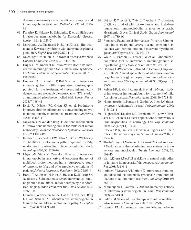
300 Plasma-derived immunoglobulins
74 Gajdos P, Chevret S, Clair B, Tranchant C, Chastang C. Clinical trial of plasma exchange and high-dose intravenous immunoglobulin in myasthenia gravis. Myasthenia Gravis Clinical Study Group. Ann Neurol 1997; 41: 789–96
75 Rønager J, Ravnborg M, Hermansen I, Vorstrup S. Immu-noglobulin treatment versus plasma exchange in patients with chronic moderate to severe myasthenia gravis. Artif Organs 2001; 25: 967–73
76 Wolfe GI, Barohn RJ, Foster BM et al. Randomized, controlled trial of intravenous immunoglobulin in myasthenia gravis. Muscle Nerve 2002; 26: 549–52
77 Hartung H-P, Mouthon L, Ahmed R, Jordan S, Laupland KB, Jolles S. Clinical applications of intravenous immu-noglobulins (IVIg) – beyond immunodeficiencies and neurology. Clin Exp Immunol 2009; 158(suppl 1): 23–33
78 Relkin NR, Szabo P, Adamiak B et al. 18-Month study of intravenous immunoglobulin for treatment of mild Alzheimer disease. Neurobiol Aging 2009; 30: 1728–36
79 Hammarström L, Hansen S, Gardulf A. Does IgG thera-py prevent Alzheimer’s disease? J Neuroimmunol 2009; 215: 122–4
80 Hughes RAC, Dalakas MC, Cornblath DR, Latov N, Wek-sler ME, Relkin N. Clinical applications of intravenous immunoglobulins in neurology. Clin Exp Immunol 2009; 158(suppl 1): 34–42
81 Crocker P R, Paulson J C, Varki A. Siglecs and their roles in the immune system. Nat Rev Immunol 2007; 7: 255–66
82 Tha-In T, Bayry J, Metselaar HJ, Kaveri SV, Kwekkeboom J. Modulation of the cellular immune system by intra-venous immunoglobulin. Trends Immunol 2008; 29: 608–15
83 Vani J, Elluru S, Negi VS et al. Role of natural antibodies in immune homeostasis: IVIg perspective. Autoimmun Rev 2008; 7: 440–4
84 Imbach P, Lazarus AH, Kühne T. Intravenous immuno-globulins induce potentially synergistic immunomod-ulations in autoimmune disorders. Vox Sang 2010; 98: 385–94
85 Nimmerjahn F, Ravetch JV. Anti-inflammatory actions of intravenous immunoglobulin. Annu Rev Immunol 2008; 26: 513–33
86 Ballow M. Safety of IGIV therapy and infusion-related adverse events. Immunol Res 2007; 38: 122–32
87 Bonilla FA. Intravenous immunoglobulin: adverse
disease: a meta-analysis on the efficacy of aspirin and immunoglobulin treatment. Pediatrics 1995; 96: 1057–61
63 Furusho K, Nakano H, Shinomiya K et al. High-dose intravenous gammaglobulin for Kawasaki disease. Lancet 1984; 2: 1055–7
64 Newburger JW, Takahashi M, Burns JC et al. The treat-ment of Kawasaki syndrome with intravenous gamma globulin. N Engl J Med 1986; 315: 341–7
65 Newburger JW, Fulton DR. Kawasaki disease. Curr Treat Options Cardiovasc Med 2007; 9: 148–58
66 Hughes RAC, Raphaël JC, Swan AV, van Doorn PA. Intra-venous immunoglobulin for Guillain-Barré syndrome. Cochrane Database of Systematic Reviews 2007; 1: CD002063
67 Hughes RAC, Donofrio P, Bril V et al. Intravenous immune globulin (10% caprylate-chromatography purified) for the treatment of chronic inflammatory demyelinating polyradiculoneuropathy (ICE study): a randomised placebo-controlled trial. Lancet Neurol 2008; 7: 136–44
68 Dyck PJ, O’Brien PC, Oviatt KF et al. Prednisone improves chronic inflammatory demyelinating polyra-diculoneuropathy more than no treatment. Ann Neurol 1982; 11: 136–41
69 van Schaik IN, van den Berg LH, de Haan R, Vermeulen M. Intravenous immunoglobulin for multifocal motor neuropathy. Cochrane Database of Systematic Reviews 2005; 2: CD004429
70 Federico P, Zochodne DW, Hahn AF, Brown WF, Feasby TE. Multifocal motor neuropathy improved by IVIg: randomised, double-blind, placebo-controlled study. Neurology 2000; 55: 1256–62
71 Léger J-M, Viala K, Cancalon F et al. Intravenous immunoglobulin as short- and long-term therapy of multifocal motor neuropathy: a retrospective study of response to IVIg and of its predictive criteria in 40 patients. J Neurol Neurosurg Psychiatry 2008; 79: 93–6
72 Harbo T, Andersen H, Hess A, Hansen K, Sindrup SH, Jakobsen J. Subcutaneous versus intravenous immu-noglobulin in multifocal motor neuropathy: a random-ized, single-blinded cross-over trial. Eur J Neurol 2009; 16: 631–8
73 Eftimov F, Vermeulen M, de Haan RJ, van den Berg LH, van Schaik IN. Subcutaneous immunoglobulin therapy for multifocal motor neuropathy. J Peripher Nerv Syst 2009; 14: 93–100
301 References
99 Winward DB, Brophy MT. Acute renal failure after administration of intravenous immunoglobulin: review of the literature and case report. Pharmacotherapy 1995; 15: 765–72
100 Soluk L, Price H, Sinclair C, Atalla-Mikhail D, Genereux M. Pathogen safety of intravenous Rh immunoglob-ulin liquid and other immune globulin products: enhanced nanofiltration and manufacturing process overview. Am J Ther 2008; 15: 435–43
101 Stucki M, Boschetti N, Schaefer W et al. Investigations of prion and virus safety of a new liquid IVIG product. Biologicals 2008; 36: 239–47
102 Male D, Brostoff J, Roth D, Roitt I. Immunology, 7th edn, 2006
103 Abbas A K. Cellular and Molecular Immunology, 6th edn, 2009
104 Lobo ED, Hansen RJ, Balthasar JP. Antibody pharma-cokinetics and pharmacodynamics. J Pharm Sci 2004; 93: 2645–68
reactions and management. J Allergy Clin Immunol 2008; 122: 1238–9
88 Aukrust P, Froland SS, Laibakk NB et al. Release of cytokines, soluble cytokine receptors, and interleukin-1 receptor antagonist after intravenous immunoglobulin administration in vivo. Blood 1994; 84: 2136–43
89 Brennan VM, Salome-Bentley NJ, Chapel HM. Prospec-tive audit of adverse reactions occurring in 459 prima-ry antibody-deficient patients receiving intravenous immunoglobulin. Clin Exp Immunol 2003; 133: 247–51
90 Horn J, Thon V, Bartonkova D et al. Anti-IgA antibodies in common variable immunodeficiencies (CVID): diagnostic workup and therapeutic strategy. Clin Immunol 2007; 122: 156–62
91 Sekul EA, Cupler EJ, Dalakas MC. Aseptic meningitis associated with high-dose intravenous immunoglobu-lin therapy: frequency and risk factors. Ann Intern Med 1994; 121: 259–62
92 Daw Z, Padmore R, Neurath D et al. Hemolytic trans-fusion reactions after administration of intravenous immune (gamma) globulin: a case series analysis. Transfusion 2008; 48: 1598–601
93 Kahwaji J, Barker E, Pepkowitz S et al. Acute hemolysis after high-dose intravenous immunoglobulin therapy in highly HLA sensitized patients. Clin J Am Soc Nephrol 2009; 4: 1993–7
94 Vo AA, Cam V, Toyoda M et al. Safety and adverse events profiles of intravenous gammaglobulin products used for immunomodulation: a single-center experience. Clin J Am Soc Nephrol 2006; 1: 844–52
95 Vecchietti G, Kerl K, Prins C, Kaya G, Saurat JH, French LE. Severe eczematous skin reactions after high-dose intravenous immunoglobulin infusion: report of 4 cases and review of the literature. Arch Dermatol 2006; 142: 213–7
96 Dickenmann M, Oettl T, Mihatsch MJ. Osmotic nephro-sis: acute kidney injury with accumulation of proximal tubular lysosomes due to administration of exogenous solutes. Am J Kidney Dis 2008; 51: 491–503
97 Anonymous. Renal insufficiency and failure associ-ated with immune globulin intravenous therapy – United States. 1985–1998. MMWR 1999; 48: 518–21
98 Chapman SA, Gilkerson KL, Davin TD, Pritzker MR. Acute renal failure and intravenous immune globulin: occurs with sucrose-stabilized, but not with D-sorbitol-stabilized formulation. Ann Pharmacother 2004; 38: 2059–67