Embed Size (px)
Citation preview

Principles of diagnsosis of ischemic heart diseaseMohammad Hashemi
Interventional cardiologist
Department of cardiology
Ischemic chest pain due to coronary artery disease (CAD)
stable angina pectoris, unstable angina, non-ST elevation myocardial infarction, and ST elevation myocardial infarction.

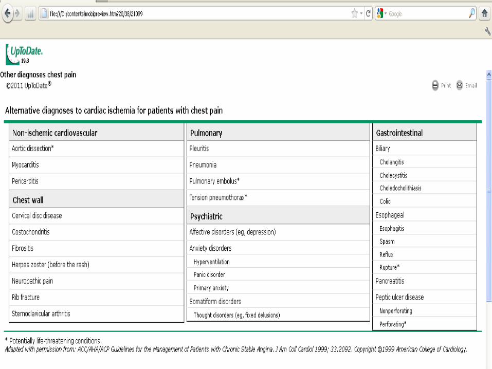
The differential diagnosis of patients presenting with chest pain is extensive
Ischemic chest pain due to coronary artery disease Patients classically complain of chest heaviness,
pressure, tightness or burning, but may vigorously deny "pain.
Other descriptions such as provocation with physical or emotional stress or cold, relief with rest, and radiation to the neck, jaw, and shoulder are common.
Ischemic pain usually lasts more than 2 but less than 20 minutes, unless a myocardial infarction is occurring.
Associated symptoms may include dyspnea, nausea and vomiting, diaphoresis, presyncope, or palpitations.
The correct diagnosis is based on
detailed history (pain description; associated symptoms; and in some cases disease risk factors) that is
supported by specific physical findings,
an electrocardiogram, and/or chest x-ray.

EVALUATION
The office evaluation of new onset chest pain in stable individuals should begin with the consideration of imminently life-threatening causes (including acute coronary syndrome, pulmonary embolus, aortic dissection, pneumothorax, and esophageal rupture).

This is usually accomplished using clinical judgement, along with ECG testing, and less frequently exercise testing, other noninvasive testing, or invasive angiography.
The clinical evaluation is also useful to estimate the pretest probability of organic causes of chest pain prior to undergoing diagnostic tests (eg, stress ECG testing to detect CHD or d-dimer, CTa-angiography or lung perfusion scanning to detect pulmonary embolism).
Pretest probability is an important component of the interpretation of these test results.
The history typical chest pain is less useful in patients suspected of having an acute coronary syndrome (ACS)
Conversely, in patients with stable, intermittent chest pain, a description of typical angina (ie, substernal discomfort, precipitated by exertion, improved with rest or nitroglycerin in less than ten minutes with many patients reporting radiation to shoulders, jaw, or inner arm) was a moderately strong predictor of obstructive epicardial CHD .
Description of chest pain
Descriptions of chest discomfort due to myocardial ischemia may differ depending on: patients' culture,
gender,
age
and presence of comorbid conditions such as diabetes.

Quality of the pain
Region or location of painRadiationProvocationSeverity Palliation

Associated symptoms
Belching, Belching,
Vomiting
Diaphoresis
Dyspnea
Cough
Syncope
Palpitations
Psychiatric symptoms
Constitutional symptoms —particularly in elderly may describe profound fatigue.
Risk factors
The clinical impression raised by the patient's description of pain must be interpreted together with other aspects of the history, including risk factors for various etiologies of chest pain

Risk factors
A history or lack of classical coronary risk factors does not usefully discriminate ischemic causes of chest pain in the acute setting
Physical examination
A brief, "core" examination may suffice to diagnose life-threatening and common etiologies of chest pain. The general appearance
A full set of vital signs
Palpation of the chest wall may evoke pain
A complete cardiac examination including auscultation and palpation
Determine if the breath sounds are symmetric
A careful examination of the abdomen is important
Focal neurologic signs
Electrocardiogram
Normal electrocardiogram A normal ECG markedly reduces
the probability that chest pain is due to acute myocardial infarction, but it does not exclude a serious cardiac etiology (particularly unstable angina).

Abnormal electrocardiogram
The ECG is valuable both for risk stratification and diagnosis of acute myocardial infarction.
Stress testing for the diagnosis of coronary heart disease
The major questions to be addressed before selecting one or more of the available diagnostic tests are:
What is the patient's pretest risk of CHD?
How accurate are the alternative tests?
What are the costs and effects on health outcomes of each test?
Do special considerations make one test more suitable in a specific patient?
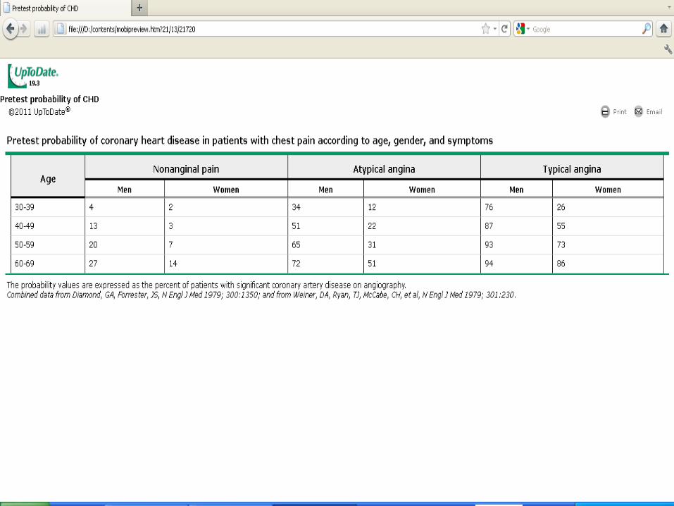
PRETEST PROBABILITY
An initial estimate of the likelihood of CHD is based upon the clinical history.
The pretest probability determines the need for noninvasive diagnostic tests or even coronary arteriography

Estimating pretest probability
Definite or classic angina — Substernal chest discomfort characterized by all of the following characteristics: a typical quality and duration, provocation by exertion or emotional stress, and relief by rest or nitroglycerin
Probable or atypical angina — Chest pain with two of the three above characteristics
Nonanginal or nonischemic chest pain — Chest pain with one or none of the above characteristic
Estimating pretest probability
It should be noted that asymptomatic patients have a low pretest probability of significant CHD, and generally are not screened by use of stress testing.
Symptomatic patients with moderate risk of CHD may be considered for stress testing .
NON-INVASIVE TESTS
Exercise ECG testing is the most commonly used noninvasive test because it is simple and inexpensive.
Echocardiography using either exercise or pharmacologic (dobutamine or dipyridamole) stress.
Radionuclide myocardial perfusion imaging using either exercise or pharmacologic stress.
Women
Diagnosing coronary artery disease is more difficult in women than men for the following reasons: Women are less likely to present with (or report
during treadmill testing) typical angina.
False positives test results during exercise ECG testing are more common.
Limitations of exercise stress
The exercise ECG cannot be interpreted in the presence of resting ST segment changes, left ventricular hypertrophy, left bundle branch block, a ventricular paced rhythm, or the Wolff Parkinson White syndrome.
The patient must also be able to exercise adequately,

Exercise ECG testing is the most commonly used noninvasive test because it is simple and inexpensive.
INVASIVE TESTS
Coronary angiography is the definitive diagnostic test for the presence of obstructive coronary artery disease.
However, it is seldoam used as the initial test because of its invasive nature and because the presence of a lesion does not prove that the patient's symptoms are due to ischemia.