Embed Size (px)
Citation preview
Principles of Anatomy and Physiology
Thirteenth Edition
Chapter 22The Lymphatic System and Immunity
Copyright © 2012 by John Wiley & Sons, Inc.
Gerard J. Tortora • Bryan H. Derrickson
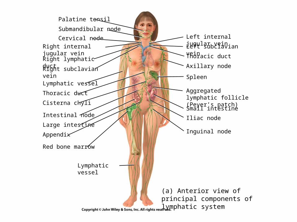
(a) Anterior view of principal components of lymphatic system
Palatine tonsil
Submandibular node
Cervical node
Right internal jugular vein
Right lymphatic duct
Right subclavian vein
Lymphatic vessel
Thoracic duct
Cisterna chyli
Intestinal node
Large intestine
Appendix
Red bone marrow
Lymphatic vessel
Left internal jugular vein
Left subclavian vein
Thoracic duct
Axillary node
Spleen
Aggregated lymphatic follicle (Peyer’s patch)
Small intestine
Iliac node
Inguinal node
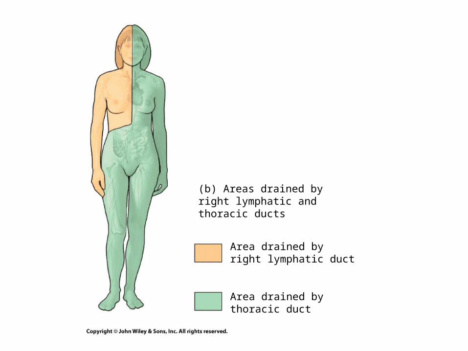
(b) Areas drained by right lymphatic and thoracic ducts
Area drained byright lymphatic duct
Area drained bythoracic duct
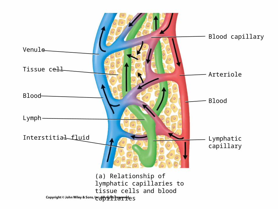
(a) Relationship of lymphatic capillaries to tissue cells and blood capillaries
Venule
Tissue cell
Lymph
Interstitial fluid
Blood
Blood capillary
Arteriole
Lymphatic capillary
Blood
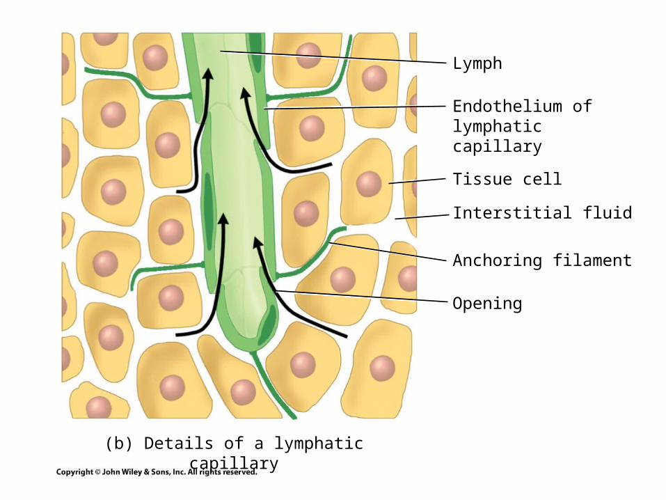
(b) Details of a lymphatic capillary
Lymph
Endothelium of lymphatic capillary
Tissue cell
Interstitial fluid
Anchoring filament
Opening
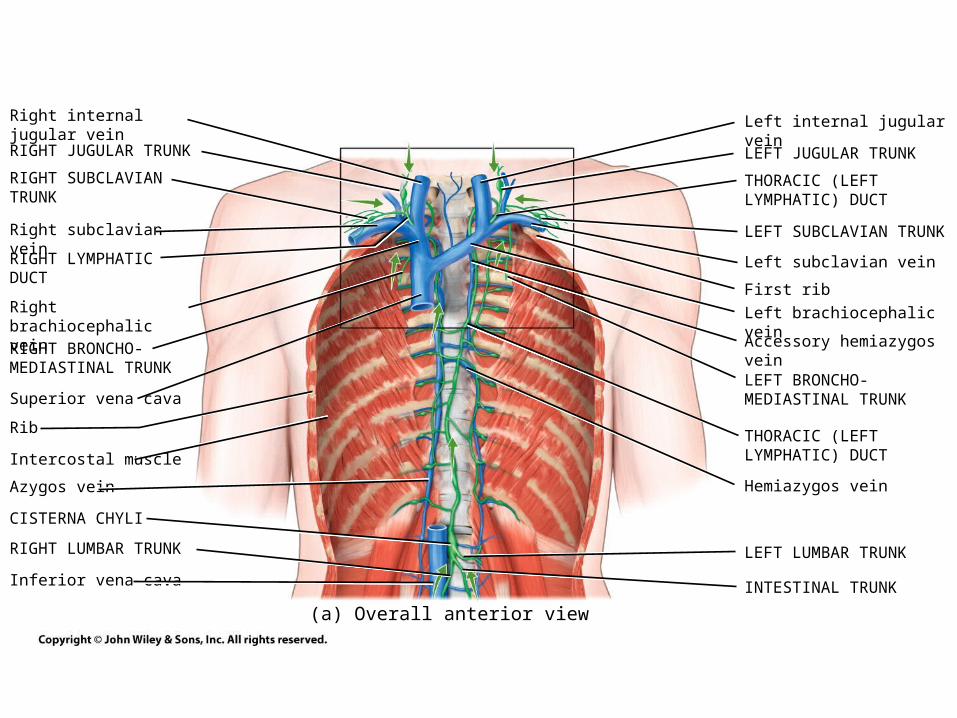
(a) Overall anterior view
Right internal jugular vein
RIGHT JUGULAR TRUNK
RIGHT SUBCLAVIAN TRUNK
Right subclavian vein
RIGHT LYMPHATIC DUCT
Right brachiocephalic vein
RIGHT BRONCHO-MEDIASTINAL TRUNK
Superior vena cava
Rib
Intercostal muscle
Azygos vein
CISTERNA CHYLI
RIGHT LUMBAR TRUNK
Inferior vena cava
Left internal jugular vein
LEFT JUGULAR TRUNK
THORACIC (LEFT LYMPHATIC) DUCT
LEFT SUBCLAVIAN TRUNK
Left subclavian vein
First rib
Left brachiocephalic vein
Accessory hemiazygos vein
LEFT BRONCHO-MEDIASTINAL TRUNK
THORACIC (LEFT LYMPHATIC) DUCT
Hemiazygos vein
LEFT LUMBAR TRUNK
INTESTINAL TRUNK
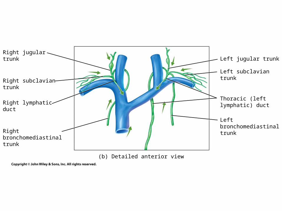
(b) Detailed anterior view
Right jugular trunk
Right subclavian trunk
Right lymphatic duct
Right bronchomediastinal trunk
Left jugular trunk
Left subclavian trunk
Thoracic (left lymphatic) duct
Left bronchomediastinal trunk
Lymphatic duct
SYSTEMIC CIRCULATION PULMONARY CIRCULATION
Subclavian vein
Lymphatic vessel
Valve
Lymph node
Lymphatic capillariesSystemic blood capillaries
Arteries
Lymphatic capillaries
Pulmonary blood capillaries
Lymph node
Veins
Heart
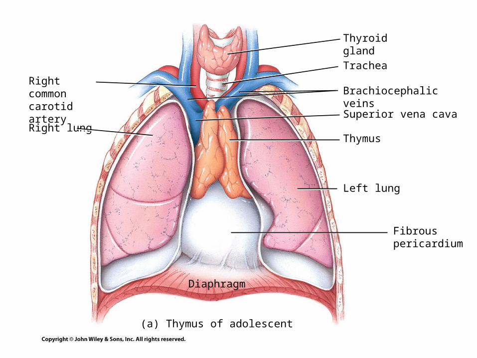
Right commoncarotid artery
(a) Thymus of adolescent
Right lung
Thyroid gland
Trachea
Superior vena cava
Thymus
Left lung
Fibrouspericardium
Diaphragm
Brachiocephalic veins
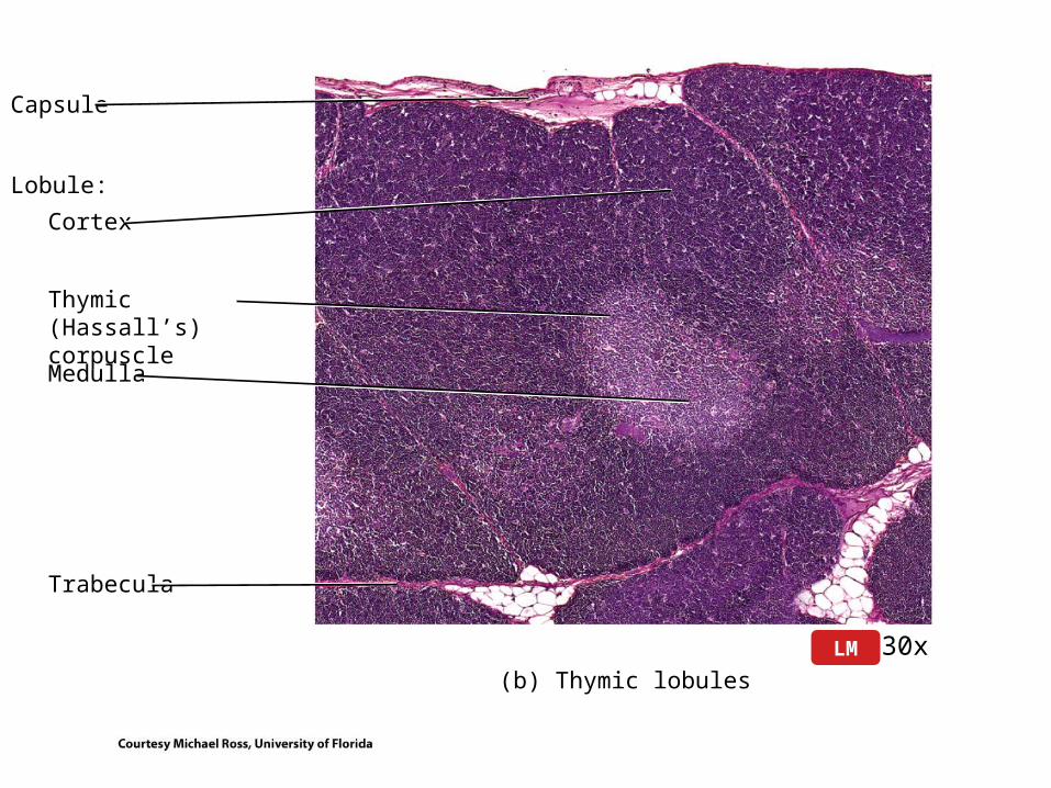
Capsule
(b) Thymic lobules
30xLM
Lobule:
Cortex
Thymic (Hassall’s)corpuscle
Medulla
Trabecula
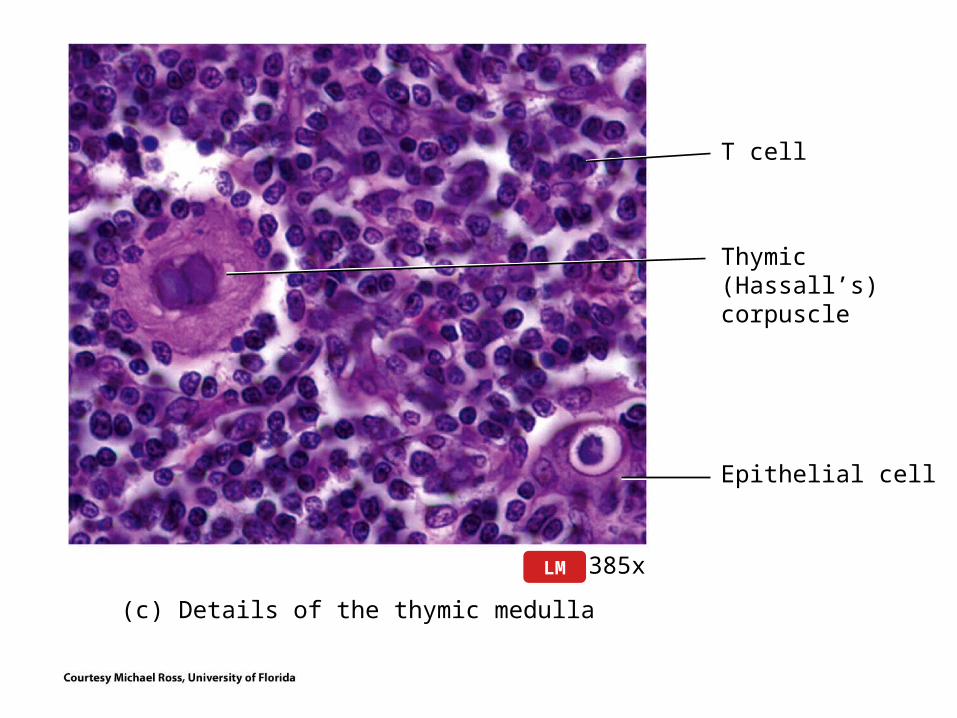
(c) Details of the thymic medulla
385xLM
T cell
Thymic (Hassall’s)corpuscle
Epithelial cell
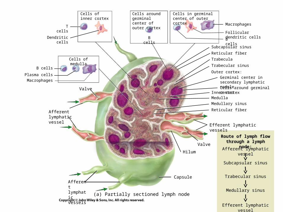
(a) Partially sectioned lymph node
Valve
Afferent lymphaticvessel
Afferent lymphatic vessels
Subcapsular sinus
Reticular fiber
Trabecula
Trabecular sinus
Outer cortex:
Germinal center in secondary lymphatic nodule
Cells around germinal centerInner cortexMedulla
Medullary sinus
Reticular fiber
Efferent lymphatic vessels
Valve
Hilum
Capsule
Cells in germinal center of outer cortex
B cells
Follicular dendritic cells
Macrophages
Cells around germinal center of outer cortex
B cells
Cells of inner cortex
T cells
Dendritic cells
Cells of medulla
B cells
Plasma cells
Macrophages
Route of lymph flowthrough a lymph node:
Afferent lymphatic vessel
Subcapsular sinus
Trabecular sinus
Efferent lymphatic vessel
Medullary sinus
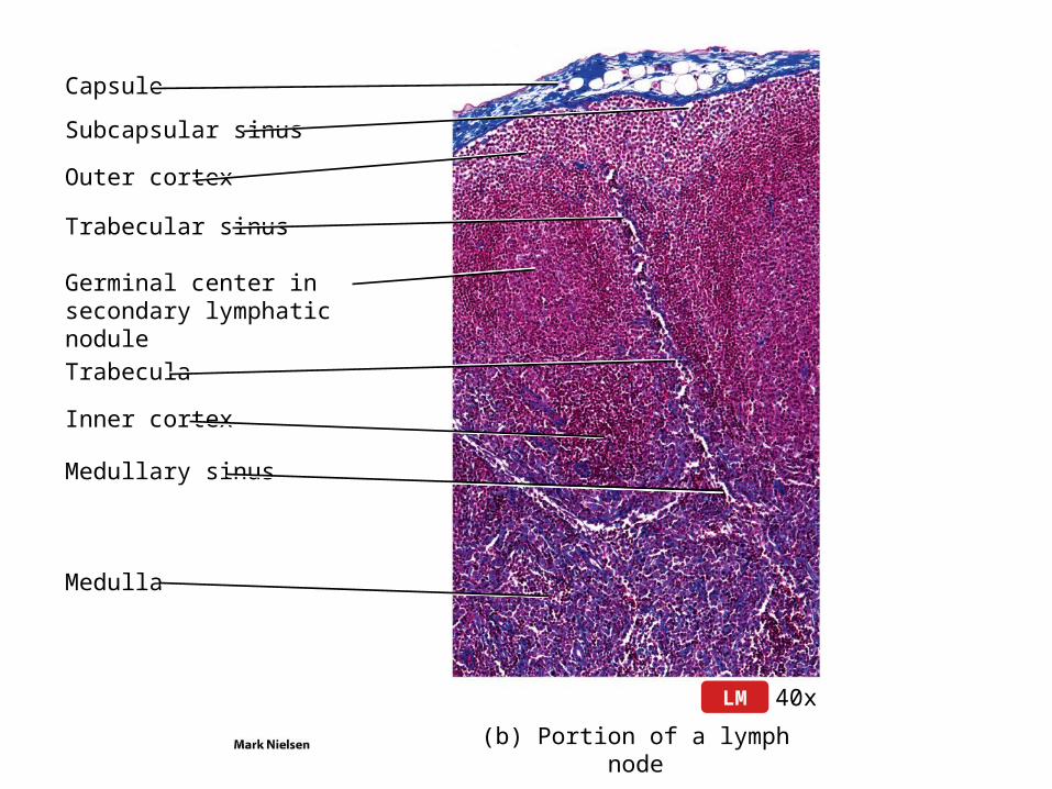
Capsule
(b) Portion of a lymph node
40xLM
Subcapsular sinus
Outer cortex
Trabecular sinus
Germinal center in secondary lymphatic nodule
Trabecula
Inner cortex
Medullary sinus
Medulla
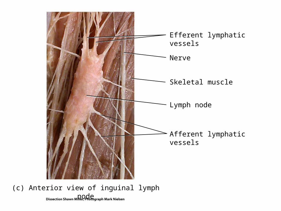
(c) Anterior view of inguinal lymph node
Efferent lymphaticvessels
Nerve
Skeletal muscle
Lymph node
Afferent lymphaticvessels
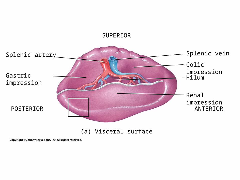
(a) Visceral surface
Splenic vein
SUPERIOR
POSTERIOR ANTERIOR
Colic impression
Hilum
Renal impression
Splenic artery
Gastric impression
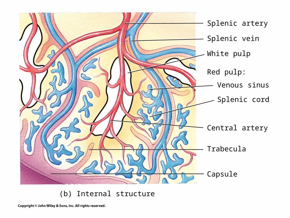
(b) Internal structure
Splenic artery
Splenic vein
White pulp
Red pulp:
Venous sinus
Splenic cord
Central artery
Trabecula
Capsule
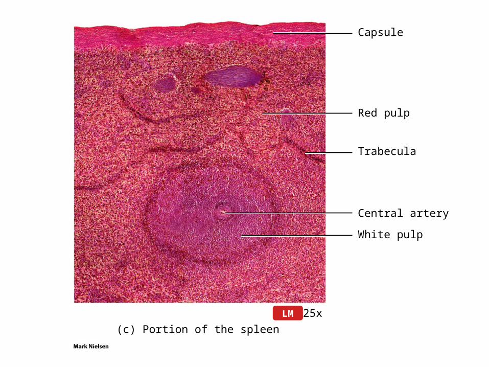
(c) Portion of the spleen
25xLM
Capsule
Red pulp
Trabecula
Central artery
White pulp
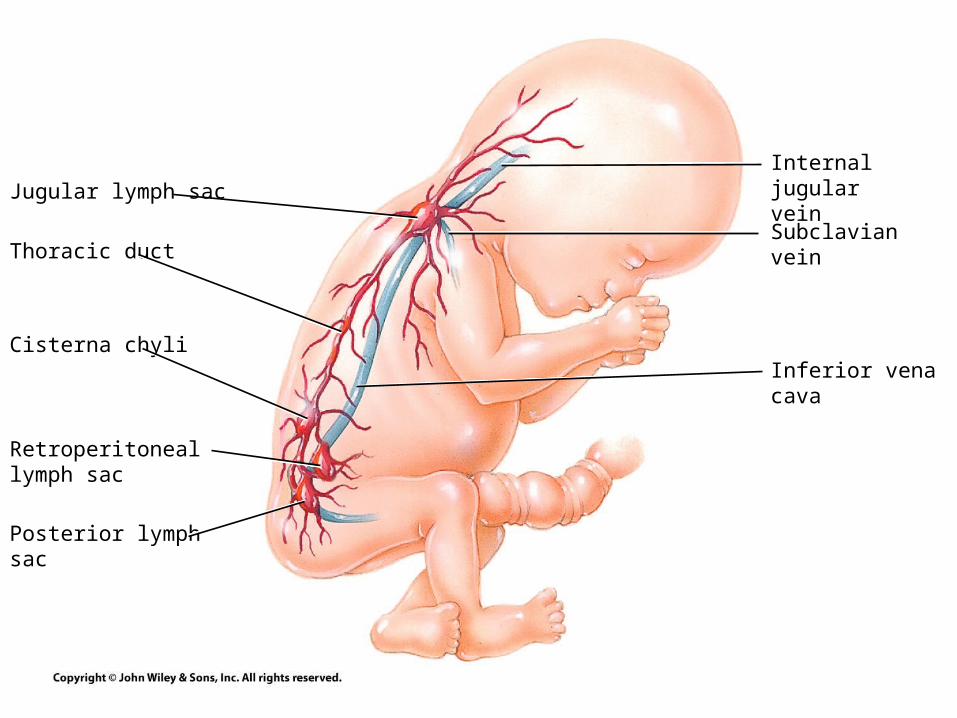
Internal jugular vein
Subclavian vein
Inferior vena cava
Jugular lymph sac
Thoracic duct
Cisterna chyli
Retroperitoneal lymph sac
Posterior lymph sac
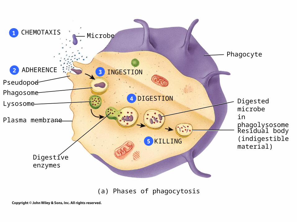
CHEMOTAXIS Microbe
ADHERENCE
Pseudopod
Phagosome
Lysosome
Plasma membrane
Digestiveenzymes
INGESTION
DIGESTION
KILLING
Phagocyte
Digested microbein phagolysosome
Residual body(indigestiblematerial)
1
2 3
4
5
(a) Phases of phagocytosis
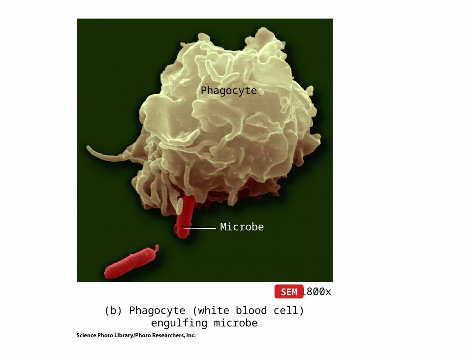
Phagocyte
Microbe
(b) Phagocyte (white blood cell) engulfing microbe
1800xSEM
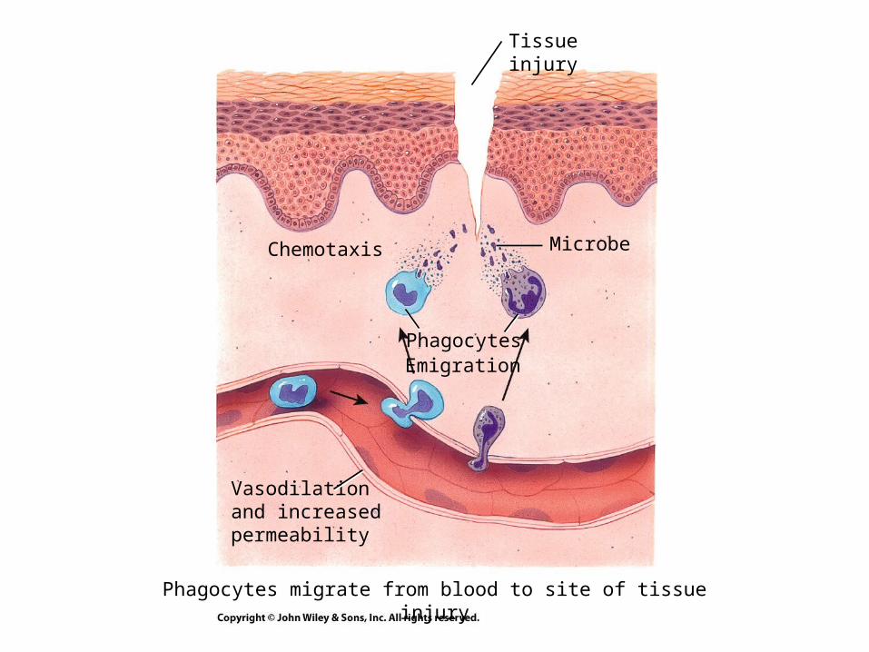
Chemotaxis Microbe
PhagocytesEmigration
Vasodilationand increasedpermeability
Tissue injury
Phagocytes migrate from blood to site of tissue injury
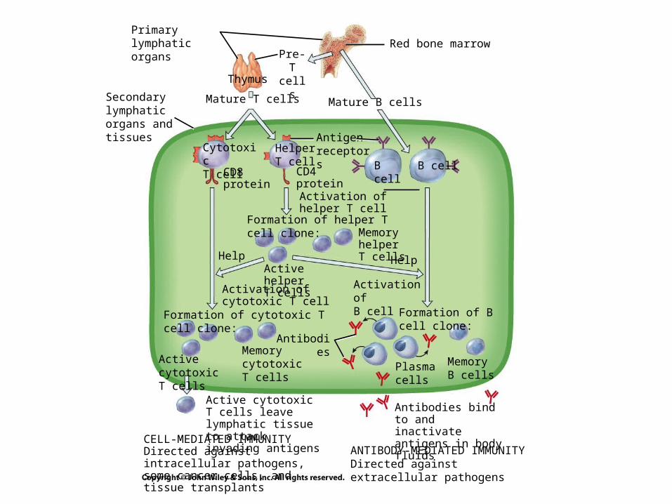
Primary lymphaticorgans Red bone marrow
Thymus
Pre-T cells
Mature T cells Mature B cells
CytotoxicT cell
HelperT cell B cell B cell
Antigenreceptors
CD4proteinActivation ofhelper T cell
Formation of helper T cell clone:MemoryhelperT cells HelpHelp
Active helperT cells
Activation ofcytotoxic T cell
Activation ofB cell
Formation of B cell clone:
Formation of cytotoxic T cell clone:
Active cytotoxicT cells
MemorycytotoxicT cells
Antibodies
Plasma cells
MemoryB cells
Antibodies bind to and inactivate antigens in body fluids
Active cytotoxic T cells leave lymphatic tissue to attack invading antigens
Secondary lymphatic organs and tissues
CD8protein
CELL-MEDIATED IMMUNITYDirected against intracellular pathogens, some cancer cells, and tissue transplants
ANTIBODY-MEDIATED IMMUNITYDirected against extracellular pathogens
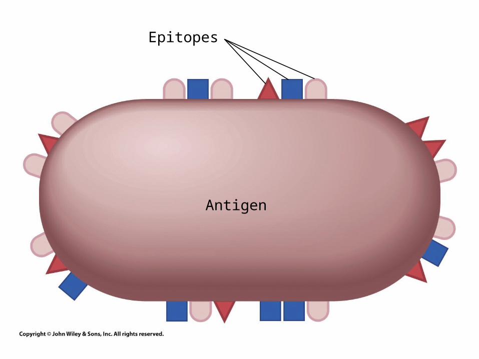
Epitopes
Antigen
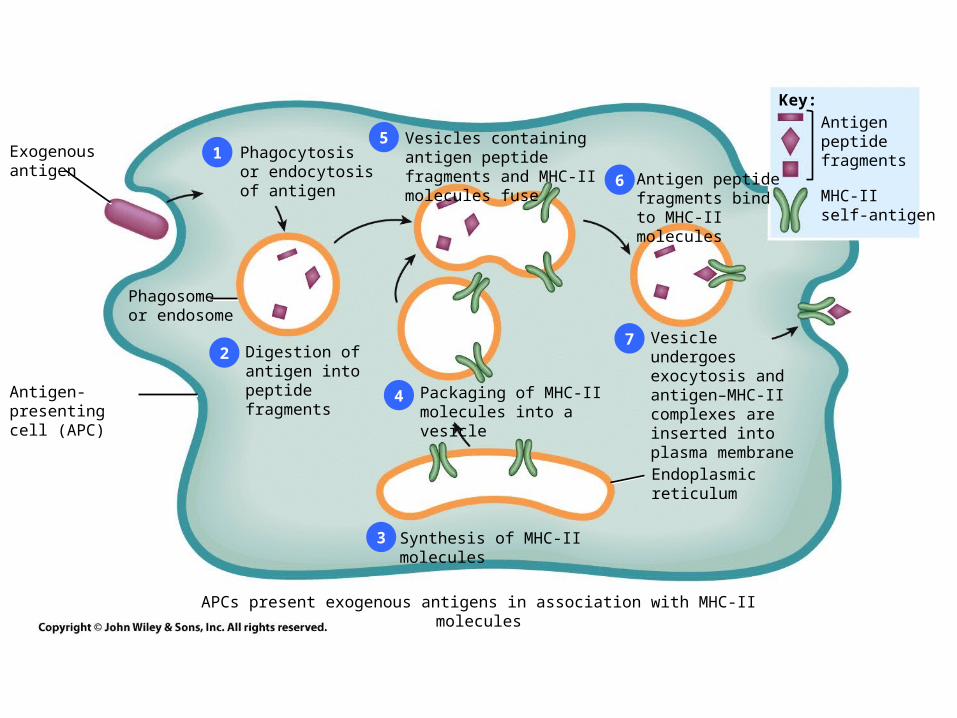
Exogenousantigen
1
2
3
4
5
Phagosomeor endosome
Digestion ofantigen intopeptide fragmentsAntigen-presenting
cell (APC)Packaging of MHC-IImolecules into a vesicle
Synthesis of MHC-II molecules
Vesicles containing antigen peptide fragments and MHC-II molecules fuse Antigen peptide
fragments bind to MHC-II molecules
Vesicle undergoesexocytosis and antigen–MHC-II complexes are inserted into plasma membrane
Endoplasmicreticulum
Key:
Antigenpeptidefragments
MHC-IIself-antigen
Phagocytosis or endocytosis of antigen
6
7
APCs present exogenous antigens in association with MHC-II molecules
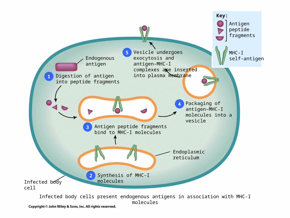
1
2
3
4
Endogenousantigen
Digestion of antigeninto peptide fragments
Antigen peptide fragmentsbind to MHC-I molecules
Synthesis of MHC-I molecules
Endoplasmicreticulum
Packaging ofantigen–MHC-Imolecules into a vesicle
Vesicle undergoes exocytosis and antigen–MHC-I complexes are inserted into plasma membrane
Infected bodycell
Key:
Antigenpeptidefragments
MHC-Iself-antigen
5
Infected body cells present endogenous antigens in association with MHC-I molecules
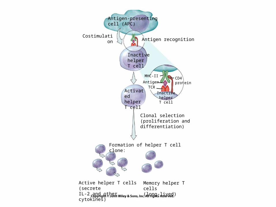
Antigen-presentingcell (APC)
CostimulationAntigen recognition
InactivehelperT cell
ActivatedhelperT cell
Formation of helper T cell clone:
Clonal selection(proliferation anddifferentiation)
Active helper T cells (secreteIL-2 and other cytokines)
Memory helper T cells(long-lived)
Inactive helperT cell
MHC-II
AntigenTCR
CD4protein
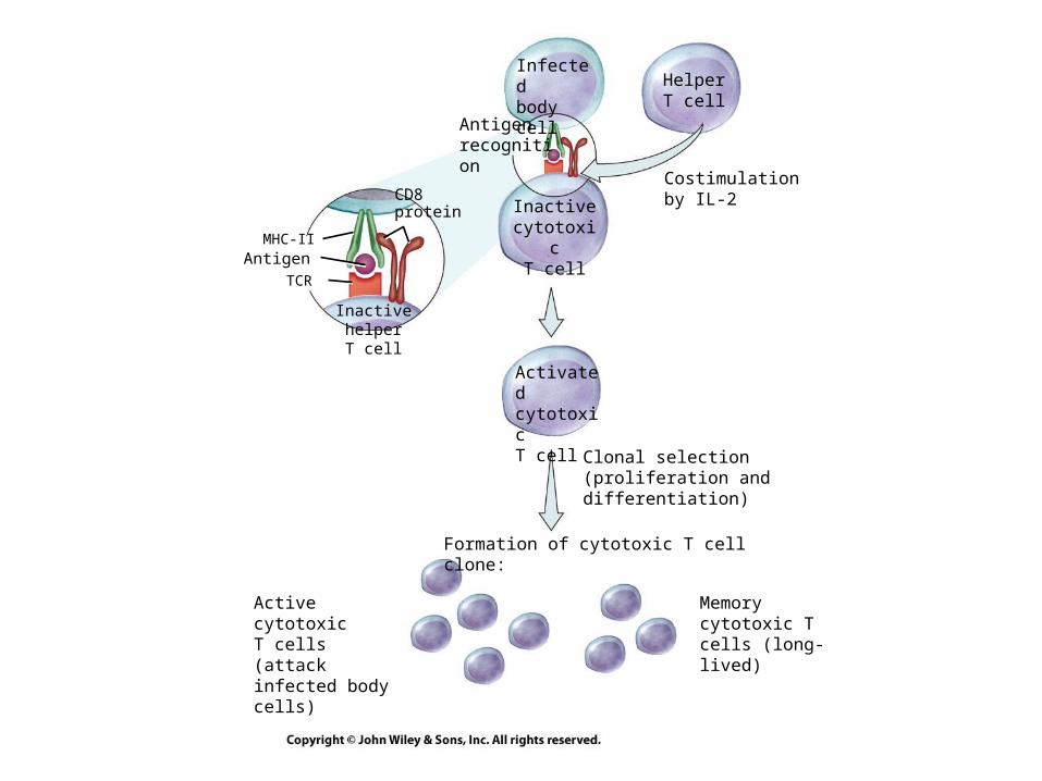
Infectedbody cell Helper T
cell
Costimulationby IL-2
Antigenrecognition
Inactive cytotoxic
T cell
ActivatedcytotoxicT cell
Clonal selection(proliferation anddifferentiation)
Formation of cytotoxic T cell clone:
Inactive helperT cell
MHC-IIAntigen
TCR
CD8protein
Active cytotoxic T cells (attack infected body cells)
Memory cytotoxic T cells (long-lived)
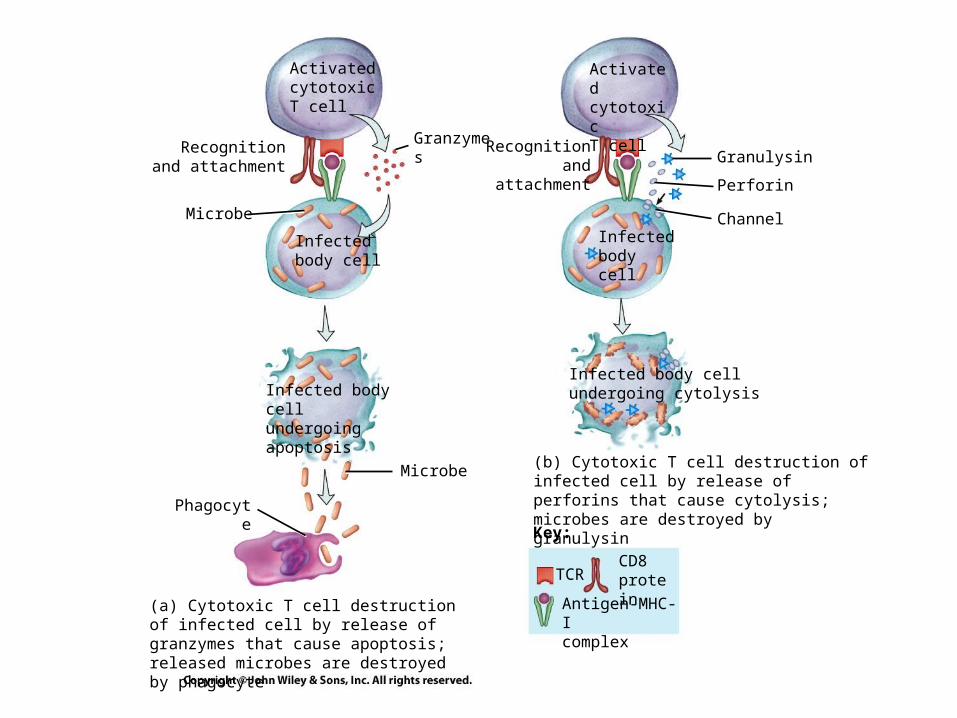
ActivatedcytotoxicT cell
ActivatedcytotoxicT cell
Recognitionand attachment
Microbe
Infectedbody cell
Infected body cellundergoing apoptosis
Microbe
Phagocyte
Granzymes Recognitionand attachment
Infectedbody cell
Infected body cellundergoing cytolysis
(b) Cytotoxic T cell destruction of infected cell by release of perforins that cause cytolysis; microbes are destroyed by granulysin
Granulysin
Perforin
Channel
Key:
CD8 protein
Antigen-MHC-Icomplex
TCR
(a) Cytotoxic T cell destruction of infected cell by release of granzymes that cause apoptosis; released microbes are destroyed by phagocyte
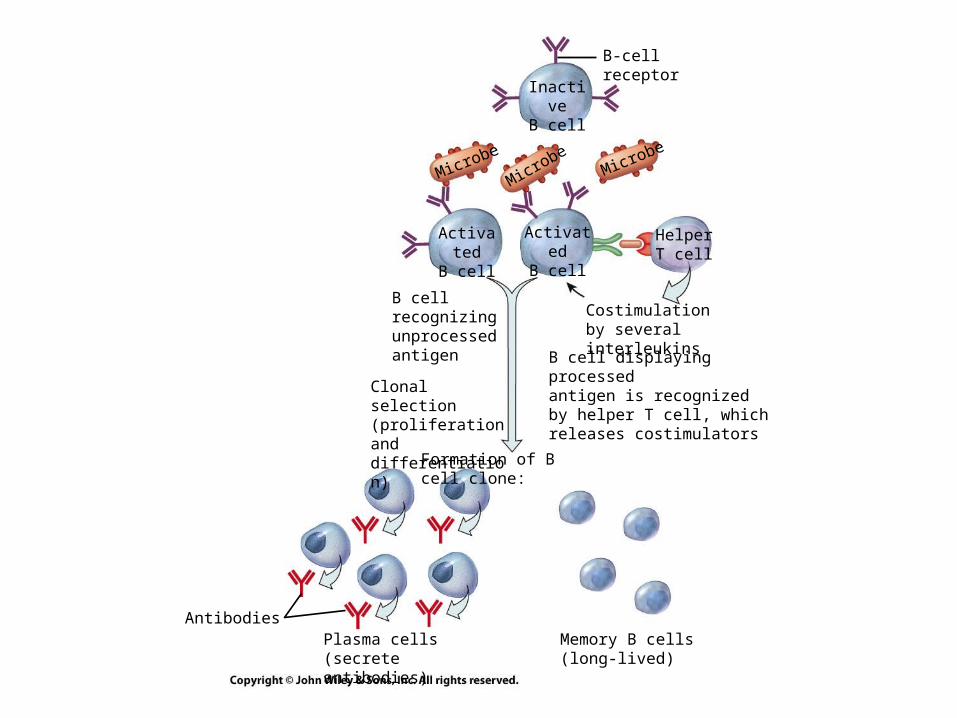
B-cellreceptor
InactiveB cell
Microbe
MicrobeMicrobe
ActivatedB cell
ActivatedB cell
HelperT cell
B cellrecognizingunprocessedantigen
Costimulationby several interleukins
B cell displaying processedantigen is recognizedby helper T cell, whichreleases costimulators
Clonal selection(proliferation anddifferentiation)
Formation of B cell clone:
AntibodiesPlasma cells(secrete antibodies)
Memory B cells(long-lived)
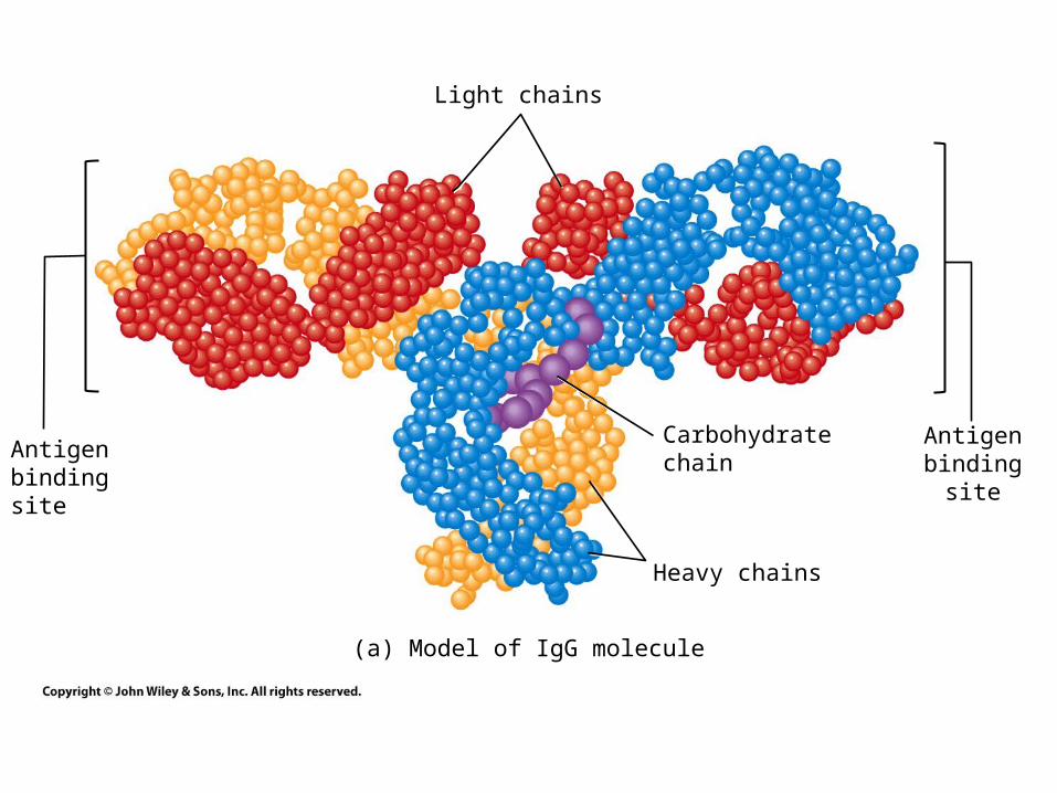
Light chains
Carbohydratechain
Heavy chains
Antigenbinding site
Antigenbindingsite
(a) Model of IgG molecule
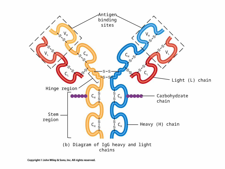
Heavy (H) chain
Hinge region
Carbohydratechain
Light (L) chain
Antigenbinding
sites
Stemregion
(b) Diagram of IgG heavy and light chains
VH
CH
CL
VL
VH
CH
CH
CHCH
CH
CL
VL
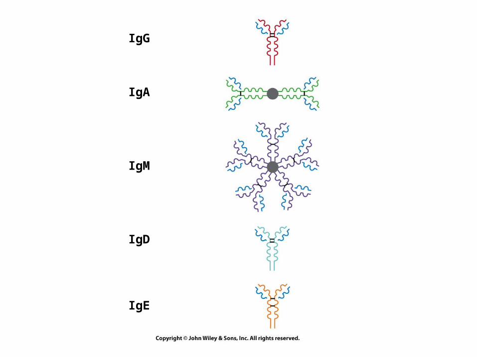
IgG
IgA
IgM
IgD
IgE
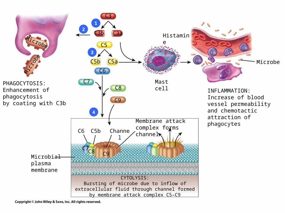
PHAGOCYTOSIS:Enhancement of phagocytosisby coating with C3b
Mic
robe
Microbe
C3
C3b C3a
C5
C5b C5a
C6
C7C8
C9
C6 C5b Channel
Membrane attack complex forms channel
CYTOLYSIS:Bursting of microbe due to inflow of extracellular fluid through
channel formed by membrane attack complex C5–C9
Microbialplasmamembrane
Microbe
INFLAMMATION:Increase of blood vessel permeability and chemotactic attraction of phagocytes
Mast cell
Histamine
C7C8 C9
12
3
4
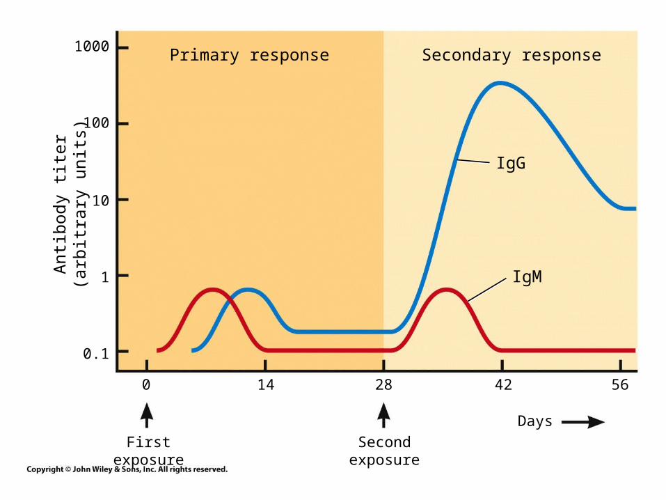
Primary response Secondary response1000
100
10
1
0.1
Ant
ibod
y ti
ter
(arb
itra
ry u
nits
)
IgG
IgM
0 14 28 42 56
First exposure Second exposure
Days
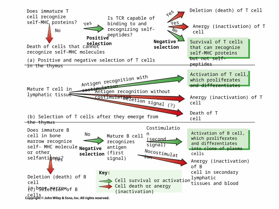
Death of cells that cannotrecognize self-MHC molecules
NoYes
Does immature T cell recognize self-MHC proteins?
(b) Selection of T cells after they emerge from the thymus
Mature T cell inlymphatic tissue
Deletion (death) of B cellin bone marrow
Does immature B cell in bone marrow recognize self- MHC molecule or other selfantigens?
(c) Selection of B cells
Cell survival or activationCell death or anergy (inactivation)
Key:
Yes
No Mature B cellrecognizes antigen(first signal)
Costimulation(second signal)
NocostimulationAnergy (inactivation) of Bcell in secondary lymphatictissues and blood
Activation of B cell, which proliferates and differentiates into clone of plasma cells
Activation of T cell, which proliferates and differentiates
Survival of T cells that can recognize self-MHC proteins but not self-peptides
Deletion (death) of T cell
Anergy (inactivation) of T cell
Anergy (inactivation) of T cell
Death of T cell
Positiveselection
Is TCR capable of binding to and recognizing self-peptides?
Negativeselection
Antigen recognition with costimulation
Antigen recognition without costimulation
Deletion signal (?)
No
Yes
Yes
(a) Positive and negative selection of T cells in the thymus
Negativeselection
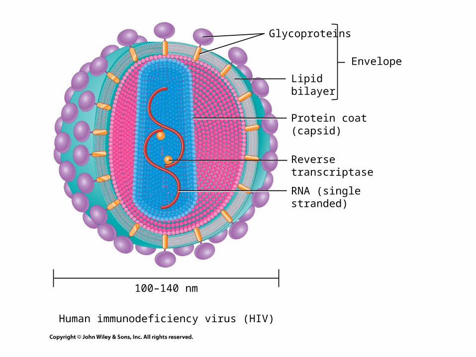
Glycoproteins
Human immunodeficiency virus (HIV)
Lipidbilayer
Protein coat (capsid)
Reverse transcriptase
RNA (single stranded)
Envelope
100–140 nm