Embed Size (px)
Citation preview
The Lymphatic System and Body Defenses
The Lymphatic SystemConsists of two semi-independent parts
Lymphatic vesselsLymphoid tissues and organs
Lymphatic system functionsTransports escaped fluids back to the bloodPlays essential roles in body defense and
resistance to disease

Lymphatic CharacteristicsLymph—excess tissue fluid carried by
lymphatic vesselsProperties of lymphatic vessels
One way system toward the heartNo pumpLymph moves toward the heart
Milking action of skeletal muscleRhythmic contraction of smooth muscle in vessel
walls
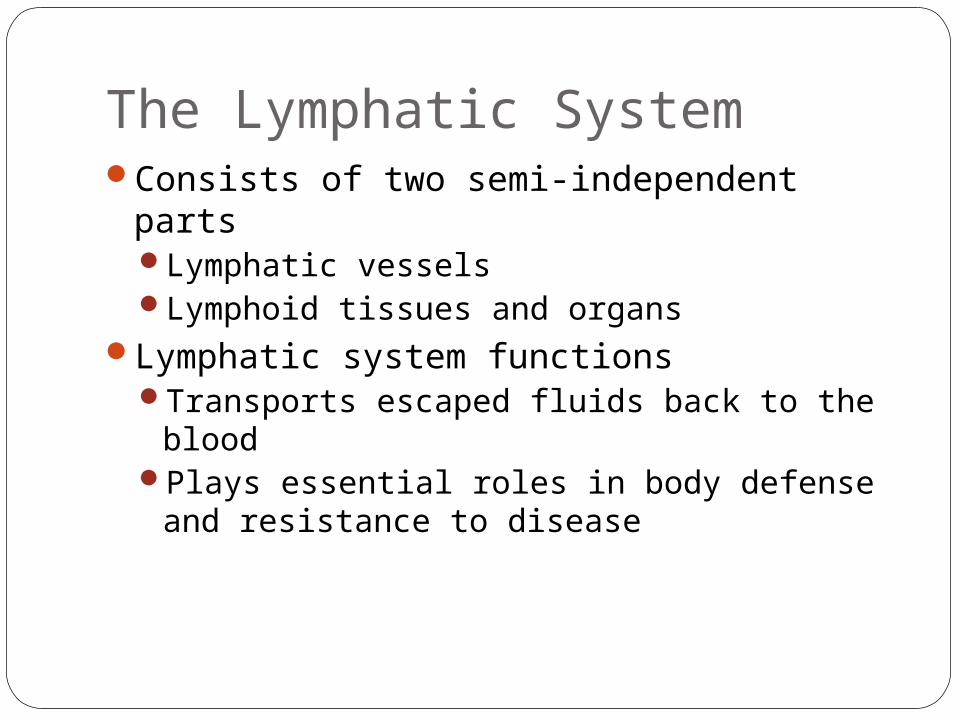
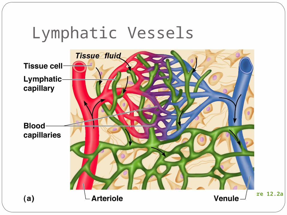
Relationship of Lymphatic Vessels to Blood Vessels
Figure 12.1
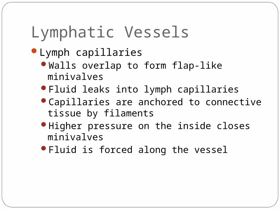
Lymphatic VesselsLymph capillaries
Walls overlap to form flap-like minivalvesFluid leaks into lymph capillariesCapillaries are anchored to connective tissue
by filamentsHigher pressure on the inside closes
minivalvesFluid is forced along the vessel
Lymphatic Vessels
Figure 12.2a
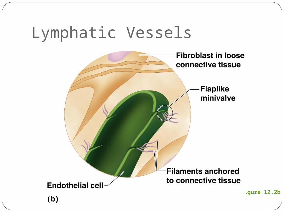
Lymphatic Vessels
Figure 12.2b
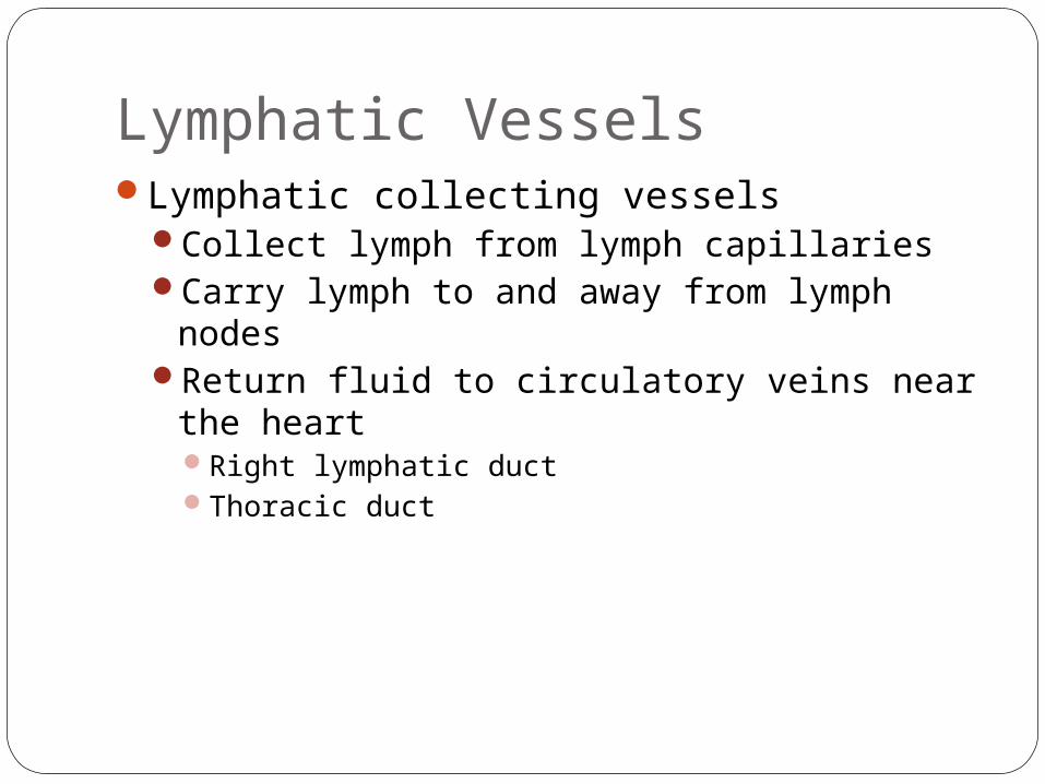
Lymphatic VesselsLymphatic collecting vessels
Collect lymph from lymph capillariesCarry lymph to and away from lymph nodesReturn fluid to circulatory veins near the
heartRight lymphatic ductThoracic duct
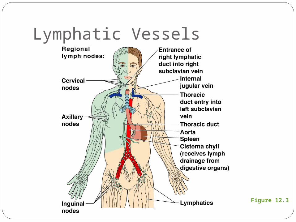
Figure 12.3
Lymphatic Vessels

LymphHarmful materials that enter lymph vessels
BacteriaVirusesCancer cellsCell debris
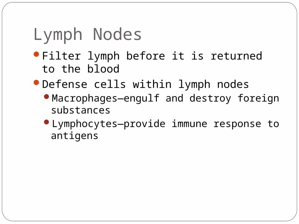
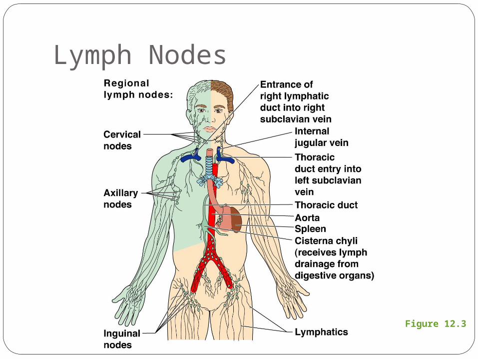
Lymph NodesFilter lymph before it is returned to the
bloodDefense cells within lymph nodes
Macrophages—engulf and destroy foreign substances
Lymphocytes—provide immune response to antigens
Lymph Nodes
Figure 12.3
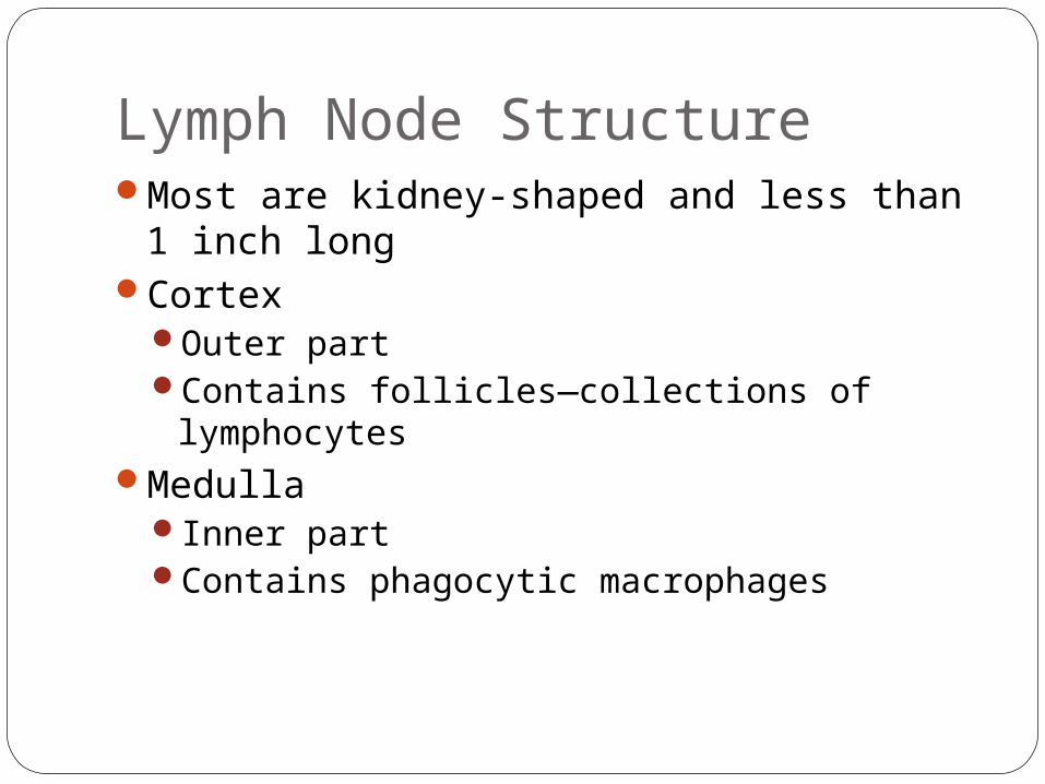
Lymph Node StructureMost are kidney-shaped and less than 1
inch longCortex
Outer partContains follicles—collections of lymphocytes
MedullaInner partContains phagocytic macrophages
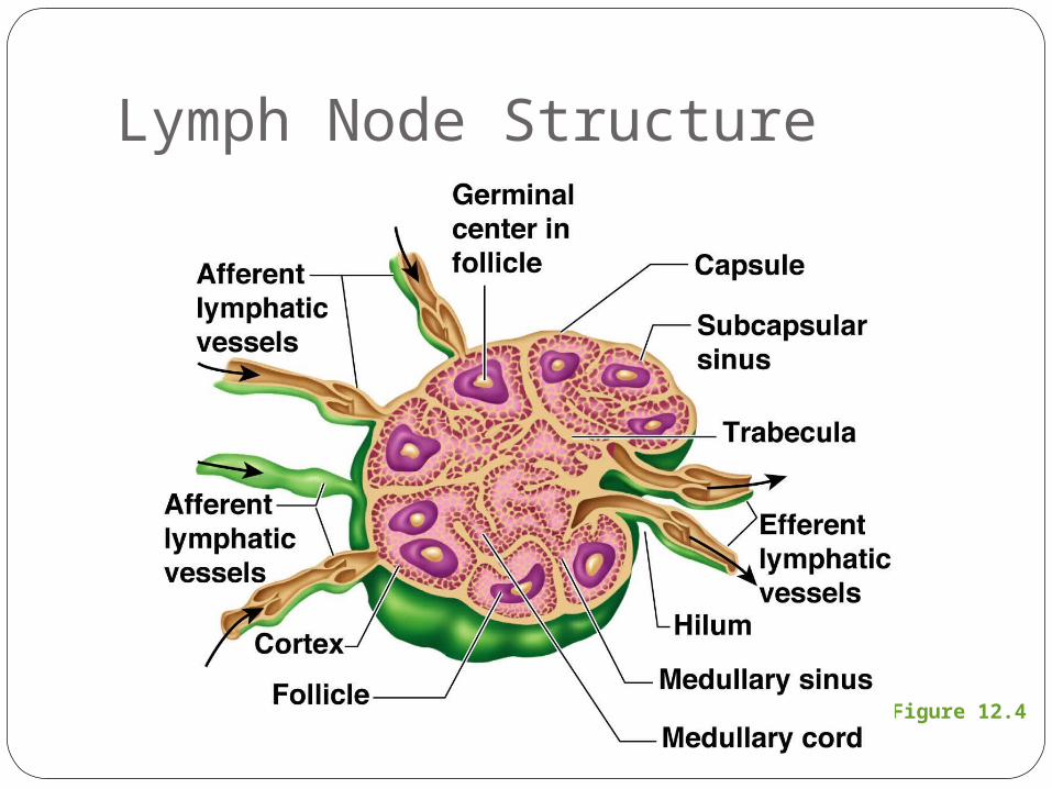
Figure 12.4
Lymph Node Structure
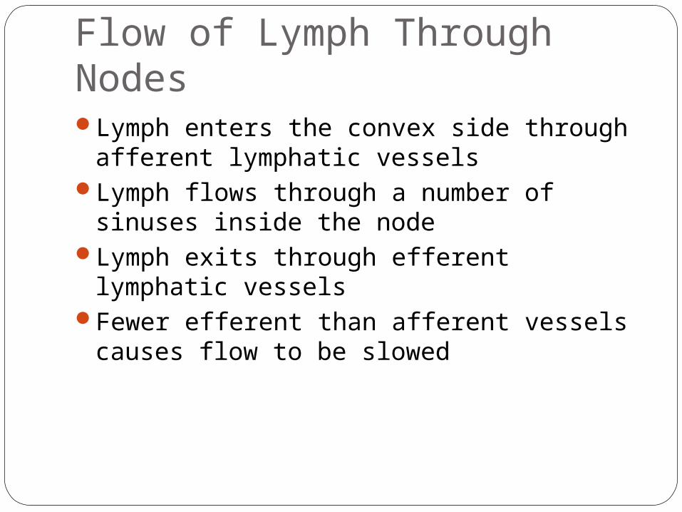
Flow of Lymph Through NodesLymph enters the convex side through
afferent lymphatic vesselsLymph flows through a number of sinuses
inside the nodeLymph exits through efferent lymphatic
vesselsFewer efferent than afferent vessels causes
flow to be slowed
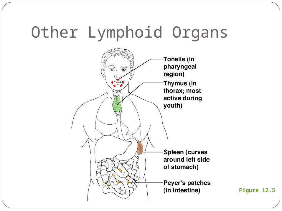
Other Lymphoid OrgansSeveral other organs contribute to
lymphatic functionSpleenThymusTonsilsPeyer’s patches
Other Lymphoid Organs
Figure 12.5
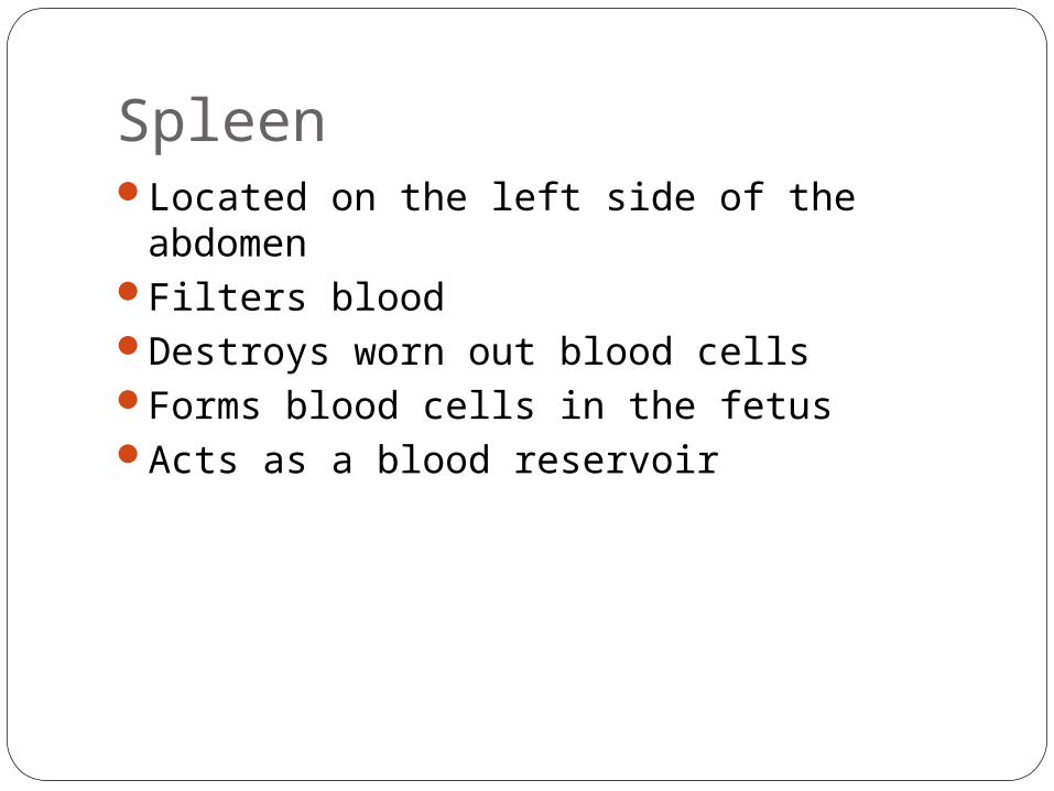
SpleenLocated on the left side of the abdomenFilters bloodDestroys worn out blood cellsForms blood cells in the fetusActs as a blood reservoir
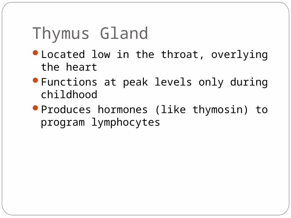
Thymus GlandLocated low in the throat, overlying the
heartFunctions at peak levels only during
childhoodProduces hormones (like thymosin) to
program lymphocytes
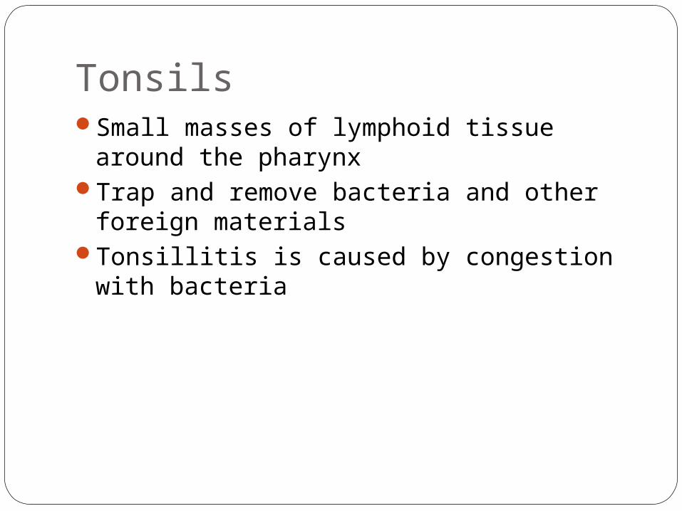
TonsilsSmall masses of lymphoid tissue around
the pharynxTrap and remove bacteria and other foreign
materialsTonsillitis is caused by congestion with
bacteria
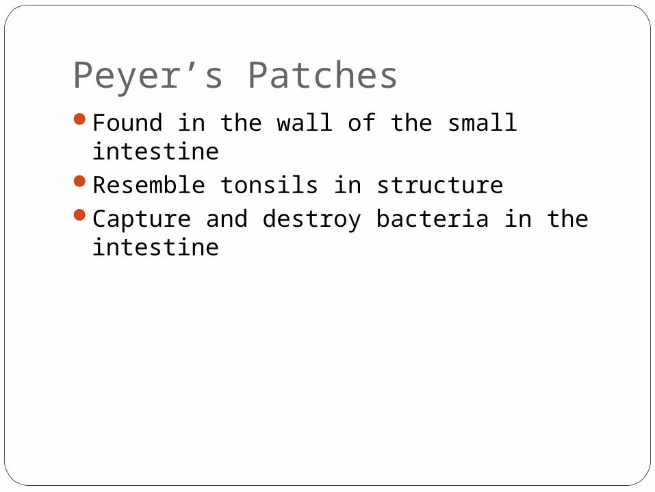
Peyer’s PatchesFound in the wall of the small intestineResemble tonsils in structureCapture and destroy bacteria in the
intestine
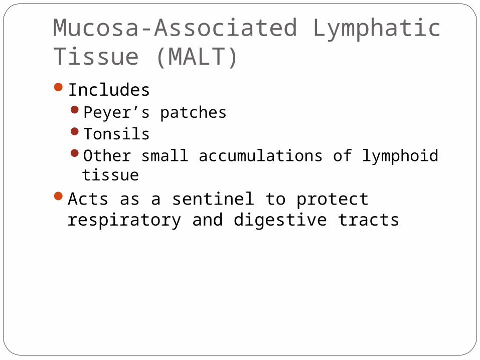
Mucosa-Associated Lymphatic Tissue (MALT)Includes
Peyer’s patchesTonsilsOther small accumulations of lymphoid tissue
Acts as a sentinel to protect respiratory and digestive tracts
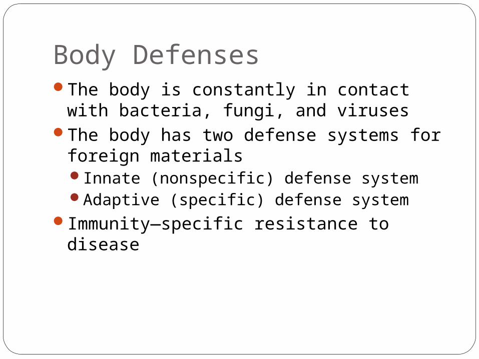
Body DefensesThe body is constantly in contact with
bacteria, fungi, and virusesThe body has two defense systems for
foreign materialsInnate (nonspecific) defense systemAdaptive (specific) defense system
Immunity—specific resistance to disease
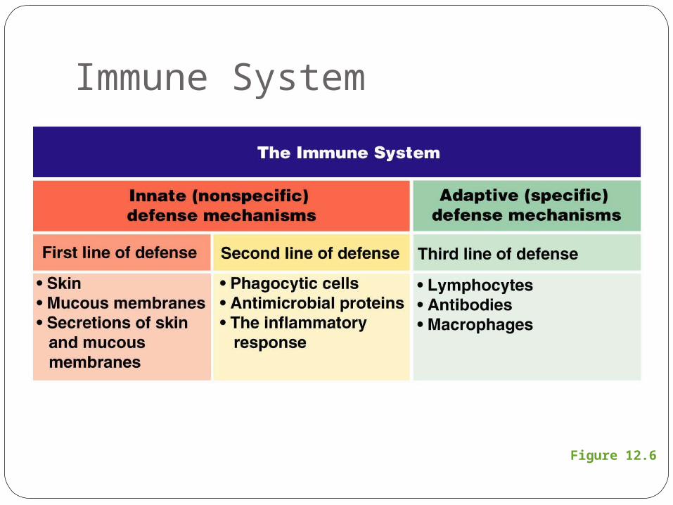
Immune System
Figure 12.6
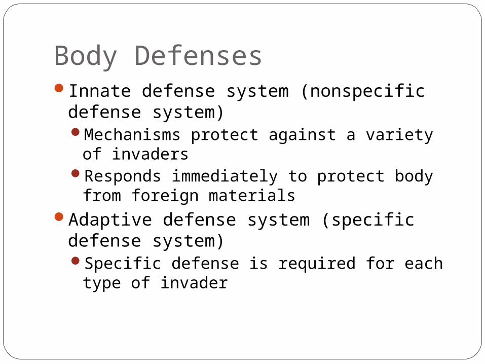
Body DefensesInnate defense system (nonspecific
defense system)Mechanisms protect against a variety of
invadersResponds immediately to protect body from
foreign materialsAdaptive defense system (specific defense
system)Specific defense is required for each type of
invader
Innate Body DefensesInnate body defenses are mechanical
barriers to pathogens such asBody surface coverings
Intact skinMucous membranes
Specialized human cellsChemicals produced by the body
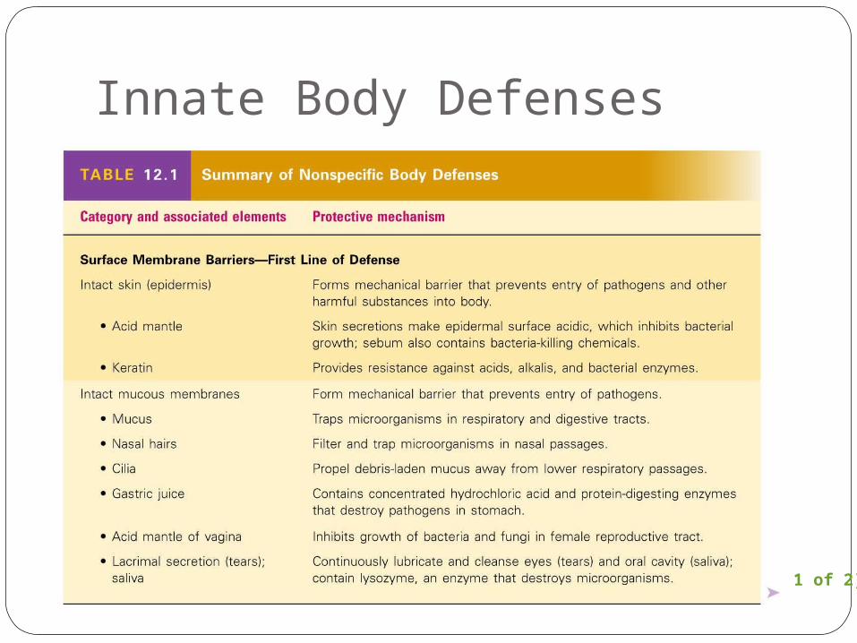
Innate Body Defenses
Table 12.1 (1 of 2)
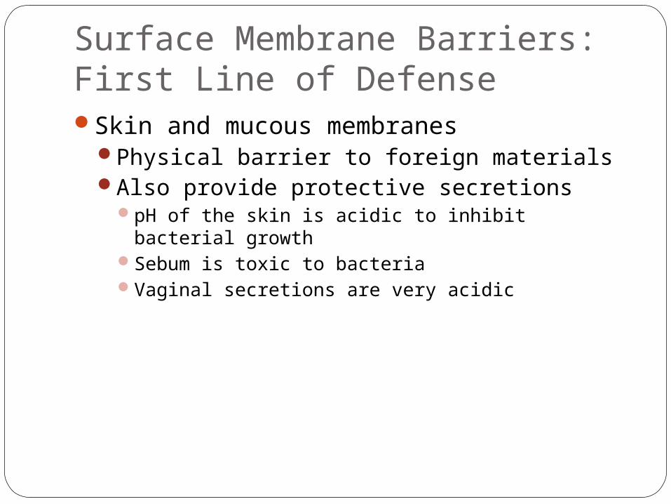
Surface Membrane Barriers:First Line of DefenseSkin and mucous membranes
Physical barrier to foreign materialsAlso provide protective secretions
pH of the skin is acidic to inhibit bacterial growthSebum is toxic to bacteriaVaginal secretions are very acidic
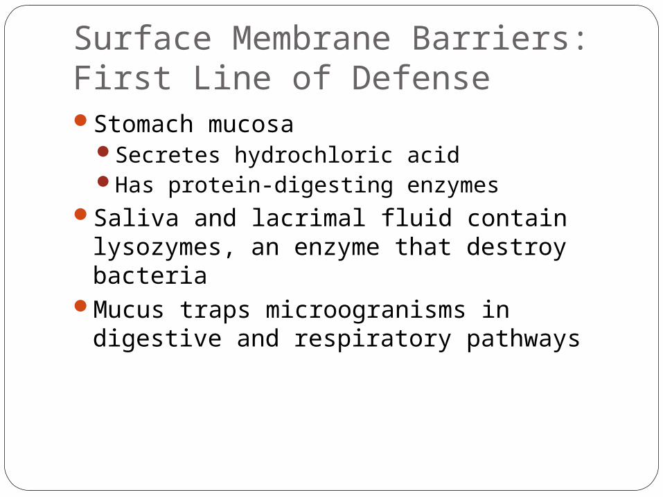
Surface Membrane Barriers:First Line of DefenseStomach mucosa
Secretes hydrochloric acidHas protein-digesting enzymes
Saliva and lacrimal fluid contain lysozymes, an enzyme that destroy bacteria
Mucus traps microogranisms in digestive and respiratory pathways
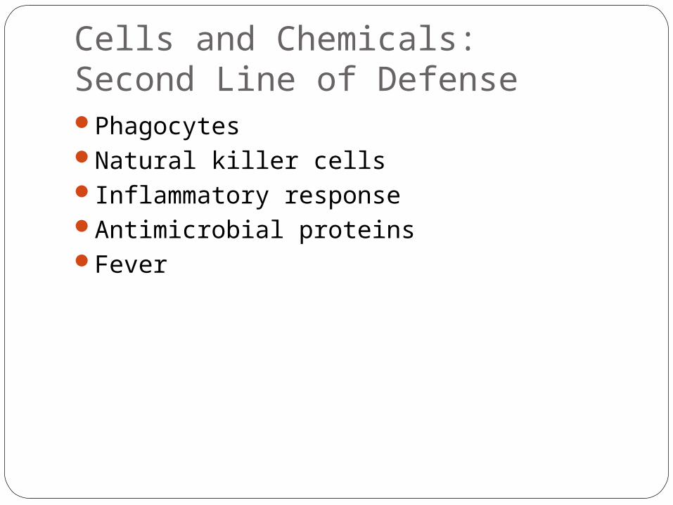
Cells and Chemicals:Second Line of DefensePhagocytesNatural killer cellsInflammatory responseAntimicrobial proteinsFever
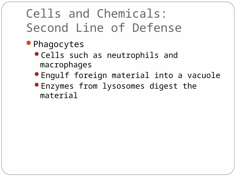
Cells and Chemicals:Second Line of DefensePhagocytes
Cells such as neutrophils and macrophagesEngulf foreign material into a vacuoleEnzymes from lysosomes digest the material
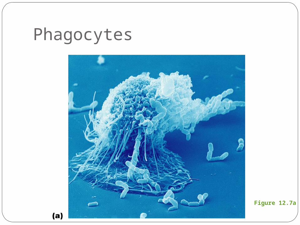
Phagocytes
Figure 12.7a
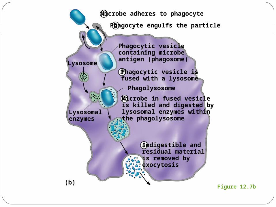
Figure 12.7b
Lysosome
Microbe adheres to phagocyte
Phagocyte engulfs the particle
Phagocytic vesicle isfused with a lysosome
Microbe in fused vesicleis killed and digested bylysosomal enzymes withinthe phagolysosome
Indigestible andresidual materialis removed byexocytosis
Phagocytic vesiclecontaining microbeantigen (phagosome)
Phagolysosome
Lysosomalenzymes
(b)
Internal Innate Defenses: Cells and ChemicalsNatural killer (NK) cells
Can lyse (disintegrate or dissolve) and kill cancer cells
Can destroy virus-infected cells
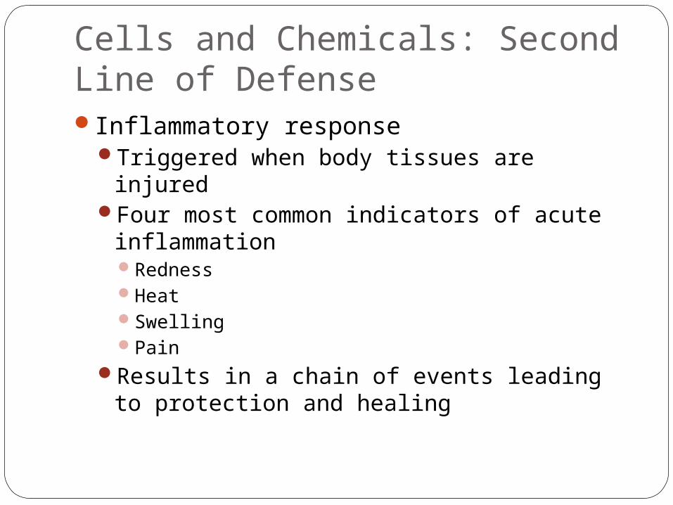
Cells and Chemicals: Second Line of DefenseInflammatory response
Triggered when body tissues are injuredFour most common indicators of acute
inflammationRednessHeatSwellingPain
Results in a chain of events leading to protection and healing
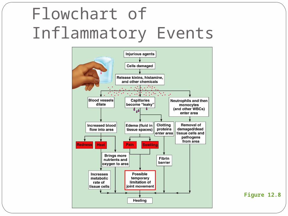
Figure 12.8
Flowchart of Inflammatory Events
Cells and Chemicals: Second Line of DefenseFunctions of the inflammatory response
Prevents spread of damaging agentsDisposes of cell debris and pathogens
through phagocytosisSets the stage for repair
Cells and Chemicals: Second Line of DefensePhagocytosis
Neutrophils move by diapedesis to clean up damaged tissue and/or pathogens
Monocytes become macrophages and complete disposal of cell debris
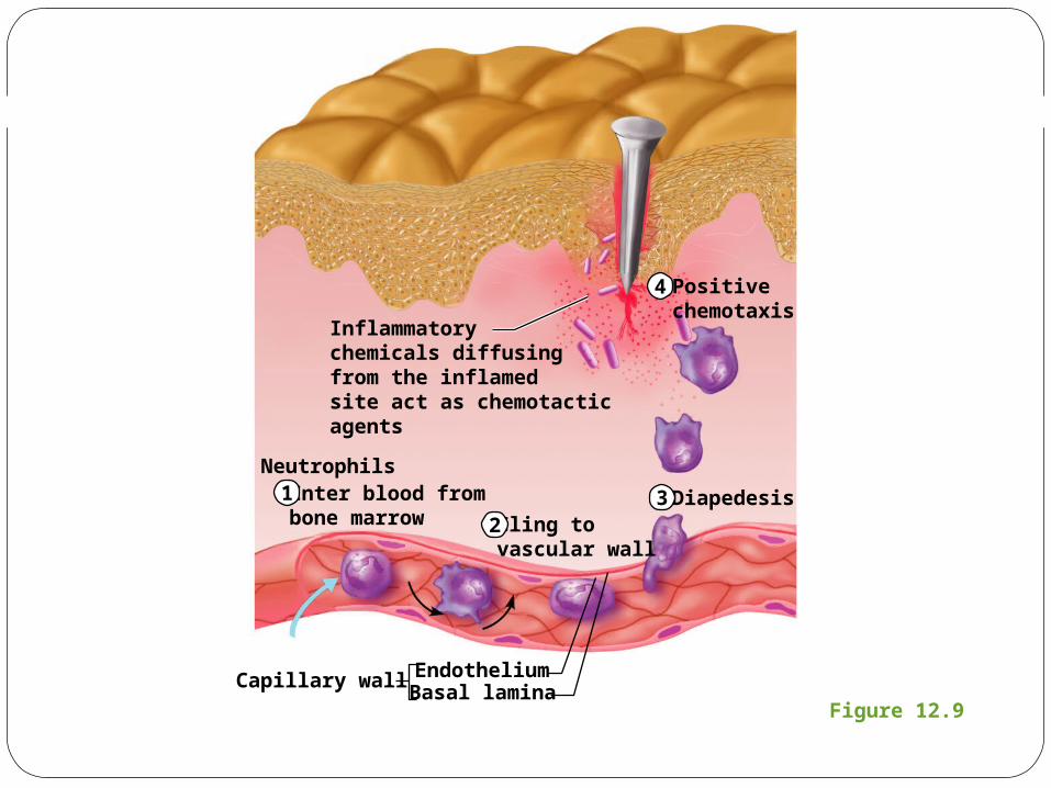
Figure 12.9
Enter blood frombone marrow
EndotheliumCapillary wall
Cling tovascular wall
Diapedesis
Positivechemotaxis
Inflammatorychemicals diffusingfrom the inflamedsite act as chemotacticagents
Basal lamina
Neutrophils1
23
4
Cells and Chemicals: Second Line of DefenseAntimicrobial proteins
Attack microorganismsHinder reproduction of microorganisms
Most importantComplement proteinsInterferon
Cells and Chemicals: Second Line of DefenseComplement proteins
A group of at least 20 plasma proteinsActivated when they encounter and attach to
cells (complement fixation)Damage foreign cell surfacesRelease vasodilators and chemotaxis
chemicals, cause opsonization
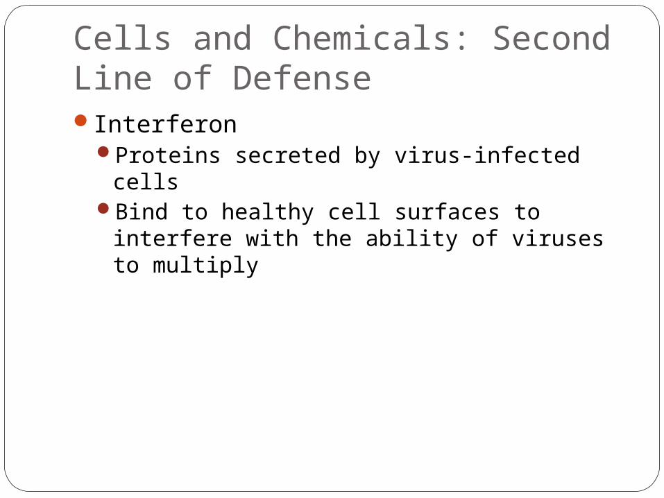
Cells and Chemicals: Second Line of DefenseInterferon
Proteins secreted by virus-infected cellsBind to healthy cell surfaces to interfere with
the ability of viruses to multiply
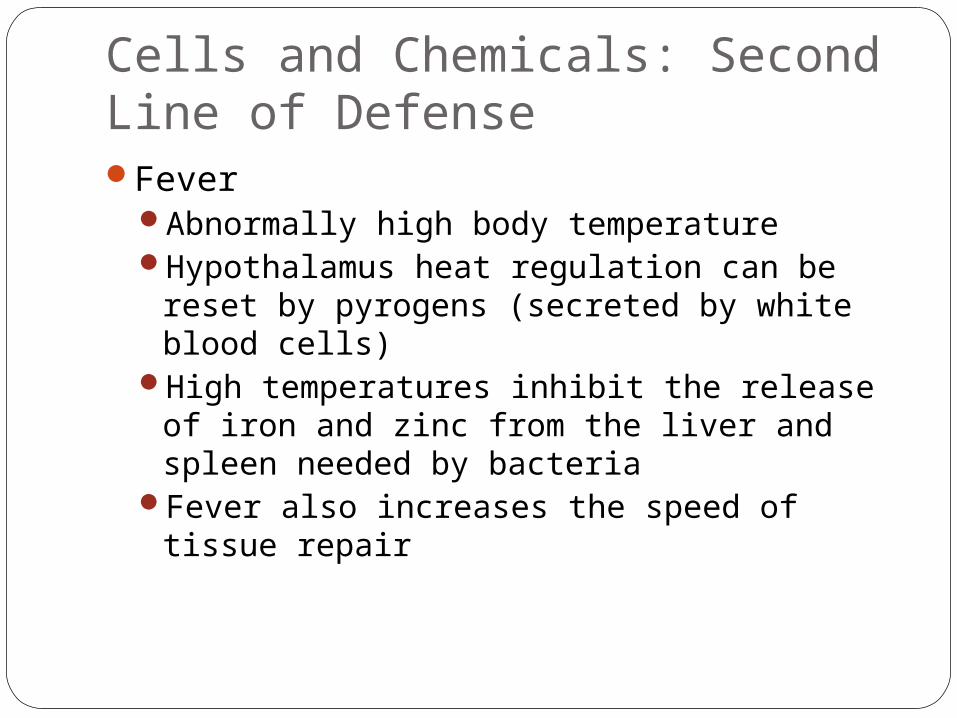
Cells and Chemicals: Second Line of DefenseFever
Abnormally high body temperatureHypothalamus heat regulation can be reset
by pyrogens (secreted by white blood cells)High temperatures inhibit the release of iron
and zinc from the liver and spleen needed by bacteria
Fever also increases the speed of tissue repair
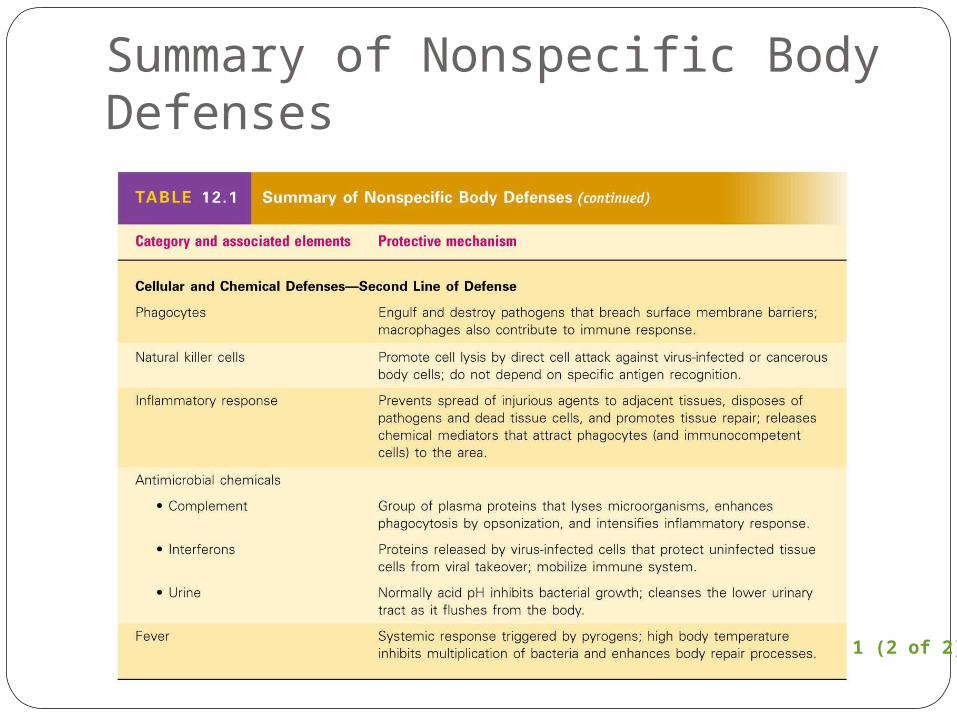
Summary of Nonspecific Body Defenses
Table 12.1 (2 of 2)