Embed Size (px)
Citation preview
Primary Neuronal CultureBrainPhys™ and NeuroCult™ Neuronal Media and Supplements
2
Table of Contents
3 Superior Primary Neuronal Culture With Standardized NeuroCult™ Reagents and BrainPhys™ Neuronal Medium
4 Traditional Neuronal Culture Increases Survival of Primary Neurons in Long-Term Culture
7 Neurophysiologically Active Neuronal Culture BrainPhys™ Neuronal Medium
10 Product Information
10 Supplementary Reagents
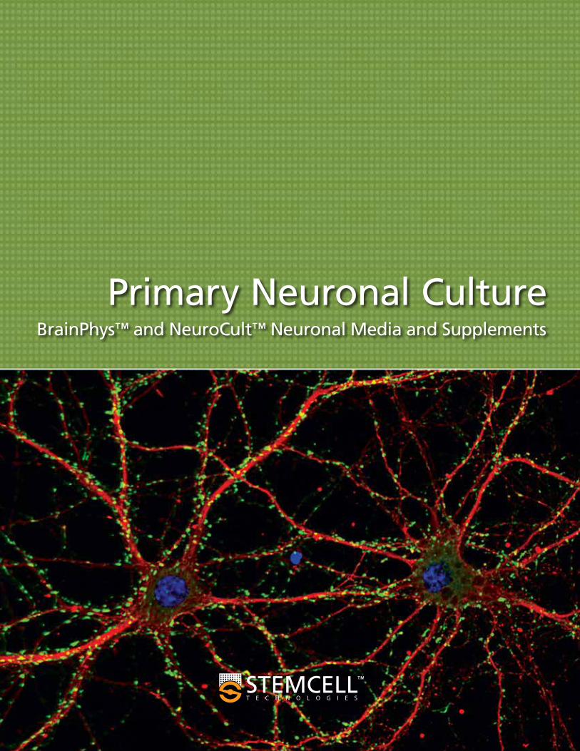
Front cover. Primary neurons cultured in NeuroCult™ SM1-supplemented NeuroCult™ Neuronal Basal Medium for 21 days.
3
Primary Neuronal Culture
Primary neuronal cultures have long been a powerful system with which to study neuronal biology in a controlled environment. To obtain healthy cultures with good morphology and cellular function, using high-quality media and supplements is crucial, because they contain numerous complex components.1-3 Variability in the quality of any of these raw materials, or in the associated manufacturing processes, results in inconsistent reagent quality, which negatively impacts sensitive neuronal cultures.3,4
NeuroCult™ SM (STEMCELL-Modified) Neuronal Supplements are based on Brewer's B27 supplement,1 and optimized to more consistently support the culture of mature, functional neurons in both short- and long-term cultures.
NeuroCult™ SM supplements may be combined with NeuroCult™ Neuronal Basal Medium or with BrainPhys™ Neuronal Medium. NeuroCult™ Neuronal Basal Medium is based on Brewer’s Neurobasal medium,1 which was designed for optimal survival of neurons, and is the ideal choice for generating traditional primary neuronal cultures. BrainPhys™ Neuronal Medium is a new neuronal basal medium designed by Dr. Cedric Bardy in Dr. Fred H. Gage’s laboratory to better support the in vitro neuronal function of both primary and human pluripotent stem cell (hPSC)-derived neurons.5
Superior Primary Neuronal CultureWith Standardized NeuroCult™ Reagents and BrainPhys™ Neuronal Medium
Optimal Neuronal Function and Survival
The BrainPhys™ Neuronal Medium and SM1 Kit includes the best of both worlds: the consistent NeuroCult™ SM1 and the functional BrainPhys™ Neuronal Medium.
STEMCELL Products For Every Step of Your Neuronal Research
• Trypsin-EDTA • BrainPhys™ Neuronal Medium
• BrainPhys™ Neuronal Medium and SM1 Kit
• NeuroCult™ SM1 Neuronal Supplement
• NeuroCult™ SM1 Neuronal Culture Kit
• NeuroCult™ SM2 Neuronal Supplement
• NeuroCult™ Neuronal Basal Medium
• Cytokines
• Antibodies
• Antibodies
Tissue Dissociation Cell Culture Characterization
4
NeuroCult™ SM1 Neuronal Supplement is designed based on the published B27 formulation,1,2 but optimized to more consistently support the culture of mature, functional neurons, whilst minimizing glial cell contamination (<1% GFAP). NeuroCult™ SM1 is available separately, as part of the NeuroCult™ SM1 Neuronal Culture Kit (in which it is complemented by NeuroCult™ Neuronal Basal Medium), or as part of the BrainPhys™ Neuronal Medium and SM1 Kit (see page 7).
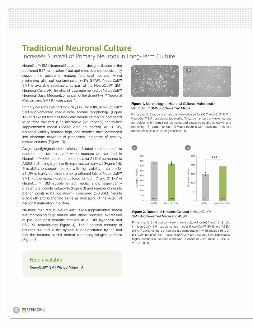
Primary neurons cultured for 7 days in vitro (DIV) in NeuroCult™ SM1-supplemented media have normal morphology (Figure 1A) and exhibit less cell body and neurite clumping, compared to neurons cultured in an alternative (Neurobasal) serum-free supplemented media (ASSM; data not shown). At 21 DIV, neuronal viability remains high, and neurites have developed into elaborate networks of processes, indicative of healthy, mature cultures (Figure 1B).
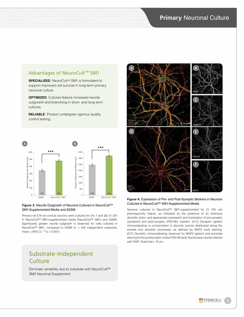
A significantly higher number of class III ß-tubulin-immunoreactive neurons can be observed when neurons are cultured in NeuroCult™ SM1-supplemented media for 21 DIV compared to ASSM, indicating significantly improved cell survival (Figure 2B). This ability to support neurons with high viability in culture for 21 DIV is highly consistent among different lots of NeuroCult™ SM1. Furthermore, neurons cultured for both 7 and 21 DIV in NeuroCult™ SM1-supplemented media show significantly greater total neurite outgrowth (Figure 3) and number of neurite branch points (data not shown), compared to ASSM. Neurite outgrowth and branching serve as indicators of the extent of neuronal maturation in culture.
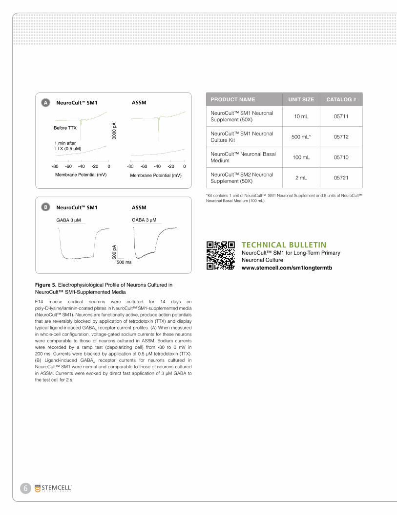
Neurons cultured in NeuroCult™ SM1-supplemented media are morphologically mature and show punctate expression of pre- and post-synaptic markers at 21 DIV (synapsin and PSD-95, respectively; Figure 4). The functional maturity of neurons cultured in this system is demonstrated by the fact that the neurons exhibit normal electrophysiological profiles (Figure 5).
Traditional Neuronal CultureIncreases Survival of Primary Neurons in Long-Term Culture
Figure 1. Morphology of Neuronal Cultures Maintained in NeuroCult™ SM1-Supplemented Media
Primary rat E18 rat cortical neurons were cultured for (A) 7 and (B) 21 DIV in NeuroCult™ SM1-supplemented media. (A) Large numbers of viable neurons are visible, with minimal cell clumping and extensive neurite outgrowth and branching. (B) Large numbers of viable neurons with developed dendritic arbors remain in culture. Magnification: 20x.
Now availableNeuroCult™ SM1 Without Vitamin A
BA
Figure 2. Number of Neurons Cultured in NeuroCult™ SM1-Supplemented Media and ASSM
Primary rat E18 rat cortical neurons were cultured for (A) 7 and (B) 21 DIV in NeuroCult™ SM1-supplemented media (NeuroCult™ SM1) and ASSM. (A) At 7 days, numbers of neurons are comparable (n = 25; mean ± 95% CI; p > 0.05) but after (B) 21 days, NeuroCult™ SM1 cultures have significantly higher numbers of neurons compared to ASSM (n = 25; mean ± 95% CI; ***p < 0.001).
BA
3,944 3,7380
500
1000
1500
2000
2500
3000
3500
4000
4500
TSFM NeuroCult™ SM1
Num
bers
of N
euro
ns
1,494 1,9220
500
1000
1500
2000
2500
TSFM NeuroCult™ SM1
Num
bers
of N
euro
ns
Num
ber o
f Neu
rons
ASSM NeuroCultTM SM1 ASSM NeuroCultTM SM1
Num
ber o
f Neu
rons
***
5
Primary Neuronal Culture
692 9610
200
400
600
800
1000
1200
TSFM NeuroCult™ SM1
Neu
rite
Out
grow
th/C
ell (
µm)
2,469 3,2110
500
1000
1500
2000
2500
3000
3500
TSFM NeuroCult™ SM1
Neu
rite
Out
grow
th/C
ell (
µm)
Figure 3. Neurite Outgrowth of Neurons Cultured in NeuroCult™ SM1-Supplemented Media and ASSM
Primary rat E18 rat cortical neurons were cultured for (A) 7 and (B) 21 DIV in NeuroCult™ SM1-supplemented media (NeuroCult™ SM1) and ASSM. Significantly greater neurite outgrowth is observed for cells cultured in NeuroCult™ SM1, compared to ASSM (n = 240 independent measures; mean ± 95% CI; ***p < 0.001).
BA
Neu
rite
Out
grow
th p
er N
euro
n (µ
m)
Neu
rite
Out
grow
th p
er N
euro
n (µ
m)
******
Figure 4. Expression of Pre- and Post-Synaptic Markers in Neurons Cultured in NeuroCult™ SM1-Supplemented Media
Neurons cultured in NeuroCult™ SM1-supplemented for 21 DIV are phenotypically mature, as indicated by the presence of an extensive dendritic arbor, and appropriate expression and localization of pre-synaptic (synapsin) and post-synaptic (PSD-95) markers. (A-C) Synapsin (green) immunolabeling is concentrated in discrete puncta distributed along the somata and dendritic processes, as defined by MAP2 (red) staining. (D-F) Dendritic immunolabeling observed for MAP2 (green) and punctate staining for the postsynaptic marker PSD-95 (red). Nuclei were counter-stained with DAPI. Scale bar= 10 µm.
B
C
E
F
A
D
ASSM NeuroCultTM SM1 ASSM NeuroCultTM SM1
Advantages of NeuroCult™ SM1
SPECIALIZED. NeuroCult™ SM1 is formulated to support improved cell survival in long-term primary neuronal culture.
OPTIMIZED. Cultures feature increased neurite outgrowth and branching in short- and long-term cultures.
RELIABLE. Product undergoes rigorous quality control testing.
Substrate-Independent CultureEliminate variability due to substrate with NeuroCult™ SM2 Neuronal Supplement
6
Figure 5. Electrophysiological Profile of Neurons Cultured in NeuroCult™ SM1-Supplemented Media
E14 mouse cortical neurons were cultured for 14 days on poly-D-lysine/laminin-coated plates in NeuroCult™ SM1-supplemented media (NeuroCult™ SM1). Neurons are functionally active, produce action potentials that are reversibly blocked by application of tetrodotoxin (TTX) and display typical ligand-induced GABAA receptor current profiles. (A) When measured in whole-cell configuration, voltage-gated sodium currents for these neurons were comparable to those of neurons cultured in ASSM. Sodium currents were recorded by a ramp test (depolarizing cell) from -80 to 0 mV in 200 ms. Currents were blocked by application of 0.5 µM tetrodotoxin (TTX). (B) Ligand-induced GABAA receptor currents for neurons cultured in NeuroCult™ SM1 were normal and comparable to those of neurons cultured in ASSM. Currents were evoked by direct fast application of 3 µM GABA to the test cell for 2 s.
NeuroCultTM SM1
1 min after TTX (0.5 µM)
Membrane Potential (mV)
-80 -60 -40 -20 0
500
pA30
00 p
A
500 ms
GABA 3 µM
NeuroCultTM SM1
ASSM
ASSM
GABA 3 µM
Membrane Potential (mV)
-80 -60 -40 -20 0
Before TTX
B
APRODUCT NAME UNIT SIZE CATALOG #
NeuroCult™ SM1 Neuronal Supplement (50X)
10 mL 05711
NeuroCult™ SM1 Neuronal Culture Kit
500 mL* 05712
NeuroCult™ Neuronal Basal Medium
100 mL 05710
NeuroCult™ SM2 Neuronal Supplement (50X)
2 mL 05721
*Kit contains 1 unit of NeuroCult™ SM1 Neuronal Supplement and 5 units of NeuroCult™ Neuronal Basal Medium (100 mL).
TECHNICAL BULLETINNeuroCult™ SM1 for Long-Term Primary Neuronal Culturewww.stemcell.com/sm1longtermtb
7
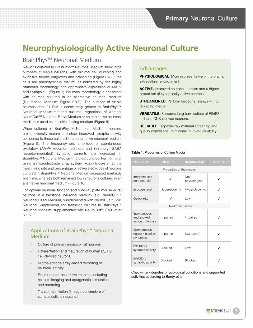
PROPERTY DMEM/F12 NEUROBASAL BRAINPHYS™
Properties of the medium
Inorganic salt concentration ✓ Not
physiological✓
Glucose level Hyperglycemic Hyperglycemic ✓
Osmolarity ✓ Low ✓
Neuronal function
Spontaneous and evoked action potentials
Impaired Impaired ✓
Spontaneous network calcium dynamics
Impaired Not tested ✓
Excitatory synaptic activity
Blocked Low ✓
Inhibitory synaptic activity
Blocked Blocked ✓
Check-mark denotes physiological conditions and supported activities according to Bardy et al. 5
Table 1. Properties of Culture Media5
Applications of BrainPhys™ Neuronal Medium
• Culture of primary mouse or rat neurons.
• Differentiation and maturation of human ES/iPS cell-derived neurons.
• Microelectrode array-based recording of neuronal activity.
• Fluorescence-based live imaging, including calcium imaging and optogenetic stimulation and recording.
• Transdifferentiation (lineage conversion) of somatic cells to neurons.5
Primary Neuronal Culture
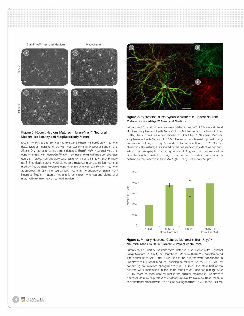
Neurons cultured in BrainPhys™ Neuronal Medium show large numbers of viable neurons, with minimal cell clumping and extensive neurite outgrowth and branching (Figure 6A,C); the cells are phenotypically mature, as indicated by the highly branched morphology and appropriate expression of MAP2 and Synapsin 1 (Figure 7). Neuronal morphology is consistent with neurons cultured in an alternative neuronal medium (Neurobasal Medium; Figure 6B,D). The number of viable neurons after 21 DIV is consistently greater in BrainPhys™ Neuronal Medium-matured cultures, regardless of whether NeuroCult™ Neuronal Basal Medium or an alternative neuronal medium is used as the initial plating medium (Figure 8).
When cultured in BrainPhys™ Neuronal Medium, neurons are functionally mature and show improved synaptic activity compared to those cultured in an alternative neuronal medium (Figure 9). The frequency and amplitude of spontaneous excitatory (AMPA receptor-mediated) and inhibitory (GABA receptor-mediated) synaptic currents are increased in BrainPhys™ Neuronal Medium-matured cultures. Furthermore, using a microelectrode array system (Axion Biosystems), the mean firing rate and percentage of active electrodes of neurons cultured in BrainPhys™ Neuronal Medium increased markedly over time, whereas both remained low in neurons cultured in an alternative neuronal medium (Figure 10).
For optimal neuronal function and survival, plate mouse or rat neurons in a traditional neuronal medium (e.g. NeuroCult™ Neuronal Basal Medium, supplemented with NeuroCult™ SM1 Neuronal Supplement) and transition cultures to BrainPhys™ Neuronal Medium, supplemented with NeuroCult™ SM1, after 5 DIV.
Neurophysiologically Active Neuronal CultureBrainPhys™ Neuronal Medium
Advantages
PHYSIOLOGICAL. More representative of the brain’s extracellular environment.
ACTIVE. Improved neuronal function and a higher proportion of synaptically active neurons.
STREAMLINED. Perform functional assays without replacing media.
VERSATILE. Supports long-term culture of ES/iPS cell-and CNS-derived neurons.
RELIABLE. Rigorous raw material screening and quality control ensure minimal lot-to-lot variability.
8
Figure 8. Primary Neuronal Cultures Matured in BrainPhys™ Neuronal Medium Have Greater Numbers of Neurons
Primary rat E18 cortical neurons were plated in either NeuroCult™ Neuronal Basal Medium (NCSM1) or Neurobasal Medium (NBSM1), supplemented with NeuroCult™ SM1. After 5 DIV, half of the cultures were transitioned to BrainPhys™ Neuronal Medium, supplemented with NeuroCult™ SM1, by performing half-medium changes every 3 - 4 days. The other half of the cultures were maintained in the same medium as used for plating. After 21 DIV, more neurons were evident in the cultures matured in BrainPhys™ Neuronal Medium, regardless of whether NeuroCult™ Neuronal Basal Medium or Neurobasal Medium was used as the plating medium. (n = 4, mean ± SEM).
Num
ber o
f Neu
rons
2000
3000
4000
5000
1000
0NBSM1 NBSM1 --->
BrainPhys™SM1NCSM1 NCSM1 --->
BrainPhys™SM1
Figure 6. Rodent Neurons Matured in BrainPhys™ Neuronal Medium are Healthy and Morphologically Mature
(A,C) Primary rat E18 cortical neurons were plated in NeuroCult™ Neuronal Basal Medium, supplemented with NeuroCult™ SM1 Neuronal Supplement. After 5 DIV, the cultures were transitioned to BrainPhys™ Neuronal Medium, supplemented with NeuroCult™ SM1, by performing half-medium changes every 3 - 4 days. Neurons were cultured for (A) 14 or (C) 21 DIV. (B,D) Primary rat E18 cortical neurons were plated and matured in an alternative neuronal medium (Neurobasal Medium), supplemented with NeuroCult™ SM1 Neuronal Supplement for (B) 14 or (D) 21 DIV. Neuronal morphology of BrainPhys™ Neuronal Medium-matured neurons is consistent with neurons plated and matured in an alternative neuronal medium.
DC
14 d
ays
21 d
ays
BA
BrainPhys™ Neuronal Medium Neurobasal
Figure 7. Expression of Pre-Synaptic Markers in Rodent Neurons Matured in BrainPhys™ Neuronal Medium
Primary rat E18 cortical neurons were plated in NeuroCult™ Neuronal Basal Medium, supplemented with NeuroCult™ SM1 Neuronal Supplement. After 5 DIV, the cultures were transitioned to BrainPhys™ Neuronal Medium, supplemented with NeuroCult™ SM1 Neuronal Supplement, by performing half-medium changes every 3 - 4 days. Neurons cultured for 21 DIV are phenotypically mature, as indicated by the presence of an extensive dendritic arbor. The pre-synaptic marker synapsin (A,B; green) is concentrated in discrete puncta distributed along the somata and dendritic processes, as defined by the dendritic marker MAP2 (A,C; red). Scale bar= 50 µm.
A B
C
9
Primary Neuronal Culture
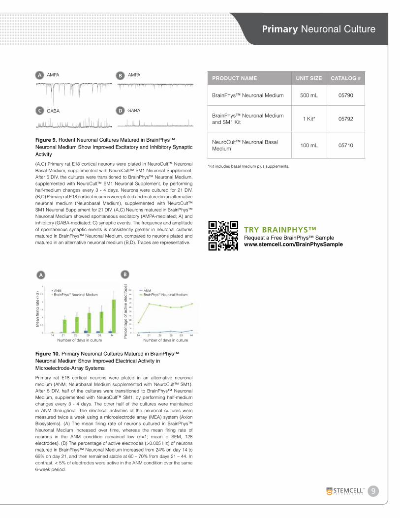
Figure 9. Rodent Neuronal Cultures Matured in BrainPhys™ Neuronal Medium Show Improved Excitatory and Inhibitory Synaptic Activity
(A,C) Primary rat E18 cortical neurons were plated in NeuroCult™ Neuronal Basal Medium, supplemented with NeuroCult™ SM1 Neuronal Supplement. After 5 DIV, the cultures were transitioned to BrainPhys™ Neuronal Medium, supplemented with NeuroCult™ SM1 Neuronal Supplement, by performing half-medium changes every 3 - 4 days. Neurons were cultured for 21 DIV. (B,D) Primary rat E18 cortical neurons were plated and matured in an alternative neuronal medium (Neurobasal Medium), supplemented with NeuroCult™ SM1 Neuronal Supplement for 21 DIV. (A,C) Neurons matured in BrainPhys™ Neuronal Medium showed spontaneous excitatory (AMPA-mediated; A) and inhibitory (GABA-mediated; C) synaptic events. The frequency and amplitude of spontaneous synaptic events is consistently greater in neuronal cultures matured in BrainPhys™ Neuronal Medium, compared to neurons plated and matured in an alternative neuronal medium (B,D). Traces are representative.
C D
A B
GABA GABA
AMPA AMPA
B
Figure 10. Primary Neuronal Cultures Matured in BrainPhys™ Neuronal Medium Show Improved Electrical Activity in Microelectrode-Array Systems
Primary rat E18 cortical neurons were plated in an alternative neuronal medium (ANM; Neurobasal Medium supplemented with NeuroCult™ SM1). After 5 DIV, half of the cultures were transitioned to BrainPhys™ Neuronal Medium, supplemented with NeuroCult™ SM1, by performing half-medium changes every 3 - 4 days. The other half of the cultures were maintained in ANM throughout. The electrical activities of the neuronal cultures were measured twice a week using a microelectrode array (MEA) system (Axion Biosystems). (A) The mean firing rate of neurons cultured in BrainPhys™ Neuronal Medium increased over time, whereas the mean firing rate of neurons in the ANM condition remained low (n=1; mean ± SEM, 128 electrodes). (B) The percentage of active electrodes (>0.005 Hz) of neurons matured in BrainPhys™ Neuronal Medium increased from 24% on day 14 to 69% on day 21, and then remained stable at 60 – 70% from days 21 – 44. In contrast, < 5% of electrodes were active in the ANM condition over the same 6-week period.
A
Mea
n fir
ing
rate
(Hz)
Number of days in culture 14 21 26 29 33 44 14 21 26 29 33 44
100
90
80
70
60
50
40
30
20
10
0
3
2.5
2
1.5
1
0.5
0
ANMBrainPhysTM Neuronal Medium
ANMBrainPhysTM Neuronal Medium
Perc
enta
ge o
f act
ive
elec
trode
s
Number of days in culture
PRODUCT NAME UNIT SIZE CATALOG #
BrainPhys™ Neuronal Medium 500 mL 05790
BrainPhys™ Neuronal Medium and SM1 Kit
1 Kit* 05792
NeuroCult™ Neuronal Basal Medium
100 mL 05710
*Kit includes basal medium plus supplements.
TRY BRAINPHYS™Request a Free BrainPhys™ Samplewww.stemcell.com/BrainPhysSample
10
CytokinesActivate, expand and differentiate cells with cytokines and growth factors. These high-quality reagents support neuronal cultures and ensure reproducibility across a variety of assays. Choose from a large selection of cytokines and growth factors to incorporate into your research workflow. To view the full list of cytokines for neuronal research visit www.stemcell.com/cytokines.
References1. Brewer GJ et al. (1993) Optimized survival of hippocampal
neurons in B27-supplemented Neurobasal, a new serum-free medium combination. J Neurosci Res. 35(5): 567-76.
2. Brewer GJ, Cotman CW. (1989) Survival and growth of hippocampal neurons in defined medium at low density: advantages of a sandwich culture technique or low oxygen. Brain Res. 494(1):65-74
3. Chen Y, Stevens B, Chang J, Milbrandt J, Barres BA, Hell JW. NS21: re-defined and modified supplement B27 for neuronal cultures. J Neurosci Methods 171(2):239-47, 2008
4. Cressey D. Neuroscientists claim growing pains. Nature 459: 19, 2009
5. Bardy C et al. (2015) Neuronal medium that supports basic synaptic functions and activity of human neurons in vitro. Proc Natl Acad Sci 112 (20) E2725-E2734.
CYTOKINE QUANTITY CATALOG #
Human Recombinant BDNF 5 µg 02519
Human Recombinant bFGF
10 µg 78003.1
50 µg 78003
1000 µg 78003.2
Human Recombinant EGF
100 µg 78006.1
500 µg 78006
1000 µg 78006.2
Human Recombinant EPO 500 U 02625
Human Recombinant IL-6
20 µg 78050.1
100 µg 78050
1000 µg 78050.2
Human Recombinant lL-1110 µg 78025.1
100 µg 78025
Human Recombinant LIF*
10 µg 78055.1
50 µg 78055
1000 µg 78055.2
Human Recombinant NT-4 5 µg 02509
Human Recombinant TGF-ß12 µg 02647
10 µg 02847
PRODUCT NAME UNIT SIZE CATALOG #
Basal Media
BrainPhys™ Neuronal Medium 500 mL 05790
NeuroCult™ Neuronal Basal Medium
100 mL 05710
Supplements
NeuroCult™ SM1 Neuronal Supplement (50X)
10 mL 05711
NeuroCult™ SM2 Neuronal Supplement (50X)
2 mL 05721
Complete Kits
BrainPhys™ Neuronal Medium and SM1 Kit
1 Kit* 05792
NeuroCult™ SM1 Neuronal Culture Kit
500 mL** 05712
NeuroCult™SM2 Neuronal Culture Kit
100 mL 05722
*Kit includes basal medium plus supplements.**Kit contains 1 unit of NeuroCult™ SM1 Neuronal Supplement and 5 units of NeuroCult™ Neuronal Basal Medium (100 mL).
Supplementary ReagentsProduct Information
11
Primary Neuronal Culture
Copyright © 2016 by STEMCELL Technologies Inc. All rights reserved including graphics and images. STEMCELL Technologies & Design, STEMCELL Shield Design and Scientists Helping Scientists and NeuroCult™ are trademarks of STEMCELL Technologies Inc. BrainPhys™ is a registered trademark of the Salk Institute for Biological Studies, used under exclusive license. While STEMCELL has made all reasonable efforts to ensure that the information provided by STEMCELL and its suppliers is correct, it makes no warranties or representations as to the accuracy or completeness of such information.
*Abbreviations: FC: Flow Cytometry; ICC: Immunocytochemistry; IF: Immunofluorescence Microscopy; IHC: Immunohistochemistry; WB: Western Blotting; FITC: Fluorescein Isothiocyanate.
TARGET ANTIGEN CLONE ISOTYPE APPLICATIONS QUANTITY CATALOG #
Neuron Markers
Microtubule Associated Protein 2 (MAP2)
AP20 Mouse IgG1 ICC
100 µg 60049
25 µg 60049.1
ß-Tubulin III TUJ1 Mouse IgG2a ICC 250 µL 60052
Tyrosine Hydroxylase TH-2 Mouse IgG1 ICC 200 µL 60058
Glial Markers
Glial Fibrillary Acidic Protein (GFAP)
- Rabbit Polyclonal IHC, WB 200 µL 60128
2E1.E9 Mouse IgG2b FC, ICC, IF, WB100 µg 60048
25 µg 60048.1
Oligodendrocyte Marker O4 81 Mouse IgM ICC 50 µg 60053
Central/Peripheral Nervous System
NGF Receptor/p75NTR (CD271)
MLR-2 Mouse IgG2a FC 100 µg 60102
MLR-2 Mouse IgG2a; FITC conjugated FC 100 µg 60102FI
192-IgG (MC192) Mouse IgG1 IHC 100 µg 60101
Neural Stem Cell Markers
Nestin Rat401 Mouse IgG1 ICC 100 µg 60051
Sox-2 Poly6519 Rabbit Polyclonal FC, ICC, IF, WB200 µL 60055
50 µL 60055.1
Antibodies Analyze cells with antibodies that are verified to work with STEMCELL’s cell culture reagents for select applications. These primary antibodies ensure consistent results for downstream applications including immunofluorescence and immunocytochemistry. Choose from a wide range of antibodies selected for neuronal research. For a complete listing of available antibodies, visit www.stemcell.com/antibodies.
Scientists Helping Scientists™ | WWW.STEMCELL.COM
TOLL FREE PHONE 1 800 667 0322 • PHONE +1 604 877 0713
[email protected] • [email protected]
FOR GLOBAL CONTACT DETAILS VISIT OUR WEBSITE
STEMCELL TECHNOLOGIES INC.’S QUALITY MANAGEMENT SYSTEM IS CERTIFIED TO ISO 13485 MEDICAL DEVICE STANDARDS.
FOR RESEARCH USE ONLY. NOT INTENDED FOR HUMAN OR ANIMAL DIAGNOSTIC OR THERAPEUTIC USES.
DOCUMENT #29898 VERSION 4.0.0 JUN 2016


![Neuronal differentiation and long-term culture of the ... · J Neural Transm (2007) [Suppl 72]: 17–28 # Springer-Verlag 2007 Printed in Austria Neuronal differentiation and long-term](https://img.pdfslide.us/doc/110x75/5e2a4ce20ca87d6a4e17c76f/neuronal-differentiation-and-long-term-culture-of-the-j-neural-transm-2007.jpg)