Embed Size (px)
Citation preview
J Neural Transm (2007) [Suppl 72]: 17–28
# Springer-Verlag 2007
Printed in Austria
Neuronal differentiation and long-term culture of the humanneuroblastoma line SH-SY5Y
R. Constantinescu1, A. T. Constantinescu2, H. Reichmann1, B. Janetzky1
1 Department of Neurology, Faculty of Medicine Carl Gustav Carus, Dresden University of Technology, Dresden, Germany2 Max-Planck-Institute of Molecular Cell Biology and Genetics, Dresden, Germany
Summary Parkinson’s disease (PD) is the second most prevalent neurode-
generative disorder in industrialized countries. Present cell culture models
for PD rely on either primary cells or immortal cell lines, neither of which
allow for long-term experiments on a constant population, a crucial requisite
for a realistic model of slowly progressing neurodegenerative diseases.
We differentiated SH-SY5Y human dopaminergic neuroblastoma cells to
a neuronal-like state in a perfusion culture system using a combination of
retinoic acid and mitotic inhibitors. The cells could be cultivated for two
months without the need for passage. We show, by various means, that the
differentiated cells exhibit, at the molecular level, many neuronal properties
not characteristic to the starting line.
This approach opens the possibility to develop chronic models, in which
the effect of perturbations and putative counteracting strategies can be
monitored over long periods of time in a quasi-stable cell population.
Keywords: Dopaminergic neurons, mitotic inhibitors, neuronal differen-
tiation, neuronal markers, perfusion culture, retinoic acid
Abbreviations
Introduction
PD is a slowly progressive degenerative neurological disorder
resulting from a degeneration of dopamine-producing neu-
rons in the substantia nigra (SN) (Dauer and Przedborski,
2003). Various in vivo and in vitro models exist for PD.
The most prevalent in vivo models rely on rodents and
primates. However, such models are inherently expensive,
there is an interspecies variability and also animal-to-ani-
mal variation in sensitivity to specific neurotoxins and
drugs used (Bove et al., 2005).
The present in vitro (cell culture) models use primary
cells or immortal cell lines. Neither cell type, however, rep-
resents a suitable model for a chronic, progressive disease
such as PD. Primary cells cannot be cultured for a suffi-
ciently long period due to the onset of replicative senes-
cence (Blander et al., 2003), while immortal cells replicate
too quickly for long-term effects on a cell to be determined.
In the latter case, the cells are typically differentiated for
2–3 days, until then they sprout neurite-like processes.
Regardless of the source, cells are treated with neurotoxins
for a short period of time, on the order of 3–5 days. This is
far from optimal if one wants to establish a chronic model.
Usage of rodent cells (be it primary or immortalized
lines, such as PC12) faces the added problem of slight
but relevant metabolic differences between rodents and
humans (Herman, 2002). Human dopaminergic neuroblas-
toma cell lines are better suited for developing PD models
araC cytosine b-D-arabinofuranosideBDNF brain derived neurotrophic factor
BrdU bromodeoxyuridine
DA dopamine
DAT dopamine transporter
DRD2 dopamine receptors type 2
FBS fetal bovine serum
FdUr 5-fluoro-20-deoxyuridineHMBS hydroxymethylbilane synthase
HRP horseradish peroxidase
MAP-2 microtubule-associated protein 2
MPTP 1-methyl-4-phenyl-1,2,3,6-tetrahydropyridine
NeuN neuronal nuclei
NeuroD1 neurogenic differentiation 1
PD Parkinson’s disease
PDL poly-D-lysine
RA retinoic acid
SN substantia nigra
TH tyrosine hydroxylase
Ur uridine
Correspondence: Dr. Bernd Janetzky, Department of Neurology, Faculty
of Medicine Carl Gustav Carus, Dresden University of Technology,
Fetscherstr. 74, 01307 Dresden, Germany
e-mail: [email protected]
because they have biochemical properties of human neu-
rons in vivo (Sherer et al., 2001). Moreover, since they are
tumor derived cell lines, they continuously divide and can
provide the required quantity of cells for different experi-
ments, without exhibiting a large variability (Biedler et al.,
1973). However, these cell lines do not have all the char-
acteristics of adult neurons in the brain, and, due to immor-
tality, still have the disadvantage of a short doubling time
(Biedler et al., 1973). One way to circumvent these short-
comings is differentiation of these cells to dopaminergic,
neuron-like, cells.
Neuronal differentiation can be induced in vitro by expo-
sure to different agents such as: tetradecanoylphorbol acetate,
brain derived neurotrophic factor (BDNF), norepinephrine,
retinoic acid (RA) etc. (Encinas et al., 2000; Laifenfeld
et al., 2002; Presgraves et al., 2004). In the case of RA-
induced differentiation, one can observe the formation of
neurites whose length increases with time of exposure.
Moreover, there is an increased synthesis of neurospecific
enzymes (such as acetylcolinesterase), neurotransmitters
(catecholamines like dopamine, DA), changes in the cyto-
skeleton markers (neurofilaments) and electrophysiologic
modifications as seen in normal neurons (Melino et al.,
1997). All these effects are due to RA induction of numer-
ous gene products, including transcription factors, struc-
tural proteins, neurotransmitters, neuropeptide hormones,
growth factors, enzymes and cell surface receptors (Maden
and Hind, 2003). After treatment with RA, cells arrest in
the G1-phase of the cell cycle, DNA synthesis is inhibited
and growth inhibition can be detected already at 48 h after
treatment (Melino et al., 1997).
Most differentiation protocols for the SH-SY5Y cell line
involve usage of RA as sole differentiation factor, with dif-
ferentiation performed over a few days. After this differ-
entiation period, cells were considered to be differentiated
based primarily on their morphology, without much addi-
tional characterization. In several studies, SH-SY5Y cells
were treated 48 h with 10 mM RA and the differentiation
was assessed by measuring the neurite length, i.e. the neur-
ites had to be longer than 50 mm (Nicolini et al., 1998). Due
to the short differentiation protocol (which is insufficient
for a terminal differentiation), the follow-up experiments
with neurotoxins had to be performed over 24 h, which
necessitated high doses of neurotoxins, not physiologically
relevant. Similarly, Maruyama et al. (1997) differentiated
SH-SY5Y cells for 3 days with 10 mM RA, but differentia-
tion was appreciated purely on the basis of morphological
changes and arrest of proliferation. It is unclear whether
cells differentiated this way accurately exhibit neuronal
characteristics without a detailed molecular analysis.
During in vivo neurodifferentiation various proteins
experience changes in their expression levels as a con-
sequence of cellular specialization. In order to compare
undifferentiated with differentiated cells, the following
neuronal markers were interesting for us. Tyrosine hydro-
xylase (TH) catalyzes the rate-limiting step in the synthesis
of DA and other catecholamines, namely the conversion
of tyrosine to dihydroxyphenylalanine. This makes TH the
marker of choice for dopaminergic neurons (Gates et al.,
2006). At the subcellular level, TH is found in small,
punctate structures (Hashemi et al., 2003). Synaptophysin,
which is an integral membrane glycoprotein, is a marker
for synaptic vesicles that store and release classical neuro-
transmitters. Thus, its presence indicates secretory activity
typical for neurons and neuroendocrine cells (Gaardsvoll
et al., 1988). Dopamine receptors type 2 (DRD2) are ex-
pressed in neurons of the midbrain, caudate and limbic
system (Nestler and Aghajanian, 1997). Dopamine trans-
porter (DAT) is a sodium-dependent DA reuptake carrier
expressed only in dopaminergic neurons and has higher
levels of expression in SN pars compacta (Storch et al.,
2004). Microtubule-associated protein 2 (MAP-2) is an
abundant neuronal cytoskeletal phosphoprotein that binds
to tubulin and stabilizes microtubules, essential for the
development and maintenance of neuronal morphology,
cytoskeleton dynamics and organelle trafficking (Binder
et al., 1985). Tau is a heterogeneous group of microtubule
stabilizing proteins associated with several diseases. In the
normal brain, Tau is localized in the axons of neurons
(Wood et al., 1986). bIII-tubulin is a neuron-specific class
of tubulin. During development, the relative abundance of
this protein increases with the rate of neuronal differentia-
tion (Lee et al., 1990). Nestin is a member of the family of
intermediate filaments and is expressed mainly in neuro-
epithelial stem cells=precursors. Nestin is not expressed in
mature cells and terminal neuronal cell differentiation is
associated with down-regulation of this protein (Duggal
and Hammond, 2002). Laminin is a major glycoprotein
component of basement membrane involved in neuronal
survival, differentiation, growth cone guidance and neurite
growth (Timpl and Brown, 1994). Neuronal nuclei (NeuN)
is a vertebrate neuron-specific nuclear antigen with un-
known function. Developmentally, NeuN immunoreactivity
is observed after the neurons become postmitotic and no
reactivity has been observed in the proliferative zones
(Mullen et al., 1992). Neurogenin is a transcription factor
that induces neurogenesis and inhibits the differentiation of
neural stem cells into astrocytes (Ma et al., 1996). Neuro-
genic differentiation 1 (NeuroD1) is a member of the basic
helix-loop-helix transcription factors family implicated in
18 R. Constantinescu et al.
growth and differentiation of neurons and is expressed in
postmitotic cells (Lee et al., 1995). A suitable model for
PD should use cells that exhibit as many of these markers
as possible.
Encinas et al. (2000) established a differentiation proto-
col for SH-SY5Y cells using RA and BDNF. They obtained
homogenous populations of fully differentiated neuronal
cells and thoroughly analyzed the differentiated cells by
different methods. This is one of the few studies (Rebhan
et al., 1994; Encinas et al., 2000; Edsjo et al., 2003) in
which the cells were differentiated up to 12 days. Also, it
is one of the rare examples where differentiated cells were
extensively characterized by analyzing different neuronal
markers. However, the system they developed would not
have been suited for the long-term, perfusion, culture sys-
tem we aimed to develop. A perfusion culture system is
characterized by constant, slow addition of fresh media and
removal, at the same rate, of the used media. This pro-
cedure has the advantage that, especially for long-term
culture, the cells are kept in a quasi-constant environment,
avoiding both sudden changes in the concentrations of
nutrients and accumulation of toxic metabolites (Minuth
et al., 1999). Since we planned to cultivate the cells for
weeks, under constant renewal of medium, the cost of BDNF
to be added to the culture medium would have been very
high. This required the establishment of a different proto-
col for differentiation, which would not rely on expensive
growth factors.
The cell culture presented in this paper yields differen-
tiated cells that are very close to primary dopaminergic
neurons. These differentiated cells present many neuronal
markers at both mRNA and protein levels. Furthermore, we
show that, as a consequence of differentiation, these cells
exhibit a decrease of the mitotic active, proliferating popu-
lation. Thus, such a culture is best suited for a long-term
chronic intoxication and treatment strategy as would be the
case for a PD model.
Materials and methods
Cell culture
SH-SY5Y cells (Deutsche Sammlung von Mikroorganismen und Zellkultu-
ren GmbH) were grown to confluence in T-25 flasks (Nunc) in Dulbecco’s
Modified Eagles Medium (DMEM) supplemented with L-glutamine, sodium
pyruvate, 1000mg=l D-glucose and aminoacids (Gibco=Invitrogen#31885)
to which were added 20% heat inactivated fetal bovine serum (FBS),
penicillin (100U=ml), streptomycin (100mg=ml) and Hepes (10mM), in a
5% CO2 humidified incubator at 37�C. Cultures were split twice a week andcells were seeded at 2.5�104 cells=cm2.
Cells were plated at 2�105 cells=coverslip in 1ml medium on 12mm
glass coverslips precoated with poly-D-lysine (PDL) (Beckton-Dickison).
Plated coverslips were maintained in 4-well dishes (Nunc), in DMEM
supplemented with 20% FBS, in a 5% CO2 incubator for two days at 37�Cin order to allow the cells to better adhere to coverslips and multiply them
to the necessary density. Primary rodent cultures were kindly provided by
G. Gille’s group and were prepared according to Gille et al. (2002). After two
days in these conditions, the coverslips were transferred into the perfusion
culture system (Minucells and Minutissue Vertriebs GmbH, Bad Abbach,
Germany). The system was connected to a peristaltic pump (Ismatec), which
was set to 1ml=h, equivalent to a total medium exchange within 3.5 h for the
6 coverslips perfusion container. Differentiation was started in L-15 medium
(Invitrogen) supplemented with 10% FBS and all-trans retinoic acid (RA,
10mM final concentration) for 14 days. After this, RA was removed from
media and mitotic inhibitors (10mM FdUr, 10mM Ur and 1mM araC) were
added for the following 10 days. After these 10 days, the medium was
supplemented only with FdUr and Ur for the rest of the time in culture. The
protocol was modeled after Pleasure et al. (1992). Treatment with these
mitotic inhibitors was typically performed for a total of 16 days.
Table 1. List of antibodies used in the present work
Antibody (reported specificity) Protein accession number
for the human counterpart
Supplier Fold dilution
WB IF
Tau (rabbit) P27348 Chemicon 1000 100
TH (mouse) P07101 Chemicon 1000 1000
MAP2 (rabbit) P11137 Chemicon – Boehringer Mannheim 2000 1000
bIII tubulin (mouse) Q13509 Sigma 1000 1000
Nestin (mouse), human specific P04179 Chemicon 1000 1000
DAT (rat) Q01959 Advanced Targeting Systems, San Diego, CA 500 NT
Synaptophysin (mouse) P08247 Chemicon 500 100
NeuN (mouse) Antigen identity unknown Chemicon 1000 500
a tubulin (DM1a mouse) NA Sigma NA 500
BrdU (rat) BU1=75 NA abcam, Cambridge, UK NA 200
Laminin B2 chain (rat) P55268 Chemicon 1000 1000
Donkey anti mouse, donkey anti rabbit,
TexasRed coupled
NA BioRad NA 100
Alexa Fluor 594 donkey anti rat NA Molecular Probes NA 500
Anti actin (mouse), monoclonal AC-40 P68032 MPI-CBG Dresden, Antibody Facility 2500 NA
HRP-coupled secondary NA BioRad 3000 NA
NA not applicable, NT not tried, WB Western blotting, IF immunofluorescence.
Differentiation of SH-SY5Y cells in a long-term perfusion culture 19

Quantitative real-time RT-PCR analysis
Total cellular RNA was extracted from undifferentiated and differentiated
cells using the RNeasy total RNA purification mini kit (Qiagen) followed
by treatment with RNAse-free DNAse. The reverse transcription was per-
formed with SuperScript III Platinum Two-Step qRT-PCR kit (Invitrogen)
and the obtained cDNA was used for the real time PCR reaction at
1mg DNA=reaction. The DNA was amplified in a MX3000P thermocycler
(Stratagene) using Brillant SYBR Green QPCR Master Mix (Stratagene)
with primers at 1mM final concentration using an annealing temperature
of 60�C. Primer sequences (forward, reverse) and expected lengths of the
amplified products are listed in the Table 2. Results are expressed relative to
the housekeeping gene hydroxymethylbilane synthase (HMBS) that is con-
sidered to be the unity.
Western blotting analysis
Differentiated cells were removed from coverslips and undifferentiated cells
were removed from flasks with trypsin=EDTA, washed with PBS and in-
cubated with hot Laemmli sample buffer supplemented with Complete
protease inhibitors (Roche) for 10min. The protein concentration was de-
termined using the BCA protein assay kit (Pierce). Ten micrograms of
protein were loaded per minigel lane and separated on a 4–20% SDS-
polyacrylamide gradient gel (Invitrogen), then electroblotted onto the
nitrocellulose membrane (Schleicher and Schuell, 0.22 mm). Blocking
was performed with a PBS=5% skimmed milk=0.5% Tween-20 solution.
Membrane strips were incubated with the primary antibody (see Table 1) at
1mg=mL, washed and incubated with the secondary, horseradish peroxidase
(HRP)-coupled antibodies. Protein bands were revealed with the Enhanced
chemiluminescence kit (Amersham) and recorded on Amersham Hyperfilm.
Gels were scanned and lane densitometry analysis was performed using the
ImageJ software (Rasband, 2006). Molecular weight was estimated using
MagicMark (Invitrogen) and Prestained Protein Marker, Broad Range (New
England Biolabs).
Immunofluorescence characterization of differentiated cells
Undifferentiated and differentiated SH-SY5Y cells and rodent primary
dopaminergic neurons, all cultivated on glass coverslips precoated with
PDL, were fixed in 4% paraformaldehyde, then permeabilized with 0.1%
Triton X-100 in PBS. Cells were incubated in blocking buffer (2% BSA in
PBS) for 20min and then incubated with various primary antibodies for
30min. Subsequently, the cells were incubated with the secondary, Texas
Red coupled antibody, and, after a brief wash, with the FITC-coupled anti-
tubulin antibody DM1a, in order to counterstain for the cytoskeleton.
Finally, the coverslips were mounted on microscope slides in mounting
medium containing p-phenylenediamine as antifade and DAPI for DNA
staining. Fluorescence images were acquired using two microscopes: a
Leica DMIRE2 inverted microscope equipped with a Leica DC350FX
camera and a Zeiss Axioplan 2 equipped with a Hamamatsu C4742-95
camera.
BrdU incorporation
Undifferentiated and differentiated SH-SY5Y cells were incubated for 72h
in media supplemented with 10mMBrdU (differentiated cells in the absence
of mitotic inhibitors). For immunofluorescence, cells were treated as above,
with the exception that the permeabilization step was followed by a DNA
denaturation using 4N HCl at room temperature for 15min in order to make
the DNA accessible to the antibody.
Statistics
Results were obtained, in general, from at least three independent experi-
ments (six for the RT-PCR analysis). Results are presented as mean values
and error bars represent SEM. For assessing difference, two-tailed Student’s
t-test or Mann–Whitney test for unpaired samples were performed using the
program InStat, with p values <0.05 considered significant (�).
Table 2. List of primers used in the present work
Gene symbol=name Primer sequence 50–30 forward=reverse Product length (bp)
DAT Fw 50-GAC TTT CTC CTG TCC GTC ATT GGC T-30 278
Rv 50-GAG AAG AGA TAG TGC AGC GCC CAG-30
Tau Fw 50-GCG GCA GTG TGC AAA TAG TCT ACA A-30 203
Rv 50-GGA AGG TCA GCT TGT GGG TTT CAA T-30
MAP2 Fw 50-CAT GGG TCA CAG GGC ACC TAT TC-30 209
Rv 50-GGT GGA GAA GGA GGC AGA TTA GCT G-30
DRD2 Fw 50-TGC AGA CCA CCA CCA ACT ACC TGA T-30 224
Rv 50-GAG CTG TAG CGC GTA TTG TAC AGC AT-30
Synaptophysin Fw 50-ATT GTG CCA ACA AGA CCG AGA GT-30 195
Rv 50-CAG GAA GAT GTA GGT GGC CAG AG-30
Laminin Fw 50-GTT TAA CGA TCC CAA AGT TCT CAA GTC C-30 208
Rv 50-GCA GGC ATT CAC TGG CAC TTT CC-30
HMBS Fw 50-TCG GGG AAA CCT CAA CAC C-30 155
Rv 50-CCT GGC CCA CAG CAT ACA T-30
Nestin Fw 50-TGG CTC AGA GGA AGA GTC TGA-30 148
Rv 50-TCC CCC ATT TAC ATG CTG TGA-30
bIII tubulin Fw 50-GGC CTC TTC TCA CAA GTA CG-30 317
Rv 50-CCA CTC TGA CCA AAG ATG AAA-30
Neurogenin1 Fw 50-GCC TAC AAC TAC ATC TGG GCT CTG-30 173
Rv 50-GGC TGG GCT ACT GGG GTC A-30
NeuroD1 Fw 50-CCG TCC GCC GAG TTT G-30 118
Rv 50-GCG GTG CCT GAG AAG ATT G-30
TH Fw 50-GCC CTAC CAA GAC CAG ACG TA -30 90
Rv 50-CGT GAG GCA TAG CTC CTG A-30
20 R. Constantinescu et al.
Results
Differentiation of the SH-SY5Y cell line
The differentiation process was performed with RA in a
perfusion system that requires the cells to be grown on
coverslips. We used PDL precoated coverslips, since cells
adhered poorly on plain glass and plastic coverslips were
not suitable for fluorescence microscopy.
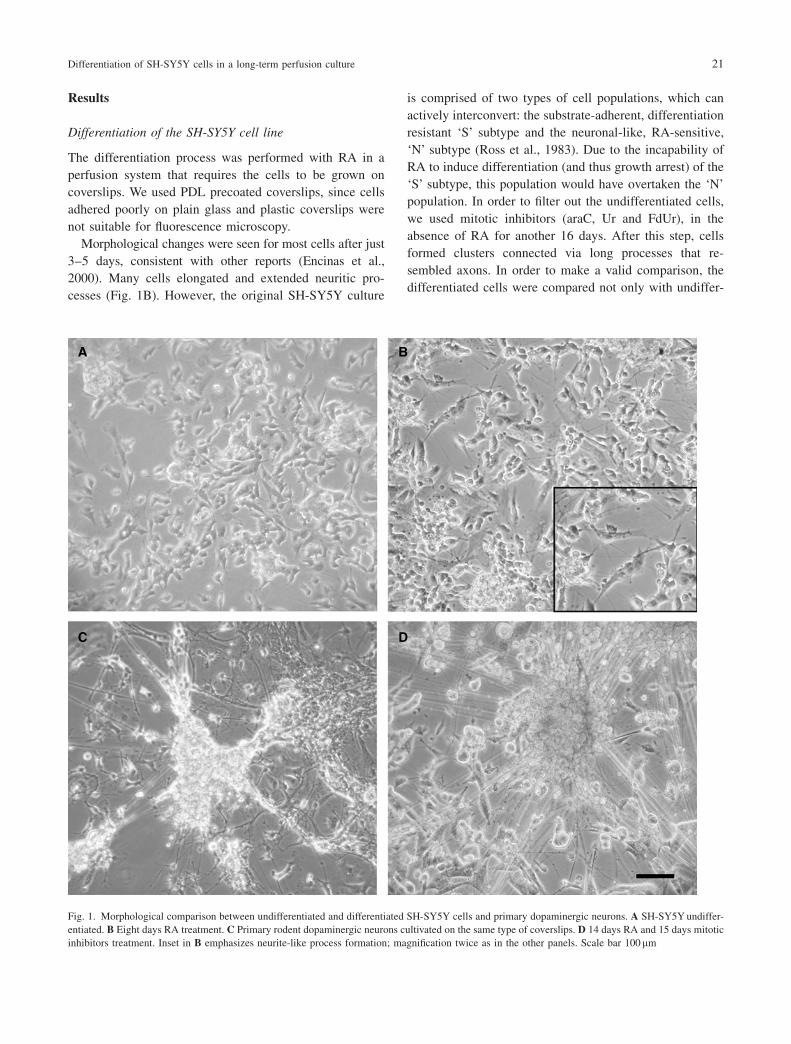
Morphological changes were seen for most cells after just
3–5 days, consistent with other reports (Encinas et al.,
2000). Many cells elongated and extended neuritic pro-
cesses (Fig. 1B). However, the original SH-SY5Y culture
is comprised of two types of cell populations, which can
actively interconvert: the substrate-adherent, differentiation
resistant ‘S’ subtype and the neuronal-like, RA-sensitive,
‘N’ subtype (Ross et al., 1983). Due to the incapability of
RA to induce differentiation (and thus growth arrest) of the
‘S’ subtype, this population would have overtaken the ‘N’
population. In order to filter out the undifferentiated cells,
we used mitotic inhibitors (araC, Ur and FdUr), in the
absence of RA for another 16 days. After this step, cells
formed clusters connected via long processes that re-
sembled axons. In order to make a valid comparison, the
differentiated cells were compared not only with undiffer-
Fig. 1. Morphological comparison between undifferentiated and differentiated SH-SY5Y cells and primary dopaminergic neurons. A SH-SY5Yundiffer-
entiated. B Eight days RA treatment. C Primary rodent dopaminergic neurons cultivated on the same type of coverslips. D 14 days RA and 15 days mitotic
inhibitors treatment. Inset in B emphasizes neurite-like process formation; magnification twice as in the other panels. Scale bar 100mm
Differentiation of SH-SY5Y cells in a long-term perfusion culture 21
entiated SH-SY5Y cells (cultivated in DMEM-20% FBS
medium) but also with rodent primary dopaminergic neu-
rons cultivated on identical coverslips (Fig. 1C and D). The
differentiated cells had a morphology similar to rodent
primary dopaminergic neurons. The differentiation process
seemed more successful in the perfusion system compared
with the classic cell culture method. As it can be seen in
Fig. 2, in the dish culture there are more apoptotic, round
cells, compared to perfusion culture (panels B and A, re-
spectively). Moreover, in panel D (dish culture), many
more fibroblast-like, undifferentiated cells can be observed
compared to panel C.
BrdU staining for proliferation control
It is widely accepted that most of the neuronal cells in the
adult brain cease dividing (Cajal, 1928; Gage, 2002). There
is evidence for new neurons in the adult mammalian brain.
However, proliferation is confined to the olfactory bulb and
dentate gyrus (Rakic, 2002). Since the cells seemed to de-
velop a neuronal morphology, and in order to test the effi-
ciency of the mitotic inhibitors treatment, cell duplication
was assessed by BrdU incorporation into cellular DNA.
Both types of cells (undifferentiated and differentiated,
the latter in the absence of mitotic inhibitors) were incubat-
Fig. 2. Comparison between perfusion and plate cultivation during differentiation of cells (14 days RA and seven days mitotic inhibitors). A and C
Perfusion. B and D dish. Note in B and D many apoptotic cells (round, bright floating cells) and many cells with a fibroblast-like morphology. Scale bar
100mm. Same magnification for A and B, respectively C and D
22 R. Constantinescu et al.
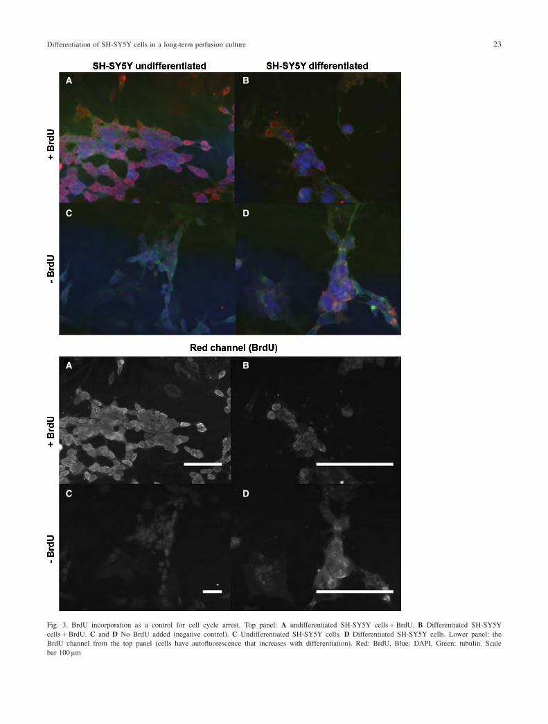
Fig. 3. BrdU incorporation as a control for cell cycle arrest. Top panel: A undifferentiated SH-SY5Y cellsþBrdU. B Differentiated SH-SY5Y
cellsþBrdU. C and D No BrdU added (negative control). C Undifferentiated SH-SY5Y cells. D Differentiated SH-SY5Y cells. Lower panel: the
BrdU channel from the top panel (cells have autofluorescence that increases with differentiation). Red: BrdU, Blue: DAPI, Green: tubulin. Scale
bar 100mm
Differentiation of SH-SY5Y cells in a long-term perfusion culture 23

ed with medium containing BrdU for 72 h and subsequently
stained for BrdU incorporation. The negative control (un-
differentiated and differentiated cells not treated with
BrdU, but stained as the other ones) showed that the cells
exhibit autofluorescence, which increases after differentia-
tion (Fig. 3, the red channel). The BrdU signal in the dif-
ferentiated cells is very close to the background (compare
panels B and D), whereas the undifferentiated cells incor-
porated BrdU and led to a strong signal (in panel A) com-
pared to their corresponding control (panel C).
Thus, we concluded that the cell divisions markedly
slowed down after the mitotic inhibitors treatment and
the differentiated cells are closer to ‘‘real’’ (slow dividing)
neurons.
RT-PCR results confirm the differentiation of the cells
To confirm differentiation, we examined several neuronal
markers. Mature neurons express specific markers that
identify their specialized role in the nervous system. From
various known neuronal markers, the twelve presented in
the introduction were chosen for this study with the ratio-
nale that an increase in their expression (with the exception
of nestin) would indicate that the cells are progressing
towards a more neuronal phenotype.
As expected, RT-PCR results showed that the mRNA
of many neuronal markers increased after differentiation
(Fig. 4). For example, a significant change (p<0.05) was
observed for Neurogenin, tau, laminin and DRD2, while
the message for other proteins (such as MAP2 and DAT)
was increased, even if not at a statistically significant
level.
Thus, the RT-PCR results suggest that the treatment with
RA and mitotic inhibitors led to an increase of the message
for many neuronal markers.
Western blotting analysis
To confirm that changes in mRNA level resulted in changes
in protein levels, we examined candidate markers by
Western blotting. The bands corresponding to the proteins
of interest (Fig. 5) were quantified using ImageJ and the
b-actin band as a reference (Fig. 6).
Since not all proteins have a commercial antibody avail-
able, some antibodies are better than others and several
large proteins are difficult to transfer, only a subset of the
Fig. 5. Western blot analysis of marker
proteins. D means differentiated SH-SY5Y
cells. U means undifferentiated SH-SY5Y
cells. Actin was used as loading control.
Numbers on the left represent the molecu-
lar weight in kDa
Fig. 4. Variation of neuronal markers after differentiation. Marker mRNA
level quantification by QPCR, normalized to undifferentiated cells and
HMBS. Reference level is one (mRNA level of marker in undifferentiated
cells). The mRNA level decreases after differentiation for NeuroD1 and
increases for all the other markers analysed. Asterisk mark statistically
significant changes, i.e. p<0.05
24 R. Constantinescu et al.
markers tested by RT-PCR could be assessed by Western
blotting. Based on the results of Western blotting, MAP2,
TH and NeuN increased following the differentiation pro-
tocol. Moreover, nestin, a marker for neuronal progenitor
cells that decreases during differentiation, was decreased
in SH-SY5Y differentiated cells (see Figs. 5 and 6), com-
pared to undifferentiated SH-SY5Y. We concluded that the
mRNA of the upregulated genes was indeed translated into
increased protein amounts in the cell.
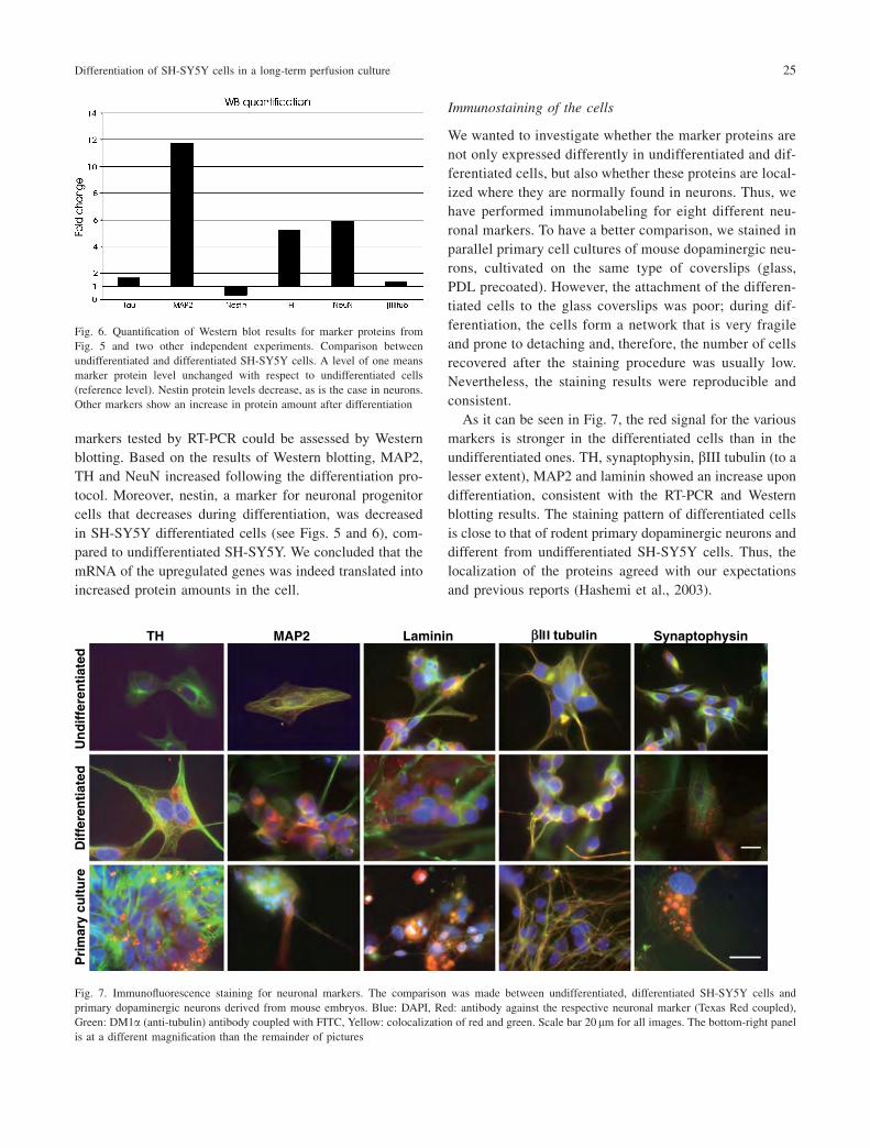
Immunostaining of the cells
We wanted to investigate whether the marker proteins are
not only expressed differently in undifferentiated and dif-
ferentiated cells, but also whether these proteins are local-
ized where they are normally found in neurons. Thus, we
have performed immunolabeling for eight different neu-
ronal markers. To have a better comparison, we stained in
parallel primary cell cultures of mouse dopaminergic neu-
rons, cultivated on the same type of coverslips (glass,
PDL precoated). However, the attachment of the differen-
tiated cells to the glass coverslips was poor; during dif-
ferentiation, the cells form a network that is very fragile
and prone to detaching and, therefore, the number of cells
recovered after the staining procedure was usually low.
Nevertheless, the staining results were reproducible and
consistent.
As it can be seen in Fig. 7, the red signal for the various
markers is stronger in the differentiated cells than in the
undifferentiated ones. TH, synaptophysin, bIII tubulin (to a
lesser extent), MAP2 and laminin showed an increase upon
differentiation, consistent with the RT-PCR and Western
blotting results. The staining pattern of differentiated cells
is close to that of rodent primary dopaminergic neurons and
different from undifferentiated SH-SY5Y cells. Thus, the
localization of the proteins agreed with our expectations
and previous reports (Hashemi et al., 2003).
Fig. 7. Immunofluorescence staining for neuronal markers. The comparison was made between undifferentiated, differentiated SH-SY5Y cells and
primary dopaminergic neurons derived from mouse embryos. Blue: DAPI, Red: antibody against the respective neuronal marker (Texas Red coupled),
Green: DM1a (anti-tubulin) antibody coupled with FITC, Yellow: colocalization of red and green. Scale bar 20 mm for all images. The bottom-right panel
is at a different magnification than the remainder of pictures
Fig. 6. Quantification of Western blot results for marker proteins from
Fig. 5 and two other independent experiments. Comparison between
undifferentiated and differentiated SH-SY5Y cells. A level of one means
marker protein level unchanged with respect to undifferentiated cells
(reference level). Nestin protein levels decrease, as is the case in neurons.
Other markers show an increase in protein amount after differentiation
Differentiation of SH-SY5Y cells in a long-term perfusion culture 25
Taken together, these results suggest that the neuronal
markers are expressed and localized as in neuronal cells.
Discussion
In the present work we show that the human dopaminergic
neuroblastoma cell line SH-SY5Y can be differentiated to
dopaminergic neurons using a specific protocol and a per-
fusion culture system. The results presented here show that
these cells can be differentiated further than has been re-
ported up to now (Nicolini et al., 1998; Maruyama et al.,
1997). We have also performed a thorough characterization
of the differentiated cells and have shown that many neu-
ronal characteristics can be attained using this protocol.
While animal models probably mimic more accurately
aspects of a disease, there are several distinct disadvan-
tages, most obviously, time and cost. In a live animal, many
variables can perturb the study of different mechanisms.
Cell culture models present the advantage that they are
more easily to perform and repeat, whilst being time- and
cost-saving. This makes them a good candidate for prelim-
inary studies on the efficiency of various substances, es-
pecially when a more controlled setting is required.
In order to have the basic cellular system for developing
new oxidative stress models of PD, a human derived cell
line was used, which is easier to cultivate than primary
neurons, relatively homogenous in composition and closely
resembling the cells affected in PD. For this purpose, the
human dopaminergic neuroblastoma cell line SH-SY5Y
was chosen as a starting point.
The SH-SY5Y cells are often used in cell culture models
of PD because they possess many of the qualities of human
neurons (Sherer et al., 2001). These cells have neuronal
origin, express TH and dopamine-b-hydroxylase, which arespecific to catecholaminergic neurons (Ross et al., 1983)
and express receptors and transporters for DA and acetyl-
choline (Biedler et al., 1978; Willets et al., 1995). These
cells also express genes associated with neuronal differen-
tiation, including neurofilament proteins and neuron spe-
cific enolase among others. Despite expressing all these
markers, they are considered immature neuroblasts at differ-
ent stages of neuronal differentiation (Biedler et al., 1973)
and have been shown to maintain the stem cell character-
istics and to proliferate in culture for a long time with no
contamination (Ross et al., 1983). This is important in the
neuroscience and neurotoxicology fields, where the conta-
minating presence of glial cells, astrocytes and other types
of cells can lead to unwanted effects. The SH-SY5Y cell
line presents also the advantage that it can be grown and
differentiated in the absence of growth factors (Nicolini
et al., 1998). The effects of neurotrophic factors used in
differentiation are confusing, especially if the cells are
further used to study drug-induced neurotoxicity (for exam-
ple antineoplastic drugs) and the effect of similar trophic
factors (Nicolini et al., 1998).
Despite these advantages, there are several differences
with respect to neurons, most notably a different expression
level of neuronal cell markers (Farooqui, 1994) and con-
firmed cell proliferation (Pahlman et al., 1995). In particu-
lar, undifferentiated SH-SY5Y cells are not an ideal model
for dopaminergic neurons as they have a low expression of
DAT (Presgraves et al., 2004). Toxicity by 1-methyl-4-phe-
nyl-1,2,3,6-tetrahydropyridine (MPTP, a neurotoxin widely
used in PD pharmacological models) requires the presence
of DAT to enter the cells and to be converted to the toxic
ion MPPþ (Presgraves et al., 2004). This implies that un-
differentiated SH-SY5Y cells are more resistant to MPTP
than normal dopaminergic neurons, and are thus not a good
starting point for an MPTP-based model of PD (Presgraves
et al., 2004). Similarly, the relatively high oxidative stress
imposed by DA synthesis makes dopaminergic neurons
more susceptible to intoxication by Complex I inhibitors
compared to other cells (Barzilai et al., 2001). This was our
main reason to generate differentiated cells in order to be
further used for a chronic PD model. Another reason to use
differentiated cells is to have a constant, non-dividing cell
population in order to establish a chronic intoxication mod-
el. This would avoid problems stemming from variations in
cell numbers and the constant renewal of the cell popula-
tion. In this respect, primary cells have the disadvantage
that they cannot be maintained in culture for very long time
whereas immortalized cell lines multiply too much.
We cultivated the cells plated on PDL precoated glass
coverslips in a long-term perfusion culture system. This per-
fusion system is more convenient to use than a normal cell
culture dish and the cells can be cultivated for a longer time
and under better conditions (Minuth et al., 1999). The perfu-
sion system is characterized by the continuous addition of
fresh medium with nutrients and the concomitant withdrawal
of the used medium with toxic metabolites. In this way, it is
possible to cultivate the cells=tissues in conditions closer to
the in vivo situation (Minuth et al., 1999, 2000).
The differentiation protocol started with the treatment
of cells with RA for 14 days. After 8 days of treatment,
cells elongated and exhibited branching similar to neurons
(Fig. 1B), as described by several other authors for a
shorter treatment (Nicolini et al., 1998; Maruyama et al.,
1997). After about two weeks of treatment with RA and
another two with mitotic inhibitors to eliminate the prolif-
erating subpopulation, the cells resembled morphologically
26 R. Constantinescu et al.

the primary rodent dopaminergic neurons cultivated on
the same type of coverslips. A BrdU incorporation assay
showed that the cells, indeed, stopped proliferating, while
RT-PCR, Western blotting and immunofluorescence were
used to show that several neuronal markers were upregu-
lated as a consequence of the differentiation protocol.
Quantification of immunofluorescence pictures is prone
to many pitfalls; in this particular case, where cells aggre-
gate, it is impossible to do a proper quantification over the
entire volume, so the results are only qualitative. Even if
the results from RT-PCR and Western blotting were not
always in perfect agreement at the quantitative level, both
methods, as well as the immunofluorescence suggested that
most of the markers tested increased following the differ-
entiation protocol. The immunofluorescence results also
show that the proteins localized as expected for a neuronal
cell. However, one has to keep in mind that SH-SY5Y cells
have neuronal origin, so it is not surprising that, even before
differentiation, they already express – albeit at lower levels –
proteins that are considered markers for a neuronal cell. Still,
there is an obvious signal increase for the above-mentioned
markers (Fig. 7). An overview of the neuronal markers var-
iation after differentiation is presented in Table 3.
Patch-clamp would be the ultimate way to prove that the
cells are differentiated. However, the differentiated cells
are fragile and entangled in a complicated network. More-
over, many cells are packed in large clusters which means
patch-clamp would be very difficult (if not impossible) to
perform.
In conclusion, in the present work we have developed a
new cell culture system using human neuroblastoma cells
differentiated in perfusion, which allows to better control
vital parameters and to maintain the culture for longer time
(i.e., weeks instead of just days) (Minuth et al., 2000).
The differentiation protocol presented here has several
advantages. Much more time is allowed for the cells to dif-
ferentiate and ‘‘filter out’’ many of the cells that do not
undergo differentiation. Other cell culture models utilized
short-term (a few days) treatment with RA with=without
neurotrophins, tetradecanoylphorbol acetate or norepine-
phrine (Singh et al., 2003; Laifenfeld et al., 2002). In the
present work, the differentiated cells were thoroughly char-
acterized at both the morphological and molecular levels.
The results presented suggest that the differentiation proto-
col was successful and the differentiated cells have a good
similarity with primary neurons.
The low division rate of the cells, taken together with our
own observations during cell handling, suggests that the
population is relatively constant for a long time. A classical
culture using cell lines would require splitting the culture
every few days, which would skew the results of any via-
bility testing. This new model gives the opportunity to try
various neurotoxins in low dose and long time in culture.
This way, the differentiated cells can be further used to mod-
el PD and other neurodegenerative disorders affecting the
dopaminergic system of the brain. Moreover, in these mod-
els new potential therapies can be tested for their long-term
effect. We are presently developing such a chronic model.
Acknowledgements
We thank G. Gille and her students for neuronal rodent primary cells,
T. Rohrmeier, Neuroprofile GmbH, Regensburg for advice regarding the
differentiation protocol, the A. Storch group for several RT-PCR primers
and antibodies, T. Hyman, MPI-CBG Dresden for access to his laboratory
equipment, the MPI-CBG Dresden Antibody facility for providing the
actin antibody and Nathan Goehring for very useful comments on this
manuscript.
The present work was supported by a MeDDrive 2004 grant from
the Faculty of Medicine of Dresden University of Technology awarded to
R. Constantinescu.
References
Barzilai A, Melamed E, Shirvan A (2001) Is there a rationale for neuro-
protection against dopamine toxicity in Parkinson’s disease? Cell Mol
Neurobiol 21: 215–235
Biedler JL, Helson L, Spengler BA (1973) Morphology and growth,
tumorigenicity, and cytogenetics of human neuroblastoma cells in
continuous culture. Cancer Res 33: 2643–2652
Table 3. Summary of the neuronal markers variation after differentiation
Neuronal
marker
Variation after
differentiation
Expected from literature
RT-PCR WB IF
TH � þ þ þ (Hashemi et al., 2003)
MAP2 þ þ þ þ (Binder et al., 1985)
bIII tubulin þ � � þ (Lee et al., 1990)
Tau þ þ NO þ (Wood et al., 1986)
Nestin þ � NO � (Duggal and Hammond,
2002)
Laminin þ NO þ þ (Timpl and Brown, 1994)
NeuN NA þ NO þ (Mullen et al., 1992)
Synaptophysin þ NO þ þ (Gaardsvoll et al., 1988)
Neurogenin1 þ NT NT þ (Ma et al., 1996)
DRD2 þ NT NT þ (Nestler and Aghajanian,
1997)
DAT þ NO NT þ (Storch et al., 2004)
NeuroD1 � NT NT þ (Lee et al., 1995)
þ¼ increase, �¼ decrease.
� no or very small variation, NO means no optimal result (problems with
the antibody or the protocol, e.g. the transfer on the nitrocellulose mem-
brane in Western blotting).
NT not tried (did not find a working antibody), NA not applicable (there is
no possibility to design primers for NeuN, as the antigen is not known).
WB Western blotting, IF immunofluorescence.
Differentiation of SH-SY5Y cells in a long-term perfusion culture 27
Biedler JL, Roffler-Tarlov S, Schachner M, Freedman LS (1978) Multiple
neurotransmitter synthesis by human neuroblastoma cell lines and
clones. Cancer Res 38: 3751–3757
Binder LI, Frankfurter A, Rebhun LI (1985) The distribution of tau in the
mammalian central nervous system. J Cell Biol 101: 1371–1378
Blander G, de Oliveira RM, Conboy CM, Haigis M, Guarente L (2003)
Superoxide dismutase 1 knock-down induces senescence in human
fibroblasts. J Biol Chem 278: 38966–38969
Bove J, Prou D, Perier C, Przedborski S (2005) Toxin-induced models of
Parkinson’s disease. NeuroRx 2: 484–494
Cajal SR y (1928) Degeneration and regeneration of the nervous system.
University Press, London
Dauer W, Przedborski S (2003) Parkinson’s disease: mechanisms and
models. Neuron 39: 889–909
Duggal N, Hammond RR (2002) Nestin expression in ganglioglioma. Exp
Neurol 174: 89–95
Edsjo A, Lavenius E, Nilsson H, Hoehner JC, Simonsson P, Culp LA,
Martinsson T, Larsson C, Pahlman S (2003) Expression of trkB in
human neuroblastoma in relation to MYCN expression and retinoic
acid treatment. Lab Invest 83: 813–823
Encinas M, Iglesias M, Liu Y, Wang H, Muhaisen A, Cena V, Gallego C,
Comella JX (2000) Sequential treatment of SH-SY5Y cells with
retinoic acid and brain-derived neurotrophic factor gives rise to ful-
ly differentiated, neurotrophic factor-dependent, human neuron-like
cells. J Neurochem 75: 991–1003
Farooqui SM (1994) Induction of adenylate cyclase sensitive dopamine
D2-receptors in retinoic acid induced differentiated human neuro-
blastoma SH-SY5Y cells. Life Sci 55: 1887–1893
Gaardsvoll H, Obendorf D, Winkler H, Bock E (1988) Demonstration of
immunochemical identity between the synaptic vesicle proteins synap-
tin and synaptophysin=p38. FEBS Lett 242: 117–120
Gage FH (2002) Neurogenesis in the adult brain. J Neurosci 22: 612–613
Gates MA, Torres EM, White A, Fricker-Gates RA, Dunnett SB (2006)
Re-examining the ontogeny of substantia nigra dopamine neurons.
Eur J Neurosci 23: 1384–1390
Gille G, Rausch WD, Hung ST, Moldzio R, Ngyuen A, Janetzky B,
Engfer A, Reichmann H (2002) Protection of dopaminergic neurons
in primary culture by lisuride. J Neural Transm 109: 157–169
Hashemi SH, Li JY, Ahlman H, Dahlstrom A (2003) SSR2(a) receptor
expression and adrenergic=cholinergic characteristics in differentiated
SH-SY5Y cells. Neurochem Res 28: 449–460
Herman GE (2002) Mouse models of human disease: lessons learned and
promises to come. ILAR J 43: 55–56
Laifenfeld D, Klein E, Ben Shachar D (2002) Norepinephrine alters the
expression of genes involved in neuronal sprouting and differentiation:
relevance for major depression and antidepressant mechanisms. J
Neurochem 83: 1054–1064
Lee JE, Hollenberg SM, Snider L, Turner DL, Lipnick N, Weintraub H
(1995) Conversion of xenopus ectoderm into neurons by neurod, a
basic helix-loop-helix protein. Science 268: 836–844
Lee MK, Tuttle JB, Rebhun LI, Cleveland DW, Frankfurter A (1990) The
expression and posttranslational modification of a neuron-specific
beta-tubulin isotype during chick embryogenesis. Cell Motil Cyto-
skeleton 17: 118–132
Ma Q, Kintner C, Anderson DJ (1996) Identification of neurogenin, a
vertebrate neuronal determination gene. Cell 87: 43–52
Maden M, Hind M (2003) Retinoic acid, a regeneration-inducing molecule.
Dev Dyn 226: 237–244
Maruyama W, Benedetti MS, Takahashi T, Naoi M (1997) A neurotoxin
N-methyl(R)salsolinol induces apoptotic cell death in differentiated
human dopaminergic neuroblastoma SH-SY5Y cells. Neurosci Lett
232: 147–150
Melino G, Thiele CJ, Knight RA, Piacentini M (1997) Retinoids and the
control of growth=death decisions in human neuroblastoma cell lines.
J Neurooncol 31: 65–83
Minuth WW, Schumacher K, Strehl R, Kloth S (2000) Physiological and
cell biological aspects of perfusion culture technique employed to
generate differentiated tissues for long term biomaterial testing and
tissue engineering. J Biomater Sci Polym Ed 11: 495–522
MinuthWW, Steiner P, Strehl R, Schumacher K, de Vries U, Kloth S (1999)
Modulation of cell differentiation in perfusion culture. Exp Nephrol 7:
394–406
Mullen RJ, Buck CR, Smith AM (1992) NeuN, a neuronal specific nuclear
protein in vertebrates. Development 116: 201–211
Nestler EJ, Aghajanian GK (1997) Molecular and cellular basis of addic-
tion. Science 278: 58–63
Nicolini G, MilosoM, Zoia C, Di Silvestro A, Cavaletti G, Tredici G (1998)
Retinoic acid differentiated SH-SY5Y human neuroblastoma cells:
an in vitro model to assess drug neurotoxicity. Anticancer Res 18:
2477–2481
Pahlman S, Hoehner JC, Nanberg E, Hedborg F, Fagerstrom S, Gestblom C,
Johansson I, Larsson U, Lavenius E, Ortoft E, Soderholm H (1995)
Differentiation and Survival Influences of Growth-Factors in Human
Neuroblastoma. Eur J Cancer 31A: 453–458
Pleasure SJ, Page C, Lee VM (1992) Pure, postmitotic, polarized human
neurons derived from NTera 2 cells provide a system for expressing
exogenous proteins in terminally differentiated neurons. J Neurosci 12:
1802–1815
Presgraves SP, Ahmed T, Borwege S, Joyce JN (2004) Terminally differ-
entiated SH-SY5Y cells provide a model system for studying neuro-
protective effects of dopamine agonists. Neurotox Res 5: 579–598
Rakic P (2002) Adult neurogenesis in mammals: an identity crisis.
J Neurosci 22: 614–618
Rasband WS (2006) Image J. U. S. National Institutes of Health, Bethesda,
Maryland, USA, http:==rsb.info.nih.gov=ij=
Rebhan M, Vacun G, Bayreuther K, Rosner H (1994) Altered ganglioside
expression by SH-SY5Y cells upon retinoic acid-induced neuronal
differentiation. Neuroreport 5: 941–944
Ross RA, Spengler BA, Biedler JL (1983) Coordinate morphological
and biochemical interconversion of human neuroblastoma cells. J Natl
Cancer Inst 71: 741–747
Sherer TB, Trimmer PA, Borland K, Parks JK, Bennett JP Jr, Tuttle JB
(2001) Chronic reduction in complex I function alters calcium signal-
ing in SH-SY5Y neuroblastoma cells. Brain Res 891: 94–105
Singh US, Pan J, Kao YL, Joshi S, Young KL, Baker KM (2003) Tissue
transglutaminase mediates activation of RhoA and MAP kinase path-
ways during retinoic acid-induced neuronal differentiation of SH-
SY5Y cells. J Biol Chem 278: 391–399
Storch A, Ludolph AC, Schwarz J (2004) Dopamine transporter: involve-
ment in selective dopaminergic neurotoxicity and degeneration.
J Neural Transm 111: 1267–1286
Timpl R, Brown JC (1994) The laminins. Matrix Biol 14: 275–281
Willets JM, Lambert DG, Lunec J, Griffiths HR (1995) Studies on
the neurotoxicity of 6,7-dihydroxy-1-methyl-1,2,3,4-tetrahydroiso-
quinoline(salsolinol) in SH-SY5Y cells. Eur J Pharmacol 293:
319–326
Wood JG, Mirra SS, Pollock NJ, Binder LI (1986) Neurofibrillary tangles
of Alzheimer disease share antigenic determinants with the axonal
microtubule-associated protein tau (tau). Proc Natl Acad Sci USA 83:
4040–4043
28 R. Constantinescu et al.: Differentiation of SH-SY5Y cells in a long-term perfusion culture


![Creating permissive microenvironments for stem cell ... · neuronal differentiation from NSPCs [33]. Elastic modulus Cell differentiation can be influenced, in part, by the mechanical](https://img.pdfslide.us/doc/110x75/5fcf1181a9c1051993304a8a/creating-permissive-microenvironments-for-stem-cell-neuronal-differentiation.jpg)