Embed Size (px)
Citation preview
Sarcoma (1998) 2, 205± 207
CASE REPORT
Primary liposarcoma of the mediastinum
JOEL GREIF,1 SILVIA MARMOR,2 OFER MERIMSKY,3 FELIX KOVNER3 &MOSHE INBAR3
1Pulmonary Division, 2Pathology Department and 3Oncology Department, Tel-Aviv Sourasky Medical Center and the
Sackler Faculty of Medicine, Tel-Aviv University, Tel-Aviv, Israel
Abstract
Patient. A 62-year-old man presented with effort dyspnea, non-productive cough and weakness of 4 month duration. Hehad no ® ndings on physical examination.Discussion. Chest X-ray revealed a large mass in the left anterior mediastinum. Computerized tomography of the chestshowed a well-delineated homogeneous mediastinal mass with fat-equivalent density and a small pleural effusion.Fiberoptic bronchoscopy revealed narrowing of the left main bronchus, secondary to external compression. The bronchialmucosa was normal and brush cytology was negative. A CT-guided ® ne needle aspiration (FNA) of the mass yieldedfragments of cells embedded in myxoid background material and closely packed atypical lipoblasts, compatible withliposarcoma. The patient underwent a left lateral thoracotomy and margibnal resection of the mass. The histopathologicalexamination con® rmed the diagnosis of mixed-type liposarcoma, consisted of myxoid and pleomorphic liposarcoma.Postoperative two-® eld radiation therapy was delivered to the mediastinum for a total midplane dose of 40 Gy. After adisease-free interval of 8 months the disease recurred in the mediastinum and pleura. Palliative chemotherapy achieved ashort duration partial response but the patient succumbed to local recurrence 2 years after the diagnosis.
Key words: primary liposarcoma, mediastinum , neop lasm.
Introduction
Liposarcomas are relatively uncommon neoplasms,
accounting for approximately 15± 20% of all sarco-
mas.1 They usually arise in the lower extremities or
retroperitoneum but have been reported in other
locations such as the abdomen, vulva and buttocks.
Primary lipoblastic tumors originating in the
mediastinum are extremely rare.
The present report concerns a patient with such a
primary mediastinal liposarcoma ® rst diagnosed by
® ne needle aspiration biopsy. The clinical course,
pathological ® ndings and therapeutic approaches are
presented and the current literature is reviewed.
Case Report
A 62 year-old man presented with dyspnea on
effort, non-productive cough and weakness of 4
months’ duration. The physical examination was
unremarkable and blood count and biochemistry
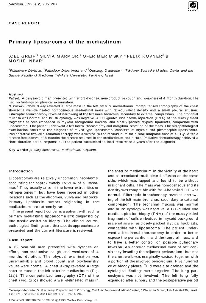
values were normal. A chest X-ray revealed a large
anterior mass in the left anterior mediastinum (Fig.
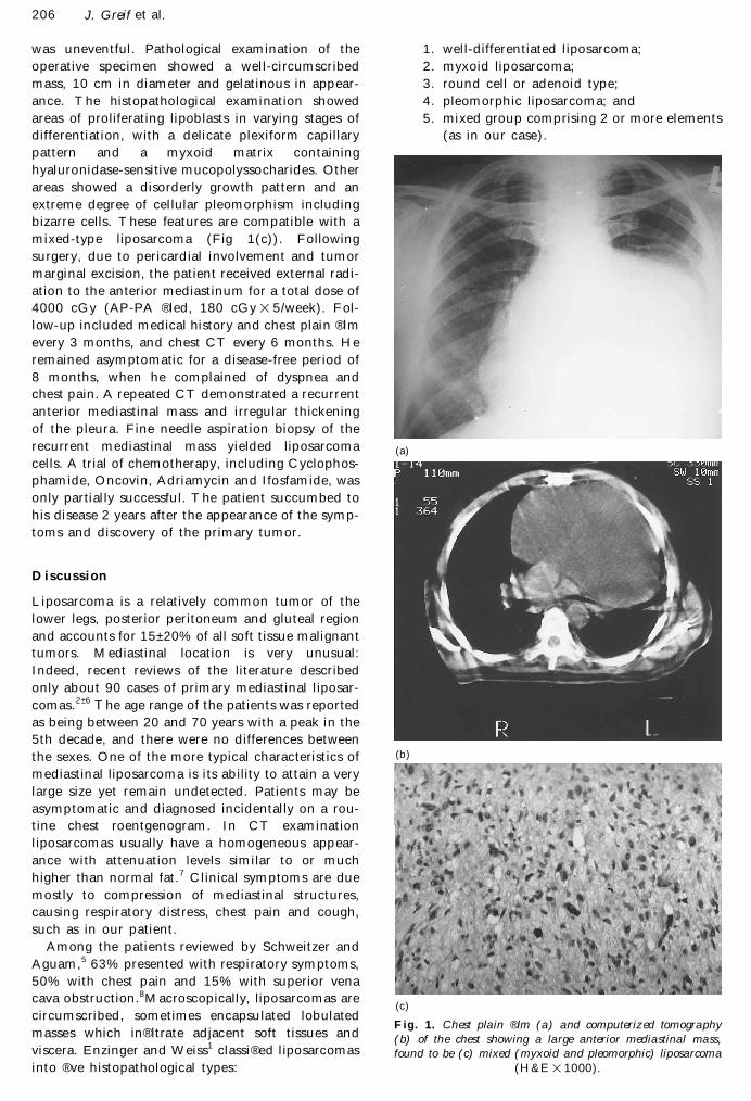
1(a)). The computerized tomography (CT) of the
chest (Fig. 1(b)) showed a well-delineated mass in
the anterior mediastinum in the vicinity of the heart
and an associated small pleural effusion on the same
side, which was lapped and found to be without
malignant cells. The mass was homogeneous end its
density was compatible with fat. Abdominal CT was
normal. Fiberoptic bronchoscopy revealed narrow-
ing of the left main bronchus, secondary to external
compression. The bronchial mucosa was normal
and brush cytology was negative. A CT-guided ® ne
needle aspiration biopsy (FNA) of the mass yielded
fragments of cells embedded in myxoid background
material as well as closely packed atypical lipoblasts,
compatible with liposarcoma. The patient under-
went a left lateral thoracotomy in order to better
expose the pericardium and the tumoral mass, and
to have a better control on possible pulmonary
invasion. An anterior mediastinal mass of soft con-
sistency invading the adjacent pericardium, but not
the chest wall, was marginally excised together with
a portion of the involved pericardium. Five hundred
cc of bloody pleural ¯ uid were also drained and the
cytological ® ndings were negative. The lung par-
enchyma was not involved. The left lung fully
expanded after surgery and the postoperative period
Correspondence to: O. Merimsky, Department of Oncology, Tel Aviv Sourasky Medical Center, 6 Weizman Street, Tel-Aviv 64239, Israel.Tel.: Int-972-3-697-483 0; Fax: Int-972-3-697-4828 .
1357-714 X/98/030205± 03 $9.00 Ó 1998 Carfax Publishing Ltd
206 J. Greif et al.
was uneventful. Pathological examination of the
operative specimen showed a well-circumscribed
mass, 10 cm in diameter and gelatinous in appear-
ance. The histopathological examination showed
areas of proliferating lipoblasts in varying stages of
differentiation, with a delicate plexiform capillary
pattern and a myxoid matrix containing
hyaluronidase-sensitive mucopolyssocharides. Other
areas showed a disorderly growth pattern and an
extreme degree of cellular pleomorphism including
bizarre cells. These features are compatible with a
mixed-type liposarcoma (Fig 1(c)). Following
surgery, due to pericardial involvement and tumor
marginal excision, the patient received external radi-
ation to the anterior mediastinum for a total dose of
4000 cGy (AP-PA ® led, 180 cGy 3 5/week). Fol-
low-up included medical history and chest plain ® lm
every 3 months, and chest CT every 6 months. He
remained asymptomatic for a disease-free period of
8 months, when he complained of dyspnea and
chest pain. A repeated CT demonstrated a recurrent
anterior mediastinal mass and irregular thickening
of the pleura. Fine needle aspiration biopsy of the
recurrent mediastinal mass yielded liposarcoma
cells. A trial of chemotherapy, including Cyclophos-
phamide, Oncovin, Adriamycin and Ifosfam ide, was
only partially successful. The patient succumbed to
his disease 2 years after the appearance of the symp-
toms and discovery of the primary tumor.
Discussion
Liposarcoma is a relatively common tumor of the
lower legs, posterior peritoneum and gluteal region
and accounts for 15± 20% of all soft tissue malignant
tumors. Mediastinal location is very unusual:
Indeed, recent reviews of the literature described
only about 90 cases of primary mediastinal liposar-
comas.2 ± 6 The age range of the patients was reported
as being between 20 and 70 years with a peak in the
5th decade, and there were no differences between
the sexes. One of the more typical characteristics of
mediastinal liposarcoma is its ability to attain a very
large size yet remain undetected. Patients may be
asymptomatic and diagnosed incidentally on a rou-
tine chest roentgenogram. In CT examination
liposarcomas usually have a homogeneous appear-
ance with attenuation levels similar to or much
higher than normal fat.7 Clinical symptoms are due
mostly to compression of mediastinal structures,
causing respiratory distress, chest pain and cough,
such as in our patient.
Among the patients reviewed by Schweitzer and
Aguam,5 63% presented with respiratory symptoms,
50% with chest pain and 15% with superior vena
cava obstruction.8Macroscopically, liposarcomas are
circumscribed, sometimes encapsulated lobulated
masses which in ® ltrate adjacent soft tissues and
viscera. Enzinger and Weiss1 classi ® ed liposarcomas
into ® ve histopathological types:
1. well-differentiated liposarcoma;
2. myxoid liposarcoma;
3. round cell or adenoid type;
4. pleomorphic liposarcoma; and
5. mixed group comprising 2 or more elements
(as in our case).
Fig. 1. Chest plain ® lm (a) and computerized tomography
(b) of the chest showing a large anterior mediastinal mass,
found to be (c) mixed (myxoid and pleomorphic) liposarcoma
(H&E 3 1000).
(a)
(b)
(c)

Primary liposarcoma of the mediastinum 207
The clinical course is characterized by frequent
recurrence of the tumor and death due to local
disease, with a mean survival of 2.5 years.4 Fine
needle aspiration will reveal a characteristic picture
and help to establish the diagnosis9,10 Ð as in our
case.
Because of the low incidence of primary mediasti-
nal sarcomas, treatment strategies are extrapolated
from similar tumors in other places. The ® rst choice
of therapy for mediastinal liposarcoma is surgical
excision of the tumor; but, because of invasion to
surrounding tissues total excision is frequently
impossib le.
Patient survival following surgery is closely related
to histological type. In cases of well-differentiated
tumors, the surgery can be curative in nearly all
patients. With poorly differentiated histological
types and invasion of adjacent organs, surgery is
mainly palliative, with the removal of the bulk of the
tumor allowing only symptomatic relief. Radiother-
apy is the treatment of choice for unresectable
tumors or in the event of local recurrence.8
Reported cases of good therapeutic responses to
high-dose radiation treatment of liposarcomas in
other locations11,12 stand in marked contrast to the
poor results reported in mediastinal tumors. It
should be mentioned that such large doses (up to
9000 rad) as used in peripheral tumors would carry
the risk of mediastinal and paricardial ® brosis.
The role of chemotherapy has yet to be deter-
mined. Based on the experience with limb lesions,
the addition of chemotherapy could be considered
in cases of high-grade malignant histologies or
incomplete resected lesions. Whether such therapy
will have an impact on survival is, as yet, unknown
at this time. Some reports suggest that chemo-
therapy is effective against this disorder,12,13 while
others found it not to be useful.14
In conclusion, primary mediastinal liposarcomas
are very rare. A presumptive diagnosis of this tumor
can be obtained by chest CT and con® rmed by
percutaneous needle biopsy. Principles of treatment
include surgical resection and adjuvant radio-
chemotherapy. Despite multimodality therapy, the
prognosis continues to be poor.
References
1 Enzinger FM, Weiss SW. Liposarcoma: in soft tissue
tumors. St Louis: CV Mosby, 1983 ; 242± 80.2 Dogan R, Ayrancioglu K, Aksu O. Primary mediasti-
nal liposarcoma. A report of a case and review of theliterature. Eur J Cardiothorac Surg 1989; 3 ; 357± 70.
3 Grewal RG, Prager K, Austin JM, Rotterdam H.Long-term survival in non-encapsulated primaryliposarcoma of the mediastinum. Thorax 1993;48 ; 1276± 7.
4 Klimstra DS, Moran CA, Perino G, Koss MN, RosalJ. Liposarcoma of the anterior mediastinum and thy-musÐ a clinicopathological study of 28 cases. Am J
Surg Pathol 1995; 19 ; 782± 91.5 Schweitzer DL, Aguam AS. Primary liposarcoma of
the mediastinum. Report of a case and review of theliterature. J Thorac Cardiovasc Surg 1977; 74 ; 83± 97.
6 Shibata K, Koga Y, Onitsuka T, Wake N, Ishil K,Sekiya P. Sumyoshi A. Primary liposarcoma of themediastinumÐ a case report and review of the litera-ture. Jpn J Surg 1986; 16 ; 277± 83.
7 Santamaria G, Serres X. Prune X. Primary mediasti-nal liposarcoma with moderately high CT attenuation.AJR 1996; 1064± 5.
8 Reltan JB, Kaalhus O. Radiotherapy of liposarcomas.Br J Radiol 1980; 53 ; 969± 75.
9 Attal H, Jensen JoA, Reyes CV. Myxoid liposarcomaof the anterior mediastinum. Diagnosis by ® ne needleaspiration biopsy. Acta Cytol 1995; 39 ; 511± 3.
10 Shattuck MC, Victor TA. Cytologic features of well-differentiated sclerosing liposarcoma in aspirated sam-ples. Acta Cytol 1988; 32 ; 896± 901.
11 Friedman M, Egan JW. Effect of irradiation onliposarcoma. Acta Radiol 1960; 64 ; 225± 31.
12 James DH. Effective chemotherapy for abdominalliposarcoma. Pediatrics 1986; 63 ; 311± 5.
13 Antman KA, Blum RH, Wilson RE. Survival ofpatients with localized high-grade soft tissue sarcomawith multimodality therapy. Cancer 1983; 51 ; 396± 40l.
14 Standerfer RJ, Armistead SH, Paneth M. Liposar-coma of the mediastinum report of two cases andreview of the literature. Thorax 1981; 36 ; 693± 4.

Submit your manuscripts athttp://www.hindawi.com
Stem CellsInternational
Hindawi Publishing Corporationhttp://www.hindawi.com Volume 2014
Hindawi Publishing Corporationhttp://www.hindawi.com Volume 2014
MEDIATORSINFLAMMATION
of
Hindawi Publishing Corporationhttp://www.hindawi.com Volume 2014
Behavioural Neurology
EndocrinologyInternational Journal of
Hindawi Publishing Corporationhttp://www.hindawi.com Volume 2014
Hindawi Publishing Corporationhttp://www.hindawi.com Volume 2014
Disease Markers
Hindawi Publishing Corporationhttp://www.hindawi.com Volume 2014
BioMed Research International
OncologyJournal of
Hindawi Publishing Corporationhttp://www.hindawi.com Volume 2014
Hindawi Publishing Corporationhttp://www.hindawi.com Volume 2014
Oxidative Medicine and Cellular Longevity
Hindawi Publishing Corporationhttp://www.hindawi.com Volume 2014
PPAR Research
The Scientific World JournalHindawi Publishing Corporation http://www.hindawi.com Volume 2014
Immunology ResearchHindawi Publishing Corporationhttp://www.hindawi.com Volume 2014
Journal of
ObesityJournal of
Hindawi Publishing Corporationhttp://www.hindawi.com Volume 2014
Hindawi Publishing Corporationhttp://www.hindawi.com Volume 2014
Computational and Mathematical Methods in Medicine
OphthalmologyJournal of
Hindawi Publishing Corporationhttp://www.hindawi.com Volume 2014
Diabetes ResearchJournal of
Hindawi Publishing Corporationhttp://www.hindawi.com Volume 2014
Hindawi Publishing Corporationhttp://www.hindawi.com Volume 2014
Research and TreatmentAIDS
Hindawi Publishing Corporationhttp://www.hindawi.com Volume 2014
Gastroenterology Research and Practice
Hindawi Publishing Corporationhttp://www.hindawi.com Volume 2014
Parkinson’s Disease
Evidence-Based Complementary and Alternative Medicine
Volume 2014Hindawi Publishing Corporationhttp://www.hindawi.com


![superior mediastinum: [Green] Inferior Mediastinum: Below the plane passing from Sternal Angle/Angle Luise Inferior mediastinum has 3 parts: Purple: anterior](https://img.pdfslide.us/doc/110x75/56649c9e5503460f9495e1bf/superior-mediastinum-green-inferior-mediastinum-below-the-plane-passing.jpg)