Embed Size (px)
Citation preview
NANO EXPRESS Open Access
Preparation of ZnO Nanoparticles with HighDispersibility Based on OrientedAttachment (OA) ProcessDingding Cao, Sheng Gong*, Xugang Shu*, Dandan Zhu and Shengli Liang
Abstract
Understanding nanoparticle growth mechanisms is crucial for the synthesis of nanocrystals with desired biologicaland chemical properties. Growth of nanocrystals by oriented attachment (OA) is frequently reported as a methodsupplementary to the classical growth by Ostwald ripening (OR) process. In this work, ZnO nanoparticles (NPs) wereprepared by wet chemical method. Size/shape evolution of ZnO NPs in ethanol solution was systematically studiedusing transmission electron microscopy (TEM), dynamic light scattering (DLS), and X-ray diffraction (XRD). Inaddition, a detailed process of the nanoparticle growth-based OA mechanism is discussed. Results revealed thatreaction conditions affect size/shape of NPs and change their surface structure: prior to OA, the surface of adjacentparticles transformed into their “rough” states. We proved that stability of the solution was significantly improved inthis state. Such a state is important to design nanoparticles with high stability and as nano-suspensions with specialphysical and/or chemical properties. This state is a critical step in enhancing OA process.
Keywords: Zinc oxide, Nanocrystals synthesis, Growth mechanism, Oriented attachment (OA), Suspension property
IntroductionZnO nanoparticles (NPs) attract a lot of attention for fun-damental studies and potential applications in differentresearch areas: from physical chemistry to biomedical sci-ences [1]. ZnO NPs represent a versatile functional mater-ial, and their superior properties find current and potentialapplications in catalysts, transducers, semiconductors, mi-croelectronics, textile, cosmetics, water treatment [2], etc.Additionally, ZnO NPs exhibit antimicrobial activity andanti-inflammation properties [3], providing more efficient,less expensive, and less toxic [4] alternatives to antibioticsand bactericides.Variety of the synthesis routes for ZnO NPs is remark-
able [5–7]. However, it is still challenging to controltheir crystalline structure, stability, and dispersibility incommon solutions such as water and ethanol [8, 9]. Ascomplexity of synthetic reactions increases, a thoroughunderstanding of nanoparticle formation mechanism isneeded [10, 11]. General mechanism is more or lessunderstood. However, major gaps in understanding of
oriented attachment (OA) as well as in the understand-ing of how particle structure changes still remain [12]. Alot of experimental data interpretation and descriptionduring OA crystallization are reported [13]. However, ef-forts to explain this phenomenon quantitatively andfrom a point of view of its mechanism started appearingin the literature only recently. Especially, understandinghow NP performance in a suspension is affected by theparticle morphology is lacking [12]. Control of ZnO NPstability, solubility, surface structure, shape, and aggrega-tion properties represents some of the key roles for ZnONP industrial and other practical applications [5]. Asnano-industry develops, long-standing and traditionalinterpretations of particle formation mechanisms mustbe revisited.This work focuses on the synthesis of highly stable sus-
pension of ZnO nanoparticles (NPs) optimized by changingpH, reaction time, and growth temperature. The growthprocess of NPs (individual as well as their clusters) wasmonitored by high-resolution transmission electron mi-croscopy (HR-TEM) and X-ray powder diffraction (XRD).This is the first study to report the effect of the reactionconditions on suspension and dispersion of ZnO NPs.
© The Author(s). 2019 Open Access This article is distributed under the terms of the Creative Commons Attribution 4.0International License (http://creativecommons.org/licenses/by/4.0/), which permits unrestricted use, distribution, andreproduction in any medium, provided you give appropriate credit to the original author(s) and the source, provide a link tothe Creative Commons license, and indicate if changes were made.
* Correspondence: [email protected]; [email protected] of Chemistry and Chemical Engineering, Zhongkai University ofAgriculture and Engineering, Guangzhou 510220, China
Cao et al. Nanoscale Research Letters (2019) 14:210 https://doi.org/10.1186/s11671-019-3038-3
Relationship between particle structure and growthkinetics was determined by studying the OA process ofcrystal growth. This study provides a better understand-ing of nanoparticles growth from a physical-chemicalpoint of view of stability, dispersibility, and suspensionmorphologies. ZnO NPs obtained in this work demon-strated excellent stability in suspensions, which can bewidely used for practical applications.
MethodsZinc acetate dihydrate (Zn(O2CCH3)2(H2O)2) and so-dium hydroxide (NaOH) were purchased from ShanghaiAladdin Biochemical Technology Co. (China). Absoluteethanol was obtained from Tianjin Damao Chemical Re-agents Co. (China). All reagents were analytically pureand used as received without any further purification.First, products were prepared at the following standard
conditions: 60 °C synthesis temperature, 2 h duration,7.22 and 3.73 mmol of NaOH and zinc acetate dihy-drate, respectively, as initial starting material quantities.To study this reaction and to obtain the best product,the synthesis procedure was modified by changing pre-cursor concentrations, reaction time, and temperature aswell as pH. Final products were white precipitates(see Additional file 5: Table S1).As it was discussed elsewhere [13–15], synthesis mix-
ture was prepared from two different solutions: solutionA and solution B; solution A contained 3.73 mmol ofzinc acetate dihydrate dissolved in 40 ml of ethanol;solution B contained 7.22 mmol of NaOH dissolved in320 μL of bi-distilled water and then in 25 mL of etha-nol. Solution B was added dropwise to solution A undervigorous and constant stirring for 2.25 h at 45, 50, 55,60, and 65 °C, after which solution was allowed to cooldown to room temperature. As-synthesized ZnO sam-ples were collected by centrifugation and washed thor-oughly with pure ethanol. This procedure was repeatedseveral times: ZnO NPs were re-dispersed in ethanol ordried at 60 °C for 2 h. All ZnO NPs were stored at roomtemperature. These samples were marked as samples 1–6,respectively. During the formation of NPs, the followingreactions occurred [16]: (Zn(O2CCH3)2(H2O)2) reactedwith NaOH in ethanol. Dehydrating properties of ethanolprevented the formation of zinc hydroxide [17].Aging experiments were performed using experi-
mental conditions of sample 4. Durations of aging ex-periments were 1, 1.5, 2.25, 6, 12, and 24 h. Sampleswere marked as samples 19–24, respectively. Anotherseries of experiments were performed with differentprecursor concentrations: 1, 4, 7, 10, 14, and 18 mmolof Zn(O2CCH3)2(H2O)2) and 3.73, 5.22, 6.34, 7.46,8.58, and 9.33 mmol of NaOH. These samples weremarked as samples 7–18, respectively.
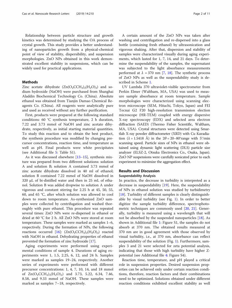
A certain amount of the ZnO NPs was taken afterwashing and centrifugation and re-dispersed into a glassbottle (containing fresh ethanol) by ultrasonication andvigorous shaking. After that, dispersion and stability ofsamples were characterized visually during aging experi-ments, which lasted for 1, 7, 14, and 21 days. To deter-mine the suspendability of the samples, the supernatantwas subjected to the light absorbance measurementsperformed at λ = 370 nm [7, 18]. The synthetic processof ZnO NPs as well as the suspendability study is de-scribed in Scheme 1.UV Lambda 370 ultraviolet-visible spectrometer from
Perkin Elmer (Waltham, MA, USA) was used to meas-ure sample absorbance at room temperature. Samplemorphologies were characterized using scanning elec-tron microscope (SEM, Hitachi, Tokyo, Japan) and FEITecnai G2 F20 high-resolution transmission electionmicroscope (HR-TEM) coupled with energy dispersiveX-ray spectroscopy (EDX) and selected area electrondiffraction (SAED) (Thermo Fisher Scientific, Waltham,MA, USA). Crystal structures were detected using Smar-tlab X-ray powder diffractometer (XRD) with Cu Kαradia-tion (λ = 1.5418 Å) in the 2θ = 20–80°range with 5°/minscanning speed. Particle sizes of NPs in ethanol were ob-tained using dynamic light scattering (DLS) particle sizeanalyzer (ELSZ-2, Otsuka Electronics Co., Osaka, Japan).ZnO NP suspensions were carefully sonicated prior to eachexperiment to minimize the aggregation effect.
Results and DiscussionSuspendability AnalysisIn practice, the decrease in turbidity is interpreted as adecrease in suspendability [19]. Here, the suspendabilityof NPs in ethanol solution was studied by turbidimetry[18]. Turbidity of different samples is clearly distinguish-able by visual turbidity (see Fig. 1). In order to betterdigitize the sample turbidity difference, spectrophoto-metric techniques are commonly used [20, 21]. Gener-ally, turbidity is measured using a wavelength that willnot be absorbed by the suspended nanoparticles [18]. Asshown in Additional file 1: Figure S1, the samples do notabsorb at 370 nm. The obtained results measured at370 nm are in good agreement with those observed byvisual turbidity, i.e., at 370 nm, absorbance can reflectsuspendability of the solution (Fig. 1). Furthermore, sam-ples 3 and 21 were selected for zeta potential analysis,indicating that those with high turbidity have higher Z-potential (see Additional file 4: Figure S4).Reaction time, temperature, and pH played a critical
role in suspension properties. Desired suspension prop-erties can be achieved only under certain reaction condi-tions, therefore, reaction factors and their combinationsneed to be optimized. Solutions obtained under differentreaction conditions exhibited excellent stability as well
Cao et al. Nanoscale Research Letters (2019) 14:210 Page 2 of 11
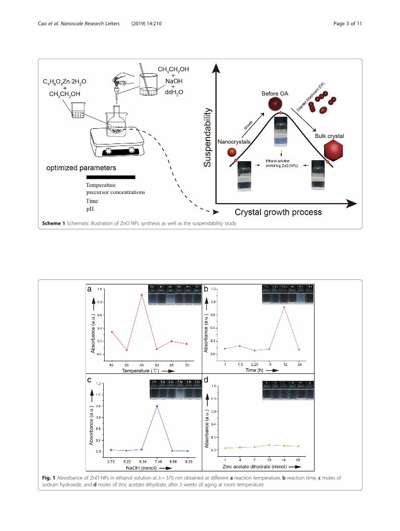
Fig. 1 Absorbance of ZnO NPs in ethanol solution at λ = 370 nm obtained at different a reaction temperature, b reaction time, c moles ofsodium hydroxide, and d moles of zinc acetate dihydrate, after 3 weeks of aging at room temperature
Scheme 1 Schematic illustration of ZnO NPs synthesis as well as the suspendability study
Cao et al. Nanoscale Research Letters (2019) 14:210 Page 3 of 11
as outstanding suspension performance (see Fig. 1a–c).When reaction conditions were 55 °C, 12 h, and7.46 mmol of initial NaOH, particles exhibited excellentlong-term suspension performance in ethanol. Furtherincrease of the reaction time, temperature, and pH valueresulted in particle precipitation and deterioration ofsuspension performance.Contrary to the previously reported results [22], sus-
pendability of ZnO NPs in this work was not affected bythe precursor concentrations (see Fig. 1d). This resultalso contradicts to the classical crystal theory since theprobability of particle collisions would be enhanced athigher concentrations. Results from this work provedthat increased precursor concentration during non-classical crystallization is not a prerequisite for particlesagglomeration.Suspension properties of ZnO NPs in ethanol demon-
strated an inverted U-shape curve as function of certainconditions. At longer reaction time, higher temperatureand higher pH values, ZnO NP ethanol suspensionsremained highly transparent. These variations further dem-onstrate changes of ZnO NPs surface structure. In general,surface characteristics of NPs strongly affect suspension ap-pearance and properties of materials. They can lead tounique suspension morphologies (see Fig. 1) and long-term
suspension performance. Our experiments proved thatthese colloids remained in a dispersed state for weeks.Thus, studying suspension morphology can provide usefulinformation on OA processes and surface structure of NPs.
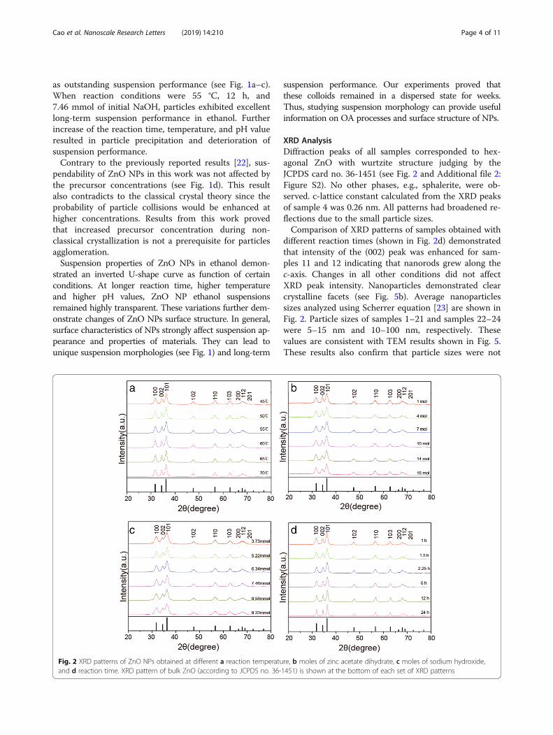
XRD AnalysisDiffraction peaks of all samples corresponded to hex-agonal ZnO with wurtzite structure judging by theJCPDS card no. 36-1451 (see Fig. 2 and Additional file 2:Figure S2). No other phases, e.g., sphalerite, were ob-served. c-lattice constant calculated from the XRD peaksof sample 4 was 0.26 nm. All patterns had broadened re-flections due to the small particle sizes.Comparison of XRD patterns of samples obtained with
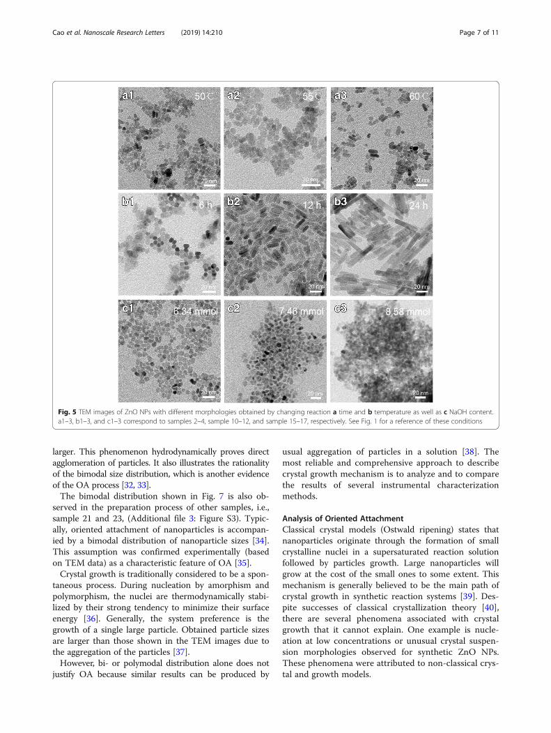
different reaction times (shown in Fig. 2d) demonstratedthat intensity of the (002) peak was enhanced for sam-ples 11 and 12 indicating that nanorods grew along thec-axis. Changes in all other conditions did not affectXRD peak intensity. Nanoparticles demonstrated clearcrystalline facets (see Fig. 5b). Average nanoparticlessizes analyzed using Scherrer equation [23] are shown inFig. 2. Particle sizes of samples 1–21 and samples 22–24were 5–15 nm and 10–100 nm, respectively. Thesevalues are consistent with TEM results shown in Fig. 5.These results also confirm that particle sizes were not
Fig. 2 XRD patterns of ZnO NPs obtained at different a reaction temperature, b moles of zinc acetate dihydrate, c moles of sodium hydroxide,and d reaction time. XRD pattern of bulk ZnO (according to JCPDS no. 36-1451) is shown at the bottom of each set of XRD patterns
Cao et al. Nanoscale Research Letters (2019) 14:210 Page 4 of 11
the major factors causing different properties of thesolution suspensions shown on Fig. 1.
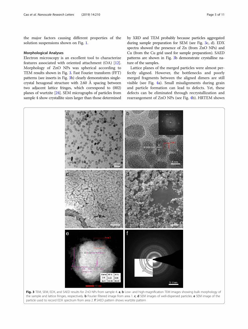
Morphological AnalysesElectron microscopy is an excellent tool to characterizefeatures associated with oriented attachment (OA) [12].Morphology of ZnO NPs was spherical according toTEM results shown in Fig. 3. Fast Fourier transform (FFT)patterns (see inserts in Fig. 3b) clearly demonstrates single-crystal hexagonal structure with 2.60 Å spacing betweentwo adjacent lattice fringes, which correspond to (002)planes of wurtzite [24]. SEM micrographs of particles fromsample 4 show crystallite sizes larger than those determined
by XRD and TEM probably because particles aggregatedduring sample preparation for SEM (see Fig. 3c, d). EDXspectra showed the presence of Zn (from ZnO NPs) andCu (from the Cu grid used for sample preparation). SAEDpatterns are shown in Fig. 3b demonstrate crystalline na-ture of the samples.Lattice planes of the merged particles were almost per-
fectly aligned. However, the bottlenecks and poorlymerged fragments between the aligned dimers are stillvisible (see Fig. 4a). Small misalignments during grainand particle formation can lead to defects. Yet, thesedefects can be eliminated through recrystallization andrearrangement of ZnO NPs (see Fig. 4b). HRTEM shown
Fig. 3 TEM, SEM, EDX, and SAED results for ZnO NPs from sample 4. a, b Low- and high-magnification TEM images showing bulk morphology ofthe sample and lattice fringes, respectively. b Fourier filtered image from area 1. c, d SEM images of well-dispersed particles. e SEM image of theparticle used to record EDX spectrum from area 2. f SAED pattern shows wurtzite pattern
Cao et al. Nanoscale Research Letters (2019) 14:210 Page 5 of 11
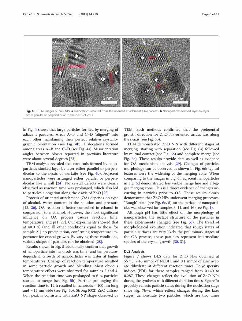
in Fig. 4 shows that large particles formed by merging ofadjacent particles. Areas A–B and C–D “aligned” intoeach other maintaining their perfect relative crystallo-graphic orientation (see Fig. 4b). Dislocations formedamong areas A–B and C–D (see Fig. 4a). Misorientationangles between blocks reported in previous literaturewere about several degrees [23].TEM analysis revealed that nanorods formed by nano-
particles stacked layer-by-layer either parallel or perpen-dicular to the c-axis of wurtzite (see Fig. 4b). Adjacentnanoparticles were arranged either parallel or perpen-dicular like a wall [24]. No crystal defects were clearlyobserved as reaction time was prolonged, which also ledto particles elongation along the c-axis of ZnO [25].Process of oriented attachment (OA) depends on type
of alcohol, water content in the solution and pressure[13, 26]. OA reaction is better controlled in ethanol incomparison to methanol. However, the most significantinfluence on OA process causes reaction time,temperature, and pH [27]. Our experiments showed thatat 40.0 °C (and all other conditions equal to those forsample 21) no precipitation, confirming temperature im-portance for crystal growth. By varying these conditions,various shapes of particles can be obtained [28].Results shown in Fig. 5 additionally confirm that growth
of nanoparticle into nanorods was time- and temperature-dependent. Growth of nanoparticles was faster at highertemperatures. Change of reaction temperature resultedin some particle growth and blending. Most obvioustemperature effects were observed for samples 2 and 4.When the reaction time was prolonged to 6 h, particlesstarted to merge (see Fig. 5b). Further prolonging thereaction time to 12 h resulted in nanorods ~ 100 nm longand ~ 15 nm wide (see Fig. 5b). Strong (002) ZnO diffrac-tion peak is consistent with ZnO NP shape observed by
TEM. Both methods confirmed that the preferentialgrowth direction for ZnO NP-oriented arrays was alongthe c-axis (see Fig. 5b).TEM demonstrated ZnO NPs with different stages of
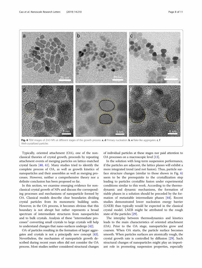
merging: starting with separation (see Fig. 6a) followedby mutual contact (see Fig. 6b) and complete merge (seeFig. 6c). These results provide data as well as evidencefor OA mechanism analysis [29]. Changes of particlesmorphology can be observed as shown in Fig. 6d: typicalfeatures were the widening of the merging zone. Whencomparing to the images in Fig. 6f, adjacent nanoparticlesin Fig. 6d demonstrated less visible merge line and a big-ger merging zone. This is a direct evidence of changes oc-curring in particles prior to OA. These results clearlydemonstrate that ZnO NPs underwent merging processes.“Rough” state (see Fig. 6c, d) on the surface of nanoparti-cles was observed for samples 3, 11, and 16 (see Fig. 1).Although pH has little effect on the morphology of
nanoparticles, the surface structure of the particles inthese experiments changed (see Fig. 5c). The trend ofmorphological evolution indicated that rough states ofparticle surfaces are very likely the preliminary stages ofthe OA process; these particles represent intermediatespecies of the crystal growth [30, 31].
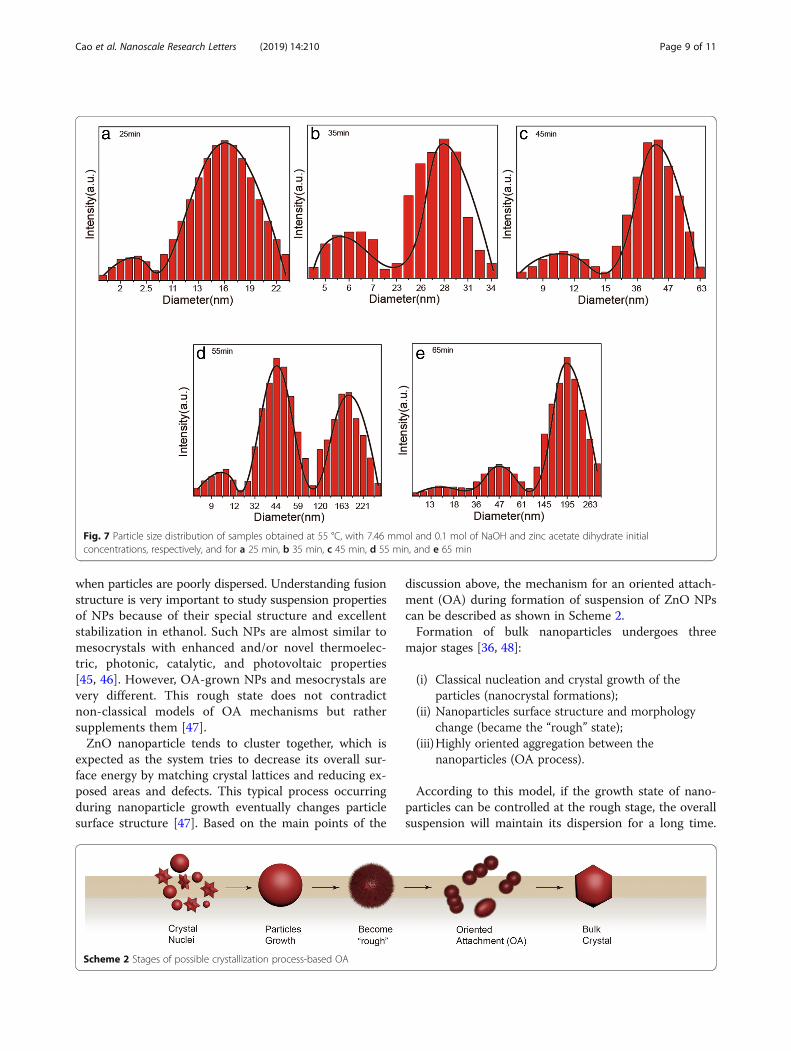
DLS AnalysisFigure 7 shows DLS data for ZnO NPs obtained at55 °C, 7.46 mmol of NaOH, and 0.1 mmol of zinc acet-ate dihydrate at different reaction times. Polydispersityindices (PDI) for these samples ranged from 0.140 to0.287. These changes reflect the evolution of ZnO NPsduring the synthesis with different duration times. Figure 7aprobably reflects particle states during the nucleation stagesince Fig. 7b–e, which reflect changes during the laterstages, demonstrate two particles, which are two times
Fig. 4 HRTEM images of ZnO NPs. a Dislocations resulted from the oriented attachment (OA) process. b Nanoparticles formed layer-by-layereither parallel or perpendicular to the c-axis of ZnO
Cao et al. Nanoscale Research Letters (2019) 14:210 Page 6 of 11
larger. This phenomenon hydrodynamically proves directagglomeration of particles. It also illustrates the rationalityof the bimodal size distribution, which is another evidenceof the OA process [32, 33].The bimodal distribution shown in Fig. 7 is also ob-
served in the preparation process of other samples, i.e.,sample 21 and 23, (Additional file 3: Figure S3). Typic-ally, oriented attachment of nanoparticles is accompan-ied by a bimodal distribution of nanoparticle sizes [34].This assumption was confirmed experimentally (basedon TEM data) as a characteristic feature of OA [35].Crystal growth is traditionally considered to be a spon-
taneous process. During nucleation by amorphism andpolymorphism, the nuclei are thermodynamically stabi-lized by their strong tendency to minimize their surfaceenergy [36]. Generally, the system preference is thegrowth of a single large particle. Obtained particle sizesare larger than those shown in the TEM images due tothe aggregation of the particles [37].However, bi- or polymodal distribution alone does not
justify OA because similar results can be produced by
usual aggregation of particles in a solution [38]. Themost reliable and comprehensive approach to describecrystal growth mechanism is to analyze and to comparethe results of several instrumental characterizationmethods.
Analysis of Oriented AttachmentClassical crystal models (Ostwald ripening) states thatnanoparticles originate through the formation of smallcrystalline nuclei in a supersaturated reaction solutionfollowed by particles growth. Large nanoparticles willgrow at the cost of the small ones to some extent. Thismechanism is generally believed to be the main path ofcrystal growth in synthetic reaction systems [39]. Des-pite successes of classical crystallization theory [40],there are several phenomena associated with crystalgrowth that it cannot explain. One example is nucle-ation at low concentrations or unusual crystal suspen-sion morphologies observed for synthetic ZnO NPs.These phenomena were attributed to non-classical crys-tal and growth models.
Fig. 5 TEM images of ZnO NPs with different morphologies obtained by changing reaction a time and b temperature as well as c NaOH content.a1–3, b1–3, and c1–3 correspond to samples 2–4, sample 10–12, and sample 15–17, respectively. See Fig. 1 for a reference of these conditions
Cao et al. Nanoscale Research Letters (2019) 14:210 Page 7 of 11
Typically, oriented attachment (OA), one of the non-classical theories of crystal growth, proceeds by repeatingattachment events of merging particles on lattice-matchedcrystal facets [40, 41]. Many studies tried to identify thecomplete process of OA, as well as growth kinetics ofnanoparticles and their assembles as well as merging pro-cesses. However, neither a comprehensive theory nor adefinite conclusion has been proposed so far.In this section, we examine emerging evidence for non-
classical crystal growth of NPs and discuss the correspond-ing processes and mechanisms of nanoparticle formed byOA. Classical models describe clear boundaries dividingcrystal particles from its monomeric building units.However, in the OA process, it becomes obvious that thisboundary is not abrupt but rather represents a broadspectrum of intermediate structures from nanoparticlesand to bulk crystals. Analysis of these “intermediate pro-cesses” converting small crystals to large crystals will helpto understand changes that nano-surfaces undergo [42].OA of particles resulting in the formation of larger aggre-
gates and crystals is not a principally new concept [43].Nevertheless, the mechanism of nanoparticle growth de-scribed during recent years often did not consider the OAprocess. Most studies neither considered structural changes
of individual particles at these stages nor paid attention toOA processes on a macroscopic level [13].In the solution with long-term suspension performance,
if the particles are adjacent, the lattice planes will exhibit amore integrated trend (and not fusion). Thus, particle sur-face structure changes (similar to those shown in Fig. 6)seem to be the prerequisite to the crystallization stepleading to particles crystallite fusion under experimentalconditions similar to this work. According to the thermo-dynamic and dynamic mechanisms, the formation ofstable phases in a solution should be preceded by the for-mation of metastable intermediate phases [44]. Recentstudies demonstrated lower nucleation energy barrier(LNEB) than typically would be expected in the classicalcrystal model. LNEB might be attributed to the roughstate of the particles [29].The interplay between thermodynamics and kinetics
leads to the main characteristics of oriented attachment(OA). Prior to the OA stage, nanoparticles grow andcoarsen. When OA starts, the particle surface becomessmooth. When particles surfaces are atomically rough, thecrystal growth rate is controlled by diffusion [29]. Suchstructural changes of nanoparticles might play an import-ant role in promoting suspension properties, especially
Fig. 6 TEM images of ZnO NPs at different stages of the growth process. a, d Primary nucleation. b, e flake-like aggregates. c, fWell-crystallized particles
Cao et al. Nanoscale Research Letters (2019) 14:210 Page 8 of 11
when particles are poorly dispersed. Understanding fusionstructure is very important to study suspension propertiesof NPs because of their special structure and excellentstabilization in ethanol. Such NPs are almost similar tomesocrystals with enhanced and/or novel thermoelec-tric, photonic, catalytic, and photovoltaic properties[45, 46]. However, OA-grown NPs and mesocrystals arevery different. This rough state does not contradictnon-classical models of OA mechanisms but rathersupplements them [47].ZnO nanoparticle tends to cluster together, which is
expected as the system tries to decrease its overall sur-face energy by matching crystal lattices and reducing ex-posed areas and defects. This typical process occurringduring nanoparticle growth eventually changes particlesurface structure [47]. Based on the main points of the
discussion above, the mechanism for an oriented attach-ment (OA) during formation of suspension of ZnO NPscan be described as shown in Scheme 2.Formation of bulk nanoparticles undergoes three
major stages [36, 48]:
(i) Classical nucleation and crystal growth of theparticles (nanocrystal formations);
(ii) Nanoparticles surface structure and morphologychange (became the “rough” state);
(iii)Highly oriented aggregation between thenanoparticles (OA process).
According to this model, if the growth state of nano-particles can be controlled at the rough stage, the overallsuspension will maintain its dispersion for a long time.
Fig. 7 Particle size distribution of samples obtained at 55 °C, with 7.46 mmol and 0.1 mol of NaOH and zinc acetate dihydrate initialconcentrations, respectively, and for a 25 min, b 35 min, c 45 min, d 55 min, and e 65 min
Scheme 2 Stages of possible crystallization process-based OA
Cao et al. Nanoscale Research Letters (2019) 14:210 Page 9 of 11
Developing crystal models for particle growth under-going similar mechanisms will improve nanomaterial syn-thesis strategies. In addition, controlling the microstructureof synthetic materials using OA mechanisms is a promisingand an insufficiently explored research area.
ConclusionsThis paper reports synthesis of ZnO NP suspension in etha-nol and at low temperature without using any surfactantsand/or dispersants. Such very stable suspensions were ob-tained by optimizing solution characteristics (temperature,aging time, precursor concentrations, and pH). Surfacestructures of ZnO NPs were mostly influenced by the reac-tion temperature, followed by reaction time and pH.This work provides strong evidence that prior to ori-
ented attachment (OA) process, the surface structure ofadjacent particles transforms into a rough state, whichchanges material properties and its suspendability in thesolution. It was shown for the first time that suspend-ability of ZnO NPs in ethanol can be controlled and fur-ther used in practical suspension-based applications.This work opens a new way for understanding how
structures of NPs influence their properties. Further anddeeper understanding of OA also promises advances invarious nanomaterial design and synthesis methods, whichcan be further used for diverse industrial applications.
Additional files
Additional file 1: Figure S1. The absorbance versus wavelength curveof samples 3 and 21 (TIF 215 kb)
Additional file 2: Figure S2. XRD patterns of a sample 21 and b sample23. XRD pattern of bulk ZnO (according to JCPDS no. 36-1451) is shownat the bottom of each set of XRD patterns (TIF 796 kb)
Additional file 3: Figure S3. Dynamic light scattering (DLS)measurements of a sample 21 and b sample 23 after 50 min (TIF 998 kb)
Additional file 4: Figure S4. Z-potentials of samples 3 and 21 (TIF 738 kb)
Additional file 5: Table S1. Sample identification (ID) as function of theirpreparation conditions. The reaction system for each sample had only aone-factor variable. Standard conditions were 60 °C, 2.25 h, 7.22 mmol ofNaOH, and 3.73 mmol of zinc acetate dihydrate (DOC 50 kb)
AbbreviationsDLS: Dynamic light scattering; EDX: Energy dispersive X-ray spectroscopy; HR-TEM: High-resolution transmission electron microscopy; LNEB: Lowernucleation energy barrier; NPs: Nanoparticles; OA: Oriented attachment;OR: Ostwald ripening; SAED: Selected area electron diffraction; SEM: Scanningelectron microscope; TEM: Transmission electron microscopy; XRD: X-raydiffraction
AcknowledgementsNot applicable.
Authors’ ContributionsDDC and SG conceived and designed the experiments. DDC, DDZ, and SLLperformed the experiment. XGS and SG analyzed the data. XGS contributedthe reagents/materials. The manuscript was written through thecontributions of all authors. All authors read and approved the final versionof the manuscript.
Authors’ InformationNot applicable.
FundingThis research was funded by Natural Science Foundation of GuangdongProvince, China (Grant No. 2018A0303130068); Guangdong Science andTechnology Department, China (grant nos. 2018050506081, 2017A050506055,2017A040405055, and 2016A010103036); Guangdong Education Department,China (Grant No. 2017KZDXM045).
Availability of Data and MaterialsThe datasets generated during and/or analyzed during the current study areavailable from the corresponding author on reasonable request.
Competing InterestsThe authors declare that they have no competing interests.
Received: 5 February 2019 Accepted: 3 June 2019
References1. Brayner R, Ferrari-Iliou R, Brivois N, Djediat S, Benedetti MF, Fie´vet F (2006)
Toxicological impact studies based on Escherichia coli bacteria in ultrafineZnO nanoparticles colloidal medium. Nano Lett. 6:866–870
2. Wang ZL (2004) Zinc oxide nanostructures: growth, properties andapplications. J Phys Condens Matter. 16:R829–R858
3. Xia T, Kovochich M, Liong M, Mädler L, Gilbert B, Shi HB, Yeh JI, Zink JI, NelAE (2008) Comparison of the Mechanism of Toxicity of Zinc Oxide andCerium Oxide Nanoparticles Based on Dissolution and Oxidative StressProperties. ACS Nano. 2:2121–2134
4. Nel A, Xia T, Mädler L, Li N (2006) Toxic potential of materials at thenanolevel. Science. 311:622–627
5. Tienes BM, Perkins RJ, Shoemaker RK, Dukovic G (2013) Layeredphosphonates in colloidal synthesis of anisotropic ZnO nanocrystals. ChemMater. 25:4321–4329
6. Shen ZC, Zhou HJ, Chen HY, Xu H, Feng CH, Zhou XH (2018) Synthesisof nano-zinc oxide loaded on mesoporous silica by coordination effectand its photocatalytic degradation property of methyl orange.Nanomaterlals-Basel. 8:317
7. Goh EG, Xu X, McCormick PG (2014) Effect of particle size on the UVabsorbance of zinc oxide nanoparticles. Scripta Mater. 78:49–52
8. Ji XH, Song XL, Li J, Bai YB, Yang WS, Peng XG (2007) Size control of goldnanocrystals in citrate reduction: the third role of citrate. J Am Chem Soc.129:13939–13948
9. Chen YF, Johnson E, Peng XG (2007) Formation of monodisperse andshape-controlled MnO nanocrystals in non-injection synthesis: self-focusingvia ripening. J Am Chem Soc. 129:10937–10947
10. Fan ZY, Lu JG (2005) Zinc oxide nanostructures: synthesis and properties. JNanosci Nanotechno. 5:1561–1573
11. Gong S, Chen HY, Zhou XH, Gunasekaran S (2017) Synthesis andapplications of MANs/poly (MMA-co-BA) nanocomposite latex byminiemulsion polymerization. Roy Soc Open Sci. 4:170844
12. Wang FD, Richards VN, Shields SP, Buhro WE (2013) Kinetics andmechanisms of aggregative nanocrystal growth. Chem Mater. 26:5–21
13. Pacholski C, Kornowski A, Weller H (2002) Self-assembly of ZnO: fromnanodots to nanorods. Angew Chem Int Edit. 41:1188–1191
14. Ancona A, Dumontel B, Garino N, Demarco B, Chatzitheodoridou D, FazziniW, Engelke H, Cauda V (2018) Lipid-coated zinc oxide nanoparticles asinnovative ROS-generators for photodynamic therapy in cancer cells.Nanomaterlals-Basel. 8:143
15. Tang X, Choo ESG, Li L, Ding J, Xue JM (2010) Synthesis of ZnONanoparticles with tunable emission colors and their cell labelingapplications. Chem Mater. 22:3383–3388
16. Zou XW, Fan HQ, Tian YM, Yan SJ (2013) Facile hydrothermal synthesis oflarge scale ZnO nanorod arrays and their growth mechanism. Mater Lett.107:269–272
17. Haase M, Weller H, Henglein A (1988) Photochemistry and radiationchemistry of colloidal semiconductors. 23. Electron storage on zinc oxideparticles and size quantization. J Phys Chem. 92:482–487
Cao et al. Nanoscale Research Letters (2019) 14:210 Page 10 of 11
18. Félix MG, Alonso A (2000) Spectroscopic techniques in the study ofmembrane solubilization, reconstitution and permeabilization by detergents.J BBA-Biomembranes 1508:51–68
19. Ramani M, Ponnusamy S, Muthamizhchelvan C, Cullen J, Krishnamurthy S,Marsili E (2013) Morphology-directed synthesis of ZnO nanostructures andtheir antibacterial activity. J Colloid Surface B. 105:24–30
20. Jiménez-Rojo N, Lete MG, Rojas E, Gil D, Valle M, Alonso A, Moya SE, GoñiFM (2015) Lipidic nanovesicles stabilize suspensions of metal oxidenanoparticles. J Chem Phys Lipids 191:84–90
21. Ramani M, Ponnusamy S, Muthamizhchelvan C (2012) From zinc oxidenanoparticles to microflowers: A study of growth kinetics and biocidalactivity. J Mat Sci Eng C-Mater 32:2381–2389
22. Xie J, Yan CZ, Zhang Y, Gu N (2013) Shape evolution of “multibranched”Mn-Zn ferrite nanostructures with high performance: a transformation ofnanocrystals into nanoclusters. Chem Mater. 25:3702–3709
23. Liao HG, Cui LK, Whitelam S, Zheng HM (2012) Real-time imaging of Pt3Fenanorod growth in solution. Science. 336:1011–1014
24. De Yoreo JJ, Gilbert PUPA, Sommerdijk NAJM, Penn RL, Whitelam S, Joester D,Zhang HZ, Rimer JD, Navrotsky A, Banfield JF et al (2015) Crystallization byparticle attachment in synthetic, biogenic, and geologic environments. Science.349:aaa6760. https://science.sciencemag.org/content/349/6247/aaa6760
25. Cheng JJ, Nicaise SM, Berggren KK, Gradecak S (2015) Dimensional tailoringof hydrothermally grown zinc oxide nanowire arrays. Nano Lett. 16:753–759
26. Buonsanti R, Llordes A, Aloni S, Helms BA, Milliron DJ (2011) Tunableinfrared absorption and visible transparency of colloidal aluminum-dopedzinc oxide nanocrystals. Nano Lett. 11:4706–4710
27. He LL, Tong ZF, Wang ZH, Chen M, Huang N, Zhang W (2018) Effects ofcalcination temperature and heating rate on the photocatalytic propertiesof ZnO prepared by pyrolysis. J Colloid Interf Sci. 509:448–456
28. Zhu R, Zhang WG, Li C, Yang R (2013) Uniform zinc oxide nanowire arraysgrown on nonepitaxial surface with general orientation control. Nano Lett.13:5171–5176
29. Yuk JM, Park J, Ercius P, Kim K, Hellebusch DJ, Crommie MF, Lee JY, Zettl A,Alivisatos AP (2012) High-resolution EM of colloidal nanocrystal growthusing graphene liquid cells. Science 336:61–64
30. Brayner R, Dahoumane SA, Yéprémian C, Djediat C, Meyer M, Couté A,Fiévet F (2010) ZnO nanoparticles: synthesis, characterization, andecotoxicological studies. Langmuir. 26:6522–6528
31. Patterson AL (1939) The Scherrer formula for X-ray particle sizedetermination. Phys Rev. 56:978–982
32. Lee EJH, Ribeiro C, Longo E, Leite ER (2005) Oriented attachment: aneffective mechanism in the formation of anisotropic nanocrystals. J PhysChem B 109:20842–20846
33. Levit AB, Rowell RL (1975) Time dependence of the size distribution,number concentration and surface area in La Mer sulfur sols. J Colloid InterfSci. 50:162–169
34. Shields SP, Richards VN, Buhro WE (2010) Nucleation control of size anddispersity in aggregative nanoparticle growth. A Study of the CoarseningKinetics of Thiolate-Capped Gold Nanocrystals. Chem Mater. 22:3212–3225
35. Abécassis B, Bouet C, Garnero C, Constantin D, Lequeux N, Ithurria S,Dubertret B, Pauw BR, Pontoni D (2015) Real-time in situ probing of high-temperature quantum dots solution synthesis. Nano Lett. 15:2620–2626
36. Evans JE, Jungjohann KL, Browning ND, Arslan I (2011) Controlled growth ofnanoparticles from solution with in situ liquid transmission electronmicroscopy. Nano Lett. 11:2809–2813
37. Dumontel B, Canta M, Engelke H, Chiodoni A, Racca L, Ancona A, LimongiT, Canavese G, Cauda V (2017) Enhanced biostability and cellular uptake ofzinc oxide nanocrystals shielded with a phospholipid bilayer. J Mater ChemB 5:8799–8813
38. Bindi L, Steinhardt PJ, Yao N, Lu PJ (2009) Natural quasicrystals. Science. 324:1306–1309
39. Vengrenovich RD, Gudyma YV, Yarema SV (2001) Ostwald ripening ofquantum-dot nanostructures. Semiconductors 35:1378–1382
40. Zhang J, Huang F, Lin Z (2010) Progress of nanocrystalline growth kineticsbased on oriented attachment. Nanoscale. 2:18–34
41. Mallavajula RK, Archer LA (2011) Nanocrystal self-assembly assisted byoriented attachment. Angew Chem Int Edit. 50:578–580
42. Olafson KN, Li R, Alamani BG, Rimer JD (2016) Engineering crystal modifiers:bridging classical and nonclassical crystallization. Chem Mater. 28:8453–8465
43. Greer HF (2014) Non-classical crystal growth of inorganic and organicmaterials. Mater Sci Tech-lond 30:611–626
44. Lee J, Yang J, Kwon SG, Hyeon T (2016) Nonclassical nucleation and growthof inorganic nanoparticles. Nat Rev Mater. 1:16034
45. Fang J, Ding BJ, Gleiter H (2011) Mesocrystals: syntheses in metals andapplications. Chem Soc Rev. 40:5347–5360
46. Bergström L, Sturm EV, Salazar-Alvarez G, Cölfen H (2015) Mesocrystals inbiominerals and colloidal arrays [J]. Accounts Chem Res. 48:1391–1402
47. Chen JS, Zhu T, Li CM, Lou XW (2011) Building hematite nanostructures byoriented attachment [J]. Angew Chem Int Edit. 50:650–653
48. Ivanov VK, Fedorov PP, Baranchikov AY, Osiko VV (2014) Orientedattachment of particles: 100 years of investigations of non-classical crystalgrowth. Russ Chem Rev+. 83:1204–1222
Publisher’s NoteSpringer Nature remains neutral with regard to jurisdictional claims inpublished maps and institutional affiliations.
Cao et al. Nanoscale Research Letters (2019) 14:210 Page 11 of 11


![SYNTHESIS OF PMMA/ZnO NANOPARTICLES COMPOSITE USED …mit.imt.si/Revija/izvodi/mit175/popovic.pdf · d. popovi] et al.: synthesis of pmma/zno nanoparticles composite used for resin](https://img.pdfslide.us/doc/110x75/5a8ef09e7f8b9a78648d6099/synthesis-of-pmmazno-nanoparticles-composite-used-mitimtsirevijaizvodimit175.jpg)