Embed Size (px)
Citation preview
Synthesis and Characterization of ZnO Nanoparticles
Ooi Sheue Lin (24809)
A project report submitted in partial fulfillment of the
Final Year Project II (STF 3015)
Supervisor: Dr. Chin Suk Fun
Resource Chemistry
Department of Chemistry
Faculty of Resource Science and Technology
University Malaysia Sarawak
2012
I
Declaration
I hereby declare that the work portrayed in this thesis was carried by me under the
supervision of Dr. Chin Suk Fun at the Department of Chemistry, Faculty of Resource
Science and Technology, Universiti Malaysia Sarawak and no portion of this dissertation
has been submitted in support of an application for another degree of qualification of this
or any other university or institution of higher learning.
Ooi Sheue Lin
Program of Resource Chemistry
Faculty of Resource Science and Technology
Universiti Malaysia Sarawak
II
Acknowledgement
First of all, I would like to forward my greatest gratitude to my supervisor, Dr. Chin Suk
Fun for providing the necessary advice, guidance, encouragement, support and facilities
throughout the project. Besides, I would like to thank my examiner, Assoc. Prof. Dr. Mohd.
Abu Affan, and the lecturers for their kind guidance and support. Additionally, I wish to
extend my appreciation to all technical staffs and lab assistances for their incessant help
and cooperation. Deepest appreciation is also offered to the postgraduate students, Rose
Chua Siaw Chin, and Lau Siu Ping for their unremitting assistance. Last but not least, I
would like to take this opportunity to express my gratefulness to my family and friends for
their continuous encouragement, moral and financial support in completing this study.
III
Table of Content
Declaration…………………………………………………………………………………..I
Acknowledgement………………………………………………………………………….II
Table of Content………………………………………………………………………….III
List of Abbreviations……………………………………………………………………….V
List of Figures…………………………………………………………………………….VII
List of Schemes……………………………………………………………………………IX
List of Tables……………………………………………………………………………….X
Abstract……………………………………………………………………………………..1
1.0 Introduction……………………………………………………………………………2
1.1 Objective………………………………………………………………………………..5
2.0 Literature Review……………………………………………………………………...6
2.1 Synthesis of ZnO nanoparticles…………………………………………………………6
2.1.1 Precipitation method…………………………………………………………..7
2.1.2 Gas condensation method…………………………………………………....11
2.1.3 Electrochemical method……………………………………………………..12
2.1.4 Sol-gel method………………………………………………………………12
2.1.5 Hydro/solvothermal method………………………………………………....14
2.1.6 Wet chemistry method……………………………………………………….15
2.2 Surfactants……………………………………………………………………………..16
2.3 Applications of ZnO nanoparticles…………………………………………………….17
2.3.1 Luminescent materials……………………………………………………….17
2.3.2 Optoelectronic devices……………………………………………………....17
2.3.3 Biological and biomedical field……………………………………………..18
2.3.4 Antimicrobial and UV absorber textiles……………………………………..18
2.3.5 Cosmetics…………………………………………………………………....19
3.0 Methodology…………………………………………………………………………..20
3.1 Reagents and Materials……………………………………………………………......20
3.2 Method………………………………………………………………………………....20
3.2.1 Synthesis of ZnO nanoparticles (Without surfactant)……………………20
3.2.2 Synthesis of ZnO nanoparticles (With surfactant)………………………….20
3.2.3 Characterization……………………………………………………………...21
3.2.3.1 Analysis by UV-Visible spectrophotometer……………………….21
IV
3.2.3.2 Analysis by FTIR spectroscopy…………………………………...21
3.2.3.3 Analysis by SEM…………………………………………………..21
3.2.3.4 Analysis by TEM…………………………………………………..22
4.0 Results and Discussions………………………………………………………………23
4.1 ZnO nanoparticles (Without surfactant)……………………………………………….23
4.1.1 Physical properties…………………………………………………………..23
4.1.2 FTIR spectroscopy analysis………………………………………………….24
4.1.3 SEM analysis………………………………………………………………...26
4.1.4 TEM analysis………………………………………………………………...27
4.2 ZnO nanoparticles (With surfactant)…………………………………………………..28
4.2.1 Succinic acid as surfactant…………………………………………………28
4.2.1.1 Physical properties………………………………………………...28
4.2.1.2 FTIR spectroscopy analysis………………………………………..29
4.2.1.3 SEM analysis………………………………………………………32
4.2.1.4 TEM analysis………………………………………………………33
4.2.2 CMC as surfactant………………………………………………………….35
4.2.2.1 Physical properties………………………………………………...35
4.2.2.2 FTIR spectroscopy analysis………………………………………..36
4.2.2.3 SEM analysis………………………………………………………39
4.2.2.4 TEM analysis………………………………………………………39
4.2.3 Adipic acid as surfactant…………………………………………………41
4.2.3.1 Physical properties………………………………………………...41
4.2.3.2 FTIR spectroscopy analysis………………………………………..42
4.2.3.3 TEM analysis………………………………………………………45
4.3 Optical properties……………………………………………………………………...47
5.0 Conclusion....................................................................................................................50
6.0 Recommendations........................................................................................................51
7.0 References……………………………………………………………………………52
8.0 Appendix……………………………………………………………………………..55
V
List of Abbreviations
Adipic acid C6H10O4
Angstrom Å
Atmospheric atm
Carboxyl group C=O
Carboxymethyl cellulose CMC
Degree Celcius °C
Electron volt eV
Ethanol C2H5OH
Fourier Transform Infrared Spectroscopy FTIR
Hydroxyl group -OH
Lithium hydroxide LiOH
Mili electron volt meV
Mili mole mmol
Nanometer nm/ 10-9
m
Polypyrrole PPy
Polyvinyl alcohol PVA
Polyvinylpyrrollidone PVP
Potassium hydroxide KOH
Scanning Electron Microscopy SEM
Sodium carbonate Na2CO3
Sodium chloride NaCl
Sodium hydroxide NaOH
Succinic acid C4H6O4
Transmission Electron Microscopy TEM
Water H2O

VI
Weight per volume w/v
X-Ray Diffraction XRD
Zinc (II) ion Zn2+
Zinc acetate dihydrate Zn(CH3COO)2·2 H2O
Zinc chloride ZnCl2
Zinc hydroxide Zn(OH)2
Zinc nitrate hydrate Zn(NO3)2·H2O
Zinc oxide ZnO
VII
List of Figures
Figures Page
Figure 1: Structure of succinic acid………………………………………………………...4
Figure 2: Structure of adipic acid…………………………………………………………..4
Figure 3: Structure of carboxymethyl cellulose……………………………………………5
Figure 4: Structure of triethanolamine……………………………………………………..8
Figure 5: Structure of n-propylamine………………………………………………………8
Figure 6: Structure of triglycerol…………………………………………………………...9
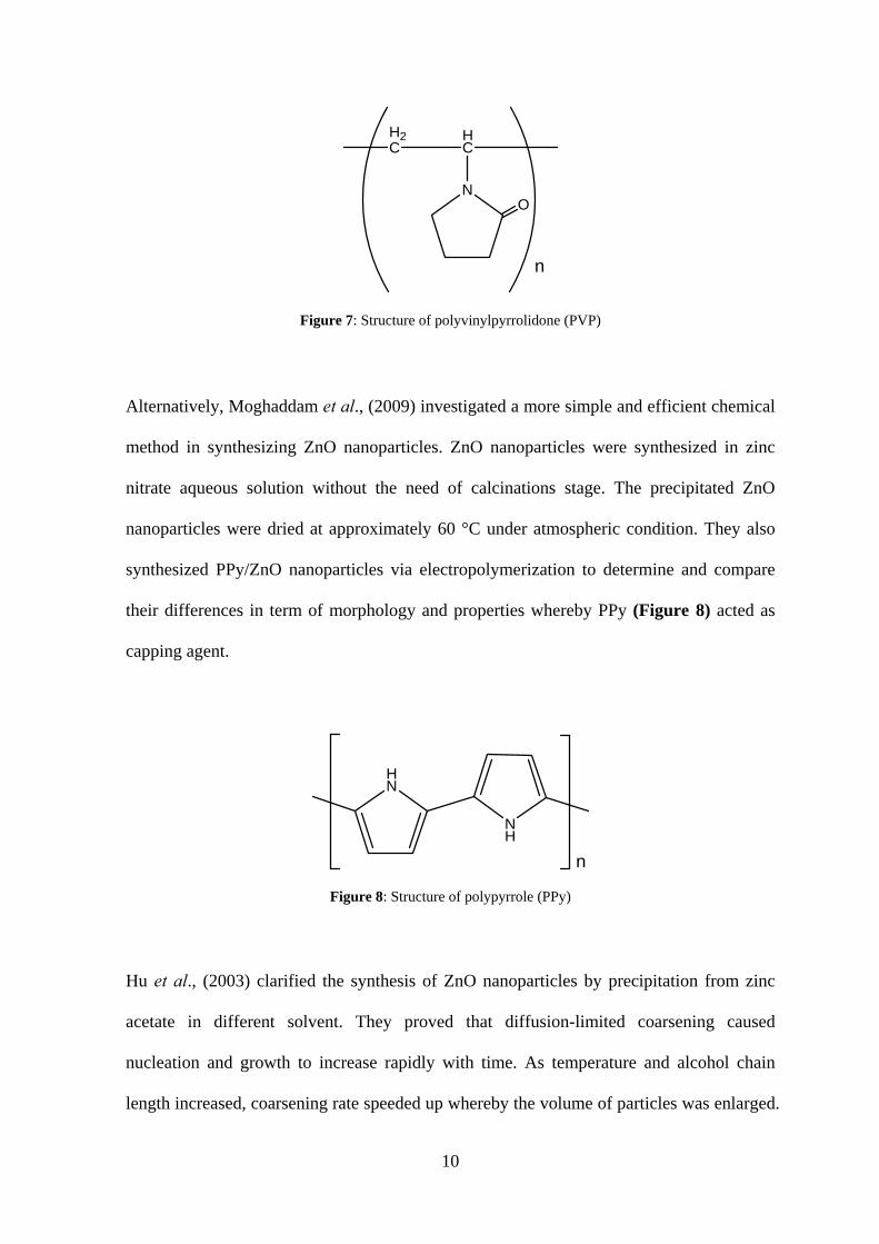
Figure 7: Structure of polyvinylpyrrolidone……………………………………………10
Figure 8: Structure of polypyrrole………………………………………………………...10
Figure 9: Structure of polyvinyl alcohol………………………………………………….13
Figure 10:IR spectrum of bare ZnO nanoparticles……………………………………….25
Figure 11: SEM image of bareZnO nanoparticles……………………………………….26
Figure 12: TEM images of bare ZnO nanoparticles………………………………………27
Figure 13:IR spectra of ZnO nanoparticles with (a) raw material, (b) 0.1% w/v,
(c) 0.5% w/v, (d) 1.0% w/v, and (e) 3.0% w/v of succinic acid
(As KBr disc)…………………………………………………………………..30
Figure 14: SEM images ofZnO nanoparticles with (a) 0.1% w/v, (b) 0.5% w/v,
(c) 1.0% w/v, and (d) 3.0% w/v of succinic acid……………………………...32
Figure 15: TEM images of ZnO nanoparticles with (a) 0.1% w/v, (b) 0.5% w/v,
(c) 1.0% w/v, and (d) 3.0% w/v of succinic acid……………………………...33
Figure 16: IR spectra of ZnO nanoparticles with (a) raw material, (b) 0.1% w/v,
(c) 0.5% w/v, (d) 1.0% w/v, and (e) 3.0% w/v of CMC
(As KBr disc)…………………………………………..………………………37
Figure 17:SEM images ofZnO nanoparticles with (a) 0.1% w/v and (b) 0.5% w/v
of CMC………………………………………………………………………...39
VIII
Figure 18: TEM images ofZnO nanoparticles with (a) 1.0% w/v and (b) 3.0% w/v
of CMC………………………………………………………………………...39
Figure 19:IR spectra of ZnO nanoparticles with (a) raw material, (b) 0.1% w/v,
(c) 0.5% w/v, (d) 1.0% w/v, and (e) 3.0% w/v of adipic acid
(As KBr disc)…………………………………………………………………..43
Figure 20: TEM images ofZnO nanoparticles with (a) 0.1% w/v, (b) 0.5% w/v,
(c) 1.0% w/v, and (d) 3.0% w/v of adipic acid………………………………...45
Figure 21:UV-Vis spectra of bare ZnO nanoparticles in comparison with modified
ZnO nanoparticles using different types of surfactants………………………..47
IX
List of Schemes
Schemes Page
Scheme 1: Synthesis of ZnO nanoparticles with oleic acid surfactant via
precipitation method…………………………………………………………….7
Scheme 2: Flow chart of the synthesis of ZnO nanoparticles by liquid precipitation……...8
Scheme 3: Flow chart of the synthesis of ZnO nanoparticles by sol-gel process…………13
Scheme 4: Flow chart of the synthesis of ZnO nanoparticlesviawet chemical method…15
X
List of Tables
Tables Page
Table 1: Physical properties of bareZnO nanoparticles………………………………….23
Table 2: IR bandsof bareZnO nanoparticles(cm-1
)……………………………………...24
Table 3: Physical properties of ZnO nanoparticles with succinic acid as surfactant……..28
Table 4: IR bands of ZnO nanoparticles with different concentration of
succinic acid(cm-1
)……………………………………………………………...29
Table 5:Physical properties of ZnO nanoparticles with CMC as surfactant……………..35
Table 6: IR bands of ZnO nanoparticles with different concentration of CMC(cm-1
)…...36
Table 7:Physical properties of ZnO nanoparticles with adipic acid as surfactant………..41
Table 8: IR bands of ZnO nanoparticles with different concentration of
adipic acid(cm-1
) ………………………………………………………………..42
1
Synthesis and Characterization of Zinc Oxide Nanoparticles
Ooi Sheue Lin
Resource Chemistry Programme Faculty of Resource Science and Technology
Universiti Malaysia Sarawak
ABSTRACT
ZnO nanoparticles have drawn a widespread attention recently due to their novel properties which contribute to various applications especially in optoelectronic devices. In this research project, the spherical ZnO nanoparticles with average size of less than 50 nm were successfully synthesized and their optical properties were measured. In order to maximize its efficiency, surface modification with surfactants is vital as ZnO nanoparticles easily agglomerate. In this study, precipitation method was applied where surfactants such as succinic acid, carboxymethyl cellulose, and adipic acid were used for enhanced properties. The addition of surfactants controlled the particle size and reduced the formation of agglomerates and at the same time helped to produce more homogenous and uniformly dispersed particles.
Keywords: zinc oxide, nanoparticles, surfactants, precipitation
ABSTRAK
Baru-baru ini, nanopartikel ZnO telah menarik perhatian ramai kerana ciri-ciri novel mereka yang menyumbang kepada pelbagai aplikasi terutamanya dalam optoelektronik peranti. Dalam projek penyelidikan ini, ZnO nanopartikel yang berbentuk sfera dengan saiz purata kurang daripada 50 nm telah berjaya disintesis dan ciri-ciri optik mereka diukur. Bagi memaksimumkan kecekapan, pengubahsuaian permukaan dengan surfaktan adalah penting kerana nanopartikel ZnO boleh menyebabkan penggumpalan. Dalam kajian ini, kaedah pemendakan telah digunakan di mana surfaktan seperti asid succinic, selulosa carboxymethyl, dan asid adipic telah digunakan untuk meningkatkan kecekapannya. Penambahan surfaktan memang membantu dalam kawalan saiz zarah dengan mengurangkan pembentukan gumpalan dan pada masa yang sama menghasilkan partikel yang lebih homogenus dan tersebar secara seragam.
Kata kunci: zink oxida, nanopartikel, surfaktan, pemendakan
2
1.0 Introduction
The idea and concept of nanotechnology was first introduced in a talk in 1959, “There’s
Plenty of Room at the Bottom”. Nanotechnology can be described as the use of
considerably enhanced nanosized structures ranging from 1 to 100 nm for the production
of materials, devices or systems (Samal et al., 2010). The synthesis of nanostructure
compound has been an extensively important research area in accordance to the
development of nanotechnology field and the exhibition of novel properties in nanoscale
materials.
Zinc oxide (ZnO) nanoparticles are hydrophobic inorganic compound existing in white
powder form. Three types of crystalline structures of ZnO nanoparticles include hexagonal
wurtzite, cubic zincblende and cubic rocksalt. Wurtzite is the most stable structure among
all. It is hexagonal and symmetrical in shape with the absence of symmetrical center. This
structure contributes to the high piezoelectricity property. ZnO nanoparticles are
categorized as II-VI semiconductor as zinc and oxygen are from the 2nd and 6th group in
periodic table, respectively. They also display features like high reflection rate, high
photoelectric and non-linear optical coefficient (Chang & Tsai, 2008). The emerging novel
optical and electronic properties of ZnO semiconductor have been a focusing issue among
researchers due to the great prospective in optoelectronic applications. Bulk ZnO possesses
wide band gap of 3.37 eV at room temperature which can be employed in the short
wavelength range, information storage and sensors. In addition, its large excitation binding
energy of 60 meV is responsible for excitonic transitions. Thus, it allows spontaneous
emission with high radiative recombination efficiency and at the same time acts as laser
emission due to its low threshold voltage (Kumbhakar et al., 2008).
3
Some modifications were implemented to enhance the applications of ZnO nanoparticles,
for instance controlling nanoparticle size, addition of surfactant, and doping with magnetic
ions. According to Moghaddam et al., (2009), the ability of ZnO to form various
nanostructures is a plus point to be advanced in photodetectors, surface acoustic wave
devices, ultravioletnanolaser, varistors, solar cells, gas sensors, biosensors, ceramics, field
emission, and nanogenerator.
Surfactant is a surface active agent which tends to reduce the surface tension of liquid and
act as dispersant. Basically, it is an amphiphilic organic compound whereby it possesses
both hydrophilic and hydrophobic characteristics. Indeed, it is proven that addition of
surfactants is essential to prevent agglomeration. The presence of water molecules in ZnO
nanoparticles lead to the formation of hard agglomerates. In order to setback this condition,
surface modification with addition of surfactants were emphasized to increase their
dispersability. Hence, the applications of ZnO nanoparticles will not be obstructed (Kang
et al., 2011).
There were a few methods in conducting the synthesis of ZnO nanoparticles. The proposed
techniques included direct precipitation (Kang et al., 2011), gas condensation method
(Chang & Tsai, 2008), electrochemical method (Nyffenegger et al., 1998), sol-gel
synthesis (Kundu et al., 2011), spray pyrolysis, thermal decomposition, molecular beam
epitaxy, chemical vapour deposition, laser ablation, and chemical synthesis (Ashtaputre et
al., 2005).
In this project, ZnO nanoparticles were synthesized via precipitation method. Zinc acetate,
sodium hydroxide, and ethanol were employed for the preparation of ZnO nanoparticles.
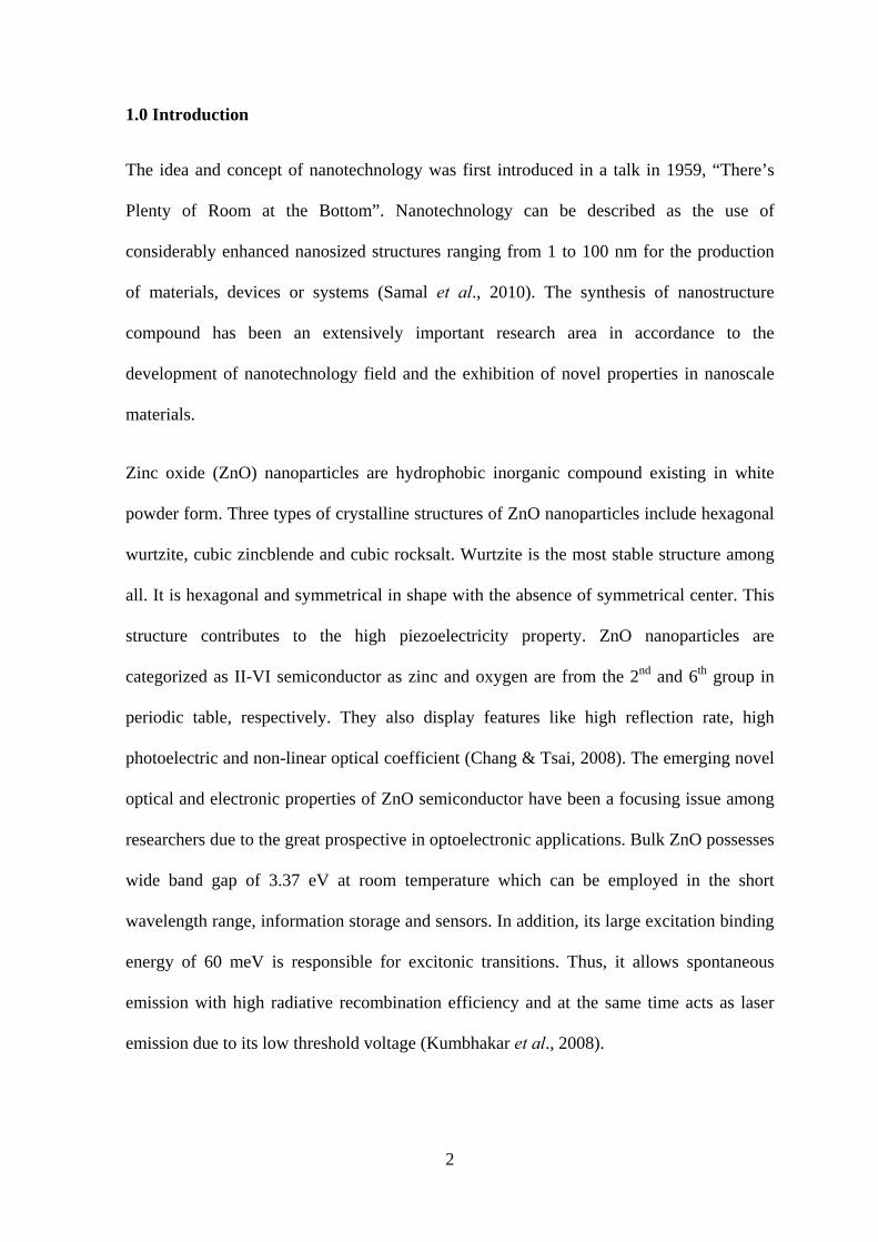
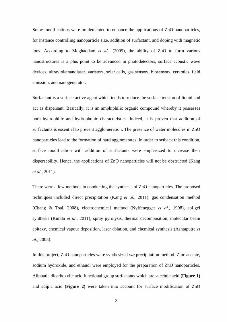
Aliphatic dicarboxylic acid functional group surfactants which are succinic acid (Figure 1)
and adipic acid (Figure 2) were taken into account for surface modification of ZnO
4
nanoparticles. These surfactants were proposed as shorter aliphatic chain and the presence
of two carboxyl groups in the compounds may enhance their surface activity and solubility
in water. In that point of view, the properties of ZnO nanoparticles were optimized for
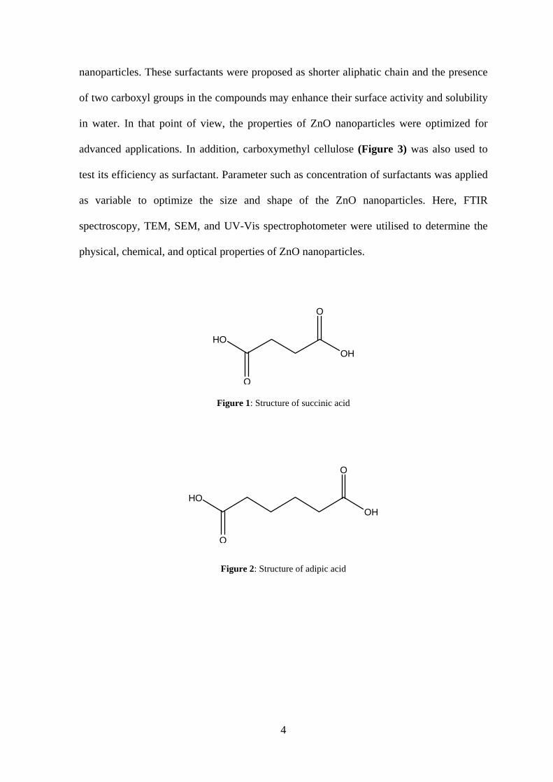
advanced applications. In addition, carboxymethyl cellulose (Figure 3) was also used to
test its efficiency as surfactant. Parameter such as concentration of surfactants was applied
as variable to optimize the size and shape of the ZnO nanoparticles. Here, FTIR
spectroscopy, TEM, SEM, and UV-Vis spectrophotometer were utilised to determine the
physical, chemical, and optical properties of ZnO nanoparticles.
O
OH
O
HO
Figure 1: Structure of succinic acid
O
OH
O
HO
Figure 2: Structure of adipic acid
5
OH
H
CH2OR
H
ORO
OR H
O
n
where R = CH2COONa
Figure 3: Structure of carboxymethyl cellulose
1.1 Objectives
1. To synthesize ZnO nanoparticles.
2. To synthesize a surface-modified ZnO nanoparticles by using different types of
surfactants.
3. To determine the synthesis parameters that will affect the morphology, size and
optical properties of the synthesized ZnO nanoparticles.
6
2.0 Literature Review
2.1 Synthesis of ZnO nanoparticles
ZnO nanoparticles have remarkable attention on the ongoing research study due to its
unique properties. No doubt, their applications especially in optoelectronic appliances can
be further advanced in size and shape dependant variation. As semiconductor nanoparticles,
their significance has encountered the economic demand in various fields and hence
becoming scientifically important materials. ZnO nanoparticles exhibit wide band gap
energy (3.37 eV) and high exciton binding energy (60 meV) at room temperature. Valued
for their novel characteristics, ZnO nanoparticles demonstrate vital applications in the short
wavelength range, which means they can be activated in the green, blue and ultraviolet
regions. Also, their ability in excitonic transitions under room temperature allows
spontaneous and laser emission (Kumbhakar et al., 2008). For optimal functions, large
intrinsic gap and nanostructure compound were applied. This implied that ZnO
nanoparticles can considerably enhanced the emission activity if compared to bulk ZnO as
tremendously small size particles lead to quantum confinement of the photo-generated
electron-hole pair. Morphologically, the thermal stability, irradiation resistance, and
flexibility of ZnO to form different nanostructures are the key to photoacoustic
applications. Moreover, the stable wurtzite structure of ZnO contributes to
photoluminescence effect with the presence of oxygen vacancies and zinc interstitials
(Moghaddam et al., 2009).
Previous research study sought out a few alternatives in synthesising ZnO nanoparticles
which include precipitation, gas condensation, electrochemical method, sol-gel synthesis,
solvothermal method, and wet chemistry methods. These experimental procedures were
carried out under various conditions.
7
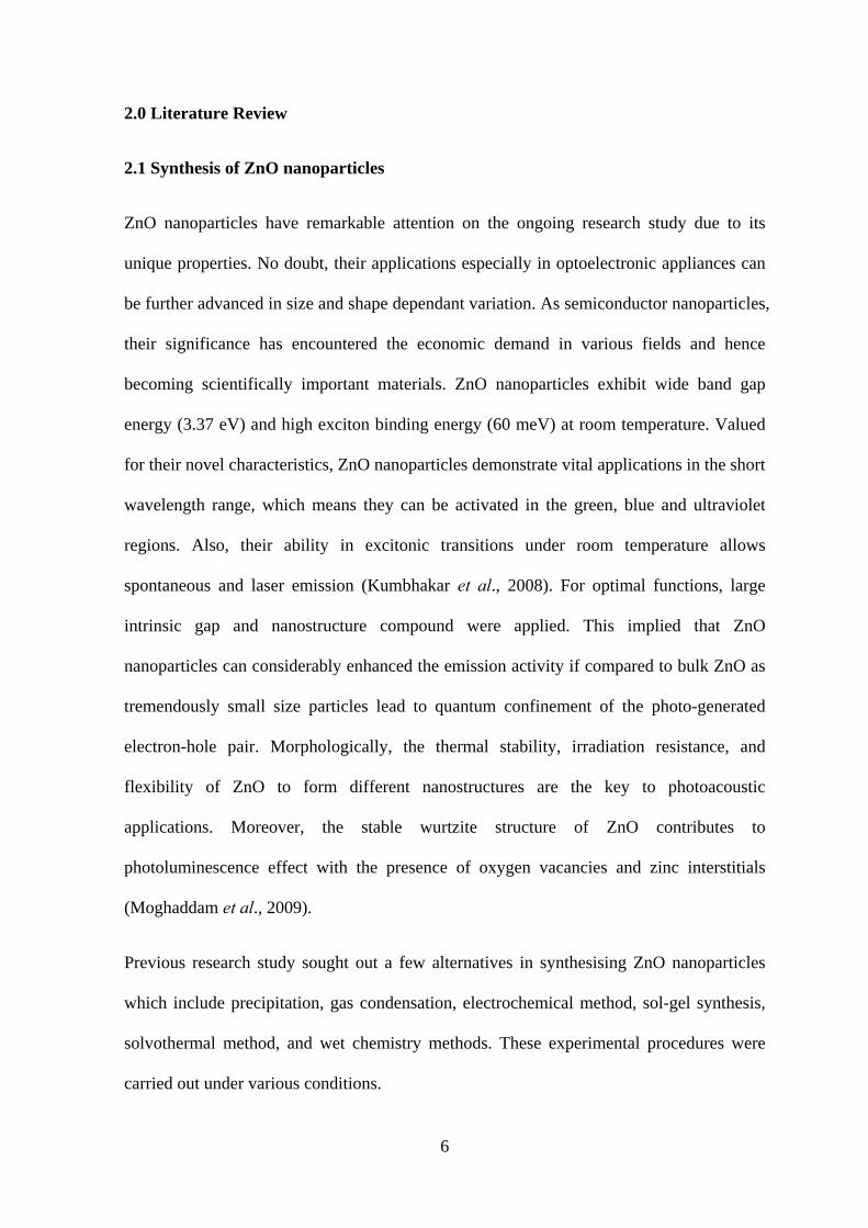
2.1.1 Precipitation method
Kang et al., (2011) practiced direct precipitation in their recent project on surface-modified
ZnO nanoparticles. In their experimental procedures, ZnCl2 and Na2CO3 solution with
surfactant were used for the synthesis of ZnO nanoparticles via precipitation process as
shown in Scheme 1. ZnO nanoparticles were finally obtained after calcinations at 400 °C.
They also verified that agglomeration may occur due to the presence of water molecules in
ZnO nanoparticles. In order to prevent this phenomenon, they suggested surface
modification of ZnO nanoparticles with oleic acid. In this case study, relatively small size
nanoparticles of approximately 29 nm were reported. The reaction occurred between the
active oxygen ions of ZnO nanoparticles and the carboxyl group of oleic acid resulted in
the formation of covalent bond between the organic layer and the inorganic nuclei. This
permitted high and stable dispersability and at the same time reduced water moiety due to
steric hinderance of the extending aliphatic carbon chain in non polar solvent.
+
ZnO
Cl
Zn
Cl
Na+Na+
sodium carbonate
O
O
O
+
Zinc cloride
OOH
oleic acid
Zinc oxide nanoparticle with surfactant
400 °C
Scheme 1: Synthesis of ZnO nanoparticles with oleic acid surfactant via precipitation method
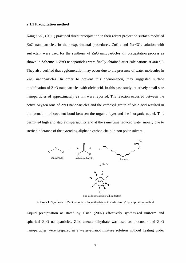
Liquid precipitation as stated by Hsieh (2007) effectively synthesized uniform and
spherical ZnO nanoparticles. Zinc acetate dihydrate was used as precursor and ZnO
nanoparticles were prepared in a water-ethanol mixture solution without heating under
8
high temperature condition. Triethanolamine (Figure 4) and n-propylamine (Figure 5)
were used as surfactants to reduce aggregation and control the size of particles produced.
The flow chart of liquid precipitation process is briefly described in Scheme 2 (Hsieh,
2007).
N
HO OH
HO H2N
Figure 4: Structure of triethanolamine Figure 5: Structure of n-propylamine
Scheme 2: Flow chart of the synthesis of ZnO nanoparticles by liquid precipitation
The chemical routes with different capping agent under various synthesis conditions were
explained by Ashtaputre et al., (2005), Kumbhakar et al., (2008), and Moghaddam et al.,
Zn(CH3COO)2 water solution TEA ethanol solution
n-propylamine
White precipitate formed
Washed with ethanol
Dried at 60 °C
Analysis for characterization
9
(2009). They discussed precipitation method in their experiments but without calcinations
at high temperature to obtain monodispersed ZnO nanoparticles.
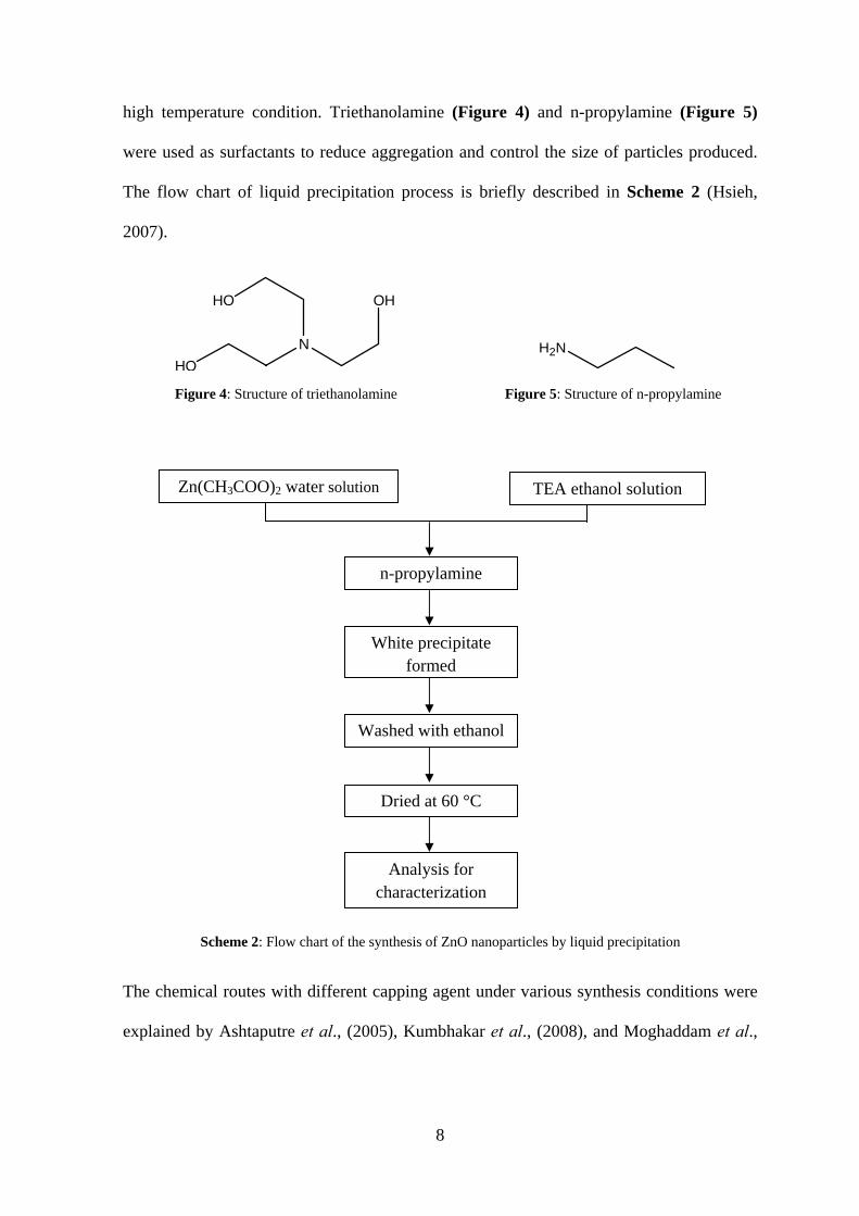
As specified by Ashtaputre et al., (2005), ZnO nanoparticles were formed by evaporating
methanol present in the precipitate at room temperature and these particles were capped
with thioglycerol (Figure 6) in alcoholic media to slow down and control the development
of ZnO nanoparticles. Zinc chloride was used as precursor. This method is size-selective
where size of particles formed can be controlled by varying the concentration of
thioglycerol. For increment in size of nanoparticles, water was added. Here, they
successfully synthesized less than 5.0 nm monodispersed nanoparticles and attained
stability by thiol capping.
SH
HO
OH
Figure 6: Structure of thioglycerol
On the other hand, Kumbhakar et al., (2008) prepared ZnO nanoparticles in double
distilled water via chemical method. Zn(NO3)2·H2O and KOH were dissolved in distilled
water under alkaline condition and PVP (Figure 7) was used as capping agent to prevent
aggregation. The precipitate deposited was centrifuged and dried at room temperature for
30 hours to obtain the ZnO nanoparticles. These steps resulted in the formation of ZnO
nanoparticles with average size of 1.9 nm.
10
H2C
HC
NO
n
Figure 7: Structure of polyvinylpyrrolidone (PVP)
Alternatively, Moghaddam et al., (2009) investigated a more simple and efficient chemical
method in synthesizing ZnO nanoparticles. ZnO nanoparticles were synthesized in zinc
nitrate aqueous solution without the need of calcinations stage. The precipitated ZnO
nanoparticles were dried at approximately 60 °C under atmospheric condition. They also
synthesized PPy/ZnO nanoparticles via electropolymerization to determine and compare
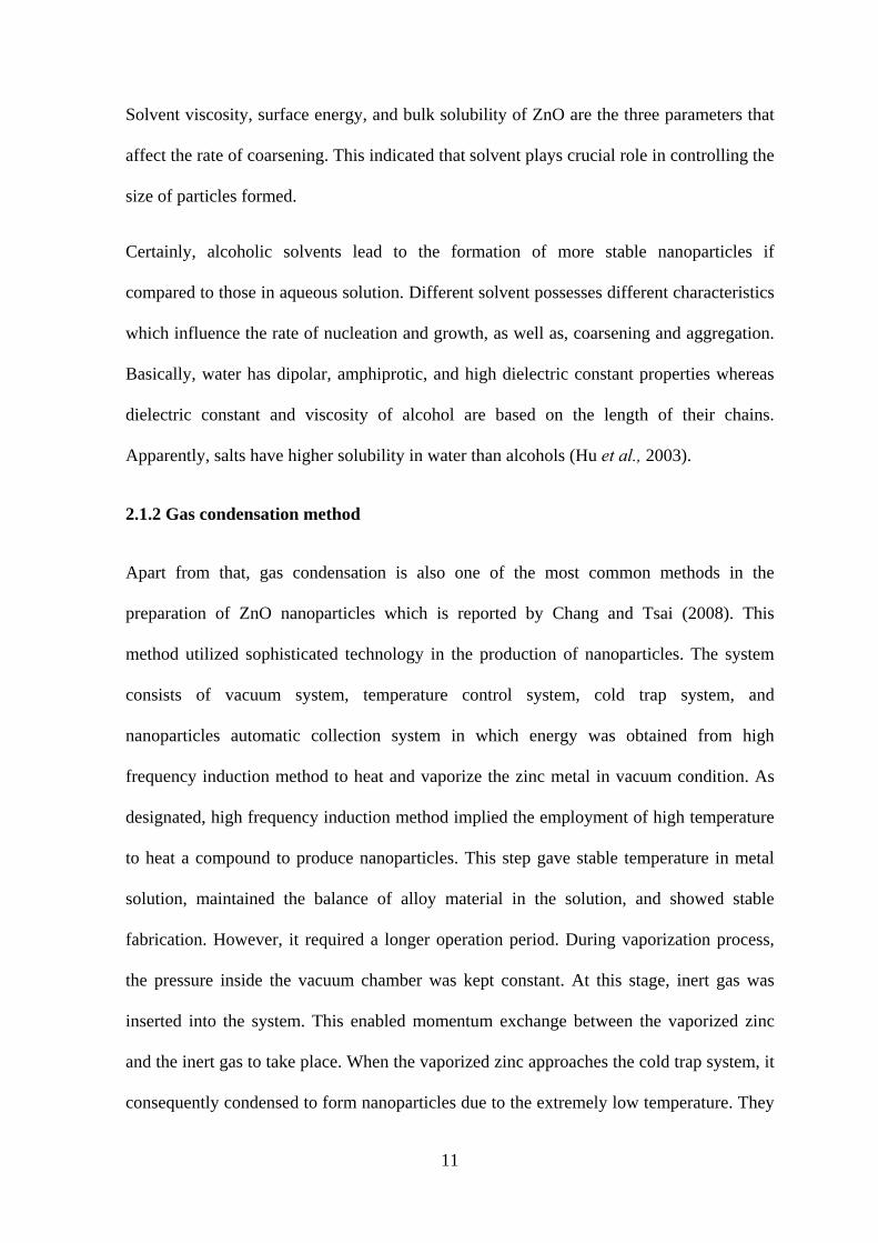
their differences in term of morphology and properties whereby PPy (Figure 8) acted as
capping agent.
HN
NH
n
Figure 8: Structure of polypyrrole (PPy)
Hu et al., (2003) clarified the synthesis of ZnO nanoparticles by precipitation from zinc
acetate in different solvent. They proved that diffusion-limited coarsening caused
nucleation and growth to increase rapidly with time. As temperature and alcohol chain
length increased, coarsening rate speeded up whereby the volume of particles was enlarged.
11
Solvent viscosity, surface energy, and bulk solubility of ZnO are the three parameters that
affect the rate of coarsening. This indicated that solvent plays crucial role in controlling the
size of particles formed.
Certainly, alcoholic solvents lead to the formation of more stable nanoparticles if
compared to those in aqueous solution. Different solvent possesses different characteristics
which influence the rate of nucleation and growth, as well as, coarsening and aggregation.
Basically, water has dipolar, amphiprotic, and high dielectric constant properties whereas
dielectric constant and viscosity of alcohol are based on the length of their chains.
Apparently, salts have higher solubility in water than alcohols (Hu et al., 2003).
2.1.2 Gas condensation method
Apart from that, gas condensation is also one of the most common methods in the
preparation of ZnO nanoparticles which is reported by Chang and Tsai (2008). This
method utilized sophisticated technology in the production of nanoparticles. The system
consists of vacuum system, temperature control system, cold trap system, and
nanoparticles automatic collection system in which energy was obtained from high
frequency induction method to heat and vaporize the zinc metal in vacuum condition. As
designated, high frequency induction method implied the employment of high temperature
to heat a compound to produce nanoparticles. This step gave stable temperature in metal
solution, maintained the balance of alloy material in the solution, and showed stable
fabrication. However, it required a longer operation period. During vaporization process,
the pressure inside the vacuum chamber was kept constant. At this stage, inert gas was
inserted into the system. This enabled momentum exchange between the vaporized zinc
and the inert gas to take place. When the vaporized zinc approaches the cold trap system, it
consequently condensed to form nanoparticles due to the extremely low temperature. They
12
established the formation of hexagonal prism shape of ZnO nanoparticles with consistent
size of 20 nm in diameter. Furthermore, they also generated even distribution and massive
amount of nanoparticles in the end of the process. Nevertheless, the collection speed of
nanoparticles was inconsistent during the initial vaporization route (Chang & Tsai, 2008).
2.1.3 Electrochemical method
In addition, Nyffenegger et al., (1998) demonstrated hybrid electrochemical/chemical (E/C)
method in synthesizing ZnO nanoparticles as control of average particle size by means of
deposition time is impossible via direct electrochemical route. As reported, there were two
stages drawn in this approach. Size-selective deposition of zinc metal onto graphite-based
surface occurred in the first place and then followed by spontaneous oxidation and
dehydration of the deposited zinc which eventually formed ZnO wurtzite structure. They
also synthesized ZnO nanoparticles via direct electrochemical procedure under three
different conditions for comparison purposes. Nyffenegger and colleagues (1998)
accounted narrow distribution of nanoparticles due to the instantaneous nucleation of zinc
on graphite surface. The E/C method is not only simple and straightforward but it can be
used to synthesis size-selective ZnO nanoparticles with diameters ranging from 15 to 100
Å. Sajanlal et al., (2011) denoted that the application of electrochemical method requires
low processing temperature, modest and cheap equipments yet generates high-quality
products.
2.1.4 Sol-gel method
Sol-gel method also known as chemical solution deposition is based on the hydrolysis of
liquid precursors and formation of colloidal sols (Kundu et al., 2011). They stipulated that
sol-gel method is a low cost chemical process if compared to physical synthesis methods.
The formation of nanosized semiconductor or metal elements permits system with peculiar
13
optical and electrical properties. In the experiment performed by Kundu and co-workers
(2011), sol-gel process can be applied in the production of uniformly dispersed ZnO
nanoparticles. They also investigated the effect of different synthesis conditions and
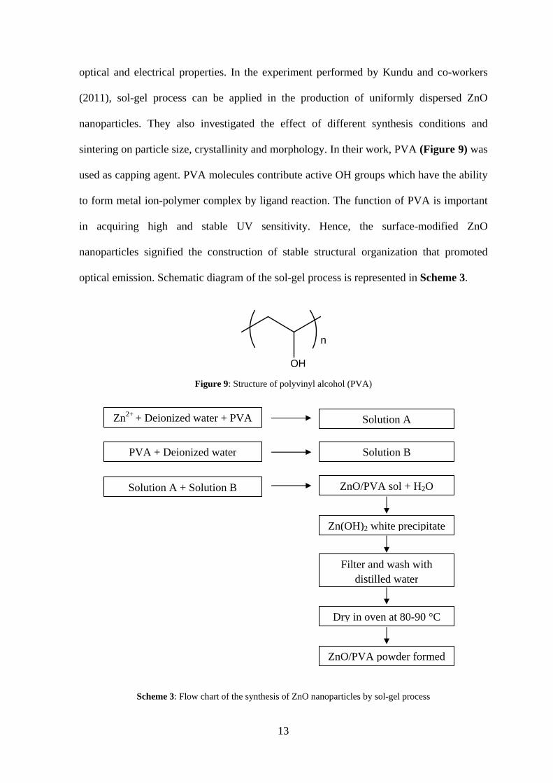
sintering on particle size, crystallinity and morphology. In their work, PVA (Figure 9) was
used as capping agent. PVA molecules contribute active OH groups which have the ability
to form metal ion-polymer complex by ligand reaction. The function of PVA is important
in acquiring high and stable UV sensitivity. Hence, the surface-modified ZnO
nanoparticles signified the construction of stable structural organization that promoted
optical emission. Schematic diagram of the sol-gel process is represented in Scheme 3.
OH
n
Figure 9: Structure of polyvinyl alcohol (PVA)
Scheme 3: Flow chart of the synthesis of ZnO nanoparticles by sol-gel process
Zn2+ + Deionized water + PVA
PVA + Deionized water
Solution A + Solution B
ZnO/PVA powder formed
Dry in oven at 80-90 °C
Filter and wash with distilled water
Zn(OH)2 white precipitate
ZnO/PVA sol + H2O
Solution B
Solution A
![SYNTHESIS OF PMMA/ZnO NANOPARTICLES COMPOSITE USED …mit.imt.si/Revija/izvodi/mit175/popovic.pdf · d. popovi] et al.: synthesis of pmma/zno nanoparticles composite used for resin](https://img.pdfslide.us/doc/110x75/5a8ef09e7f8b9a78648d6099/synthesis-of-pmmazno-nanoparticles-composite-used-mitimtsirevijaizvodimit175.jpg)