Embed Size (px)
Citation preview
Go to Section:
Chapter 37
Circulatory and Respiratory
Systems
Go to Section:
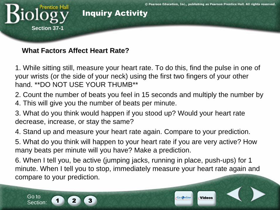
What Factors Affect Heart Rate?
1. While sitting still, measure your heart rate. To do this, find the pulse in one of
your wrists (or the side of your neck) using the first two fingers of your other
hand. **DO NOT USE YOUR THUMB**
2. Count the number of beats you feel in 15 seconds and multiply the number by
4. This will give you the number of beats per minute.
3. What do you think would happen if you stood up? Would your heart rate
decrease, increase, or stay the same?
4. Stand up and measure your heart rate again. Compare to your prediction.
5. What do you think will happen to your heart rate if you are very active? How
many beats per minute will you have? Make a prediction.
6. When I tell you, be active (jumping jacks, running in place, push-ups) for 1
minute. When I tell you to stop, immediately measure your heart rate again and
compare to your prediction.
Section 37-1
Inquiry Activity

Go to Section:
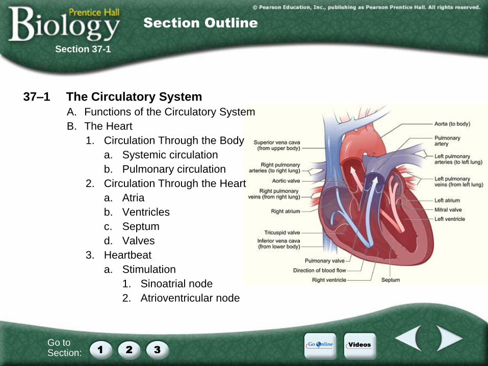
37–1 The Circulatory System
A. Functions of the Circulatory System
B. The Heart
1. Circulation Through the Body
a. Systemic circulation
b. Pulmonary circulation
2. Circulation Through the Heart
a. Atria
b. Ventricles
c. Septum
d. Valves
3. Heartbeat
a. Stimulation
1. Sinoatrial node
2. Atrioventricular node
Section 37-1
Section Outline
Go to Section:
Section 37-1
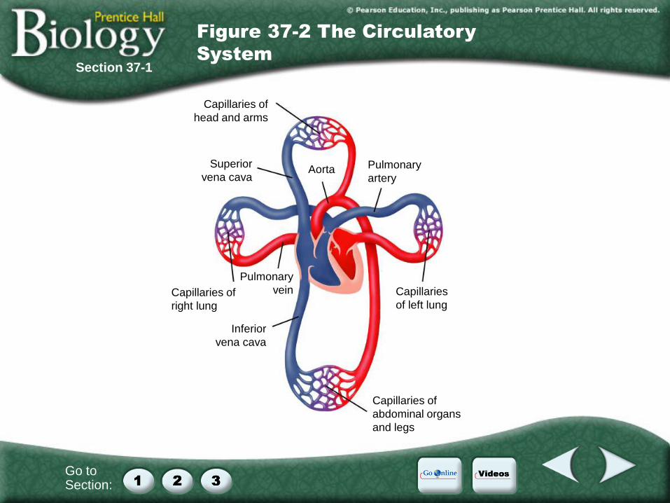
Figure 37-2 The Circulatory
System
Capillaries of
head and arms
Capillaries of
abdominal organs
and legs
Inferior
vena cava
Pulmonary
vein Capillaries of
right lung
Superior
vena cava Aorta Pulmonary
artery
Capillaries
of left lung

Go to Section:
37–1 The Circulatory System
A. Functions of the Circulatory System
B. The Heart
1. Circulation Through the Body
a. Systemic circulation
b. Pulmonary circulation
2. Circulation Through the Heart
a. Atria
b. Ventricles
c. Septum
d. Valves
3. Heartbeat
a. Stimulation
1. Sinoatrial node
2. Atrioventricular node
Section 37-1
Section Outline
Go to Section:
Section 37-1
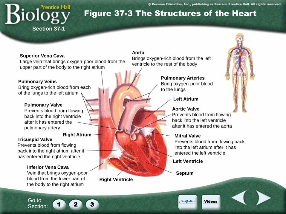
Figure 37-3 The Structures of the Heart
Right Ventricle
Right Atrium
Left Atrium
Inferior Vena Cava
Vein that brings oxygen-poor
blood from the lower part of
the body to the right atrium
Tricuspid Valve
Prevents blood from flowing
back into the right atrium after it
has entered the right ventricle
Pulmonary Valve
Prevents blood from flowing
back into the right ventricle
after it has entered the
pulmonary artery
Pulmonary Veins
Bring oxygen-rich blood from each
of the lungs to the left atrium
Superior Vena Cava
Large vein that brings oxygen-poor blood from the
upper part of the body to the right atrium
Aorta
Brings oxygen-rich blood from the left
ventricle to the rest of the body
Pulmonary Arteries
Bring oxygen-poor blood
to the lungs
Aortic Valve
Prevents blood from flowing
back into the left ventricle
after it has entered the aorta
Mitral Valve
Prevents blood from flowing back
into the left atrium after it has
entered the left ventricle
Left Ventricle
Septum
Go to Section:
37–1 The Circulatory System
A. Functions of the Circulatory System
B. The Heart
1. Circulation Through the Body
a. Systemic circulation
b. Pulmonary circulation
2. Circulation Through the Heart
a. Atria
b. Ventricles
c. Septum
d. Valves
3. Heartbeat
a. Stimulation
1. Sinoatrial node
2. Atrioventricular node
Section 37-1
Section Outline
Go to Section:
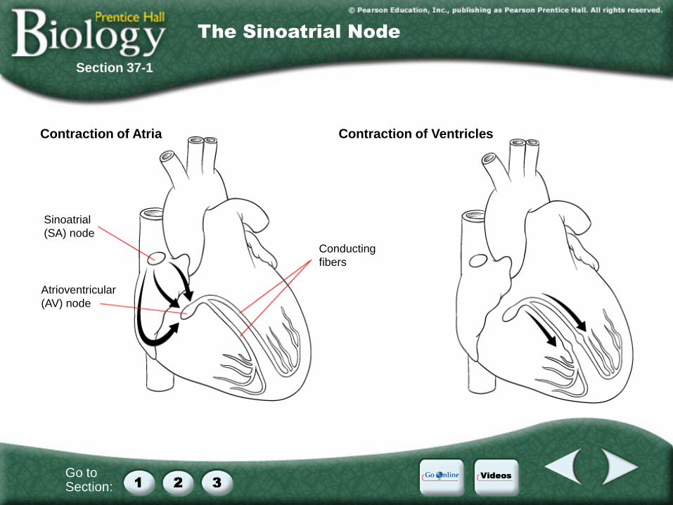
Sinoatrial
(SA) node
Atrioventricular
(AV) node
Conducting
fibers
Contraction of Atria Contraction of Ventricles
Section 37-1
The Sinoatrial Node
Go to Section:
37–1 The Circulatory System
C. Blood Vessels
1. Arteries
2. Capillaries
3. Veins
D. Blood Pressure
1. Systolic
2. Diastolic
E. Diseases of the Circulatory System
1. High Blood Pressure
2. Consequences of Atherosclerosis
3. Circulatory System Health
Section 37-1
Section Outline
Go to Section:
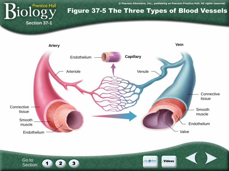
Section 37-1
Figure 37-5 The Three Types of Blood Vessels
Capillary
Connective
tissue
Connective
tissue
Smooth
muscle
Smooth
muscle
Endothelium
Endothelium
Valve
Venule
Endothelium
Arteriole
Vein Artery
Go to Section:
37–1 The Circulatory System
C. Blood Vessels
1. Arteries
2. Capillaries
3. Veins
D. Blood Pressure
1. Systolic
2. Diastolic
E. Diseases of the Circulatory System
1. High Blood Pressure
2. Consequences of Atherosclerosis
3. Circulatory System Health
Section 37-1
Section Outline
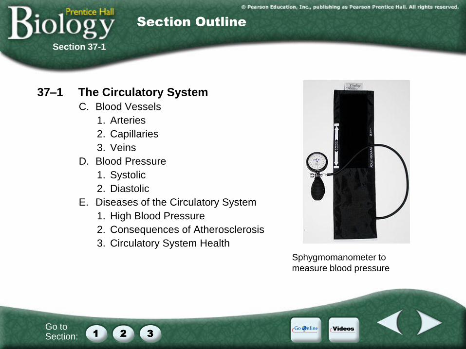
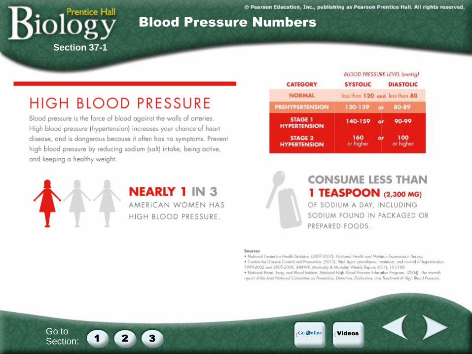
Sphygmomanometer to
measure blood pressure
Go to Section:
Section 37-1
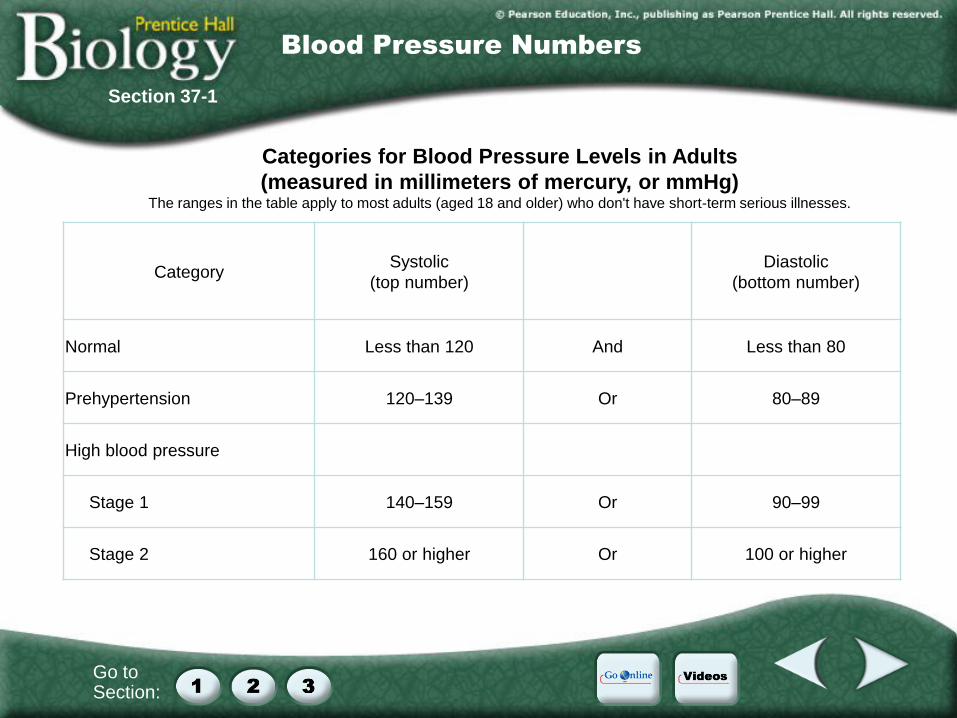
Blood Pressure Numbers
Category Systolic
(top number)
Diastolic
(bottom number)
Normal Less than 120 And Less than 80
Prehypertension 120–139 Or 80–89
High blood pressure
Stage 1 140–159 Or 90–99
Stage 2 160 or higher Or 100 or higher
Categories for Blood Pressure Levels in Adults
(measured in millimeters of mercury, or mmHg) The ranges in the table apply to most adults (aged 18 and older) who don't have short-term serious illnesses.
Go to Section:
Section 37-1
Blood Pressure Numbers
Go to Section:
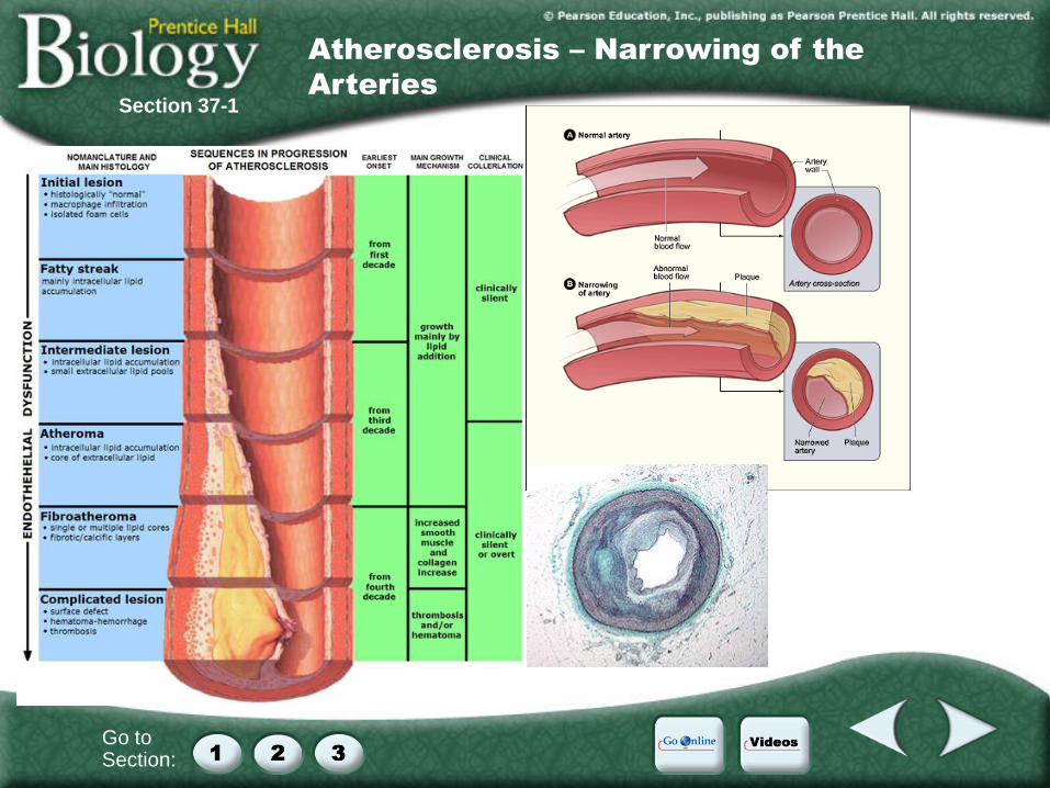
Section 37-1
Atherosclerosis – Narrowing of the
Arteries

Go to Section:
37–2 Blood and the Lymphatic System
A. Blood Plasma **transfusion**
1. Contents of plasma
2. Proteins: Albumins, Globulins, Fibrinogens
B. Blood Cells
1. Red Blood Cells (erythrocytes) **centrifuge**
2. White Blood Cells (leukocytes) **types**
a. neutrophils, basophils, lymphocytes,
eosinophils, monocytes
b. Granulocytes & agranulocytes
3. Platelets and Blood Clotting **diagram**
Thromboplastin, prothrombin, thrombin,
fibrinogen, fibrin
C. The Lymphatic System **pathways**
Section 37-2
Section Outline
Go to Section:
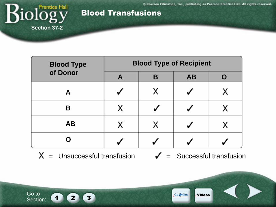
Blood Type
of Donor
A B AB O
Blood Type of Recipient
A B AB O
Unsuccessful transfusion Successful transfusion
Section 37-2
Blood Transfusions

Go to Section:
37–2 Blood and the Lymphatic System
A. Blood Plasma **transfusion**
1. Contents of plasma
2. Proteins: Albumins, Globulins, Fibrinogens
B. Blood Cells
1. Red Blood Cells (erythrocytes) **centrifuge**
2. White Blood Cells (leukocytes) **types**
a. neutrophils, basophils, lymphocytes,
eosinophils, monocytes
b. Granulocytes & agranulocytes
3. Platelets and Blood Clotting **diagram**
Thromboplastin, prothrombin, thrombin,
fibrinogen, fibrin
C. The Lymphatic System **pathways**
Section 37-2
Section Outline
Go to Section:
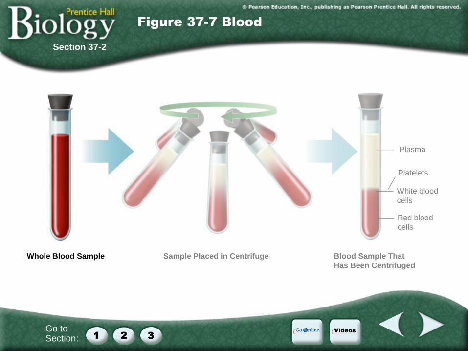
Section 37-2
Figure 37-7 Blood
Whole Blood Sample
Red blood
cells
White blood
cells
Platelets
Plasma
Sample Placed in Centrifuge Blood Sample That
Has Been Centrifuged
Go to Section:
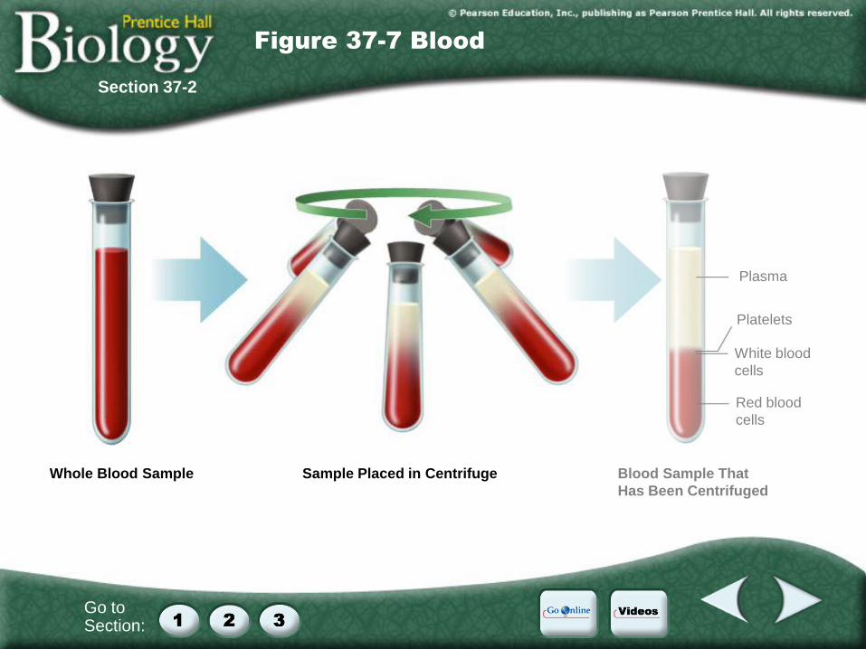
Section 37-2
Figure 37-7 Blood
Whole Blood Sample
Red blood
cells
White blood
cells
Platelets
Plasma
Sample Placed in Centrifuge Blood Sample That
Has Been Centrifuged
Go to Section:
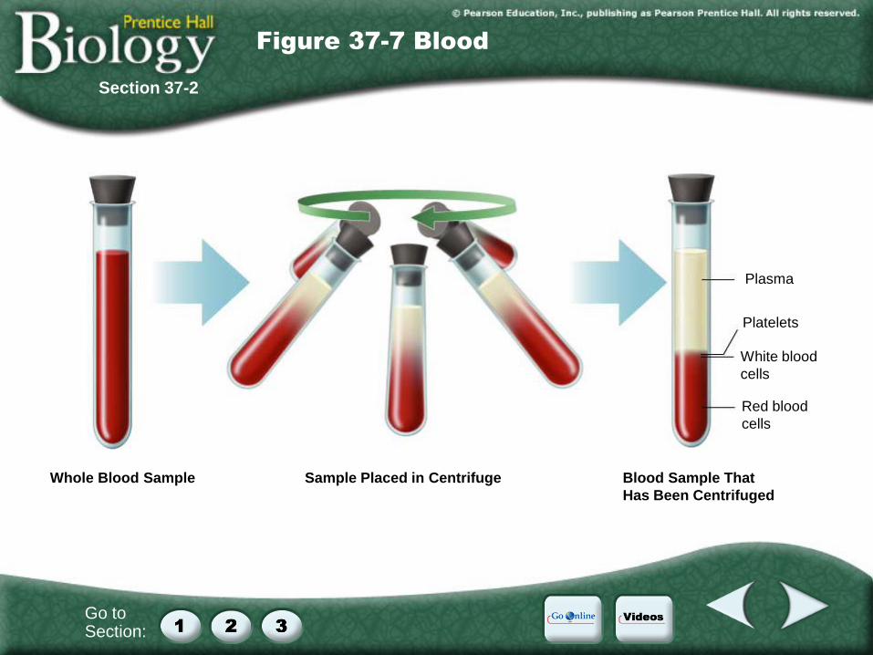
Section 37-2
Figure 37-7 Blood
Whole Blood Sample
Red blood
cells
White blood
cells
Platelets
Plasma
Sample Placed in Centrifuge Blood Sample That
Has Been Centrifuged

Go to Section:
37–2 Blood and the Lymphatic System
A. Blood Plasma **transfusion**
1. Contents of plasma
2. Proteins: Albumins, Globulins, Fibrinogens
B. Blood Cells
1. Red Blood Cells (erythrocytes) **centrifuge**
2. White Blood Cells (leukocytes) **types**
a. neutrophils, basophils, lymphocytes,
eosinophils, monocytes
b. Granulocytes & agranulocytes
3. Platelets and Blood Clotting **diagram**
Thromboplastin, prothrombin, thrombin,
fibrinogen, fibrin
C. The Lymphatic System **pathways**
Section 37-2
Section Outline
Go to Section:
Section 37-2
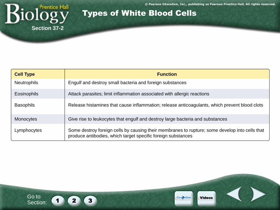
Types of White Blood Cells
Cell Type Neutrophils
Eosinophils
Basophils
Monocytes
Lymphocytes
Function Engulf and destroy small bacteria and foreign substances
Attack parasites; limit inflammation associated with allergic reactions
Release histamines that cause inflammation; release anticoagulants, which prevent blood clots
Give rise to leukocytes that engulf and destroy large bacteria and substances
Some destroy foreign cells by causing their membranes to rupture; some develop into cells that
produce antibodies, which target specific foreign substances
Go to Section:
37–2 Blood and the Lymphatic System
A. Blood Plasma **transfusion**
1. Contents of plasma
2. Proteins: Albumins, Globulins, Fibrinogens
B. Blood Cells
1. Red Blood Cells (erythrocytes) **centrifuge**
2. White Blood Cells (leukocytes) **types**
a. neutrophils, basophils, lymphocytes,
eosinophils, monocytes
b. Granulocytes & agranulocytes
3. Platelets and Blood Clotting **diagram**
Thromboplastin, prothrombin, thrombin,
fibrinogen, fibrin
C. The Lymphatic System **pathways**
Section 37-2
Section Outline
Go to Section:
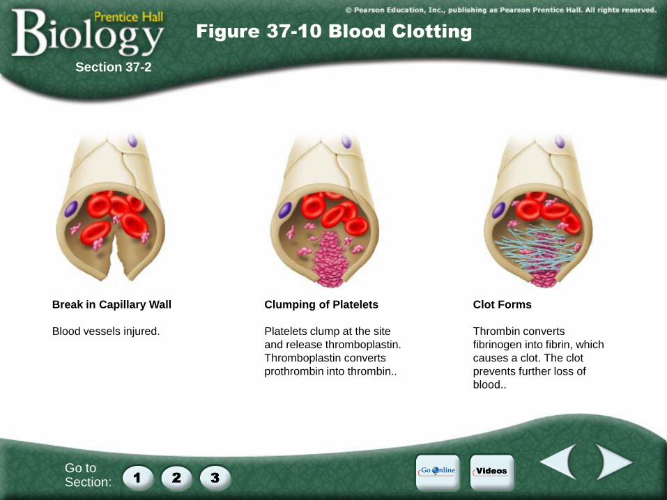
Section 37-2
Figure 37-10 Blood Clotting
Break in Capillary Wall
Blood vessels injured.
Clumping of Platelets
Platelets clump at the site
and release thromboplastin.
Thromboplastin converts
prothrombin into thrombin..
Clot Forms
Thrombin converts
fibrinogen into fibrin, which
causes a clot. The clot
prevents further loss of
blood..

Go to Section:
37–2 Blood and the Lymphatic System
C. The Lymphatic System **pathways**
1. Collects plasma fluid
2. Returns to circulatory system at superior
vena cava (flowing into right atrium)
3. Nodes along system filter out bacteria and
other microorganisms
a. During many infections you may
experience “swollen glands” which are
really your lymph nodes
4. Also absorbs fats & certain vitamins
5. Blocked lymph vessels leads to edema
6. Thymus & Spleen also instrumental
Section 37-2
Section Outline
Go to Section:
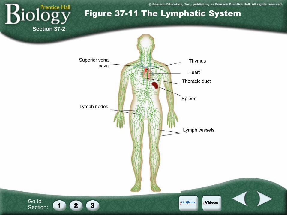
Section 37-2
Figure 37-11 The Lymphatic System
Superior vena
cava
Lymph nodes
Thymus
Heart
Thoracic duct
Spleen
Lymph vessels
Go to Section:
37–2 Blood and the Lymphatic System
C. The Lymphatic System **pathways**
1. Collects plasma fluid
2. Returns to circulatory system at superior
vena cava (flowing into right atrium)
3. Nodes along system filter out bacteria and
other microorganisms
a. During many infections you may
experience “swollen glands” which are
really your lymph nodes
4. Also absorbs fats & certain vitamins
5. Blocked lymph vessels leads to edema
6. Thymus & Spleen also instrumental
Section 37-2
Section Outline
Go to Section:
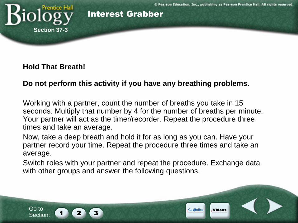
Hold That Breath!
Do not perform this activity if you have any breathing problems.
Working with a partner, count the number of breaths you take in 15 seconds. Multiply that number by 4 for the number of breaths per minute. Your partner will act as the timer/recorder. Repeat the procedure three times and take an average.
Now, take a deep breath and hold it for as long as you can. Have your partner record your time. Repeat the procedure three times and take an average.
Switch roles with your partner and repeat the procedure. Exchange data with other groups and answer the following questions.
Section 37-3
Interest Grabber
Go to Section:
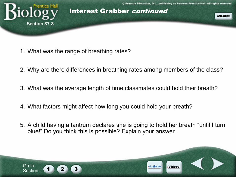
1. What was the range of breathing rates?
2. Why are there differences in breathing rates among members of the class?
3. What was the average length of time classmates could hold their breath?
4. What factors might affect how long you could hold your breath?
5. A child having a tantrum declares she is going to hold her breath “until I turn blue!” Do you think this is possible? Explain your answer.
Section 37-3
Interest Grabber continued
Go to Section:
37–3 The Respiratory System
A. What Is Respiration? **video**
B. The Human Respiratory System
*pathways & closeups*
C. Gas Exchange
D. Breathing *mechanics*
E. How Breathing Is Controlled
F. Tobacco and the Respiratory System
1. Substances in Tobacco
2. Diseases Caused by Smoking
3. Smoking and the Nonsmoker
4. Dealing With Tobacco
Section 37-3
Section Outline
Go to Section:
Section 37-3
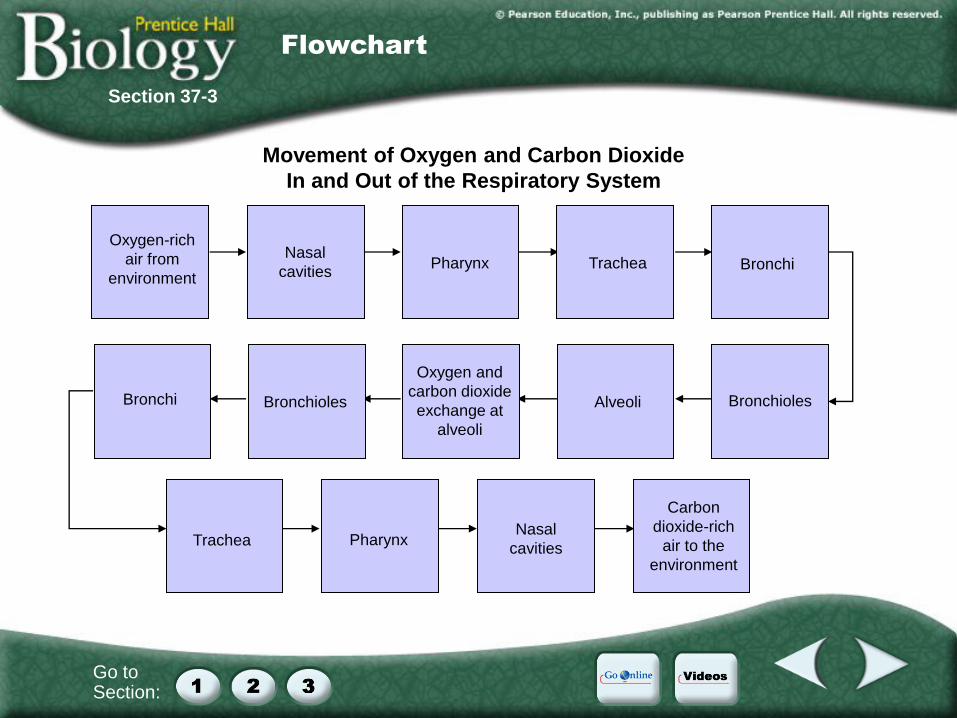
Flowchart
Movement of Oxygen and Carbon Dioxide
In and Out of the Respiratory System
Oxygen and
carbon dioxide
exchange at
alveoli
Oxygen-rich
air from
environment
Bronchioles
Nasal
cavities
Pharynx
Trachea Bronchi
Bronchioles
Alveoli
Pharynx
Nasal
cavities
Carbon
dioxide-rich
air to the
environment
Bronchi
Trachea
Go to Section:
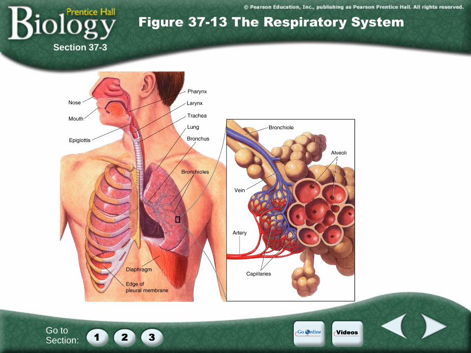
Section 37-3
Figure 37-13 The Respiratory System
Go to Section:
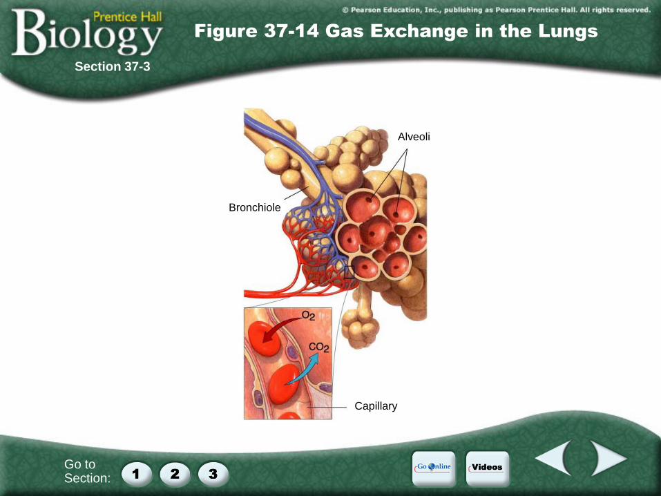
Alveoli
Bronchiole
Capillary
Section 37-3
Figure 37-14 Gas Exchange in the Lungs
Go to Section:
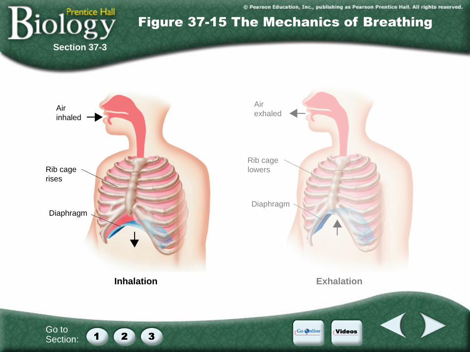
Air
inhaled
Diaphragm
Rib cage
rises
Air
exhaled
Diaphragm
Rib cage
lowers
Inhalation Exhalation
Section 37-3
Figure 37-15 The Mechanics of Breathing
Go to Section:
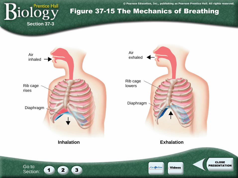
Air
inhaled
Diaphragm
Rib cage
rises
Air
exhaled
Diaphragm
Rib cage
lowers
Inhalation Exhalation
Section 37-3
Figure 37-15 The Mechanics of Breathing
Video Contents
Videos
Click a hyperlink to choose a video.
Human Circulation
Human Respiration
Video 1
Click the image to play the video segment.
Video 1
Human Circulation
Video 2
Click the image to play the video segment.
Video 2
Human Respiration
Internet
Go Online
Career links on respiratory care practitioners
Interactive test
For links on the cardiovascular system, go to www.SciLinks.org and enter the Web Code as follows: cbn-0371.
For links on blood cells, go to www.SciLinks.org and enter the Web Code as follows: cbn-0372.
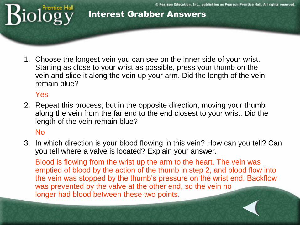
Section 1 Answers
Interest Grabber Answers
1. Choose the longest vein you can see on the inner side of your wrist. Starting as close to your wrist as possible, press your thumb on the vein and slide it along the vein up your arm. Did the length of the vein remain blue?
Yes
2. Repeat this process, but in the opposite direction, moving your thumb along the vein from the far end to the end closest to your wrist. Did the length of the vein remain blue?
No
3. In which direction is your blood flowing in this vein? How can you tell? Can you tell where a valve is located? Explain your answer.
Blood is flowing from the wrist up the arm to the heart. The vein was emptied of blood by the action of the thumb in step 2, and blood flow into the vein was stopped by the thumb’s pressure on the wrist end. Backflow was prevented by the valve at the other end, so the vein no longer had blood between these two points.
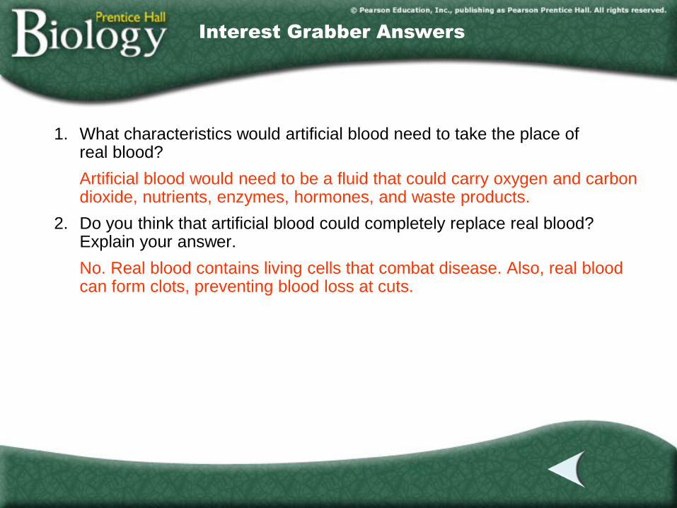
Section 2 Answers
Interest Grabber Answers
1. What characteristics would artificial blood need to take the place of real blood?
Artificial blood would need to be a fluid that could carry oxygen and carbon dioxide, nutrients, enzymes, hormones, and waste products.
2. Do you think that artificial blood could completely replace real blood? Explain your answer.
No. Real blood contains living cells that combat disease. Also, real blood can form clots, preventing blood loss at cuts.
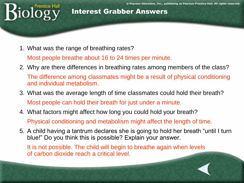
Section 3 Answers
Interest Grabber Answers
1. What was the range of breathing rates?
Most people breathe about 16 to 24 times per minute.
2. Why are there differences in breathing rates among members of the class?
The difference among classmates might be a result of physical conditioning and individual metabolism.
3. What was the average length of time classmates could hold their breath?
Most people can hold their breath for just under a minute.
4. What factors might affect how long you could hold your breath?
Physical conditioning and metabolism might affect the length of time.
5. A child having a tantrum declares she is going to hold her breath “until I turn blue!” Do you think this is possible? Explain your answer.
It is not possible. The child will begin to breathe again when levels of carbon dioxide reach a critical level.

End of Custom Shows
This slide is intentionally blank.