Embed Size (px)
Citation preview
Respiration, Circulatory, & Excretory Systems
Chapter 37Circulatory System: Week 4/23 - 5/1Respiratory System: Week 5/4 - 5/8Excretory Systems (Kidneys): Week 5/11 - 5/14
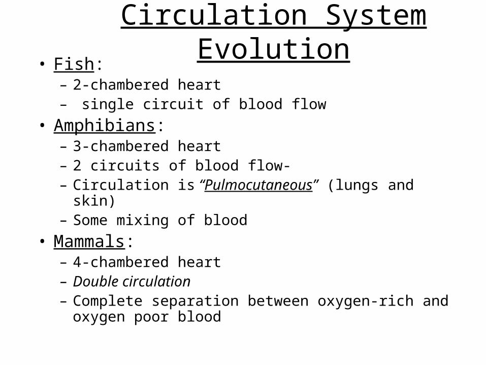
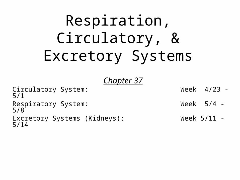
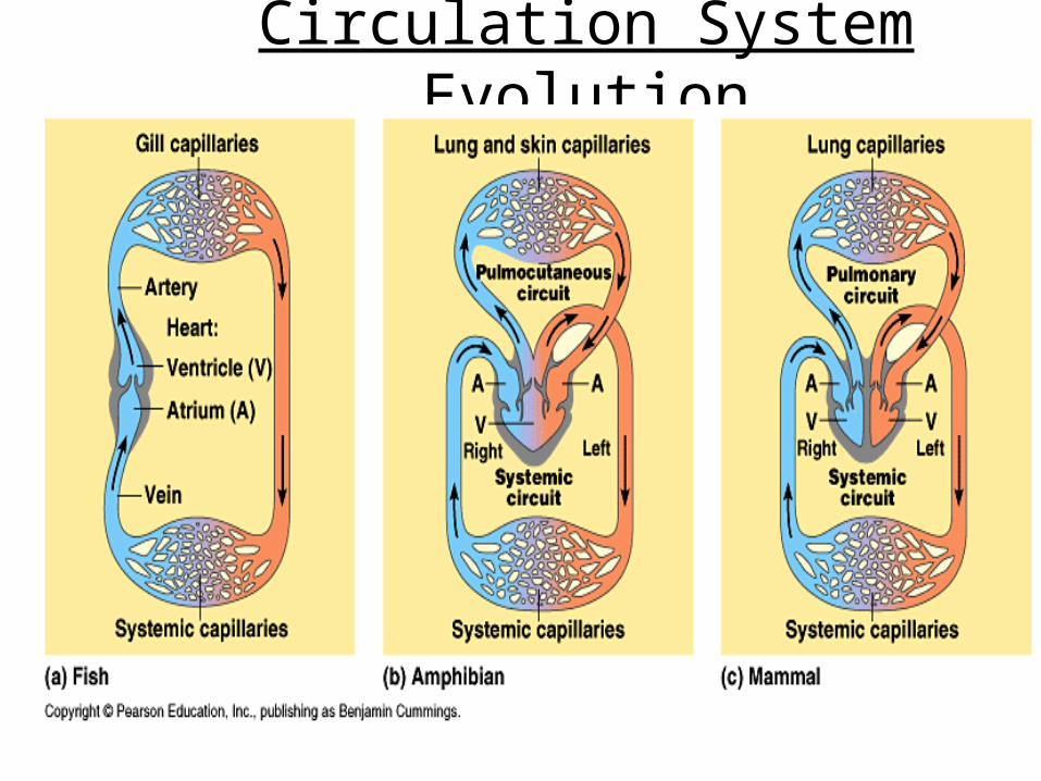
Circulation System Evolution• Fish:
– 2-chambered heart– single circuit of blood flow
• Amphibians: – 3-chambered heart – 2 circuits of blood flow- – Circulation is “Pulmocutaneous” (lungs and skin)– Some mixing of blood
• Mammals: – 4-chambered heart – Double circulation – Complete separation between oxygen-rich and
oxygen poor blood
Circulation System Evolution
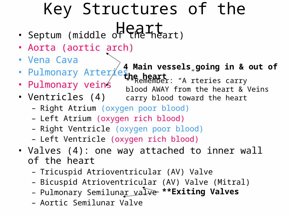
Key Structures of the Heart • Septum (middle of the heart)• Aorta (aortic arch)• Vena Cava• Pulmonary Arteries• Pulmonary veins• Ventricles (4)
– Right Atrium (oxygen poor blood)– Left Atrium (oxygen rich blood)– Right Ventricle (oxygen poor blood)– Left Ventricle (oxygen rich blood)
• Valves (4): one way attached to inner wall of the heart– Tricuspid Atrioventricular (AV) Valve– Bicuspid Atrioventricular (AV) Valve (Mitral)– Pulmonary Semilunar valve– Aortic Semilunar Valve
4 Main vessels going in & out of the heart
**Remember: “A”rteries carry blood AWAY from the heart & Veins carry blood toward the heart
**Exiting Valves
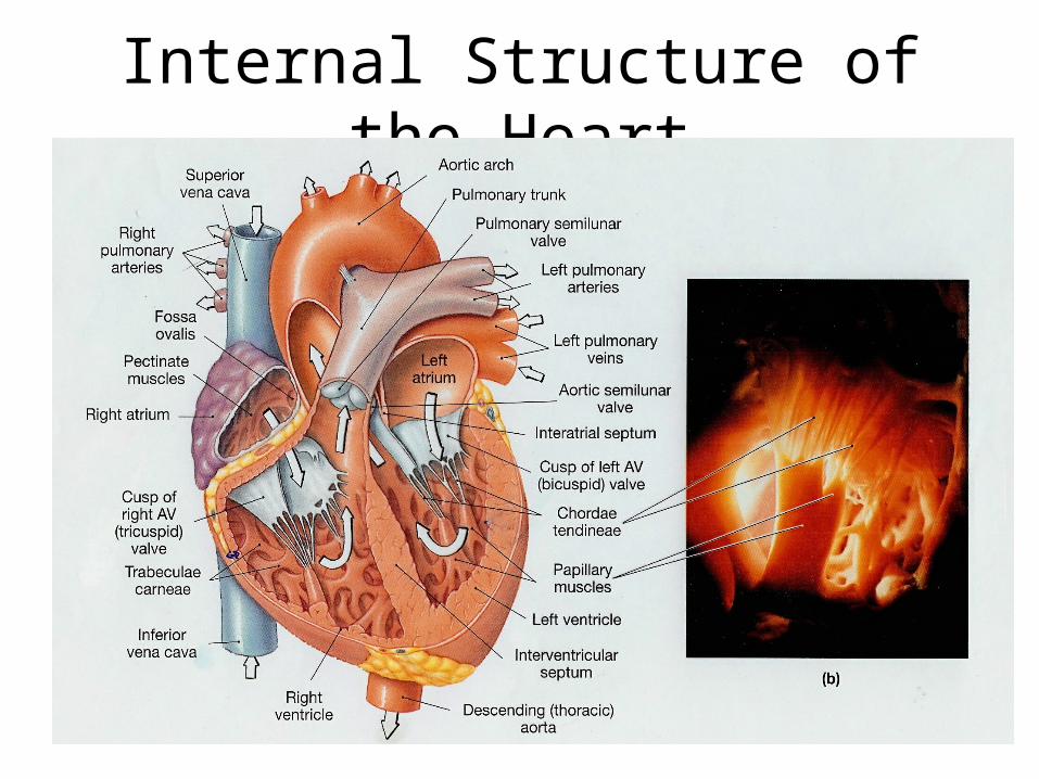
Internal Structure of the Heart
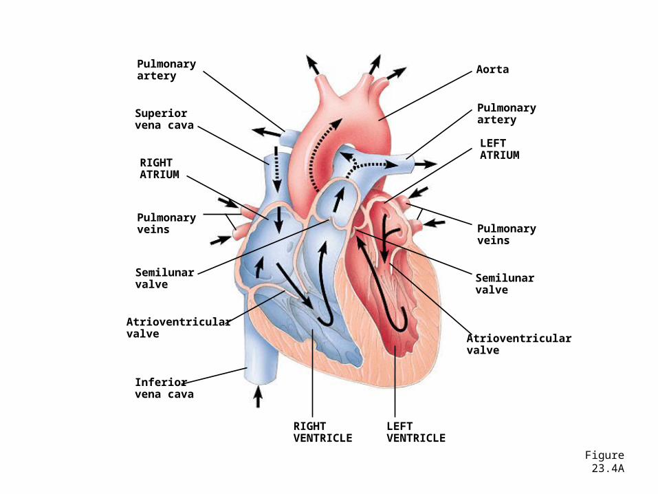
Figure 23.4A
Pulmonaryartery
Superiorvena cava
RIGHTATRIUM
Pulmonaryveins
Semilunarvalve
Atrioventricularvalve
Inferiorvena cava
Aorta
Pulmonaryartery
LEFTATRIUM
Pulmonaryveins
Semilunarvalve
Atrioventricularvalve
RIGHTVENTRICLE
LEFTVENTRICLE
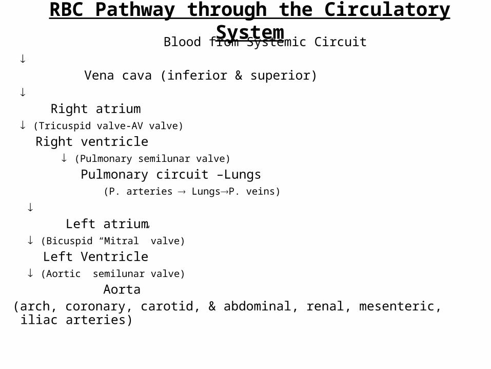
RBC Pathway through the Circulatory System Blood from Systemic Circuit
Vena cava (inferior & superior)
Right atrium
(Tricuspid valve-AV valve)
Right ventricle (Pulmonary semilunar valve)
Pulmonary circuit –Lungs (P. arteries LungsP. veins)
Left atrium
(Bicuspid “Mitral” valve)
Left Ventricle (Aortic semilunar valve)
Aorta (arch, coronary, carotid, & abdominal, renal, mesenteric, iliac arteries)

Video#2 : “A Heart Attack”Write 10 Key Statements
Introductory Questions #1• The heart has four valves in it. Name
them. Name the blood vessel that carries oxygen poor blood from the heart to the lungs to pick up more oxygen.
• Name the two large veins that bring blood to the heart from the rest of the body.
• In your textbook (pg. 975) Name the four components of blood. What % of your blood is composed of red blood cells? What about white blood cells?

Figure 23.4A
Pulmonaryartery
Superiorvena cava
RIGHTATRIUM
Pulmonaryveins
Semilunarvalve
Atrioventricularvalve
Inferiorvena cava
Aorta
Pulmonaryartery
LEFTATRIUM
Pulmonaryveins
Semilunarvalve
Atrioventricularvalve
RIGHTVENTRICLE
LEFTVENTRICLE5. chamber
1. vessel 10. vessel
11. vessel
9. vessels
8. valve
7. valve
2. chamber
3. valve
4. vessel
6. chamber
IQ #2
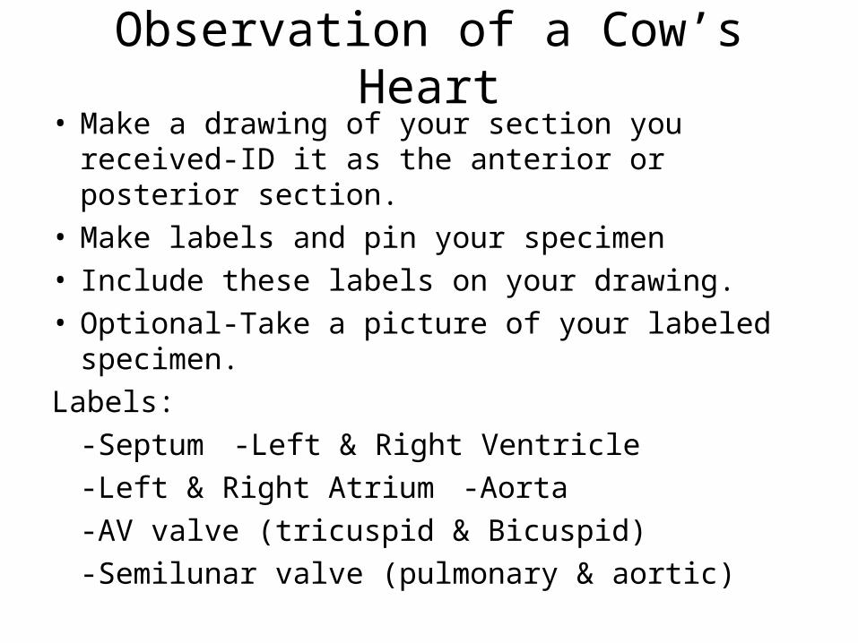
Observation of a Cow’s Heart• Make a drawing of your section you received-ID it
as the anterior or posterior section.• Make labels and pin your specimen• Include these labels on your drawing.• Optional-Take a picture of your labeled specimen.
Labels:
-Septum -Left & Right Ventricle
-Left & Right Atrium -Aorta
-AV valve (tricuspid & Bicuspid)
-Semilunar valve (pulmonary & aortic)
Video #1: Circulation: River of Life (Ch. 37.2) 1. What is the primary function of the circulatory
system?2. How is an open circulatory system different a
closed? Give an example of an organism that has an open circulatory system.
3. How is a vein different from an artery? Give two differences.
4. Name FOUR chambers and the four valves within the heart.
**Write the title for this segment and give FIVE statements.
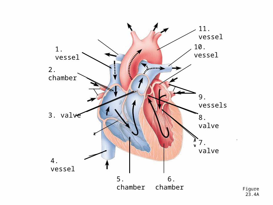
Figure 23.4A
Pulmonaryartery
Superiorvena cava
RIGHTATRIUM
Pulmonaryveins
Semilunarvalve
Atrioventricularvalve
Inferiorvena cava
Aorta
Pulmonaryartery
LEFTATRIUM
Pulmonaryveins
Semilunarvalve
Atrioventricularvalve
RIGHTVENTRICLE
LEFTVENTRICLE5. chamber
1. vessel 10. vessel
11. vessel
9. vessels
8. valve
7. valve
2. chamber
3. valve
4. vessel
6. chamber
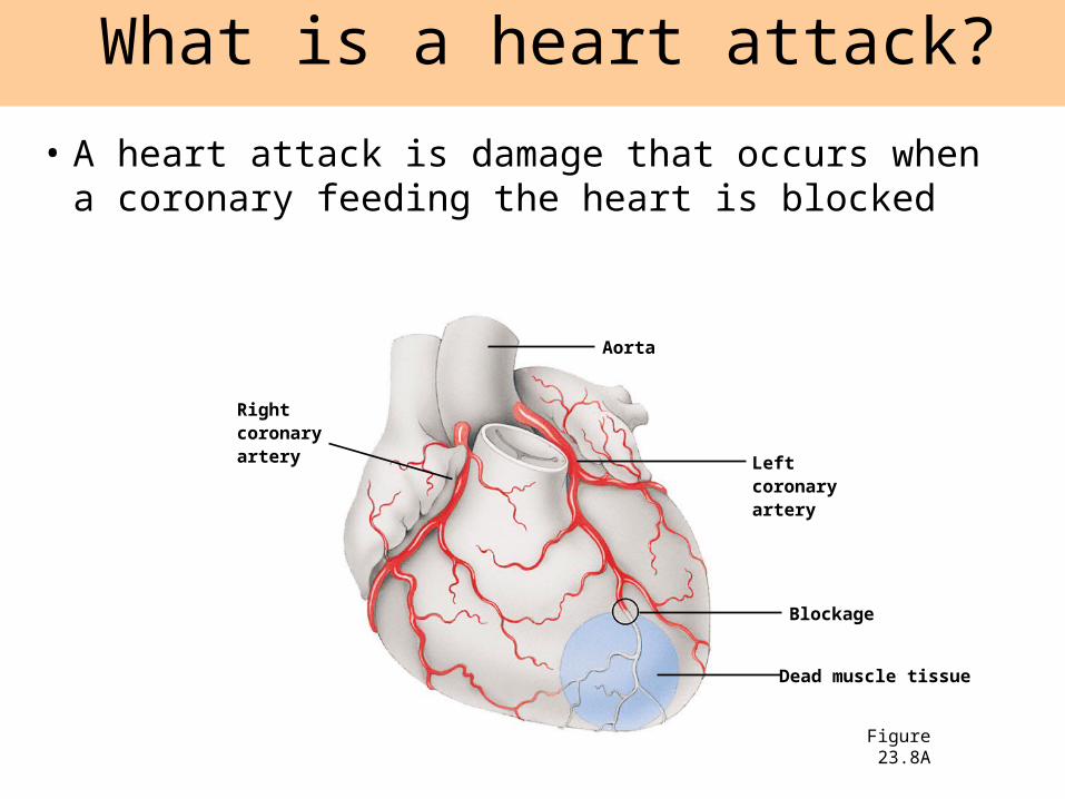
• A heart attack is damage that occurs when a coronary feeding the heart is blocked
What is a heart attack?
Figure 23.8A
Rightcoronaryartery
Aorta
Leftcoronaryartery
Blockage
Dead muscle tissue
Reading Assignment
• Using the handout read and review some of the key aspects of the circulatory system.
• On a separate sheet paper answer Questions on Pgs 25 & 26 from the second handout.
Label & Color your Hear Diagram
• Be sure to use RED for all areas that contains oxygen rich blood and BLUE for areas with oxygen poor blood.
• All valves must be correctly labeled
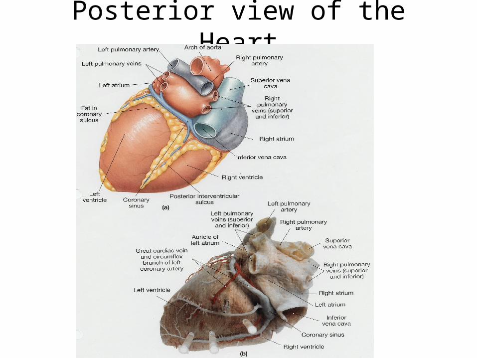
Posterior view of the Heart
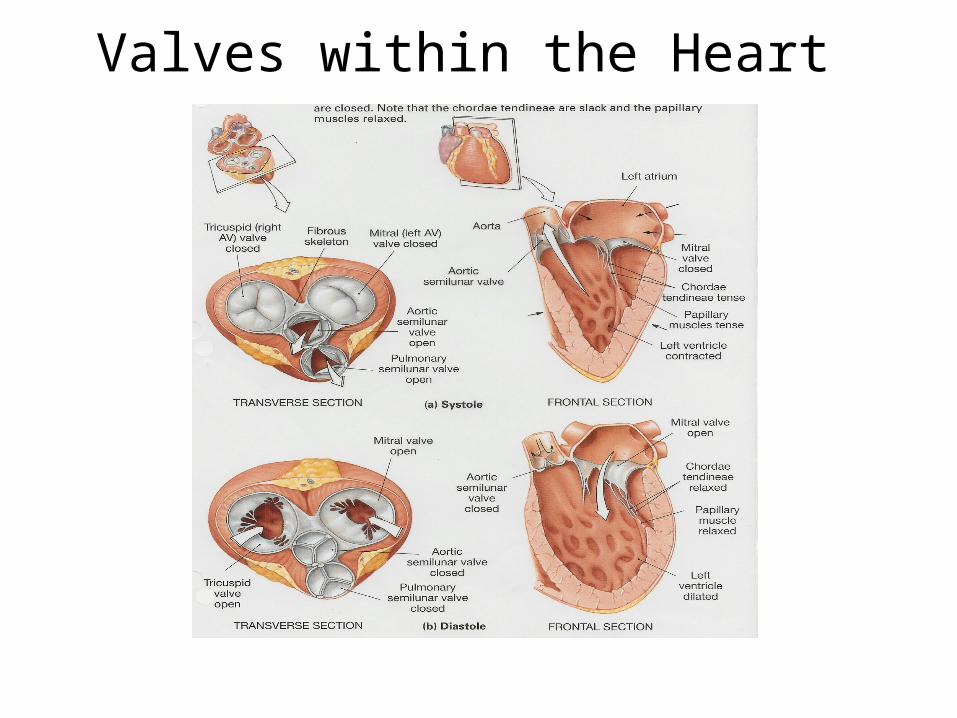
Valves within the Heart

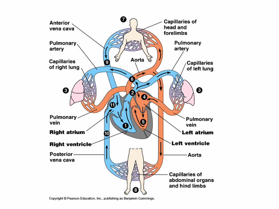
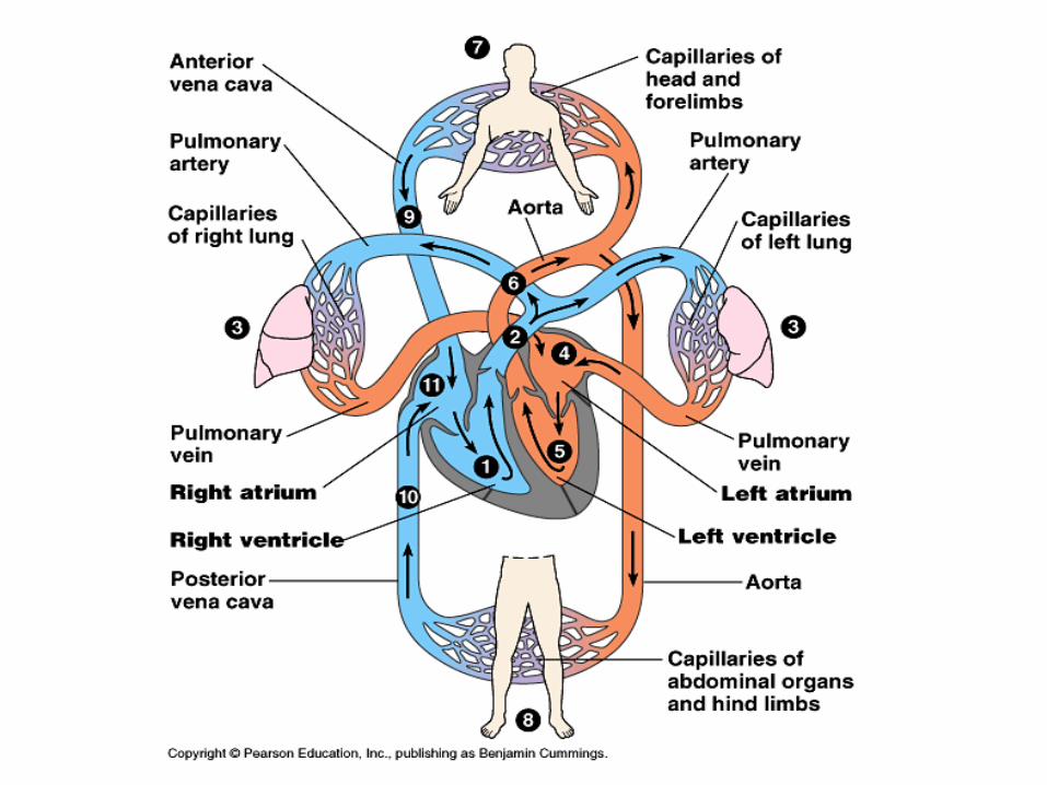
Double Circulation
• From right ventricle to lungs via pulmonary arteries through semilunar valve (pulmonary circulation)
• Capillary beds in lungs to left atrium via pulmonary veins
• Left atrium to left ventricle (through atrioventricular valve) to aorta
• Aorta to coronary arteries; then systemic circulation
• Back to heart via two venae cavae (superior and inferior); right atrium

Introductory Questions #1• The heart has four valves in it. Name
them. Name the blood vessel that carries oxygen poor blood from the heart to the lungs to pick up more oxygen.
• Name the two large veins that bring blood to the heart from the rest of the body.
• In your textbook (pg. 975) Name the four components of blood. What % of your blood is composed of red blood cells? What about white blood cells?

Figure 23.13
Withdrawblood
Place in tube
PLASMA 55%
CONSTITUENT MAJOR FUNCTIONS
WaterSolvent forcarrying othersubstances
Salts
Osmotic balance,pH buffering, andregulation ofmembranepermeability
SodiumPotassiumCalciumMagnesiumChlorideBicarbonate
Plasma proteins
Osmotic balance,pH bufferingClottingImmunity
Albumin
FibrinogenImmunoglobins(antibodies)
Substances transported by bloodNutrients (e.g., glucose, fatty acids, vitamins)Waste products of metabolismRespiratory gases (O2 and CO2)Hormones
Centrifuge
CELLULAR ELEMENTS 45%
CELL TYPE NUMBER(per mm3 of blood)
FUNCTIONS
Erythrocytes(red blood cells) 5–6 million Transport of
oxygen (and carbon dioxide)
Leukocytes(white blood cells) 5,000–10,000
Defense andimmunity
Basophil
Eosinophil
Neutrophil
Lymphocyte
Monocyte
Platelets 250,000–400,000
Blood clotting
Pg. 880
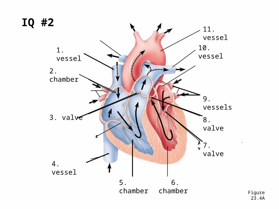
Figure 23.4A
Pulmonaryartery
Superiorvena cava
RIGHTATRIUM
Pulmonaryveins
Semilunarvalve
Atrioventricularvalve
Inferiorvena cava
Aorta
Pulmonaryartery
LEFTATRIUM
Pulmonaryveins
Semilunarvalve
Atrioventricularvalve
RIGHTVENTRICLE
LEFTVENTRICLE5. chamber
1. vessel 10. vessel
11. vessel
9. vessels
8. valve
7. valve
2. chamber
3. valve
4. vessel
6. chamber
IQ #2
Introductory Questions #3• Name the cell fragments that aide in the
process of blood clotting. (pg. 977)
• What substances are found in the plasma of blood? (pg. 975)
• Name the proteins that are found on the surface of red blood cells.
Introductory Questions #4• Give three differences between arteries
and veins.
• Why must blood slow down as it reaches a capillary bed?
• Where in the heart is the “pacemaker” and what role does it serve?
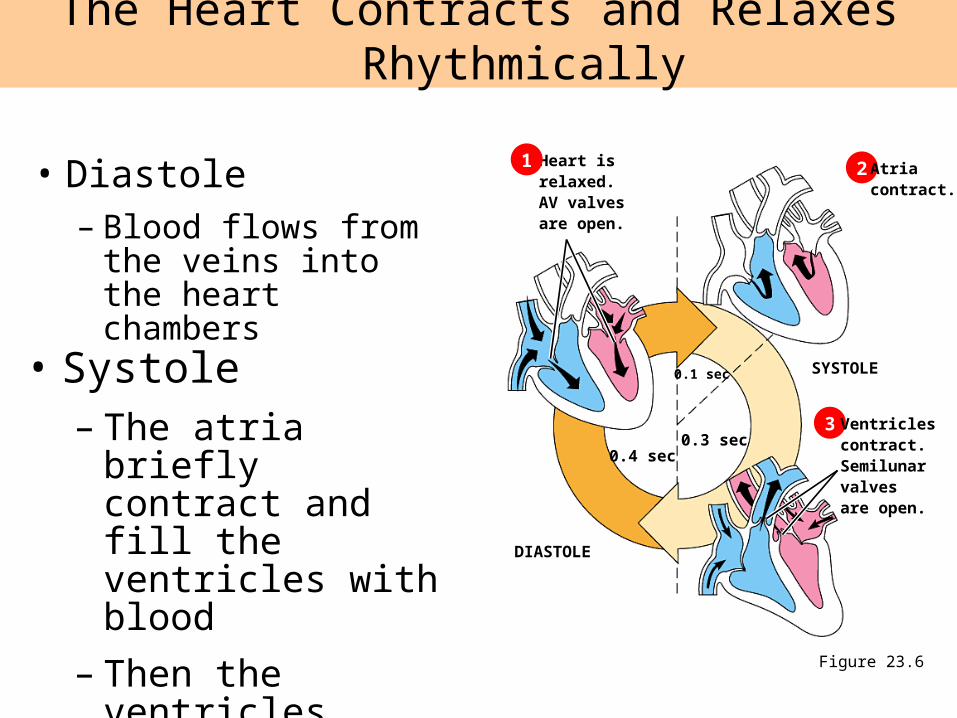
• Diastole– Blood flows from the
veins into the heart chambers
The Heart Contracts and Relaxes Rhythmically
Figure 23.6
Heart isrelaxed.AV valvesare open.
1 2
3
Atriacontract.
Ventriclescontract.Semilunarvalvesare open.
SYSTOLE
DIASTOLE
0.4 sec
0.1 sec
0.3 sec
• Systole– The atria briefly
contract and fill the ventricles with blood
– Then the ventricles contract and propel blood out
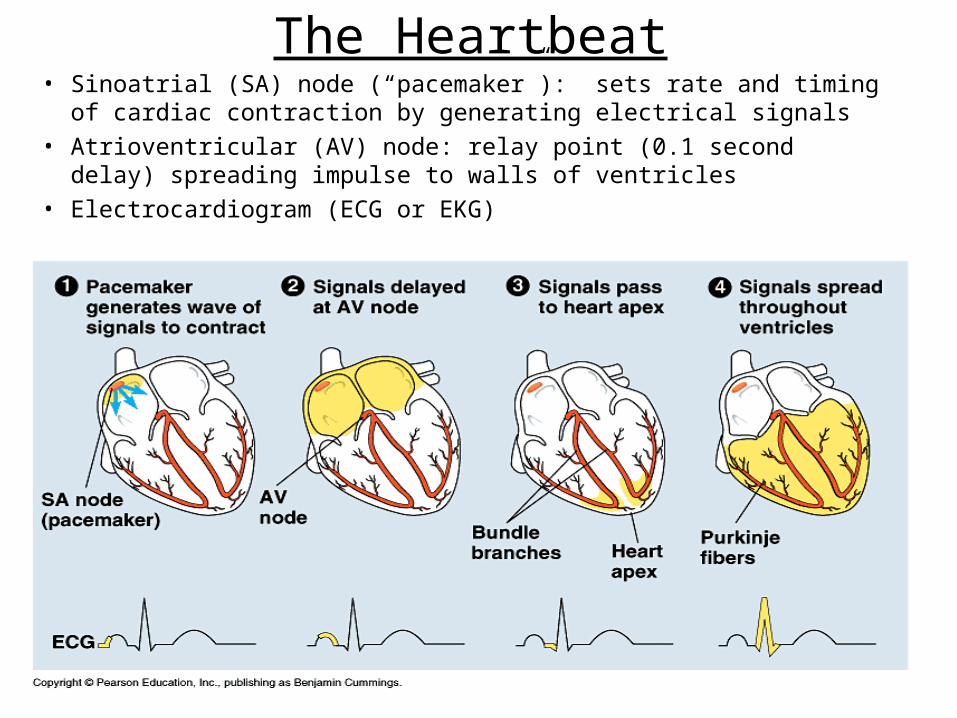
The Heartbeat• Sinoatrial (SA) node (“pacemaker”): sets rate and timing of
cardiac contraction by generating electrical signals
• Atrioventricular (AV) node: relay point (0.1 second delay) spreading impulse to walls of ventricles
• Electrocardiogram (ECG or EKG)
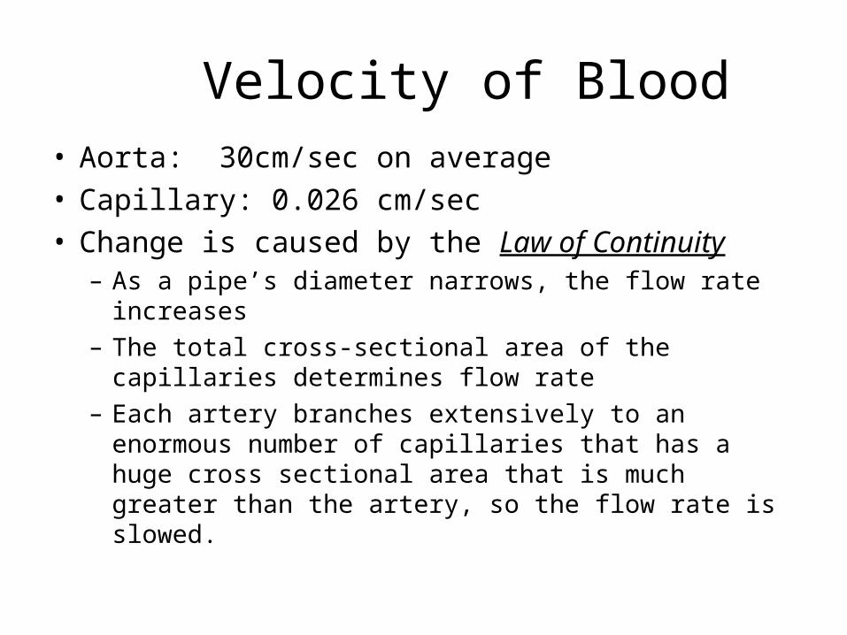
Velocity of Blood• Aorta: 30cm/sec on average • Capillary: 0.026 cm/sec• Change is caused by the Law of Continuity
– As a pipe’s diameter narrows, the flow rate increases– The total cross-sectional area of the capillaries
determines flow rate– Each artery branches extensively to an enormous
number of capillaries that has a huge cross sectional area that is much greater than the artery, so the flow rate is slowed.
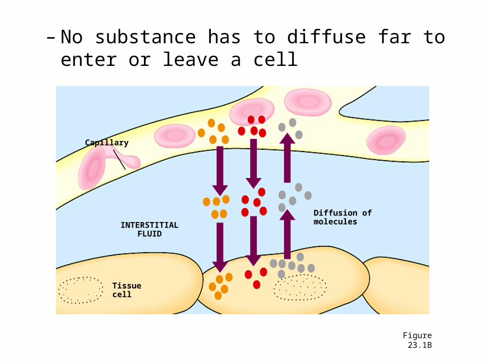
– No substance has to diffuse far to enter or leave a cell
Figure 23.1B
Capillary
INTERSTITIALFLUID
Tissuecell
Diffusion ofmolecules

Figure 23.13
Withdrawblood
Place in tube
PLASMA 55%
CONSTITUENT MAJOR FUNCTIONS
WaterSolvent forcarrying othersubstances
Salts
Osmotic balance,pH buffering, andregulation ofmembranepermeability
SodiumPotassiumCalciumMagnesiumChlorideBicarbonate
Plasma proteins
Osmotic balance,pH bufferingClottingImmunity
Albumin
FibrinogenImmunoglobins(antibodies)
Substances transported by bloodNutrients (e.g., glucose, fatty acids, vitamins)Waste products of metabolismRespiratory gases (O2 and CO2)Hormones
Centrifuge
CELLULAR ELEMENTS 45%
CELL TYPE NUMBER(per mm3 of blood)
FUNCTIONS
Erythrocytes(red blood cells) 5–6 million Transport of
oxygen (and carbon dioxide)
Leukocytes(white blood cells) 5,000–10,000
Defense andimmunity
Basophil
Eosinophil
Neutrophil
Lymphocyte
Monocyte
Platelets 250,000–400,000
Blood clotting
Pg. 880
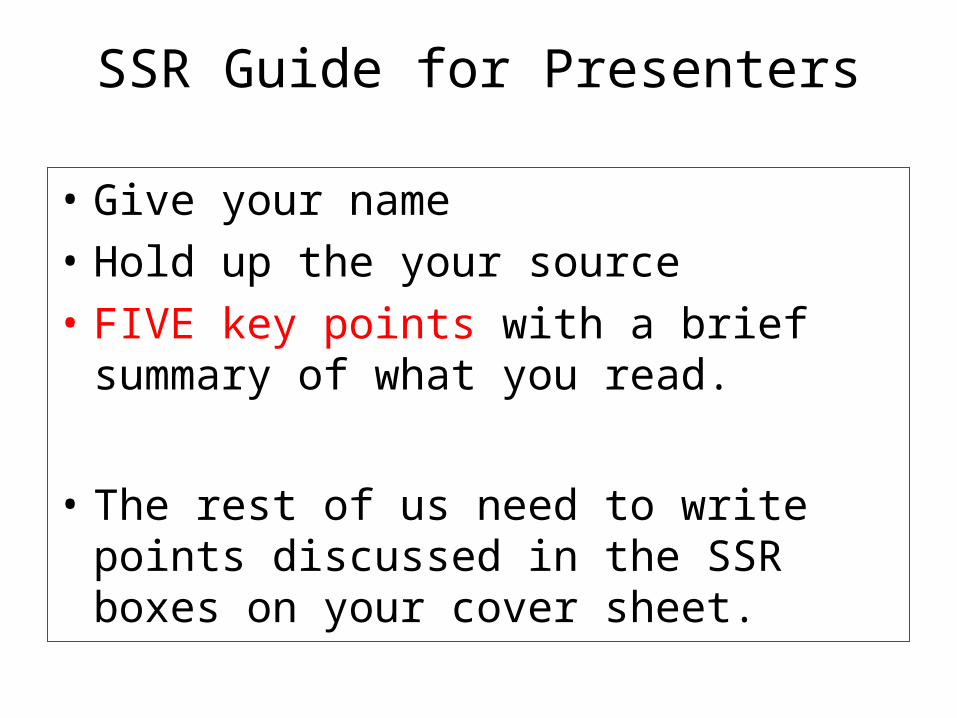
SSR Guide for Presenters
• Give your name
• Hold up the your source
• FIVE key points with a brief summary of what you read.
• The rest of us need to write points discussed in the SSR boxes on your cover sheet.
Assignment Packet
• Cover sheet
• Circulatory System Handout Questions
• Video Notes (x3)
• Heart Diagram (ID &* Color)
• Mini-lab Activity: Cow’s Heart (w/stamp)
Introductory Questions #1• What type of instrument is used to
measure a person’s blood pressure? Why is knowing a person’s blood pressure important?
• What is “normal” or average blood pressure at rest? What about heart rate?
• What does the top number and bottom number represent when reading blood pressure?
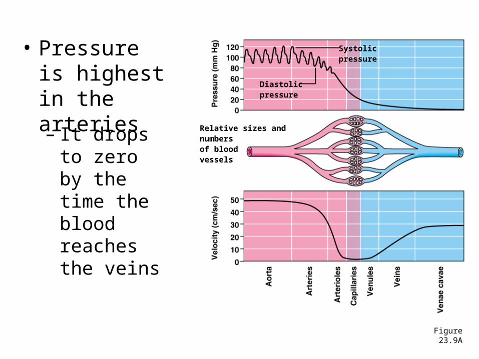
• Pressure is highest in the arteries
– It drops to zero by the time the blood reaches the veins
Figure 23.9A
Diastolicpressure
Systolicpressure
Relative sizes andnumbersof blood vessels

Velocity of Blood• Aorta: 30cm/sec on average • Capillary: 0.026 cm/sec• Change is caused by the Law of Continuity
– As a pipe’s diameter narrows, the flow rate increases– The total cross-sectional area of the capillaries
determines flow rate– Each artery branches extensively to an enormous
number of capillaries that has a huge cross sectional area that is much greater than the artery, so the flow rate is slowed.
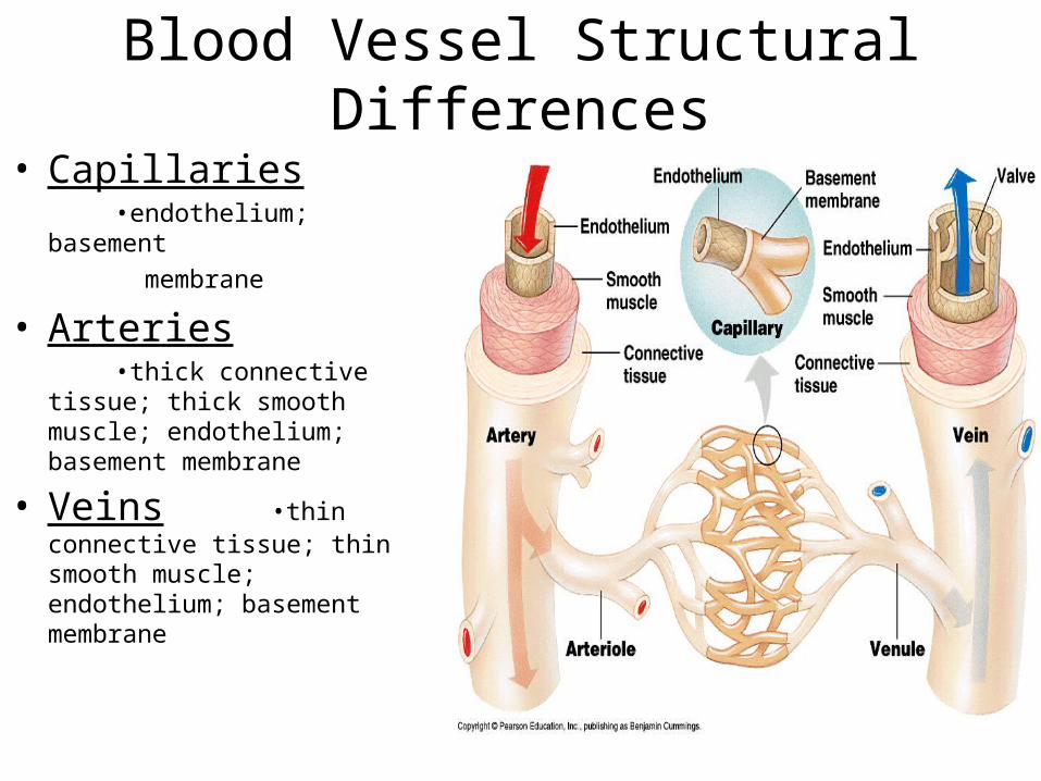
Blood Vessel Structural Differences
• Capillaries •endothelium; basement
membrane
• Arteries •thick connective tissue; thick
smooth muscle; endothelium; basement membrane
• Veins •thin connective tissue; thin smooth muscle; endothelium; basement membrane
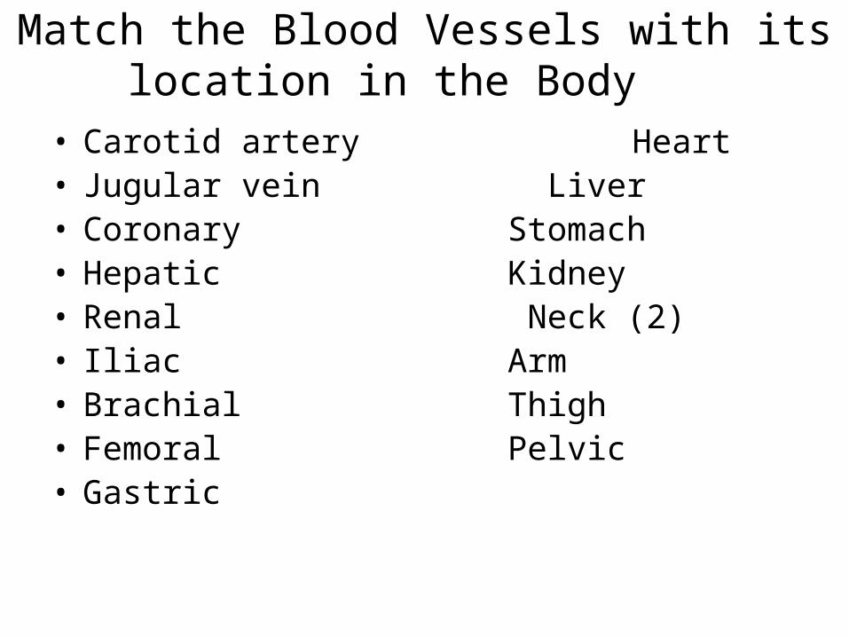
Match the Blood Vessels with its location in the Body
• Carotid artery Heart • Jugular vein Liver• Coronary Stomach• Hepatic Kidney• Renal Neck (2)• Iliac Arm• Brachial Thigh• Femoral Pelvic• Gastric
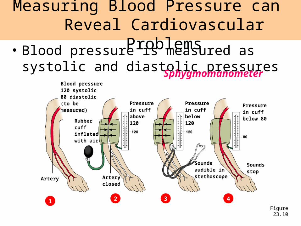
• Blood pressure is measured as systolic and diastolic pressures
Measuring Blood Pressure can Reveal Cardiovascular Problems
Figure 23.10
Blood pressure120 systolic80 diastolic(to be measured)
1 2 3 4
Rubber cuffinflated with air
Pressurein cuffbelow120
Pressurein cuffbelow 80
Artery
Pressurein cuffabove120
Soundsaudible instethoscope
Soundsstop
Arteryclosed
Sphygmomanometer
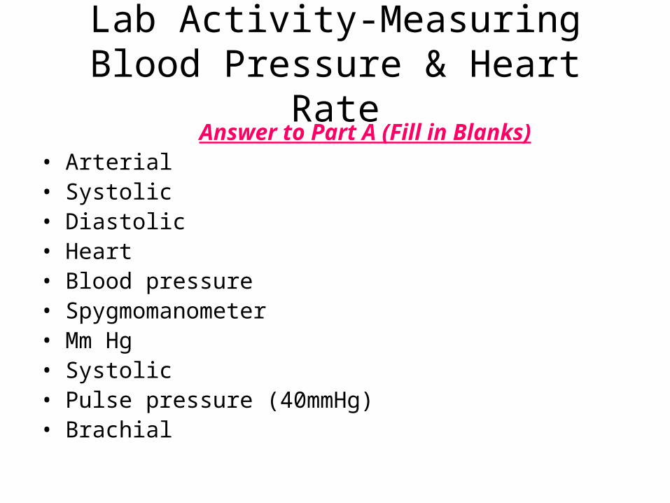
Lab Activity-Measuring Blood Pressure & Heart Rate
Answer to Part A (Fill in Blanks)• Arterial• Systolic• Diastolic• Heart• Blood pressure• Spygmomanometer• Mm Hg• Systolic• Pulse pressure (40mmHg)• Brachial
Today’s Activities
• Start IQ #3 ****stamp Hmwk: Pg. 974 #1-4
• Discuss the homework
• Discuss Lab Questions from yesterday
• Lect/Disc: Respiratory system– Answer IQ #2 & IQ #3
**Begin Video #1: The Respiratory System
Introductory Questions #3• Name the two types of muscles that allow
you to breath in and out. (pg. 973)
• When a person inhales, what happens to the diaphragm? What about when you exhale?
• When the space in the chest cavity (volume) increases which way does the air move? The textbook states that it creates a slight ____________. (see pg. 974)

Introductory Questions #2• Reading pg. 971 (Ch. 37), place these
terms in the correct order that tracks air as you breath:-Pharynx -nose & mouth -bronchi
-alveoli -bronchioles -trachea
• How does your body protect itself from the millions of particles in the air as you breath? (see pg. 972)
• What are the tiny alveoli sacs surrounded by?
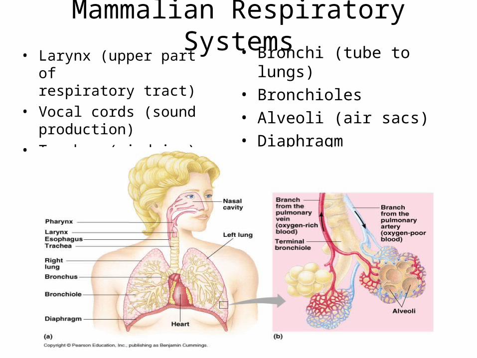
Mammalian Respiratory Systems• Larynx (upper part of
respiratory tract)
• Vocal cords (sound production)
• Trachea (windpipe)
• Bronchi (tube to lungs)• Bronchioles • Alveoli (air sacs)• Diaphragm (breathing
muscle)
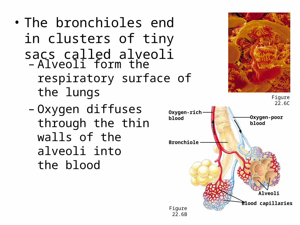
• The bronchioles end in clusters of tiny sacs called alveoli– Alveoli form the respiratory
surface of the lungs– Oxygen diffuses
through the thin walls of the alveoli into the blood
Figure 22.6C
Figure 22.6B
Oxygen-richblood Oxygen-poor
blood
Alveoli
Blood capillaries
Bronchiole
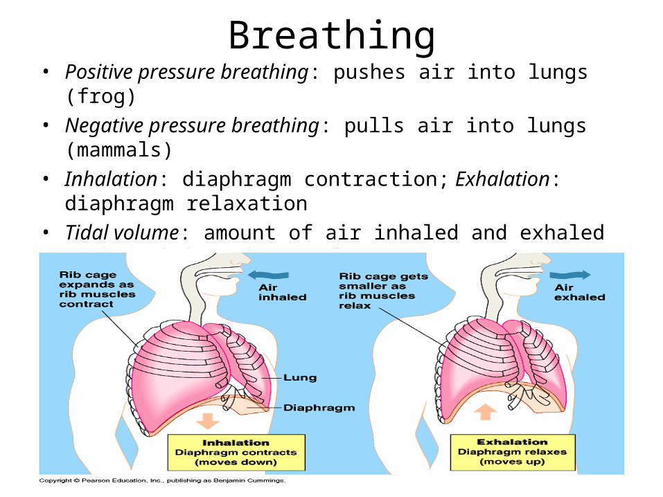
Breathing• Positive pressure breathing: pushes air into lungs (frog)
• Negative pressure breathing: pulls air into lungs (mammals)
• Inhalation: diaphragm contraction; Exhalation: diaphragm relaxation
• Tidal volume: amount of air inhaled and exhaled with each breath (500ml)
• Vital capacity: maximum tidal volume during forced breathing Regulation: CO2 concentration in blood (medulla oblongata)
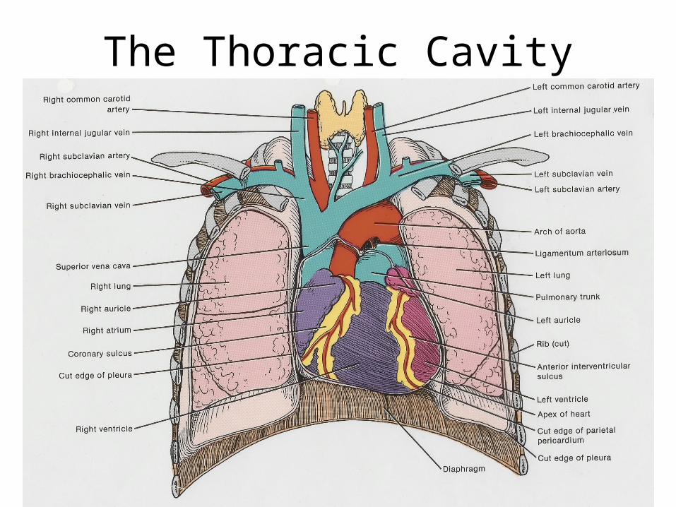
The Thoracic Cavity
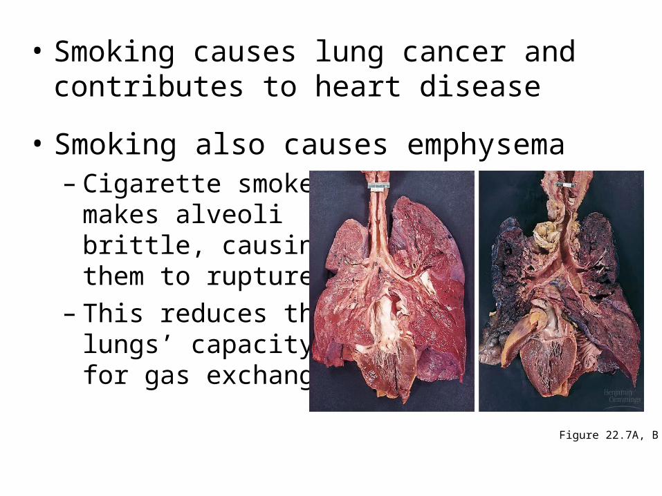
• Smoking causes lung cancer and contributes to heart disease
• Smoking also causes emphysema– Cigarette smoke
makes alveoli brittle, causing them to rupture
– This reduces thelungs’ capacity for gas exchange
Figure 22.7A, B
Video: Gas Exchange
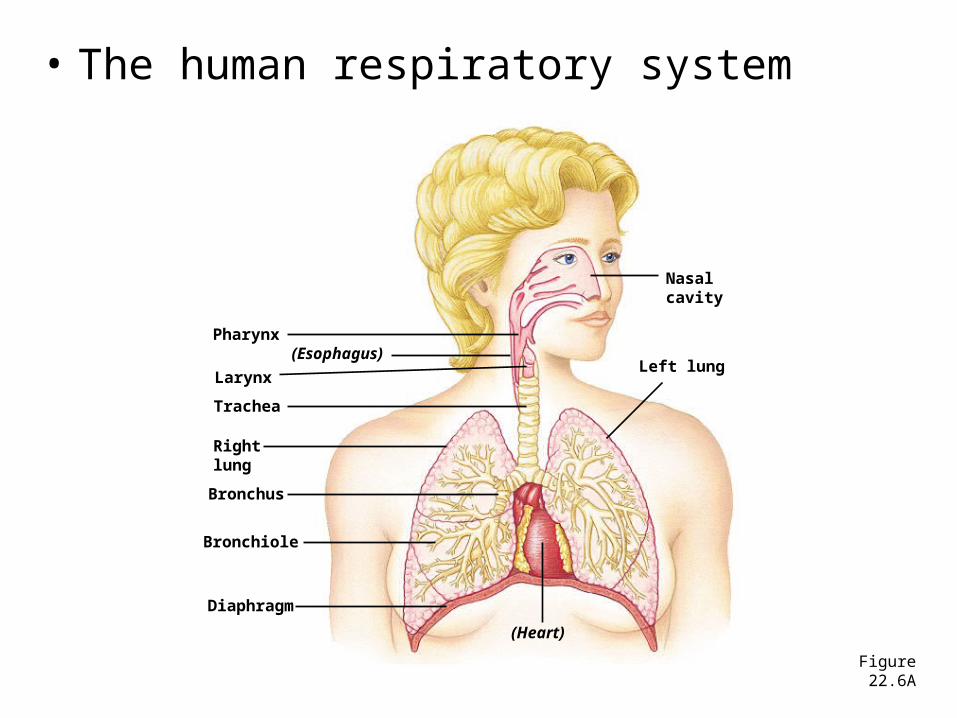
• The human respiratory system
Figure 22.6A
Nasalcavity
Left lung
Pharynx(Esophagus)
Larynx
Trachea
Bronchus
Bronchiole
Diaphragm
(Heart)
Rightlung
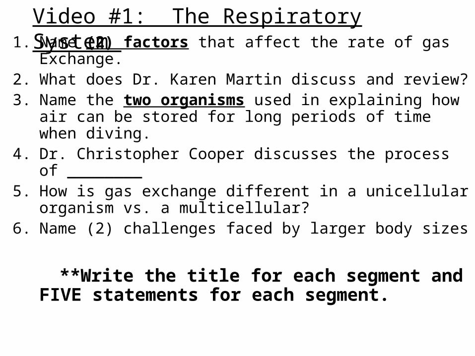
1. Name (2) factors that affect the rate of gas Exchange.2. What does Dr. Karen Martin discuss and review?3. Name the two organisms used in explaining how air
can be stored for long periods of time when diving. 4. Dr. Christopher Cooper discusses the process of
________5. How is gas exchange different in a unicellular
organism vs. a multicellular?6. Name (2) challenges faced by larger body sizes
**Write the title for each segment and FIVE statements for each segment.
Video #1: The Respiratory System
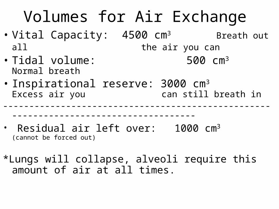
Volumes for Air Exchange • Vital Capacity: 4500 cm3 Breath out all
the air you can
• Tidal volume: 500 cm3 Normal breath
• Inspirational reserve: 3000 cm3 Excess air you can still breath in
--------------------------------------------------------------------------------------• Residual air left over: 1000 cm3 (cannot be forced out)
*Lungs will collapse, alveoli require this amount of air at all times.
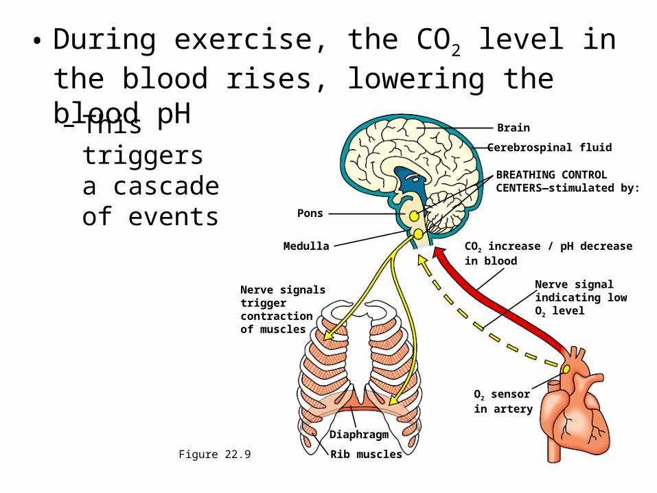
• During exercise, the CO2 level in the blood rises, lowering the blood pH
– This triggers a cascade of events
Figure 22.9
Brain
Cerebrospinal fluid
BREATHING CONTROLCENTERS—stimulated by:
CO2 increase / pH decreasein blood
Nerve signalindicating lowO2 level
O2 sensorin artery
Pons
Medulla
Nerve signalstriggercontractionof muscles
Diaphragm
Rib muscles
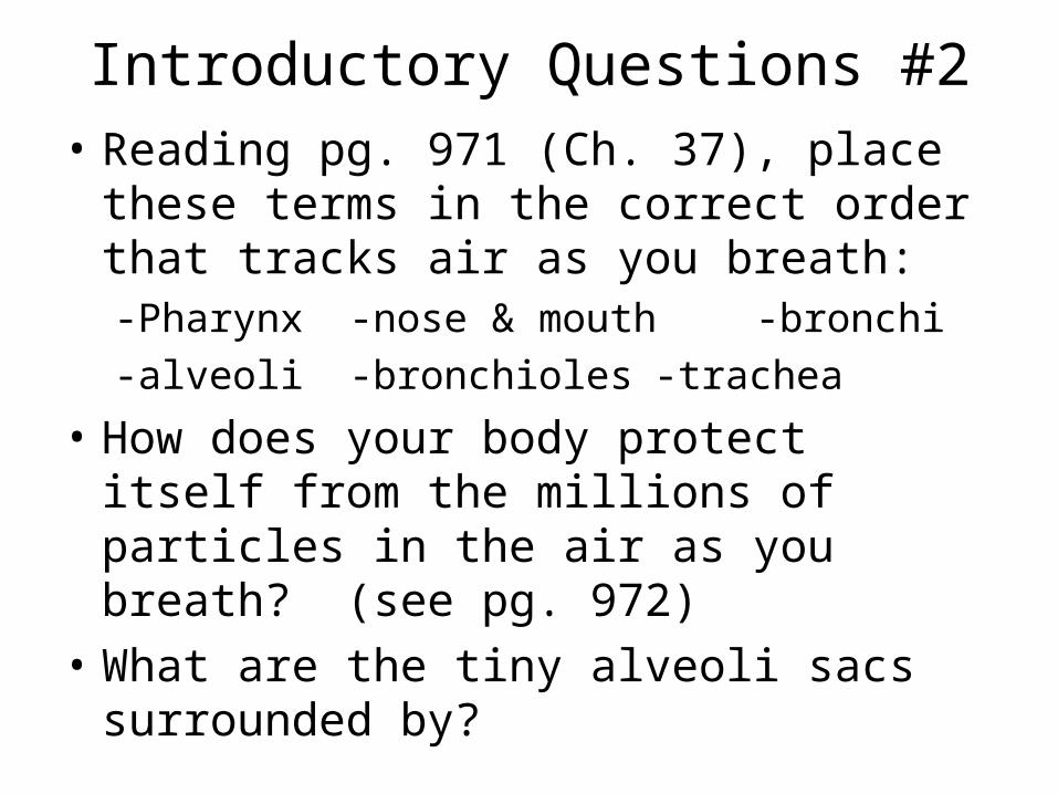
Introductory Questions #2• Reading pg. 971 (Ch. 37), place these
terms in the correct order that tracks air as you breath:-Pharynx -nose & mouth -bronchi
-alveoli -bronchioles -trachea
• How does your body protect itself from the millions of particles in the air as you breath? (see pg. 972)
• What are the tiny alveoli sacs surrounded by?

Introductory Questions #3• Name the two types of muscles that allow
you to breath in and out. (pg. 973)
• When a person inhales, what happens to the diaphragm? What about when you exhale?
• When the space in the chest cavity (volume) increases which way does the air move? The textbook states that it creates a slight ____________. (see pg. 974)
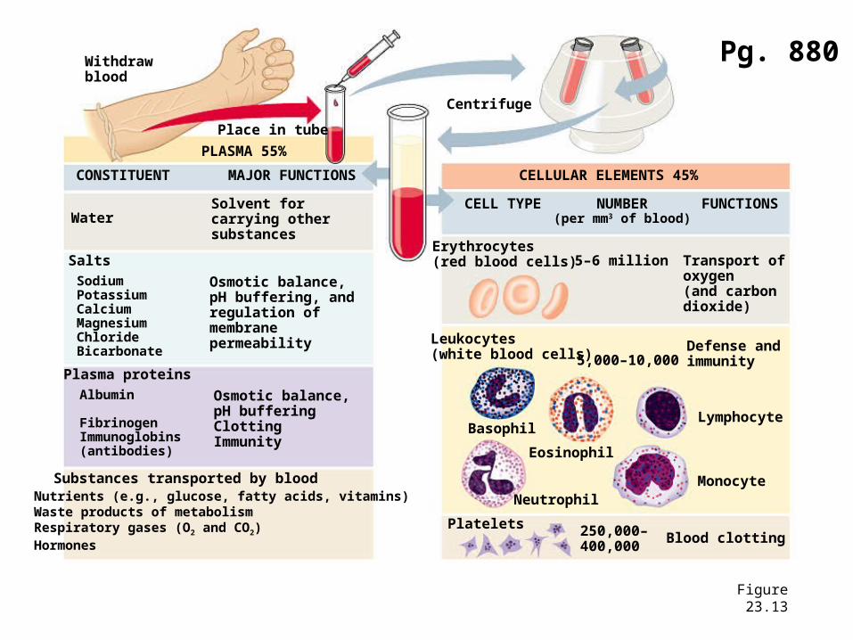
Figure 23.13
Withdrawblood
Place in tube
PLASMA 55%
CONSTITUENT MAJOR FUNCTIONS
WaterSolvent forcarrying othersubstances
Salts
Osmotic balance,pH buffering, andregulation ofmembranepermeability
SodiumPotassiumCalciumMagnesiumChlorideBicarbonate
Plasma proteins
Osmotic balance,pH bufferingClottingImmunity
Albumin
FibrinogenImmunoglobins(antibodies)
Substances transported by bloodNutrients (e.g., glucose, fatty acids, vitamins)Waste products of metabolismRespiratory gases (O2 and CO2)Hormones
Centrifuge
CELLULAR ELEMENTS 45%
CELL TYPE NUMBER(per mm3 of blood)
FUNCTIONS
Erythrocytes(red blood cells) 5–6 million Transport of
oxygen (and carbon dioxide)
Leukocytes(white blood cells) 5,000–10,000
Defense andimmunity
Basophil
Eosinophil
Neutrophil
Lymphocyte
Monocyte
Platelets 250,000–400,000
Blood clotting
Pg. 880
Introductory Questions #4• Name the flap of cartilage that covers the
entrance to the trachea when you swallow food. (see pg. 971)
• Where does the actual exchange (diffusion) of O2 and CO2 gases occur? Why does it occur at this place?
• Which part of the brain controls the rate and depth of breathing by sending impulses (signals) to the diaphragm and rib muscles?
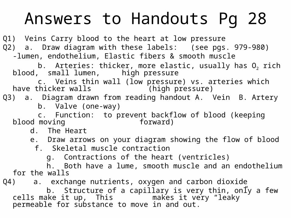
Answers to Handouts Pg 28Q1) Veins Carry blood to the heart at low pressureQ2) a. Draw diagram with these labels: (see pgs. 979-980)
-lumen, endothelium, Elastic fibers & smooth muscle b. Arteries: thicker, more elastic, usually has O2 rich blood, small lumen,
high pressure c. Veins thin wall (low pressure) vs. arteries which have thicker walls
(high pressure)Q3) a. Diagram drawn from reading handout A. Vein B. Artery b. Valve (one-way) c. Function: to prevent backflow of blood (keeping blood moving
forward) d. The Heart e. Draw arrows on your diagram showing the flow of blood f. Skeletal muscle contraction
g. Contractions of the heart (ventricles) h. Both have a lume, smooth muscle and an endothelium for the wallsQ4) a. exchange nutrients, oxygen and carbon dioxide b. Structure of a capillary is very thin, only a few cells make it up, This
makes it very “leaky” permeable for substance to move in and out.
Answers to Handouts Pg 29
Q1)
Q2)
Q3)
Q4)