Embed Size (px)
Citation preview
PREGNANCY
Edyta Mądry MD, PhD
Department of Physiology
Medical University
Poznań
1
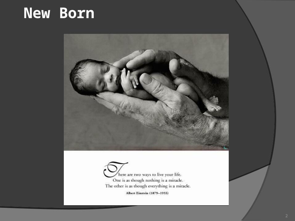
New Born
2

Edyta Mądrycantact data: [email protected]
Please note! The information marked with a red star are the most important in the process of preparing up for the final exam.
3
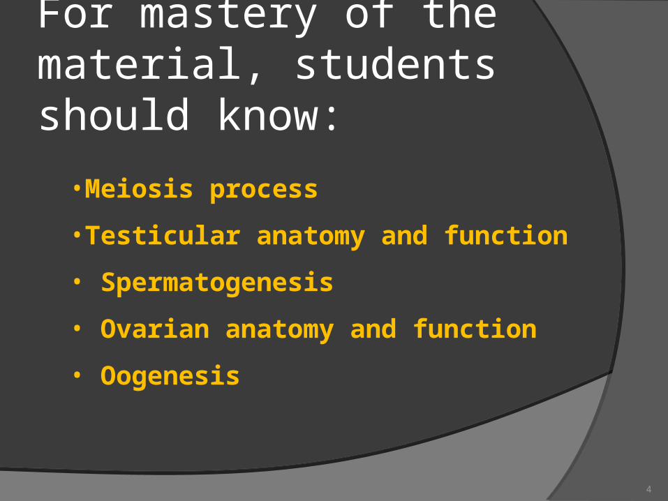
For mastery of the material, students should know:
•Meiosis process
•Testicular anatomy and function
• Spermatogenesis
• Ovarian anatomy and function
• Oogenesis
4
Pregnancy DefinitionThe period from conception to birth.
5
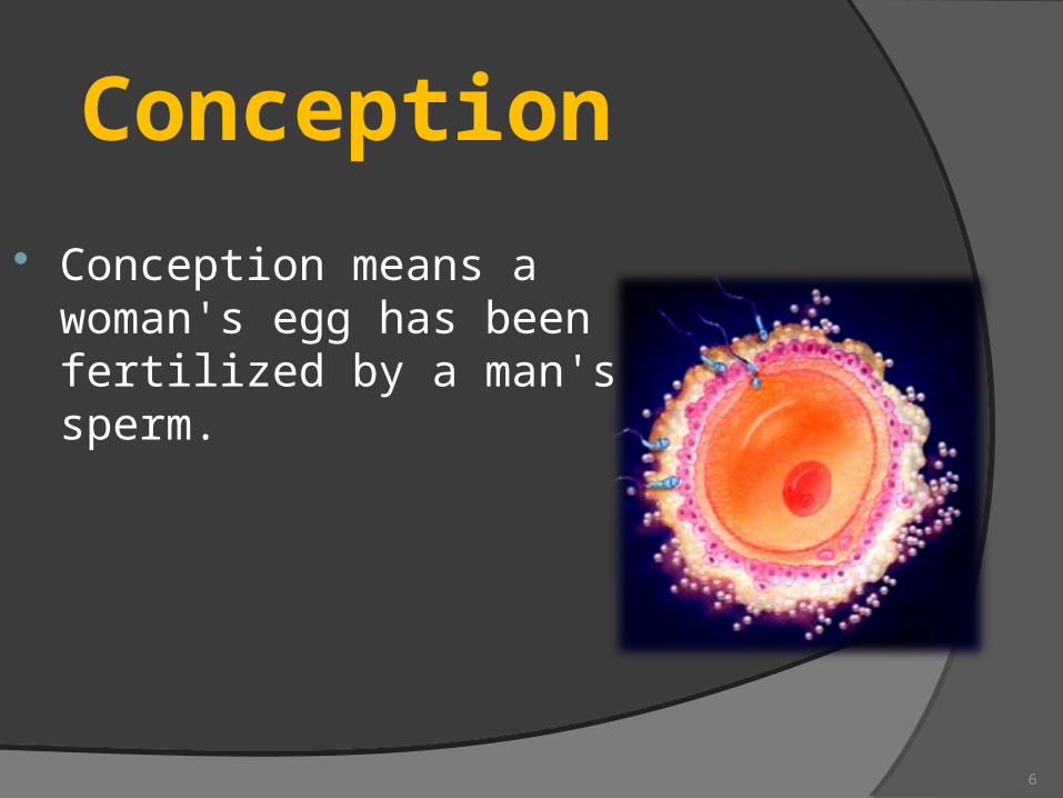
Conception
Conception means a woman's egg has been fertilized by a man's sperm.
6
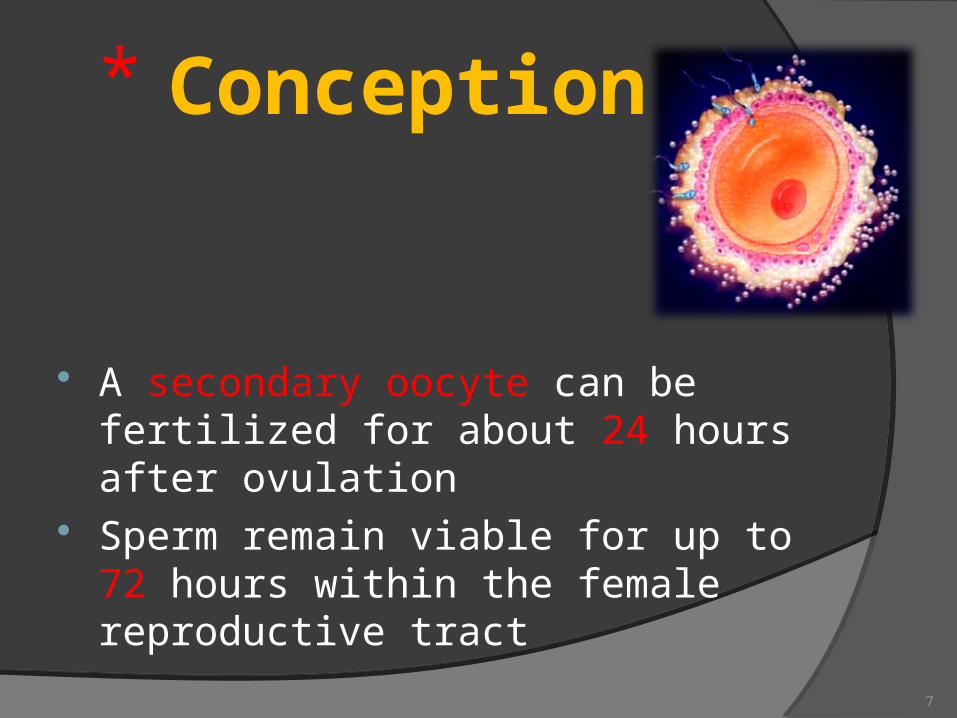
Conception
A secondary oocyte can be fertilized for about 24 hours after ovulation
Sperm remain viable for up to 72 hours within the female reproductive tract
7
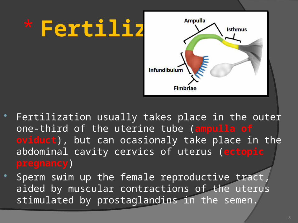
Fertilization
Fertilization usually takes place in the outer one-third of the uterine tube (ampulla of oviduct), but can ocasionaly take place in the abdominal cavity cervics of uterus (ectopic pregnancy)
Sperm swim up the female reproductive tract, aided by muscular contractions of the uterus stimulated by prostaglandins in the semen.
8
9
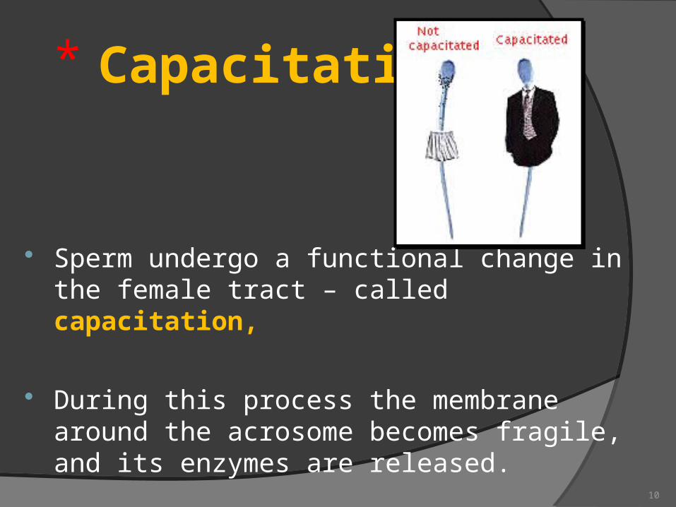
Capacitation
Sperm undergo a functional change in the female tract – called capacitation,
During this process the membrane around the acrosome becomes fragile, and its enzymes are released.
10
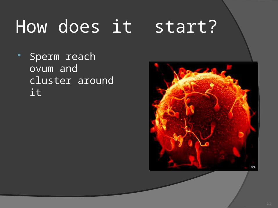
How does it start? Sperm reach ovum
and cluster around it
11
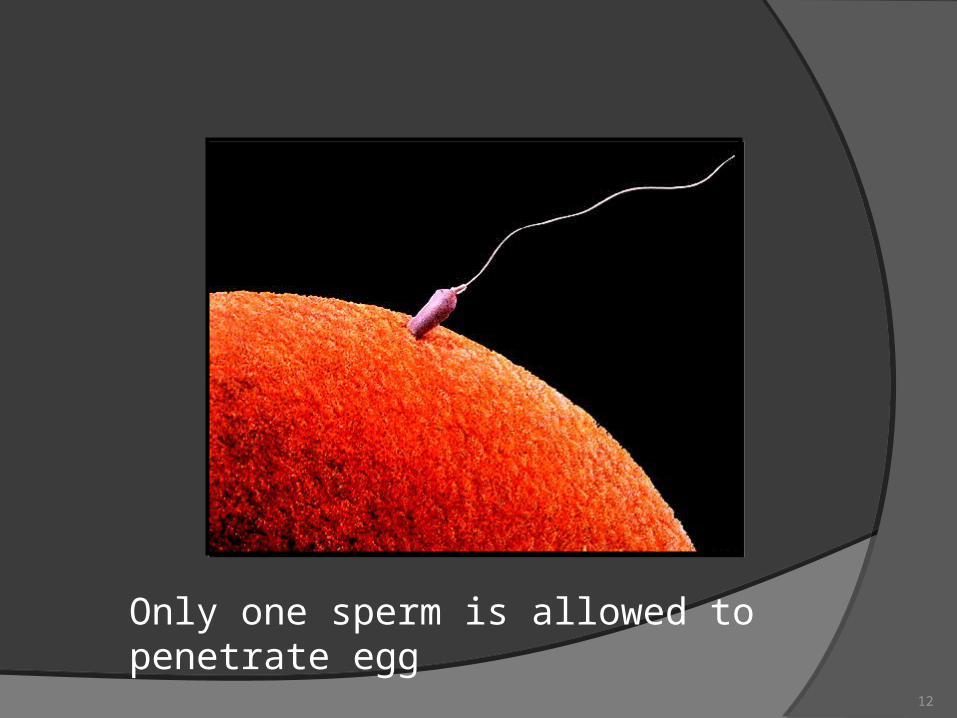
Only one sperm is allowed to penetrate egg
12
Gestation
Assumption:
Fertilized 14 days after the first day of LNMP
Gives birth 38 weeks after fertilization (+ or - 2 weeks)
Gestation in singleton pregnancies lasts an average of 40 weeks (280 days) from the first day of the last normalmenstrual period (LNMP).
10 lunar months = 9 calendar months = 280 days
(+ or – 2 weeks)
13
How to calculate the time of birthNaegele's rule 1. Determine first day of LNMP
2. Add one year3. Subtract three months4. Add seven days
For example: first day of LNMP April 21st, 2015
Resulting data of estimated data of birth : January 28 , 2016(+ or – 2 weeks )
14
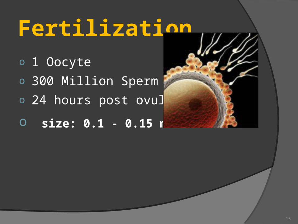
Fertilization
o 1 Oocyteo 300 Million Spermo 24 hours post ovulation
o size: 0.1 - 0.15 mm
15
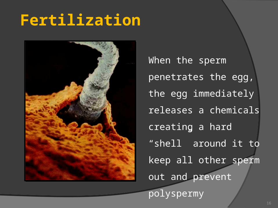
When the sperm
penetrates the egg,
the egg immediately
releases a chemicals
creating a hard “shell”
around it to keep all
other sperm out and
prevent polyspermy
Fertilization
16
Fertilization
FERTILIZED EGG = ZYGOTE The fertilization process takes about 24
hours.It takes about ten hours to navigate the
female productive track, moving up the vaginal canal, through the cervix, and into the fallopian tube where fertilization begins.
17
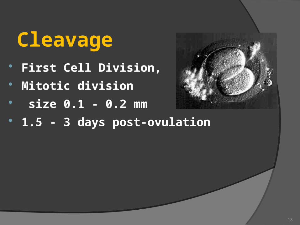
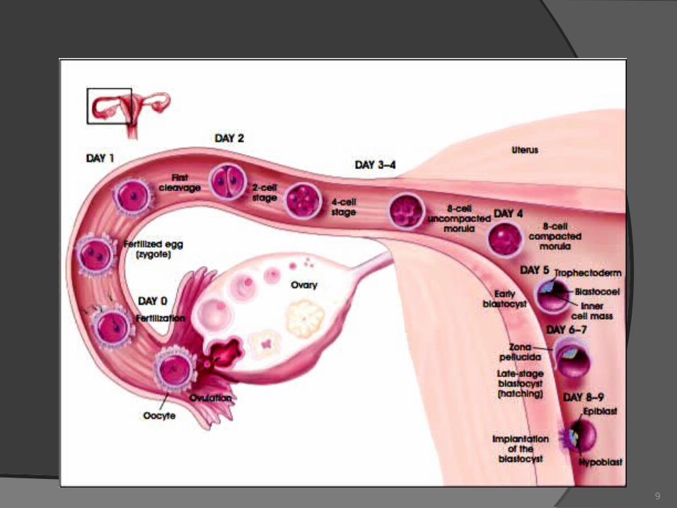
Cleavage First Cell Division, Mitotic division size 0.1 - 0.2 mm 1.5 - 3 days post-ovulation
18
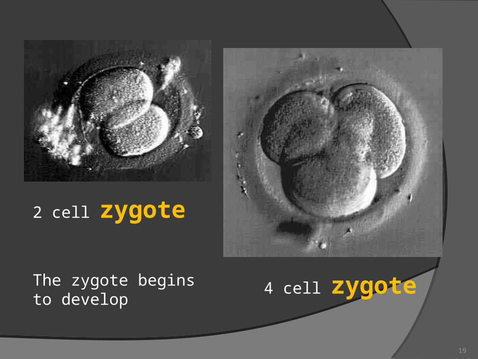
4 cell zygote
2 cell zygote
The zygote begins to develop
19
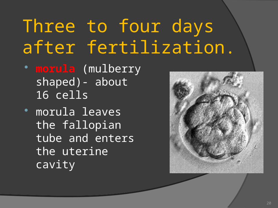
Three to four days after fertilization. morula (mulberry
shaped)- about 16 cells
morula leaves the fallopian tube and enters the uterine cavity
20
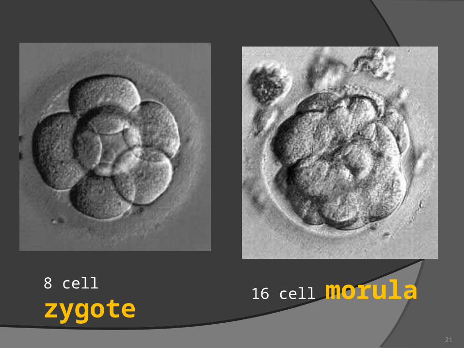
16 cell morula 8 cell zygote
21
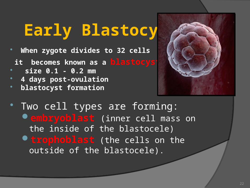
Early Blastocyst When zygote divides to 32 cells
it becomes known as a blastocyst size 0.1 - 0.2 mm 4 days post-ovulation blastocyst formation
Two cell types are forming:embryoblast (inner cell mass on the inside of
the blastocele)trophoblast (the cells on the outside of the
blastocele).
22
Implantation Begins Human Chorionic Gonadotropin
(hCG) level rise 0.1 - 0.2 mm
5 - 6 days post-ovulation The trophoblast cells secretes an enzyme which erodes the
epithelial uterine lining and creates an implantation site for the blastocyst (see slide number 22)
23
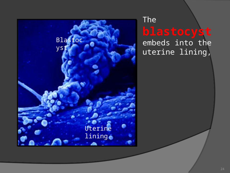
Blastocyst
Uterine lining
The blastocyst embeds into the uterine lining,
24
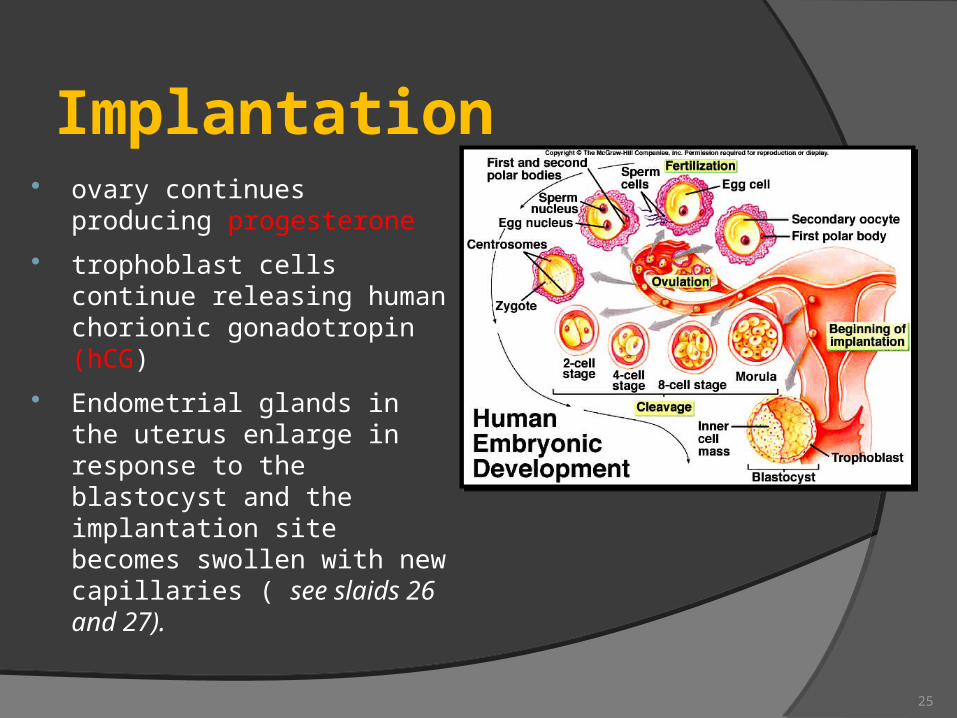
Implantation ovary continues producing
progesterone
trophoblast cells continue releasing human chorionic gonadotropin (hCG)
Endometrial glands in the uterus enlarge in response to the blastocyst and the implantation site becomes swollen with new capillaries ( see slaids 26 and 27).
25
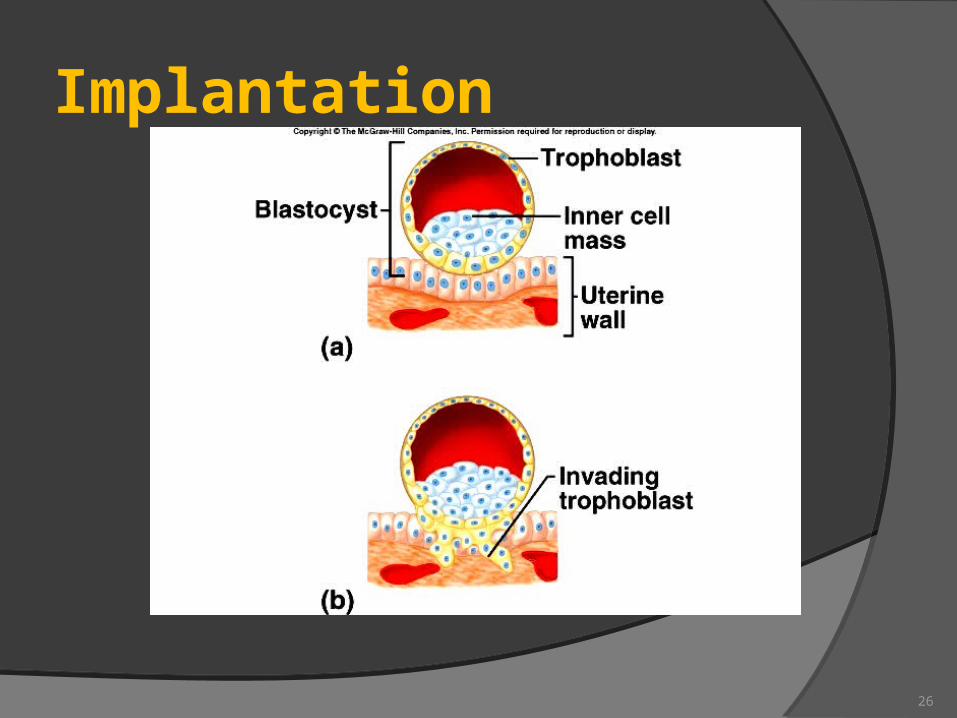
Implantation
26
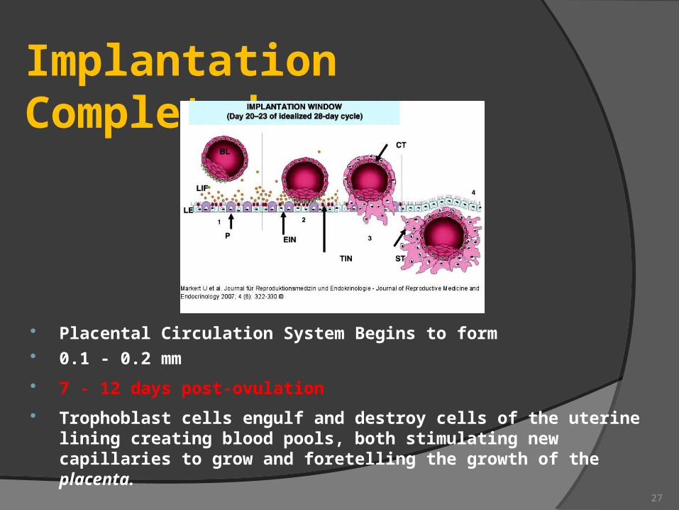
Implantation Completed
Placental Circulation System Begins to form 0.1 - 0.2 mm
7 - 12 days post-ovulation
Trophoblast cells engulf and destroy cells of the uterine lining creating blood pools, both stimulating new capillaries to grow and foretelling the growth of the placenta.
27
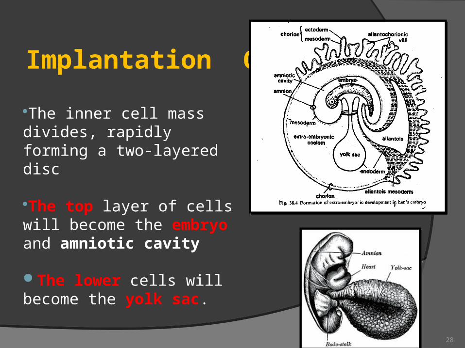
Implantation Completed
The inner cell mass divides, rapidly forming a two-layered disc
The top layer of cells will become the embryo and amniotic cavity
The lower cells will become the yolk sac.
28
Inner cell mass forms two cavities:
yolk sacamniotic cavity
In humans the yolk sac produces blood cells and future sex cells
The amniotic cavity becomes the cavity in which the embryo floats.
Fluid is produced from fetal urine, and secretions from the skin, respiratory tract, and amniotic membranes.
29
30
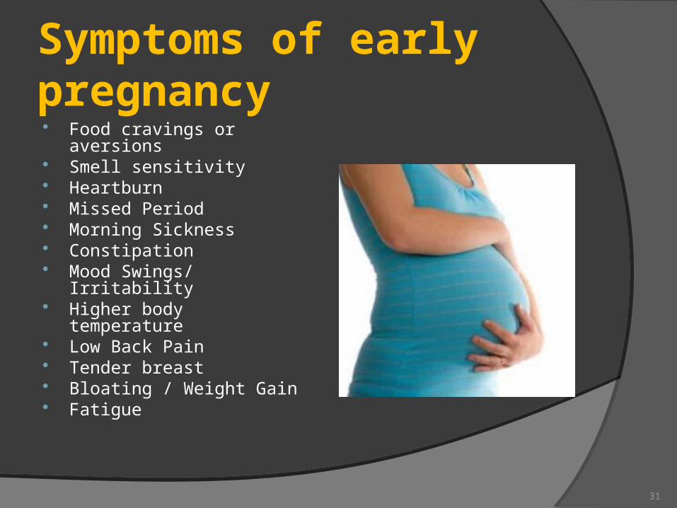
Symptoms of early pregnancy Food cravings or
aversions Smell sensitivity Heartburn Missed Period Morning Sickness Constipation Mood Swings/ Irritability Higher body temperature Low Back Pain Tender breast Bloating / Weight Gain Fatigue
31
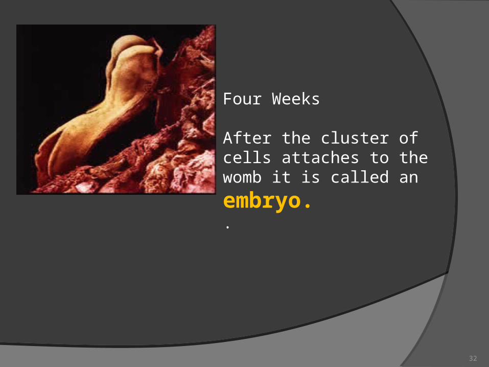
Four Weeks
After the cluster of cells attaches to
the womb it is called an embryo. .
32
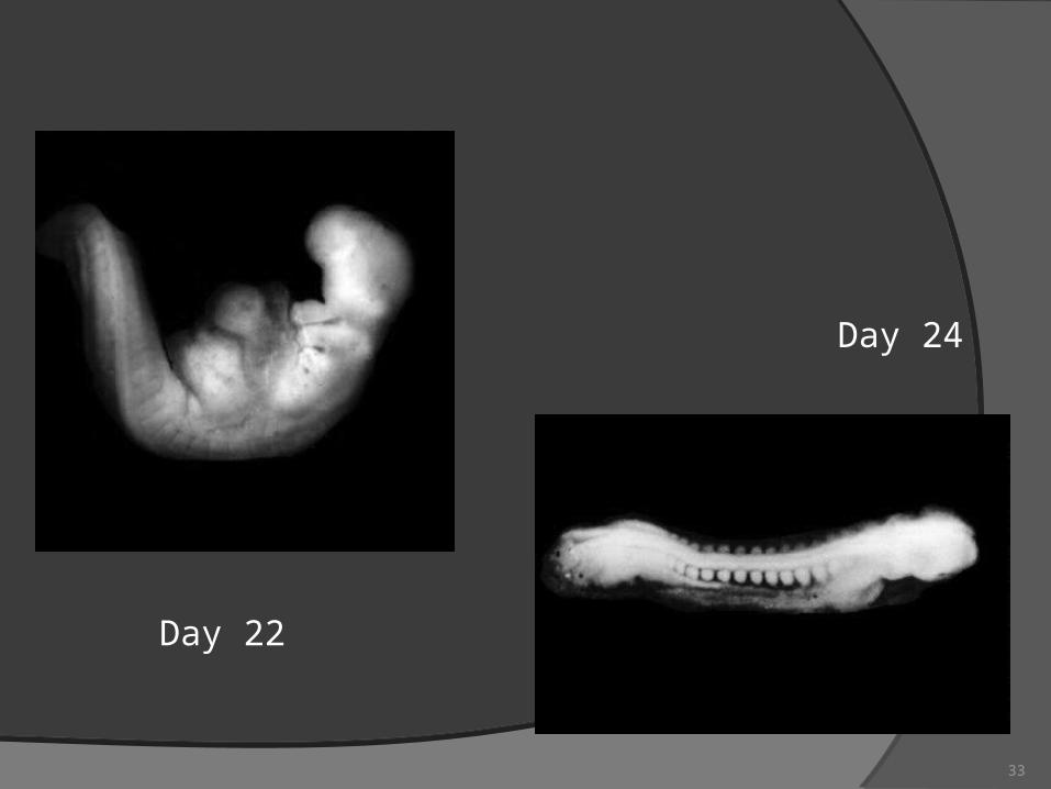
Day 22
Day 24
33
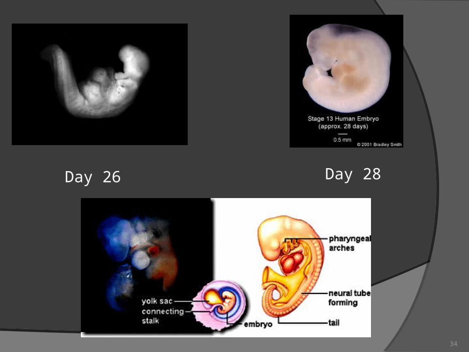
Day 26 Day 28
34
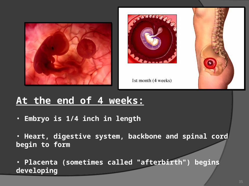
At the end of 4 weeks:
• Embryo is 1/4 inch in length
• Heart, digestive system, backbone and spinal cord begin to form
• Placenta (sometimes called "afterbirth") begins developing
35
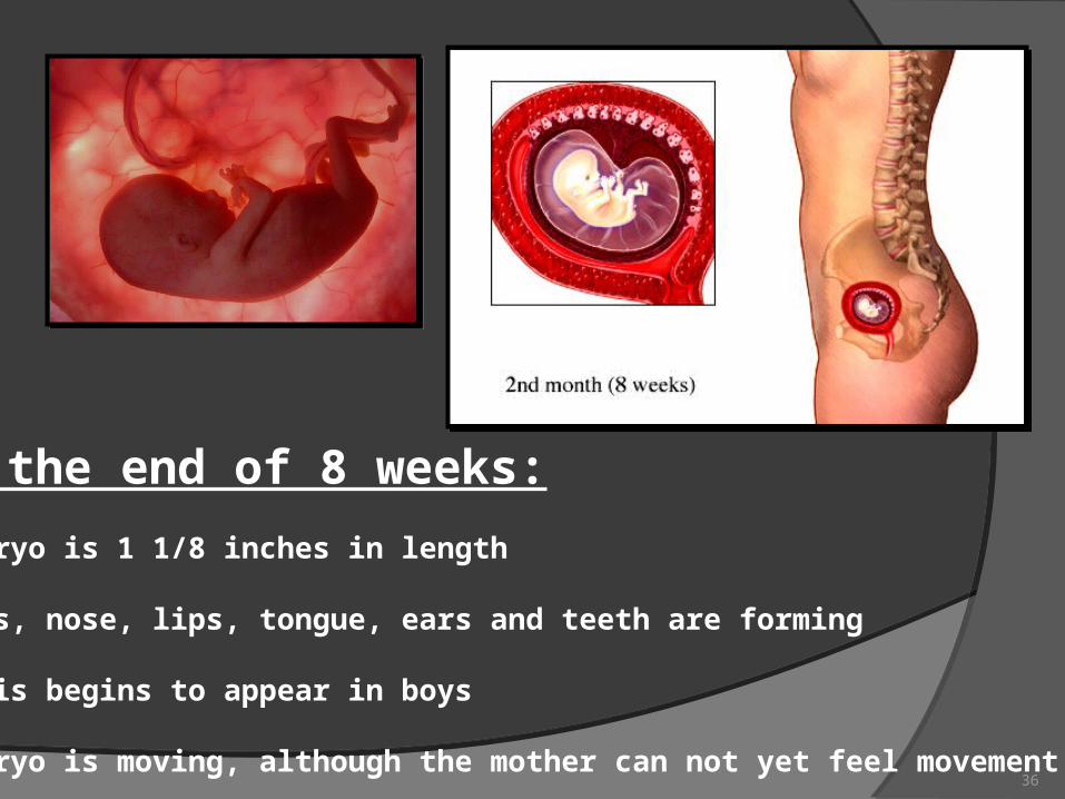
At the end of 8 weeks:
•Embryo is 1 1/8 inches in length
•Eyes, nose, lips, tongue, ears and teeth are forming
•Penis begins to appear in boys
•Embryo is moving, although the mother can not yet feel movement36
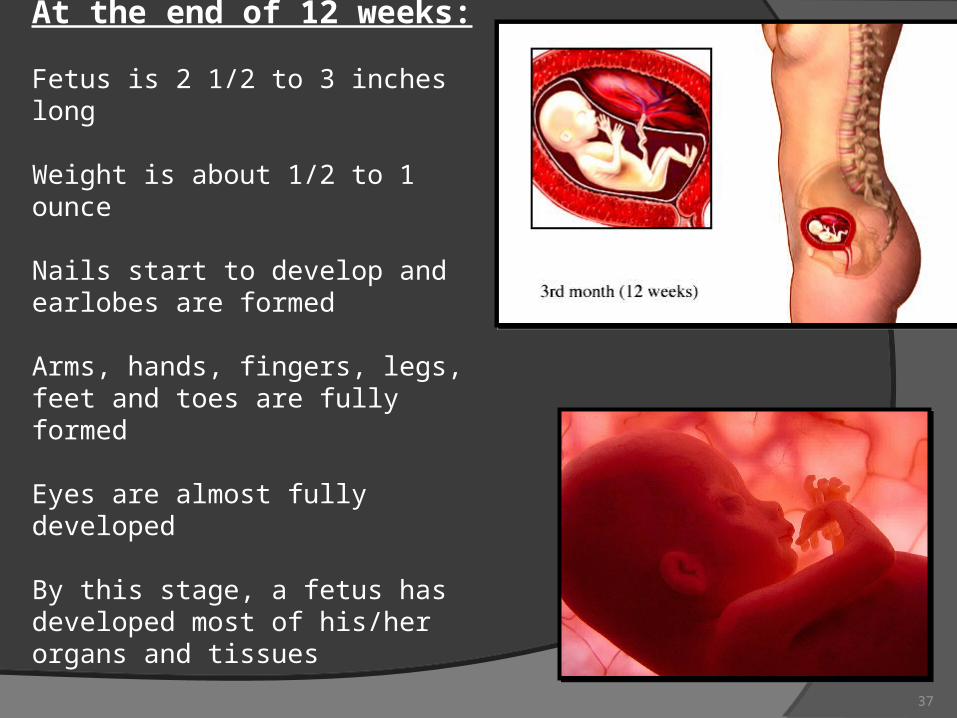
At the end of 12 weeks:
Fetus is 2 1/2 to 3 inches long
Weight is about 1/2 to 1 ounce
Nails start to develop and earlobes are formed
Arms, hands, fingers, legs, feet and toes are fully formed
Eyes are almost fully developed
By this stage, a fetus has developed most of his/her organs and tissues
37
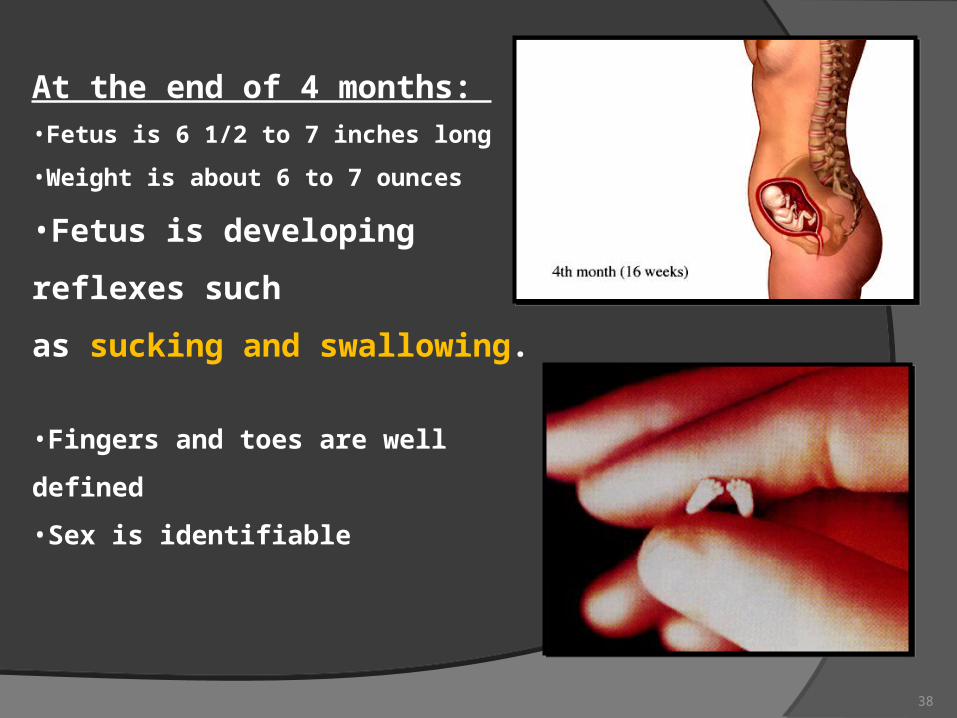
At the end of 4 months: •Fetus is 6 1/2 to 7 inches long
•Weight is about 6 to 7 ounces
•Fetus is developing reflexes such
as sucking and swallowing.
•Fingers and toes are well defined
•Sex is identifiable
38
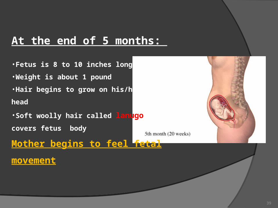
At the end of 5 months:
•Fetus is 8 to 10 inches long
•Weight is about 1 pound
•Hair begins to grow on his/her
head
•Soft woolly hair called lanugo
covers fetus body
Mother begins to feel fetal movement
39
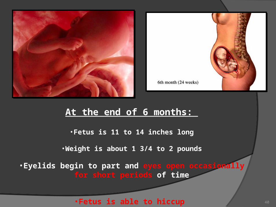
At the end of 6 months:
•Fetus is 11 to 14 inches long
•Weight is about 1 3/4 to 2 pounds
•Eyelids begin to part and eyes open occasionally for short periods of time
•Fetus is able to hiccup 40
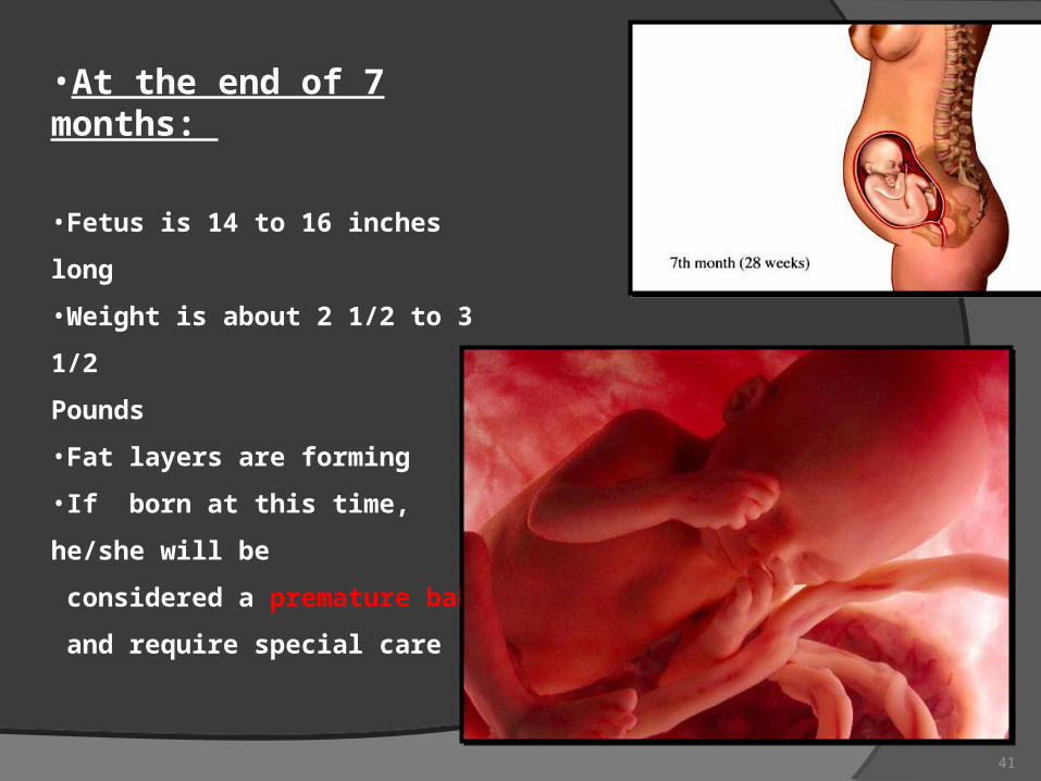
•At the end of 7 months:
•Fetus is 14 to 16 inches long
•Weight is about 2 1/2 to 3 1/2
Pounds
•Fat layers are forming
•If born at this time, he/she will be
considered a premature baby
and require special care
41
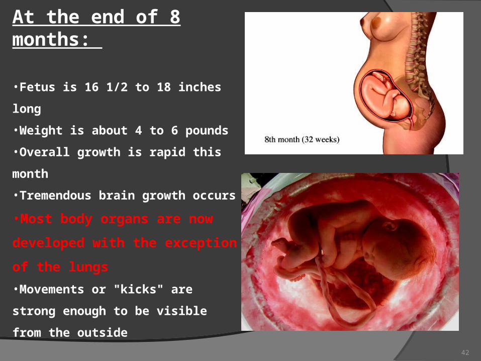
At the end of 8 months:
•Fetus is 16 1/2 to 18 inches long
•Weight is about 4 to 6 pounds
•Overall growth is rapid this month
•Tremendous brain growth occurs
•Most body organs are now developed
with the exception of the lungs
•Movements or "kicks" are strong enough
to be visible from the outside
42
•
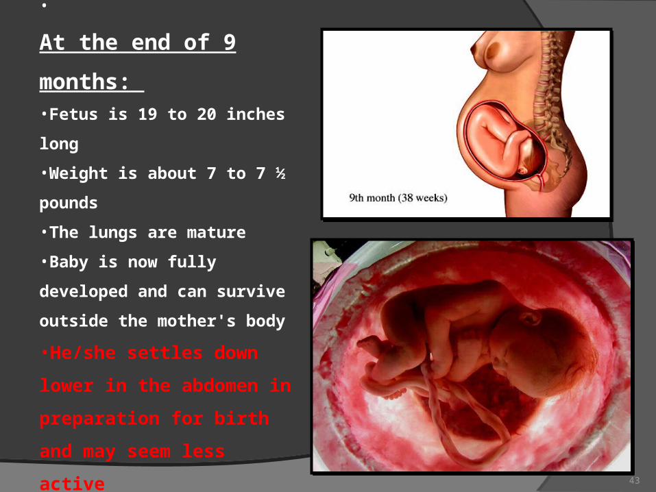
At the end of 9 months: •Fetus is 19 to 20 inches long
•Weight is about 7 to 7 ½
pounds
•The lungs are mature
•Baby is now fully developed and
can survive outside the mother's
body
•He/she settles down lower in the
abdomen in preparation for
birth and may seem less active
43
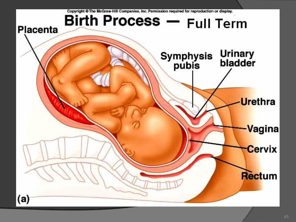
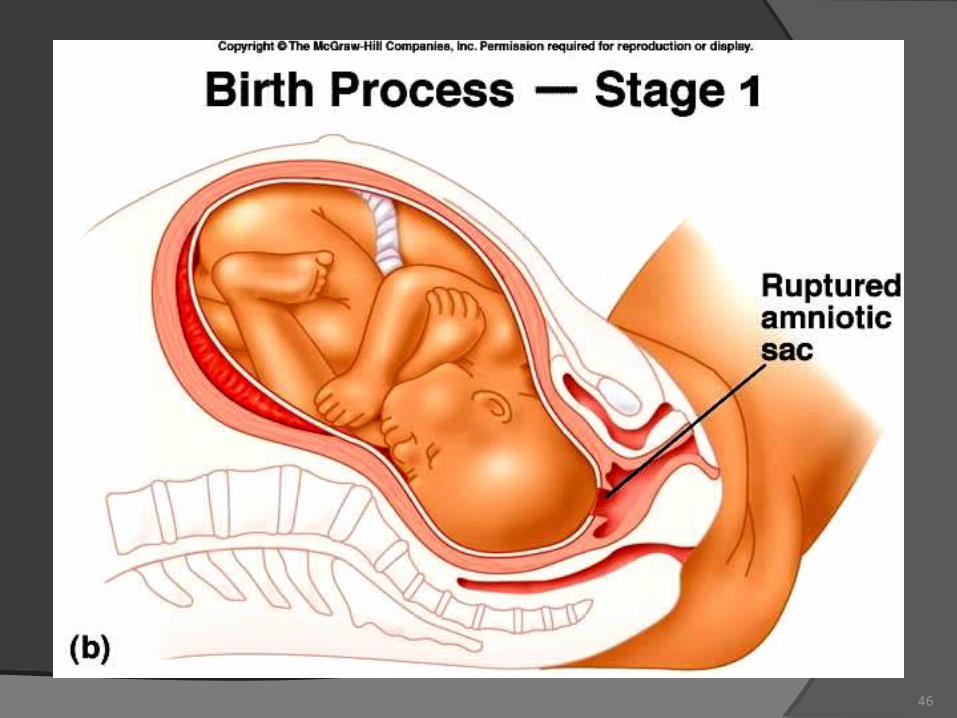
Labor (parturition)- Stage one
the period from the onset of true labor contractions until the cervix is completely dilated at 10 cm.
The uterine contractions cause the cervix to dilate, and the amniotic sac may rupture.
Usually lasts 6 – 24 hours depending on the number of previous deliveries.
44
45
46
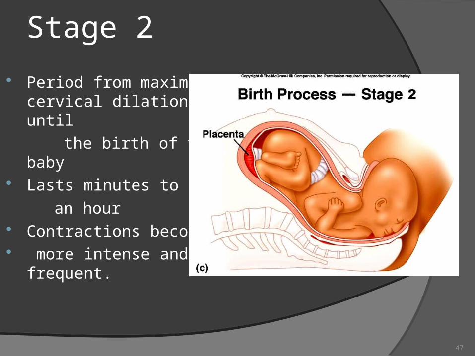
Stage 2
Period from maximal cervical dilation until
the birth of the baby Lasts minutes to
an hour Contractions become more intense and
frequent.
47
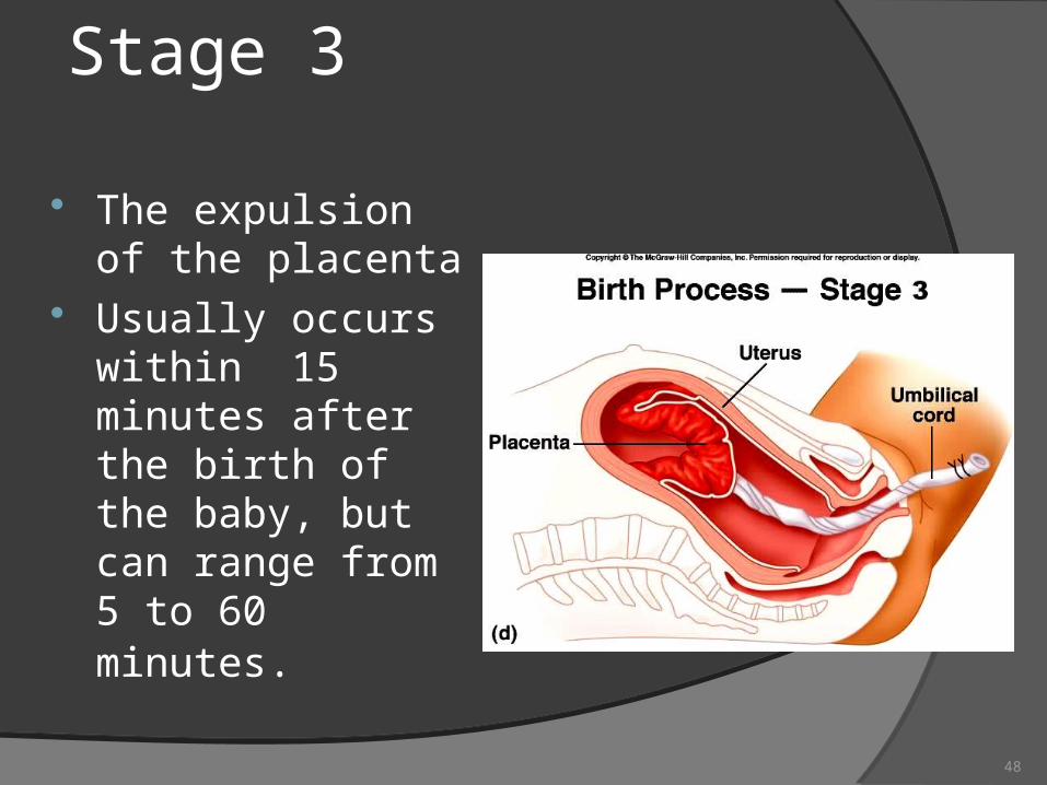
Stage 3
The expulsion of the placenta
Usually occurs within 15 minutes after the birth of the baby, but can range from 5 to 60 minutes.
48
49
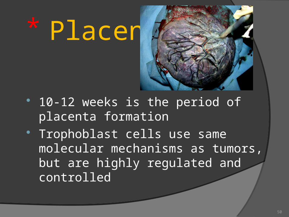
Placenta
10-12 weeks is the period of placenta formation
Trophoblast cells use same molecular mechanisms as tumors, but are highly regulated and controlled
50
Placenta FETAL SIDE MATERNAL SIDE They provide…
protectionnutritionrespirationexcretionhormone production
51
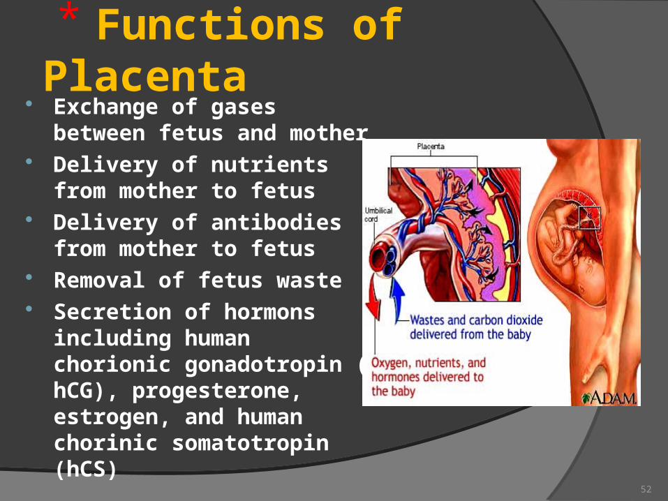
Functions of Placenta Exchange of gases between
fetus and mother Delivery of nutrients from
mother to fetus Delivery of antibodies from
mother to fetus Removal of fetus waste Secretion of hormons
including human chorionic gonadotropin ( hCG), progesterone, estrogen, and human chorinic somatotropin (hCS)
52
Placental barrier Maternal and fetal blood
do not mix- “placental barrier”
53
Metabolic Functions of the Placenta Glycogen synthesis and storage
Cholesterol synthesis: placental cholesterol is precursor for placental progesterone and estrogens
Protein production
54
Endocrine Functions Placenta Produces Peptide hormones
Human Chorionic gonodotrophin (hCG) - secreted early and helps to maintain synthesis of progesterone
Human placental lactogen (hPL): increase supply of glucose to future by decreasing maternal stores of fatty acids by altering maternal secretion of insulin
Insulin-like growth factors (IGF): IGF signaling system is a major regulator of growth in fetus and infant
55
Endocrine Functions Steroid hormones
Progesterone: produced by placenta, needed to maintain non-contractile uterus
Estrogen: produced by placenta drives many processes in pregnancy
56
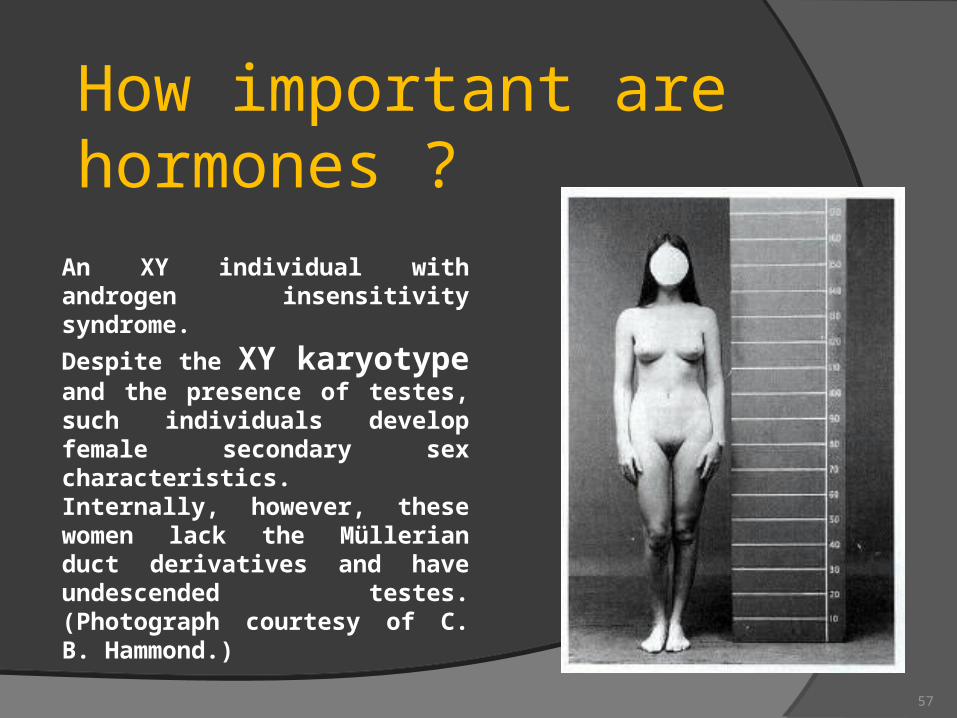
An XY individual with androgen insensitivity syndrome.
Despite the XY karyotype and the presence of testes, such individuals develop female secondary sex characteristics. Internally, however, these women lack the Müllerian duct derivatives and have undescended testes. (Photograph courtesy of C. B. Hammond.)
How important are hormones ?
57
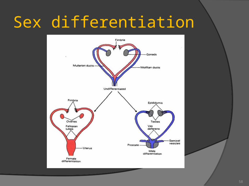
Sex differentiation
58
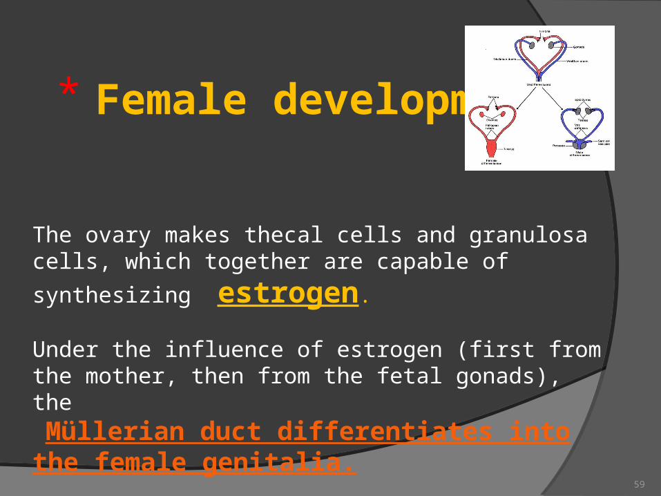
The ovary makes thecal cells and granulosa cells, which
together are capable of synthesizing estrogen.
Under the influence of estrogen (first from the mother, then from the fetal gonads), the Müllerian duct differentiates into the female genitalia.
Female development
59
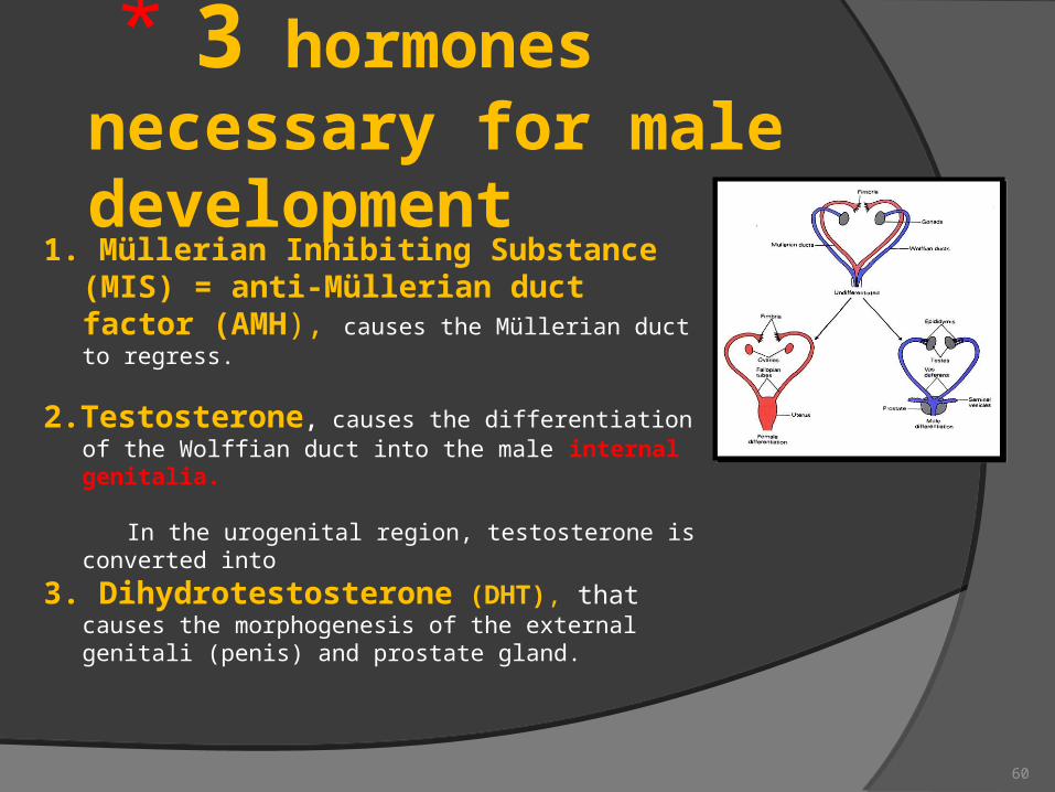
3 hormones necessary for male development
1. Müllerian Inhibiting Substance (MIS) = anti-Müllerian duct factor (AMH), causes the Müllerian duct to regress.
2.Testosterone, causes the differentiation of the Wolffian duct into the male internal genitalia.
In the urogenital region, testosterone is converted into
3. Dihydrotestosterone (DHT), that causes the morphogenesis of the external genitali (penis) and prostate gland.
60
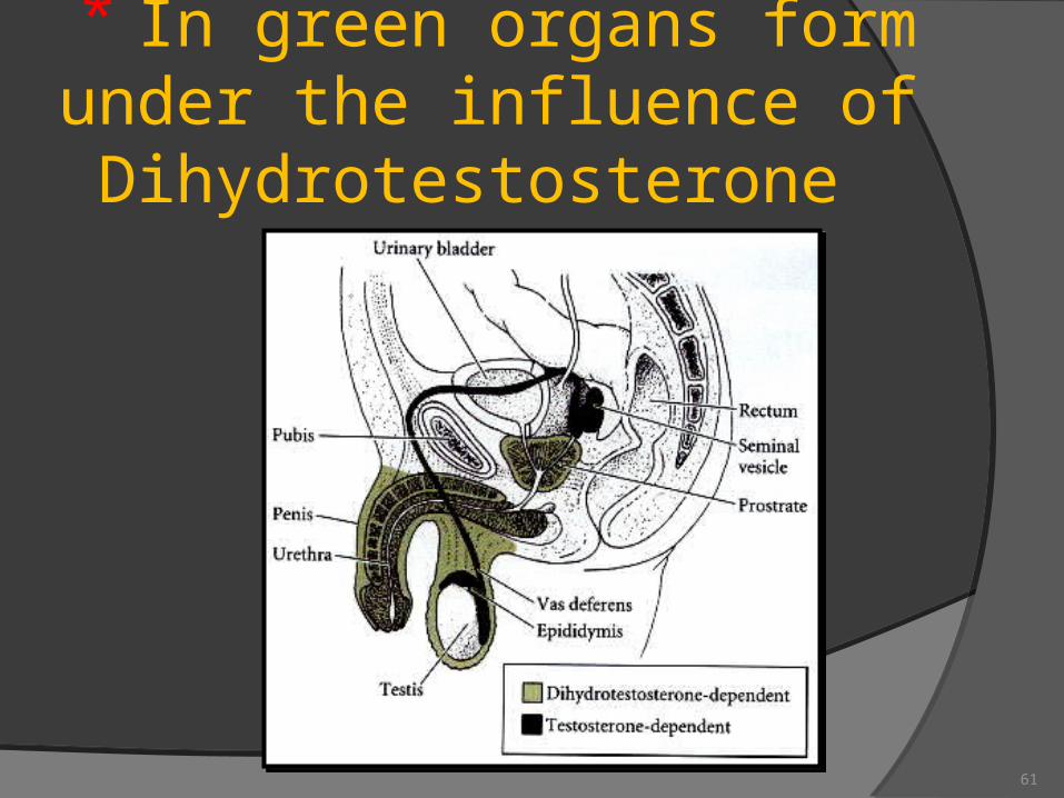
In green organs form under the influence of Dihydrotestosterone
61
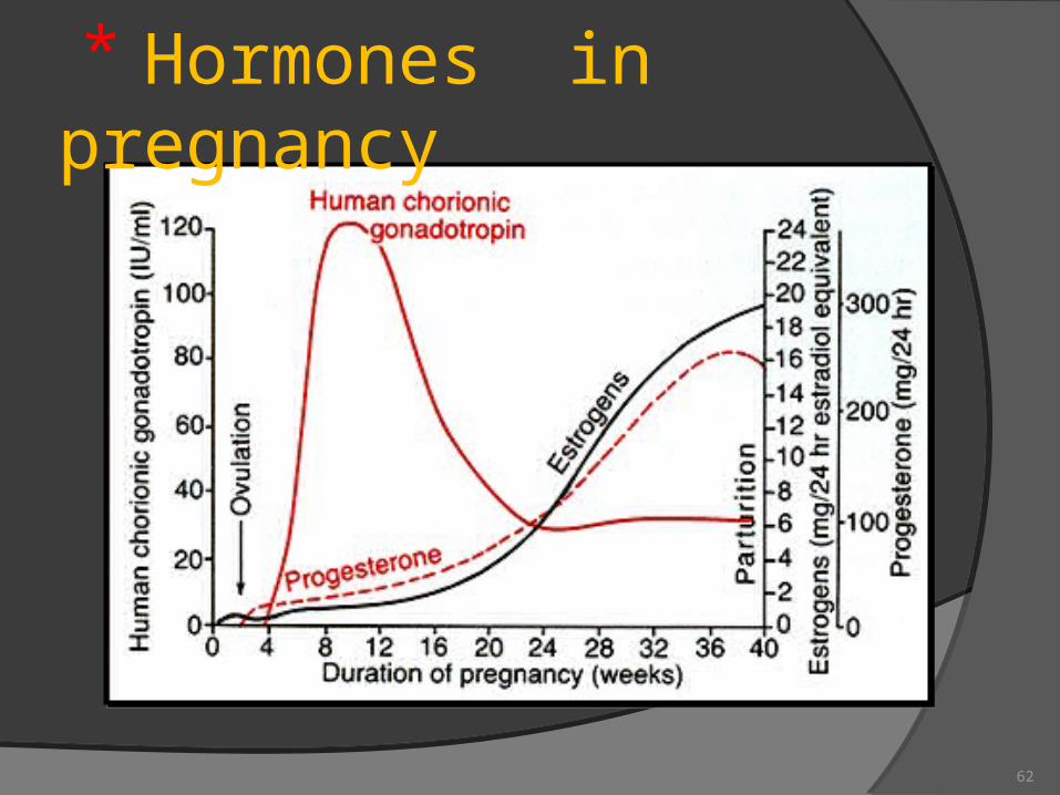
Hormones in pregnancy
62
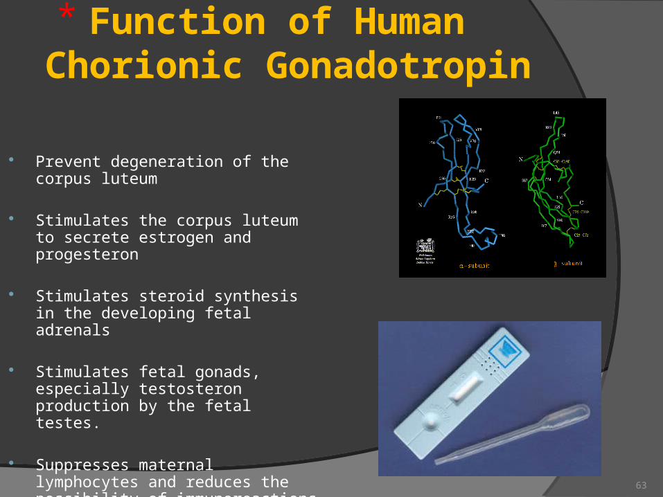
Function of Human Chorionic Gonadotropin
Prevent degeneration of the corpus luteum
Stimulates the corpus luteum to secrete estrogen and progesteron
Stimulates steroid synthesis in the developing fetal adrenals
Stimulates fetal gonads, especially testosteron production by the fetal testes.
Suppresses maternal lymphocytes and reduces the possibility of immunoreactions against the fetus.
63
What does progesterone do?
•It maintains the lining of the uterus which makes it possible for a fertilized egg to attach and survive
•Makes cervical mucous accessible by sperm
•Prevents immune rejection of the developing baby
•Increases libido around ovulation
•Protects against endometrial, breast, ovarian and prostrate cancer
•Normalizes blood clotting
•Incease body temperature64

Edyta Mądrycantact data: [email protected]
Please note! The information marked with a red star are the most important in the process of preparing up for the final exam.
65


![· Web viewPolska Agencja Prasowa [Polish news agency], Gazeta Wyborcza, Głos Wielkopolski Polska The Times, TVN ( Poznań branch), POLSAT (Poznań), TVP (Poznań …](https://img.pdfslide.us/doc/110x75/6133a6dfdfd10f4dd73b3a0e/web-view-polska-agencja-prasowa-polish-news-agency-gazeta-wyborcza-gos-wielkopolski.jpg)