Embed Size (px)
Citation preview
Potential Role of CTCF in Differential
Papillomavirus Gene Expression
A Major Qualifying Project:
Submitted to the Faculty
of the
WORCESTER POLYTECHNIC INSTITUTE
in partial fulfillment of the requirements for the
Degree of Bachelor of Science
By
_______________________ Alyson Talbot
Date: April 30, 2009
Approved
____________________________________ Professor Destin W. Heilman, Advisor
i
Abstract
Upon cellular differentiation the late genes of the Human Papillomavirus (HPV) are
expressed. Recently, the CTCF DNA-binding protein has been found to be transcription
factor and chromatin insulator. We examined if CTCF plays a role in viral transcription
and late gene expression. Through ChIP analysis it was found that CTCF binds to two
distinct regions of the Bovine Papillomavirus-1 genome. It was observed that the E2
protein and CTCF protein interact by co-immunoprecipitation. Using RNA interference,
the effect of CTCF on viral transcription was examined. It was shown that E2 and L1
RNA levels are knocked out with lower levels of CTCF. Further studies should
investigate if the type of interactions between E2 and CTCF, and the mechanism in which
CTCF controls viral transcription.

ii
Acknowledgements
I would like to thank Elliot Androphy, Carol Jolly, Suzanne Melanson, and all the
members of the Androphy Lab (Department of Medicine at UMass Medical) for their
non-stop guidance. Also, I owe a thank you to Professor Heilman who helped me with
the entire MQP process.
iii
TableofContents
Abstract ........................................................................................................................i
Acknowledgements......................................................................................................ii
Tables .........................................................................................................................iv
Figures .........................................................................................................................v
1.0Background ............................................................................................................11.1‐HumanPapillomavirusOverview .................................................................................. 11.2‐HPVViralProteins......................................................................................................... 5
1.2.1‐E6andE7Oncoproteins ............................................................................................... 51.2.2‐E1andE2Protein .......................................................................................................... 71.2.3‐L1andL2CapsidProteins.............................................................................................. 8
1.3‐HPVLateGeneActivation ............................................................................................. 91.3.1‐CohesinComplex .......................................................................................................... 91.3.2‐CohesinandCTCFAssociation.................................................................................... 101.3.3‐E2InteractionswithCohesin....................................................................................... 121.3.4‐CTCFProtein................................................................................................................ 121.3.5‐CTCFStructure............................................................................................................. 131.3.6‐CTCFFunctions ............................................................................................................ 14
2.0Methods...............................................................................................................18
3.0Results .................................................................................................................213.1‐LocationofCTCFbindingsiteswithintheBPVGenome............................................... 213.2‐CTCFBindingtotheBPV‐1DNA ................................................................................... 223.3‐InteractionsofCTCFandE2......................................................................................... 233.4‐CTCFEffectonViralTranscription ............................................................................... 24
4.0Discussion ............................................................................................................32
References .................................................................................................................36
iv
TablesTable 1: Primers used for ChIP Analysis.......................................................................... 19Table 2: Primers used for analysis of CTCF knockdown on viral transcription .............. 20
v
FiguresFigure 1: Three-dimensional surface display of the HPV virion . ...................................... 1Figure 2: The genomes for BPV-1 and the high risk HPV-16 ........................................... 2Figure 3: The life cycle of the Human Papillomavirus ...................................................... 4Figure 4: The secondary structure of BPV-1 highlighting the HR1 and HR3 domains. .... 6Figure 5: Cleavage of cohesin by seperase ......................................................................... 8Figure 6: Loop mechanism for CTCF and cohesin............................................................. 9Figure 7: Structure of CTCF zinc-fingers......................................................................... 11Figure 8: In silico study of CTCF binding site in the BPV-1 genome ............................. 26Figure 9: ChIP analysis for in vivo analysis of CTCF binding sites................................. 27Figure 10: Co-immunoprecipitation for E2 and CTCF association.................................. 28Figure 11: Process of how shRNAs work to knockdown expression of proteins............. 29Figure 12: shRNA knockdowns of CTCF levels in BPV transformed cells..................... 30Figure 13:The effect of CTCF shRNAs knockdown on viral transcription and late gene
expression ................................................................................................................ 31
1
1.0Background
1.1HumanPapillomavirusOverviewHPV is the most frequently spread sexually transmitted disease, although most
forms are not dangerous there are some high risk forms that can develop into cervical
cancer, other epithelial cancers, or genital warts (18). Also, high risk HPVs can cause
head and neck cancer although it is not as common as cervical cancer (16). The
transformation of HPV-infected cells to cancer is relatively rare, but as of 2006 cervical
cancer is the fifth deadliest cancer in women worldwide (17). It has also been found that
in 99.7% of cervical cancer patients there is a presence of HPV DNA, which means that
HPV infection is necessary for the development of cervical cancer (18). On average,
genital HPV infections last 12-18 months prior to being cleared by the immune system,
but the small number of women whose immune systems do not clear the infection are at
risk for cervical cancer (17). This suggests that HPV is able to overcome the immune
system’s response. One proposed method of how HPV can surpass the body’s immune
system is that it modifies the body’s cell-mediated immune responses and it can also alter
the innate immune system (7).
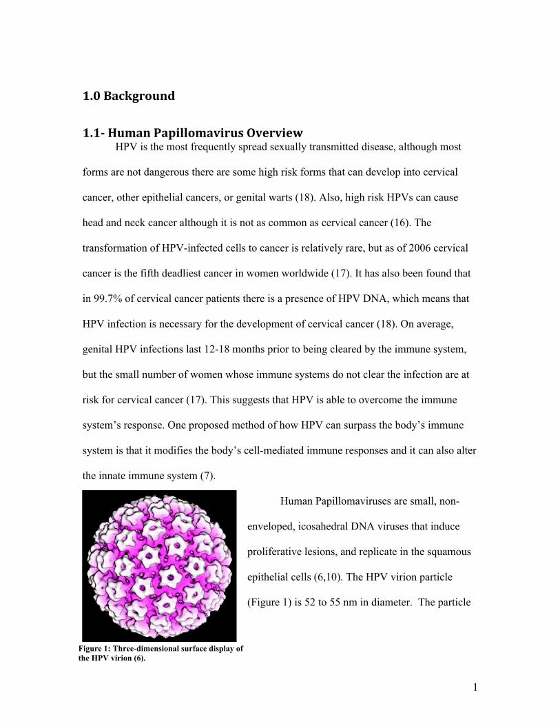
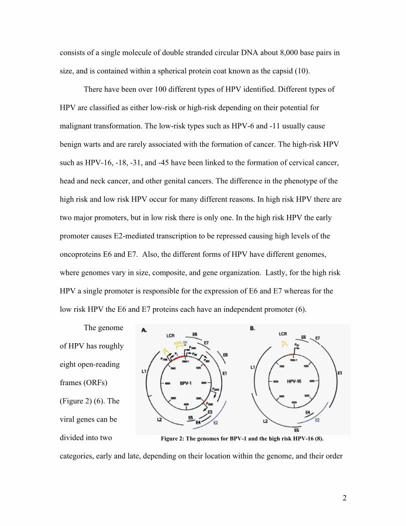
Human Papillomaviruses are small, non-
enveloped, icosahedral DNA viruses that induce
proliferative lesions, and replicate in the squamous
epithelial cells (6,10). The HPV virion particle
(Figure 1) is 52 to 55 nm in diameter. The particle
Figure 1: Three-dimensional surface display of the HPV virion (6).
2
consists of a single molecule of double stranded circular DNA about 8,000 base pairs in
size, and is contained within a spherical protein coat known as the capsid (10).
There have been over 100 different types of HPV identified. Different types of
HPV are classified as either low-risk or high-risk depending on their potential for
malignant transformation. The low-risk types such as HPV-6 and -11 usually cause
benign warts and are rarely associated with the formation of cancer. The high-risk HPV
such as HPV-16, -18, -31, and -45 have been linked to the formation of cervical cancer,
head and neck cancer, and other genital cancers. The difference in the phenotype of the
high risk and low risk HPV occur for many different reasons. In high risk HPV there are
two major promoters, but in low risk there is only one. In the high risk HPV the early
promoter causes E2-mediated transcription to be repressed causing high levels of the
oncoproteins E6 and E7. Also, the different forms of HPV have different genomes,
where genomes vary in size, composite, and gene organization. Lastly, for the high risk
HPV a single promoter is responsible for the expression of E6 and E7 whereas for the
low risk HPV the E6 and E7 proteins each have an independent promoter (6).
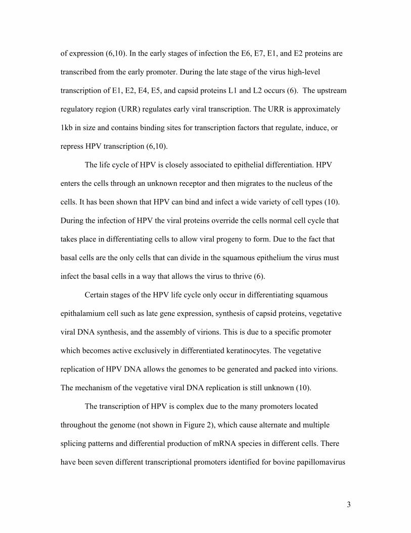
The genome
of HPV has roughly
eight open-reading
frames (ORFs)
(Figure 2) (6). The
viral genes can be
divided into two
categories, early and late, depending on their location within the genome, and their order
Figure 2: The genomes for BPV-1 and the high risk HPV-16 (8).
3
of expression (6,10). In the early stages of infection the E6, E7, E1, and E2 proteins are
transcribed from the early promoter. During the late stage of the virus high-level
transcription of E1, E2, E4, E5, and capsid proteins L1 and L2 occurs (6). The upstream
regulatory region (URR) regulates early viral transcription. The URR is approximately
1kb in size and contains binding sites for transcription factors that regulate, induce, or
repress HPV transcription (6,10).
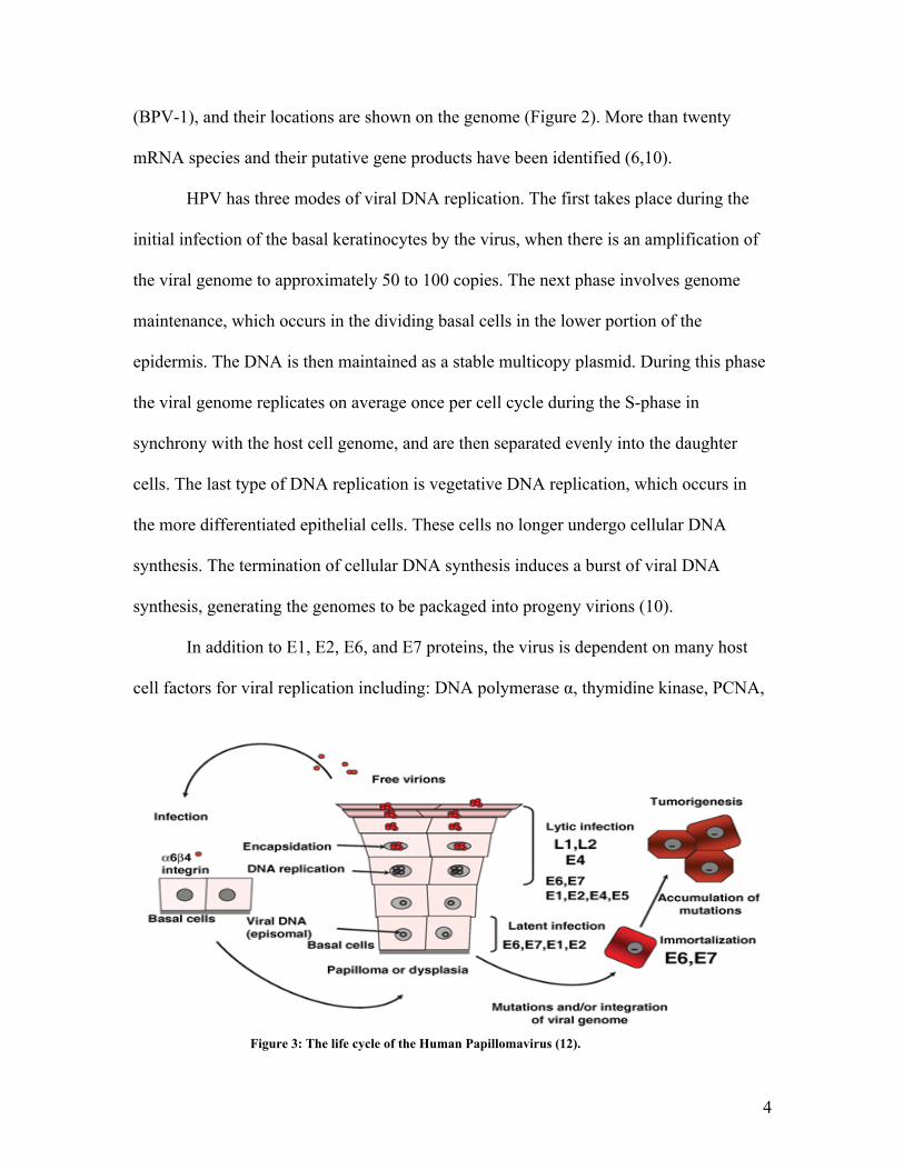
The life cycle of HPV is closely associated to epithelial differentiation. HPV
enters the cells through an unknown receptor and then migrates to the nucleus of the
cells. It has been shown that HPV can bind and infect a wide variety of cell types (10).
During the infection of HPV the viral proteins override the cells normal cell cycle that
takes place in differentiating cells to allow viral progeny to form. Due to the fact that
basal cells are the only cells that can divide in the squamous epithelium the virus must
infect the basal cells in a way that allows the virus to thrive (6).
Certain stages of the HPV life cycle only occur in differentiating squamous
epithalamium cell such as late gene expression, synthesis of capsid proteins, vegetative
viral DNA synthesis, and the assembly of virions. This is due to a specific promoter
which becomes active exclusively in differentiated keratinocytes. The vegetative
replication of HPV DNA allows the genomes to be generated and packed into virions.
The mechanism of the vegetative viral DNA replication is still unknown (10).
The transcription of HPV is complex due to the many promoters located
throughout the genome (not shown in Figure 2), which cause alternate and multiple
splicing patterns and differential production of mRNA species in different cells. There
have been seven different transcriptional promoters identified for bovine papillomavirus
4
(BPV-1), and their locations are shown on the genome (Figure 2). More than twenty
mRNA species and their putative gene products have been identified (6,10).
HPV has three modes of viral DNA replication. The first takes place during the
initial infection of the basal keratinocytes by the virus, when there is an amplification of
the viral genome to approximately 50 to 100 copies. The next phase involves genome
maintenance, which occurs in the dividing basal cells in the lower portion of the
epidermis. The DNA is then maintained as a stable multicopy plasmid. During this phase
the viral genome replicates on average once per cell cycle during the S-phase in
synchrony with the host cell genome, and are then separated evenly into the daughter
cells. The last type of DNA replication is vegetative DNA replication, which occurs in
the more differentiated epithelial cells. These cells no longer undergo cellular DNA
synthesis. The termination of cellular DNA synthesis induces a burst of viral DNA
synthesis, generating the genomes to be packaged into progeny virions (10).
In addition to E1, E2, E6, and E7 proteins, the virus is dependent on many host
cell factors for viral replication including: DNA polymerase α, thymidine kinase, PCNA,
1506 doi: 10.1111/j.1349-7006.2007.00546.x© 2007 Japanese Cancer Association
environment suitable for viral DNA replication, which sometimesinduces host cellular DNA synthesis and prevents apoptosis. Inthe outer layers of the epithelium, viral DNA is packaged intocapsids and progeny virions are released to re-initiate infection.Because the highly immunogenic virions are synthesized at theupper layers of stratified squamous epithelia they undergo onlyrelatively limited surveillance by cells of the immune system.In addition, E6 and E7 inactivate interferon (IFN) regulatoryfactor (IRF),(7,8) so that HPV viruses can remain as persistent,asymptomatic infections.
HPV infection and HPV-induced transformation
Cervical cancers originate from the lining of the cervix, thelower part of the uterus. The squamocolumnar junction, wherethe stratified non-keratinizing squamous epithelium from theexocervix and the columnar epithelium from the endocervixmeet, is the most important cytologic and colposcopic landmark,as this is highly susceptible to HPV infection and is the sitewhere more than 90% of lower genital tract neoplasia arises.Infection with high-risk HPV is associated with cervical
dysplasia or cervical intraepithelial neoplasia (CIN), andcervical cancers are thought to arise from these lesions afterlong persistent infection.(9,10) CIN I (mild dysplasia) and CIN II(moderate dysplasia) lesions show relatively low levels ofE6 and E7 expression in which the viral genomes replicateepisomally, whereas CIN III (severe dysplasia, carcinoma insitu) and invasive cancer lesions often display high-levelexpression of E6 and E7, in most cases with the integrationof viral DNA into the host cell genome whereby neoplasticdevelopment is believed to be initiated.(11)
Although HPV infections are common and the life-time riskof infection is approximately 80% for productive women, inmost cases they are resolved spontaneously by an effectiveimmune response. The ultimate development of cervical canceris rarely accompanied by high expression of E6 and E7 proteins.Thus the authors speculate that the integration of the viralgenome into the host cell is a very rare event, but after it hashappened carcinogenic transformation progresses rapidly(Fig. 3). However, epidemiological studies and experimentaldata indicate that the viral presence is not enough to inducecervical cancer and additional genetic and epigenetic events are
Fig. 2. Human papillomavirus (HPV) use a uniquestrategy for propagation, limited to stratifiedflattened epithelial tissue of mucosa and skin.Initially, HPV must infect stem cells or basal cells ofthe tissue where a phase of latent infection isestablished in which viral DNA replicate with-out making virions. In the upper layer, as cellsdifferentiate, vegetative replication of viral DNAcoordinates with expression of capsid proteins tomake virions that are then freed to search for newhost cells. Expression levels of E6 and E7 in basalcells are considered to be quite low. However, assuch infections can continue for years and even fordecades, cells may acquire high-level expression ofE6 and E7 through mutations and integration ofthe viral genome. Such cells could becomeimmortal and tumorigenic with further geneticand epigenetic events.
Fig. 3. E6 and E7 cooperatively function inthe development of cervical cancer. Multistepcarcinogenesis for human papillomavirus (HPV)-induced cervical cancer. The authors would like toemphasize that the bottleneck step to cancer isthe overexpression of E6 and E7, which is usuallyachieved by accidental integration of a viralgenome into a host chromosome. Once E6 and E7genes are overexpressed, subsequent events (inthe dark box) might be expected to occur withina short period of time because E6 and E7 cancooperatively induce chromosomal instability.
Figure 3: The life cycle of the Human Papillomavirus (12).
5
and many others. HPV also has evolved a mechanism to activate cellular genes necessary
for replication in the vegetative DNA replication. This is important because most of the
host cell factors are present only during DNA replication not when the cells are highly
differentiated (10).
1.2HPVViralProteins
1.2.1E6andE7Oncoproteins
The E6 protein of the high risk forms of HPV contains approximately 150 amino
acids including four Cys-X-X-Cys motifs, which are responsible for binding zinc
(6,10,12). The E6 protein is localized to the nucleus and cytoplasm of the infected cells.
The main function of the E6 protein is interfering with p53 mediated cell cycle regulation
(12). p53 is a tumor suppressor that plays an essential role in the cells response to DNA
damage and other cellular stress by activating several regulators of the apoptotic and
senescence pathways (5). The E6 protein binds to p53 reducing the steady-state levels of
p53. The reduction of steady-state levels of p53 allows viral replication and ends the
transcriptional transactivation of p53 (6,10,12). This means that E6 has an anti-apoptotic
function. The E6 protein is able to degrade the p53 function by inducing the ubiquitin
dependent proteolysis of p53 by forming a complex with E6AP, an ubiquitin-protein
ligase, which then can bind to p53. E6 has also been shown to interact with proteins that
have PDZ domains, which are involved in cell signaling and cell-cell adhesion. This
interaction plays a major role in the HPV life cycle because a mutation to the E6-PDZ
binding domain has shown a reduction in growth, episomal maintenance, and early
transcription. E6 can also extend the life span of keratinocytes and lead to outgrowth of
immortalized clones, which are resistant to terminal differentiation. The E6 protein is
6
also responsible for the activation of telomerase in infected cells, which causes an
increase of length of the telomeres (30). In normal cells telomeres gradual decrease in
size that leads to chromosomal instability and cellular senescence and apoptosis (5).
The E7 oncoprotein has approximately 98 amino acids and three conserved
regions, CR1, CR2, and CR3. The CR1 domain is part of the amino terminus, CR2, has
an LXCXE motif that controls binding of E7 to the retinoblastoma (Rb) tumor suppressor
protein family, and CR3 consists of two zinc finger motifs (6). A main role of the E7
protein is the binding and degradation of the Rb proteins (12). The Rb family of proteins
are major regulators of the cell cycle. When Rb is hypophoshorylated it controls the
transition of the cell cycle at G1/S phase by binding the E2F family of transcription
factors. The E2F transcription factors activate the transcription of many cellular
components during S-phase replication. The binding of Rb is regulated by two separate
regions of the protein, the LXCXE motif in CR2 is necessary for the binding of the Rb
proteins, and the N-terminus has the important residues for the degradation of the Rb
proteins. The binding and degradation of the Rb proteins by E7 causes the release of the
E2F complex (6). E7 also effects the E2F stimulated transcription by interacting with the
class I histone deacetylases (HDACs). HDACs act as transcriptional corepressors, a
protein that works with transcription factors to decrease the rate of gene transcription, by
inducing chromatin remodeling by the deacetylation of histones. E7 binds the HDACs
indirectly by an interaction that is mediated through sequences in the zinc-finger region.
The binding of E7 to HDACs has been shown to increase levels of the E2F mediated
transcription in differentiated cells that causes them to proceed into S-phase. The
expression of E7 also causes genomic instability by causing the infected cells to exhibit
7
centrosomal irregularities, such as abnormal number of centromeres. The genomic
instability is common in many malignancies (6). In the low risk forms of HPV, the E7
protein binds pRb with 10-fold lower efficiency than in the high risk types. Also, low risk
forms of HPV E7 are functionally inefficient in cellular transformation (10).
1.2.2E1andE2Protein
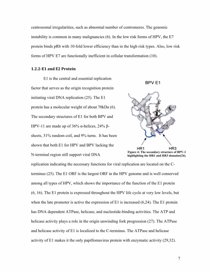
E1 is the central and essential replication
factor that serves as the origin recognition protein
initiating viral DNA replication (25). The E1
protein has a molecular weight of about 70kDa (6).
The secondary structures of E1 for both BPV and
HPV-11 are made up of 36% α-helices, 24% β-
sheets, 31% random coil, and 9% turns. It has been
shown that both E1 for HPV and BPV lacking the
N-terminal region still support viral DNA
replication indicating the necessary functions for viral replication are located on the C-
terminus (25). The E1 ORF is the largest ORF in the HPV genome and is well conserved
among all types of HPV, which shows the importance of the function of the E1 protein
(6, 16). The E1 protein is expressed throughout the HPV life cycle at very low levels, but
when the late promoter is active the expression of E1 is increased (6,24). The E1 protein
has DNA dependent ATPase, helicase, and nucleotide-binding activities. The ATP and
helicase activity plays a role in the origin unwinding fork progression (27). The ATPase
and helicase activity of E1 is localized to the C-terminus. The ATPase and helicase
activity of E1 makes it the only papillomavirus protein with enzymatic activity (29,32).
Figure 4: The secondary structure of BPV-1 highlighting the HR1 and HR3 domains(24).
8
The E2 protein functions both in viral replication and regulation of HPV
transcription. The E2 protein has a molecular weight of approximately 50 kDa. E2
consists of a β-barrel that forms dimeric complexes and binds to DNA (6). The N-
terminus of E2 has a transactivation domain and the C-terminus is involved in the
interaction with the oncoprotein E1 and DNA binding (11). The URRs (upstream
regulatory region) of high risk HPV contain four E2 binding sites that have the
palindromic sequence ACCN6GGT (20). Three of those sequences are the E1
recognition sequences at the viral origin, and the fourth site is located near the center of
the L1 ORF (6). At low levels E2 activates viral transcription, but at high levels it
represses viral transcription. This suggests that E2 serves as a regulator of the E6 and E7
proteins mediated cell cycle activities. Therefore, the loss of E2 expression is associated
with increased cellular proliferation induced by E6 and E7 leading to cervical cancer
(11). The E2 protein also plays a role in the early promoter expression by altering
chromatin remodeling through the recruitment of histone acetyltransferases (HATs).
Once differentiation takes places transcription of the viral genes switches from the E2-
regulated early promoter to the E2 independent late promoter, this results in high levels
of E1 and E2 transcription leading to viral amplification (6). Also, E2 does not play a
direct role in the viral DNA replication; it only plays a role in the pre-initiation complex
(10). Lastly, E2 has been shown to play a role in episomal maintenance and possibly
tether viral genomes to mitotic chromosomes during cell division (6).
1.2.3L1andL2CapsidProteins
L1 is about 55kDa in size, and L2 has a molecule weight of 70kDa (10). The L1
and L2 capsid proteins are expressed late in the viral life cycle in highly differentiated
9
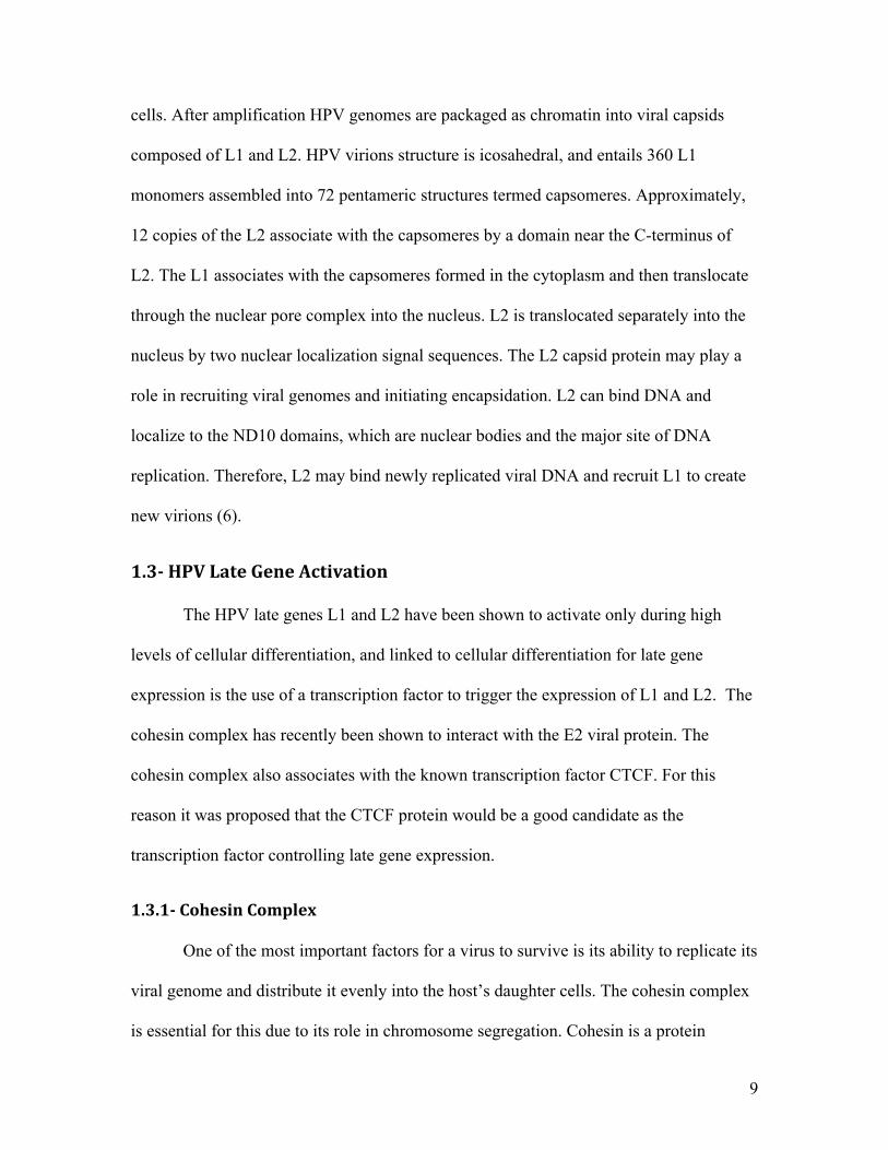
cells. After amplification HPV genomes are packaged as chromatin into viral capsids
composed of L1 and L2. HPV virions structure is icosahedral, and entails 360 L1
monomers assembled into 72 pentameric structures termed capsomeres. Approximately,
12 copies of the L2 associate with the capsomeres by a domain near the C-terminus of
L2. The L1 associates with the capsomeres formed in the cytoplasm and then translocate
through the nuclear pore complex into the nucleus. L2 is translocated separately into the
nucleus by two nuclear localization signal sequences. The L2 capsid protein may play a
role in recruiting viral genomes and initiating encapsidation. L2 can bind DNA and
localize to the ND10 domains, which are nuclear bodies and the major site of DNA
replication. Therefore, L2 may bind newly replicated viral DNA and recruit L1 to create
new virions (6).
1.3HPVLateGeneActivation
The HPV late genes L1 and L2 have been shown to activate only during high
levels of cellular differentiation, and linked to cellular differentiation for late gene
expression is the use of a transcription factor to trigger the expression of L1 and L2. The
cohesin complex has recently been shown to interact with the E2 viral protein. The
cohesin complex also associates with the known transcription factor CTCF. For this
reason it was proposed that the CTCF protein would be a good candidate as the
transcription factor controlling late gene expression.
1.3.1CohesinComplex
One of the most important factors for a virus to survive is its ability to replicate its
viral genome and distribute it evenly into the host’s daughter cells. The cohesin complex
is essential for this due to its role in chromosome segregation. Cohesin is a protein
10
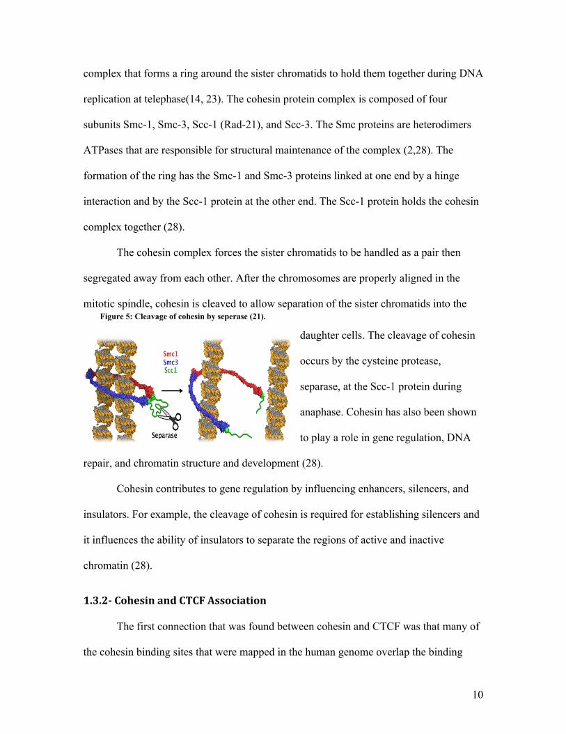
Figure 5: Cleavage of cohesin by seperase (21).
complex that forms a ring around the sister chromatids to hold them together during DNA
replication at telephase(14, 23). The cohesin protein complex is composed of four
subunits Smc-1, Smc-3, Scc-1 (Rad-21), and Scc-3. The Smc proteins are heterodimers
ATPases that are responsible for structural maintenance of the complex (2,28). The
formation of the ring has the Smc-1 and Smc-3 proteins linked at one end by a hinge
interaction and by the Scc-1 protein at the other end. The Scc-1 protein holds the cohesin
complex together (28).
The cohesin complex forces the sister chromatids to be handled as a pair then
segregated away from each other. After the chromosomes are properly aligned in the
mitotic spindle, cohesin is cleaved to allow separation of the sister chromatids into the
daughter cells. The cleavage of cohesin
occurs by the cysteine protease,
separase, at the Scc-1 protein during
anaphase. Cohesin has also been shown
to play a role in gene regulation, DNA
repair, and chromatin structure and development (28).
Cohesin contributes to gene regulation by influencing enhancers, silencers, and
insulators. For example, the cleavage of cohesin is required for establishing silencers and
it influences the ability of insulators to separate the regions of active and inactive
chromatin (28).
1.3.2CohesinandCTCFAssociation
The first connection that was found between cohesin and CTCF was that many of
the cohesin binding sites that were mapped in the human genome overlap the binding
11
sites of the CTCF protein (8,23). Two of these regions are the H19 imprinting control
region and the LCR region. Also, it was found that the CTCF and Scc-1 sequences are
very similar to one another (8,19).
CTCF is required for the localization of cohesin at its binding sites. This was
shown by the effect of removing CTCF and the result was disruption of the positioning of
the Scc-1 and Smc-3 proteins of the cohesin complex (23). It was revealed that CTCF is
not required for loading of cohesin onto DNA, showing that both cohesin and CTCF can
associate with DNA independently. Although CTCF is required for cohesin localization,
cohesin is not required for CTCF localization (3,14,23).
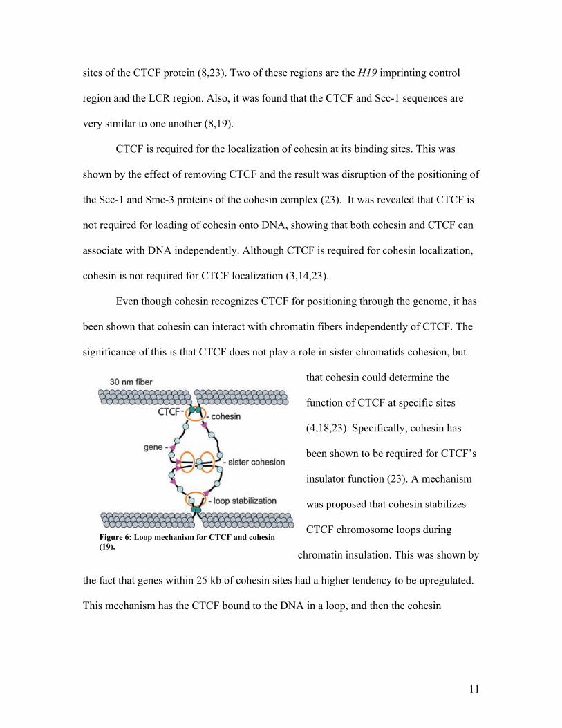
Even though cohesin recognizes CTCF for positioning through the genome, it has
been shown that cohesin can interact with chromatin fibers independently of CTCF. The
significance of this is that CTCF does not play a role in sister chromatids cohesion, but
that cohesin could determine the
function of CTCF at specific sites
(4,18,23). Specifically, cohesin has
been shown to be required for CTCF’s
insulator function (23). A mechanism
was proposed that cohesin stabilizes
CTCF chromosome loops during
chromatin insulation. This was shown by
the fact that genes within 25 kb of cohesin sites had a higher tendency to be upregulated.
This mechanism has the CTCF bound to the DNA in a loop, and then the cohesin
Using this technique, multiple laboratories demonstrated
CTCF-dependent loops in the H19-Igf2 locus in which trans-
criptional enhancers contact the insulator region, preventing
interaction of the enhancers with the Igf2 promoter.(17–19)
CTCF is also required to organize long-range loops between
different CTCF-binding sites in the mouse b globin locus,(20)
and between the XL9 control element and theHLA-DRB1 and
HLA-DQA1 MHCII genes.(21) Strikingly, using 3C and fluo-
rescence in situ hybridization (FISH), CTCF was also found to
mediate interchromosomal interactions between the mouse
Igf2-H19 locus on chromosome 7 and theWsb1-Nf1 locus on
chromosome 11.(22) CTCF also facilitates homologous inter-
actions between X chromosomes that are required for X
inactivation.(23)
It has been proposed that long-range interactions between
different CTCF sites may involve CTCF self-interactions, or
interactions between CTCF and other proteins such as
nucleophosmin,(10) but the finding that cohesin accumulates
at CTCF-binding sites now allows one to speculate that
cohesin stabilizes CTCF-mediated long-range interactions
using mechanisms similar to those by which it holds sister
chromatids together (Fig. 3). Multiple cohesin rings likely bind
to each cohesin-binding region and thus, during G2, it is
possible that some cohesin is diverted from a role in sister
chromatid cohesion to support long-range intrachromosomal
interactions that form looped-out chromosomal domains
(Fig. 3).
Recent evidence from Drosophila is consistent with a
connection between cohesin and insulators in defining func-
tional chromatin domains. Misulovin et al. mapped the binding
of cohesin and the Nipped-B loading factor throughout the
Drosophila genome, finding that they co-localize and bind
preferentially to actively transcribed regions.(24) An intrigu-
ing correlation with insulators is apparent in the Abd-B gene
that regulates segmental identity during development.
The regulatory sequences flanking Abd-B contain multiple
Figure 2. ACTCF-dependent insulator (In, red box) is positioned between theH19 and Igf2 genes, just upstream ofH19.(10) CTCF bindson the maternally derived chromosome (!), preventing activation of the Igf2 gene by an enhancer (En) located downstream ofH19. On thepaternally inherited chromosome ("), the insulator DNA is methylated, which prevents CTCF binding. The insulator is thereby inactivated,permitting the downstream enhancer to activate Igf2. Wendt et al. found by chromatin immunoprecipitation that cohesin (Rad21) co-localizes with CTCF at the insulator in HeLa cells (upper graphs), and that reduction of either CTCF or Rad21 in HeLa cells by siRNAincreases Igf2 transcripts and reduces H19 transcripts, indicating that cohesin contributes to insulator activity.(1)
Figure 3. Speculativemodel for howcohesinmight help formand stabilize CTCF-dependent long-range chromosome loops.Cohesin positioned by CTCF and not participating in sisterchromatid cohesion could interact with CTCFat other locationsin the chromosome, or even mediate ‘‘cohesion’’ between twoCTCF sites as it does between sister chromatids.
What the papers say
BioEssays 30.8 717
Figure 6: Loop mechanism for CTCF and cohesin (19).
12
encircles the two CTCF loops to hold the CTCF to the DNA, similar to how cohesin
holds sister chromatids together (19).
1.3.3E2InteractionswithCohesin
In mitosis the separation of the papillomavirus genes, is mediated by the C-
terminal DNA binding domain of the E2 protein. The E2 protein binds and tethers the
viral genome to the host cell chromosomes by protein-protein interactions. Those protein-
protein interactions require N-terminal transcactivation domain of E2, which E2 binds
with the Scc-1 protein of the cohesin complex. The binding appears to be independent of
any other cellular factors (21).
1.3.4CTCFProtein
CTCF is a ubiquitously expressed nuclear protein with an 11 zinc-finger DNA-
binding domain. It was first identified by its ability to bind a number of dissimilar
regulatory sequences in the regions close to the promoters of the chicken, mouse, and
human MYC oncogenes (1,8). CTCF is a highly conserved protein, between avian and
mammalian 93% of the amino acids of CTCF are identical, but 100% of the amino acids
are identical in the 11 zinc-finger region. Also, the ORFs of CTCF have not changed in
over 300 million years, and are conserved through a variety of species. This indicates that
the CTCF protein is adapted and has had no environmental need to mutate and change it
sequence (1,13).
CTCF is present as a nuclear extract at 130kDa on SDS-PAGE, but the theoretical
molecular weight is 82kDa. This is because CTCF is encoded in a 4.1kb mRNA, with the
largest ORF predicting a 728 amino acid protein. Possibilities for this contradiction are: a
missing exon in the cDNA due to alternative splicing, posttranslational processing, and
13
certain amino acid compositions could lead to CTCF abnormal electrophoretic migration.
Also, there is a truncated version of CTCF at 70kDa, which represents only the N-
terminal domain of the protein due to premature termination of translation (9).
1.3.5CTCFStructure
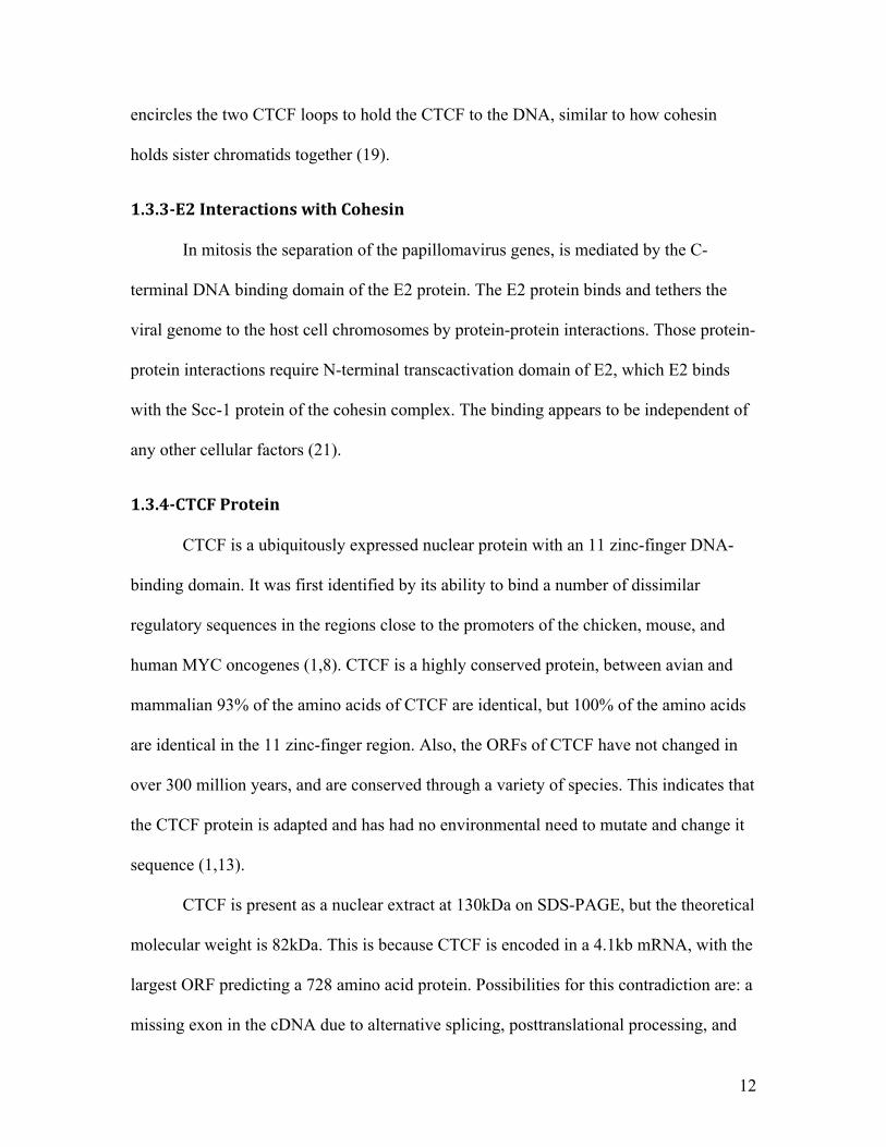
The CTCF protein is organized into three domains: the N-terminal domain, zinc-
finger domain, and the C-terminal
domain. The N- and C-termini account for
two-thirds or the protein, but the zinc-
finger domain is where most of the
functionality of CTCF occurs. The zinc
fingers of CTCF are capable of binding
either DNA or proteins (13,19). The
CTCF zinc fingers are unique in that they
can bind different DNA sequences by each zinc finger group or one zinc finger motif can
bind DNA independently while another zinc finger can bind a protein independently.
The first ten zinc-fingers are approximately 30 residues long containing a pair of cysteine
residues, which are separated by 12 amino acids from a pair of histidine residues. These
amino acids are coordinated with zinc to form an α-helix structure that recognizes DNA.
The α-helix structure allows the zinc-fingers to insert into the major groove of the DNA.
The recognition of the CTCF protein to different DNA sequences is mediated by varying
zinc-fingers; therefore, certain sets of zinc-fingers are necessary for binding to certain
sequences of DNA, but are dispensable for binding to a different sequence. Also, not all
zinc-fingers that bind to DNA behave in a similar manner. For example some zinc-fingers
TRENDS in Genetics Vol.17 No.9 September 2001
http://tig.trends.com
521Review
domains. T hese domains are individual proteinmodules tha t , when tested as fusion proteins wi th a heterologous D N A-binding domain , confert ranscr ipt ional repression . Inhibi t ion oft ranscr ipt ion is media ted by the centra l Z F domainand by sequences both N-terminal and C-terminal to the Z F region6,17. Repression by the N-terminaldomain is regula ted in a cel l-type-specific manner17.
Between avian and mammalian C T C F proteins,93% of amino acids are ident ical. However, theident i ty r ises to 100% for the region containing the11 Z Fs6. T he fi rst ten Z Fs are typical units of~30 residues containing a pair of cysteine residuesinvariant ly separated by 12 amino acids from the pairof hist idines. T hese four residues are coordinatedthrough zinc to form a compact st ructure with a D N A-recognit ion !-helix. T his type of Z F inser ts in to themajor groove of D N A to make specific contacts withnucleot ides by amino acid side chains at posit ions "1,+2, +3 and +6, marked in F ig. 1c (reviewed inRefs 18,19). T he 11th C-terminal C2 H C-type Z F isstructural ly similar to the C2 H C-type Z Fs of theF r iend of G ATA-1 (F O G) proteins that bind G ATAproteins using this type of Z F 20.
Combinatorial use of CTCF zinc fingersSequent ia l delet ion of each of the C T C F Z Fs fromeither end genera ted a panel of mutant C T C Fproteins for band-shift exper iments to assess Z Fut i l iza t ion6,21,22. T his approach suggested tha trecogni t ion by nat ive C T C F of different D N Asequences is media ted by varying contr ibut ions ofindividual Z Fs6,10,21–23. T hus, cer ta in sets of Z Fsappear to be necessary for C T C F binding to onetarget sequence, but are dispensable for binding to another (F ig. 2). However, due to the possiblein terdependence of D N A-binding proper t ies of theindividual Z Fs, and/or to addi t ional st ructura lfea tures tha t could be added to the C2 H 2 Z F fold bythe in ter-finger l in kers (reviewed in Refs 24,25), the‘missing finger ’ exper iments probably provided anincomplete picture of the contr ibut ion to targetspecifici ty of individual Z Fs tha t usual ly act in thecontext of the complete 11 Z F ar ray. Never theless,these resul ts suggest tha t each C T C F Z F might beselect ively involved in binding to some targets anddispensible for binding to others.
Co-crystal st ructures of several t ranscr ipt ionfactors with mult iple Z Fs bound to D N A have helpedto understand the posit ioning and nature of aminoacids responsible for folding and stabil i ty of theC2 H 2 class Z Fs, as well as of amino acids establishingD N A contacts19,26,27. Recent ly, extended D N A siterecognit ion by mult iple Z Fs was shown in theco-crystal st ructure of the six Z Fs of T F I I I A bound to31 bp of target D N A27. Not a l l of the Z Fs that bound toD N A behaved al i ke. Some Z Fs were posit ioned in themajor groove to contact base pairs, whereas other Z Fstraversed the D N A minor groove mak ing few or nocontacts with the D N A backbone27.
Zn Zn
ZF7
C-terminus
N-terminus
DNA-binding domain
ZF3
Zn
Zn
Zn
Zn
Zn
Zn
Zn
Zn
Zn
Zn
Zn
TRENDS in Genetics
AT
GAT
GN
te
rmin
us
ZF
1+
ZF
2Z
F1
+Z
F2
ZF
4+
ZF
5
ZF
3+
Z
F4
ZF
6+
Z
F7
ZF
3+
ZF
5
PX
XP
-mo
tifs
,
AT-
ho
ok
PX
XP
-mo
tifs
,
AT-
ho
ok
ST
OP
co
do
n
ST
OP
co
do
n
pA
sig
na
ls
pA
sig
na
ls
ZF
6+
Z
F7
ZF
7+
ZF
S 8
, 9
, 1
0,
11
+ N
LS
+ C
KII
site
s
1 2
1 2 1 2
1 2
ZF
11
+N
LS
,
CK
II s
ite
s
1 2
1 2
ZF
7+
ZF
8+
Z
F9
1 2
1 2
ZF
9+
ZF
10
+
ZF
11
1 2
1 2
E1Exon
Promoter
Size (bp) 179 797
0.6
~10 >50 0.3 4.0 4.0 0.7 5.0 1.8 1.0 10.910.0 3#UTR
3#UTR0.8 0.8 1.9 2.5 1.1
177 259 152 480 168 1569
E2 E3 E4 E5 E6 E7 E8
E0Exon
Promoter
Size (bp) 164 116 792 171 134 121 150 161 183 136 162 1490
E0 E1 E2 E3 E4 E5 E6 E7 E8 E9 E10new
(c) Structural features of CTCF and tumor-specific amino acid substitutions in zinc fingers
(a) Chicken CTCF genomic locus
(b) Human CTCF genomic locus
Fig. 1. CTCF and its prote in product. Genom ic organ ization of the ch icken (a) and human (b) CTCFgenes. F illed boxes, prote in cod ing exons; open boxes, untranslated exons; arrow , transcriptionstart sites. Estimated sizes of introns are in kilobases. The e leven ZFs of mam ma lian CTCF ared istributed in exons E2 to E8, w ith severa l ZFs be ing sp lit across ne ighboring exons15. Structure ofthe av ian CTCF gene is shown accord ing to Klenova et a l.15. (c) Structura l features of CTCF andtumor-specific am ino acid substitutions in ZFs. The comp lete am ino acid sequence of the w ild-typehuman CTCF prote in shows the DN A-b ind ing doma in , wh ich is composed of ten C2H2-class ZFs(ZFs 1–10) and one C2HC-class ZF (C-term ina l ZF11). Red , functiona lly sign ificant sites for CKIIphosphory lation7 and the RGRP-type AT-hook motif 30; green , po l II-interacting doma in . Ma jor base-contacting residues in ZF3 and ZF7 defined by stud ies of co-crysta l structures of mu lti-ZF factorsbound to DN A and tumor-specific m issense mutations characterized by G . F ilippova and co-workers(unpub lished) are ind icated on the en largements.
Figure 7: Structure of CTCF zinc-fingers (13).
14
will bind to the major groove to contact the base pairs, while others will migrate to the
minor groove making few or no contacts with the DNA base pairs. It has also been shown
that the same zinc finger can behave differently when CTCF is bound to certain target
sequences. When CTCF is bound to DNA both their polypeptides allosterically customize
their conformation to engage different zinc-fingers, either for making base contacts or to
make a target-specific surface that determines interactions with other nuclear proteins
(13).
The regions flanking the zinc-finger domain are the N- and C-terminus. The C-
terminus has a role in the trans-repressing function of CTCF, whereas the role of the N-
terminus domain is not as well classified. These domains show no significant similarities
to any previously described protein modules, except for three short motifs that are all
located in the C-terminus. The first motif expressed is KRRGRP-type AT-hook that
possibly has a role in DNA binding and protein-protein interactions in chromatin.
Another motif that is strictly conserved is the SKKEDSSDSE motif in the trans-repressor
region. In this region the protein is phosphorylated on four serines by casein kinase II. A
third motif is located between the AT-hook and phosphorylation sites. This motif
contains two repeats of the PXXP-signature characteristic of the SH3-domain binding
proteins (13).
1.3.6CTCFFunctions
CTCF is a versatile protein that has been shown to have many different cellular
functions. The best described and studied functions of CTCF are its ability to be a
transcription factor, chromatin insulator protein, and its role in epigenetics and cancer.
Other areas where CTCF could possibly play a role in are gene activation and as a tumor
15
suppressor (8,13).
CTCF was first characterized as a transcription factor by its ability to bind to the
metazoan silencing elements. For example, binding of CTCF to the promoters and
upstream silencer elements of the chicken lysozyme gene and the chicken and human
MYC gene results in transcriptional repression of the lysozyme and MYC gene. Further
studies of CTCF found that other CTCF target sites identified transcriptional response
elements that function in gene repression and activation. Also, CTCF shares many
common traits with enhancers: it independently functions of position and orientation, it
compromises functional modules that synergize in transcriptional control, and it acts
directly on a promoter. An example is that CTCF can act as transcription activator or
repressor on the lysozome gene. CTCF’s role depends on the presence of the thyroid
hormone receptor, if this hormone is bound along with CTCF it leads to synergistic
activation of the lysozome gene, but if it is not bound CTCF causes synergistic repression
of the gene. CTCF also contains an independent silencing domain that mediates
transcriptional repression. The discovery of this domain led to the identification of
CTCF-interacting co-repressors that recruit histone deacetylase (HDAC) activity.
Silencing by CTCF and its co-repressors could act directly on the transcriptional start
site. The ability of CTCF to recruit HDACs leads to CTCF inhibiting transcription by,
interfering with the transcription initiation complex and/or modifying the promoter
nucleosomes (13).
CTCF has a pivotal role in many chromatin insulators, and is the only major
protein implicated in establishment of insulators in vertebrates. Protein insulators operate
by blocking the communication between pivotal cis regulatory elements, gene promoters,
16
and enhancers or silencers. Cis-regulatory DNA elements are a region of DNA that
controls the expression of genes on the same strand. These elements mediate the level of
gene expression by recruiting trans-acting factors that influence transcription. The
regulatory elements used for the insulation are often distant from one another on the
linear genome. This makes the insulators position-dependent, where the insulator must be
positioned between the enhancer and its target promoter (8). This suggests that the
insulator prevents propagation of signals along the chromatin fiber without continuously
engaging enhancer or silencing factors. CTCF may mediate the chromatin interaction
without the need of co-factors due to the finding that CTCF can dimerize when bound to
DNA and connect two separate DNA molecules. An example of CTCF as a chromatin is
its interaction with the core insulator sequences in vertebrate insulators, β-globin FII
insulator, Xenopus repeat organizer elements, and the BEAD-A insulator. CTCF flanks
the entire region of theβ-globin gene cluster, and it presumably protects this domain
from effects of the adjacent regulatory elements (13).
One of the most characterized interactions of CTCF describes its role in both
chromatin insulation and epigenetics. CTCF plays a central part in the H19 imprinting
control region (ICR) where CTCF affects the downstream Igf2 gene. The region is
maternally unmethylated and paternally methylated. It regulates the expression of the
maternal allele of the Igf2 gene by blocking the Igf2 promoters and enhancer from
communicating with each other. CTCF associates only with the maternal unmethylated
allele of H19 ICR. The CTCF protein prevents the activation of the Igf2 gene in this
circumstance. When this region is methylated CTCF cannot bind though, allowing the
Igf2 gene to be expressed. Also, the loss of imprinting on the H19 and Igf2 locus have
17
been found to cause Beckwith-Wiedemann Syndrome with increased tumor formation
(1,8,13).
Due to CTCF’s ability to read epigenetic marks and the common occurrence of
epigenetic disturbances in cancer, the function of CTCF in cancer has also been studied.
Gene mutations to CTCF were found in patients with breast, prostates, and Wilm’s
tumor’s. These mutations were located in the zinc-finger finger region of the protein,
which affects the binding and interaction of DNA with other genes. Also, in the PXXP
motif region a common tumor suppressor was found, the MYC-binding protein BIN1. It
was shown that if mutations are made to the prolines in this region, it causes the
elimination of binding to BIN1, and affects the trans-repressing activity of the C-terminus
region (1,13).
CTCF is a unique protein with roles in transcription, insulation, tumor
suppression, and gene activation. All of these roles could be essential in the HPV
genome. Also, CTCF and cohesin have been shown to interact with one another and be
necessary for one another to function, and E2 and cohesin have been shown to interact
within HPV. Therefore, it was examined if CTCF is not only found within the HPV
genome, but also if it plays a role in late gene expression and viral transcription.
18
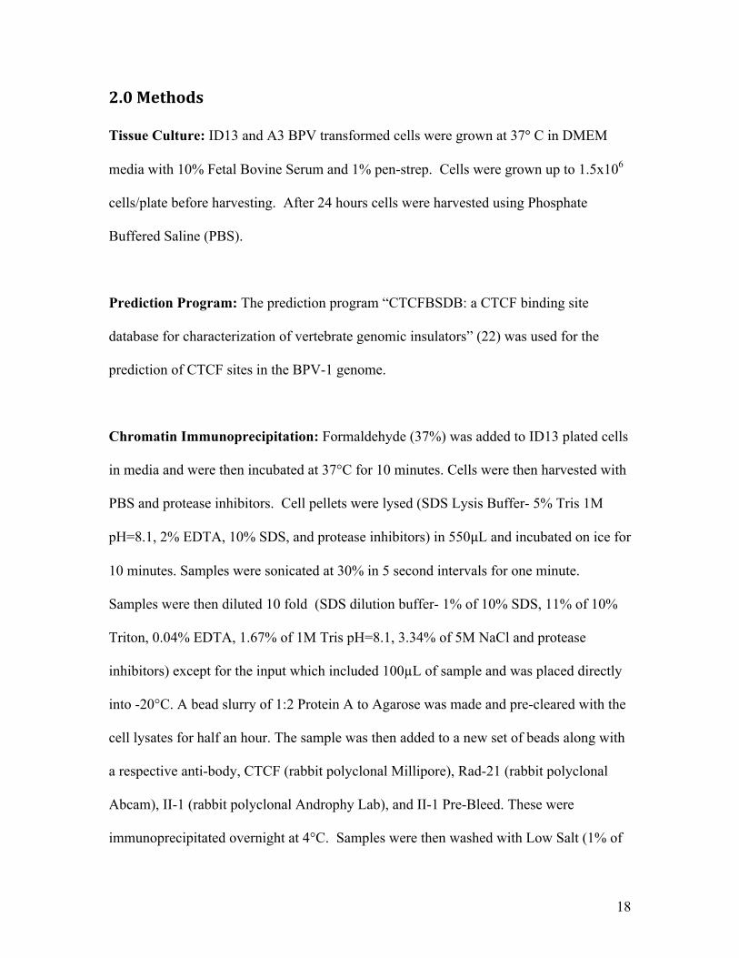
2.0Methods Tissue Culture: ID13 and A3 BPV transformed cells were grown at 37° C in DMEM
media with 10% Fetal Bovine Serum and 1% pen-strep. Cells were grown up to 1.5x10⁶
cells/plate before harvesting. After 24 hours cells were harvested using Phosphate
Buffered Saline (PBS).
Prediction Program: The prediction program “CTCFBSDB: a CTCF binding site
database for characterization of vertebrate genomic insulators” (22) was used for the
prediction of CTCF sites in the BPV-1 genome.
Chromatin Immunoprecipitation: Formaldehyde (37%) was added to ID13 plated cells
in media and were then incubated at 37°C for 10 minutes. Cells were then harvested with
PBS and protease inhibitors. Cell pellets were lysed (SDS Lysis Buffer- 5% Tris 1M
pH=8.1, 2% EDTA, 10% SDS, and protease inhibitors) in 550µL and incubated on ice for
10 minutes. Samples were sonicated at 30% in 5 second intervals for one minute.
Samples were then diluted 10 fold (SDS dilution buffer- 1% of 10% SDS, 11% of 10%
Triton, 0.04% EDTA, 1.67% of 1M Tris pH=8.1, 3.34% of 5M NaCl and protease
inhibitors) except for the input which included 100µL of sample and was placed directly
into -20°C. A bead slurry of 1:2 Protein A to Agarose was made and pre-cleared with the
cell lysates for half an hour. The sample was then added to a new set of beads along with
a respective anti-body, CTCF (rabbit polyclonal Millipore), Rad-21 (rabbit polyclonal
Abcam), II-1 (rabbit polyclonal Androphy Lab), and II-1 Pre-Bleed. These were
immunoprecipitated overnight at 4°C. Samples were then washed with Low Salt (1% of
19
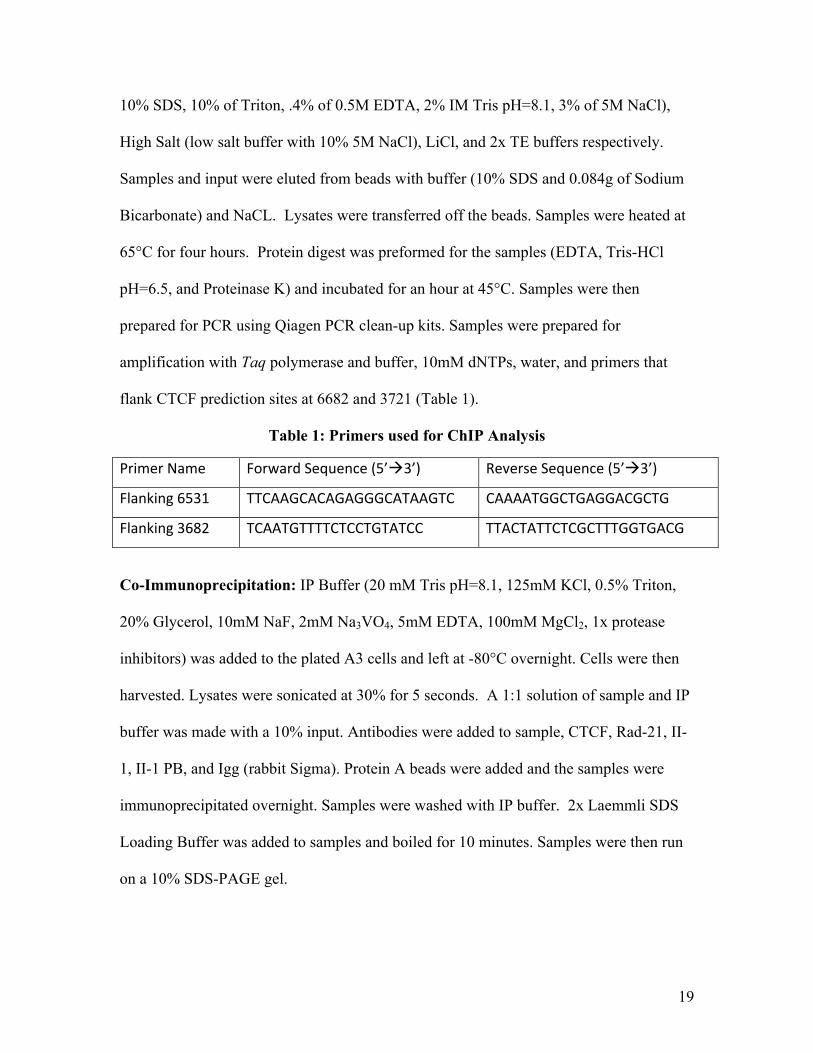
10% SDS, 10% of Triton, .4% of 0.5M EDTA, 2% IM Tris pH=8.1, 3% of 5M NaCl),
High Salt (low salt buffer with 10% 5M NaCl), LiCl, and 2x TE buffers respectively.
Samples and input were eluted from beads with buffer (10% SDS and 0.084g of Sodium
Bicarbonate) and NaCL. Lysates were transferred off the beads. Samples were heated at
65°C for four hours. Protein digest was preformed for the samples (EDTA, Tris-HCl
pH=6.5, and Proteinase K) and incubated for an hour at 45°C. Samples were then
prepared for PCR using Qiagen PCR clean-up kits. Samples were prepared for
amplification with Taq polymerase and buffer, 10mM dNTPs, water, and primers that
flank CTCF prediction sites at 6682 and 3721 (Table 1).
Table 1: Primers used for ChIP Analysis
PrimerName ForwardSequence(5’3’) ReverseSequence(5’3’)
Flanking6531 TTCAAGCACAGAGGGCATAAGTC CAAAATGGCTGAGGACGCTG
Flanking3682 TCAATGTTTTCTCCTGTATCC TTACTATTCTCGCTTTGGTGACG
Co-Immunoprecipitation: IP Buffer (20 mM Tris pH=8.1, 125mM KCl, 0.5% Triton,
20% Glycerol, 10mM NaF, 2mM Na3VO4, 5mM EDTA, 100mM MgCl2, 1x protease
inhibitors) was added to the plated A3 cells and left at -80°C overnight. Cells were then
harvested. Lysates were sonicated at 30% for 5 seconds. A 1:1 solution of sample and IP
buffer was made with a 10% input. Antibodies were added to sample, CTCF, Rad-21, II-
1, II-1 PB, and Igg (rabbit Sigma). Protein A beads were added and the samples were
immunoprecipitated overnight. Samples were washed with IP buffer. 2x Laemmli SDS
Loading Buffer was added to samples and boiled for 10 minutes. Samples were then run
on a 10% SDS-PAGE gel.
20
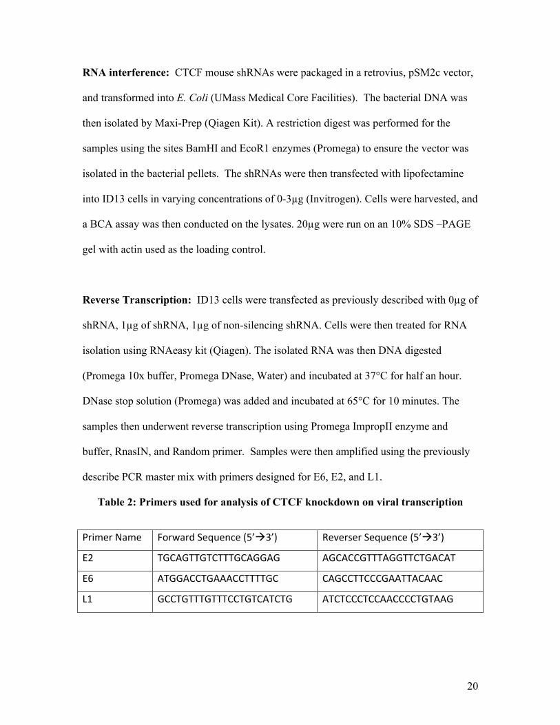
RNA interference: CTCF mouse shRNAs were packaged in a retrovius, pSM2c vector,
and transformed into E. Coli (UMass Medical Core Facilities). The bacterial DNA was
then isolated by Maxi-Prep (Qiagen Kit). A restriction digest was performed for the
samples using the sites BamHI and EcoR1 enzymes (Promega) to ensure the vector was
isolated in the bacterial pellets. The shRNAs were then transfected with lipofectamine
into ID13 cells in varying concentrations of 0-3µg (Invitrogen). Cells were harvested, and
a BCA assay was then conducted on the lysates. 20µg were run on an 10% SDS –PAGE
gel with actin used as the loading control.
Reverse Transcription: ID13 cells were transfected as previously described with 0µg of
shRNA, 1µg of shRNA, 1µg of non-silencing shRNA. Cells were then treated for RNA
isolation using RNAeasy kit (Qiagen). The isolated RNA was then DNA digested
(Promega 10x buffer, Promega DNase, Water) and incubated at 37°C for half an hour.
DNase stop solution (Promega) was added and incubated at 65°C for 10 minutes. The
samples then underwent reverse transcription using Promega ImpropII enzyme and
buffer, RnasIN, and Random primer. Samples were then amplified using the previously
describe PCR master mix with primers designed for E6, E2, and L1.
Table 2: Primers used for analysis of CTCF knockdown on viral transcription
PrimerName ForwardSequence(5’3’) ReverserSequence(5’3’)
E2 TGCAGTTGTCTTTGCAGGAG AGCACCGTTTAGGTTCTGACAT
E6 ATGGACCTGAAACCTTTTGC CAGCCTTCCCGAATTACAAC
L1 GCCTGTTTGTTTCCTGTCATCTG ATCTCCCTCCAACCCCTGTAAG
21
3.0Results
3.1LocationofCTCFbindingsiteswithintheBPVGenome
To determine if CTCF is involved in BPV it was first examined in silco if there
were any CTCF binding sites on the BPV-1 genome. To accomplish this an online
prediction program for CTCF binding sites was used (22). The prediction site is a
statistical analysis of the core motifs of CTCF that are represented by position weight
matrices (PWM). The program focuses on the four PWM that provide the strongest range
of conserved motifs.
22
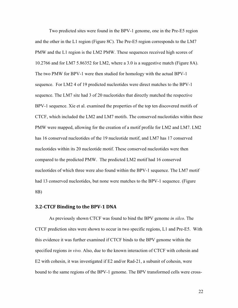
Two predicted sites were found in the BPV-1 genome, one in the Pre-E5 region
and the other in the L1 region (Figure 8C). The Pre-E5 region corresponds to the LM7
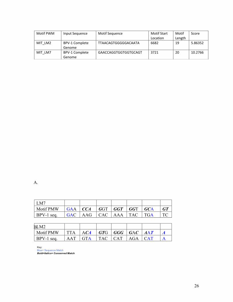
PMW and the L1 region is the LM2 PMW. These sequences received high scores of
10.2766 and for LM7 5.86352 for LM2, where a 3.0 is a suggestive match (Figure 8A).
The two PMW for BPV-1 were then studied for homology with the actual BPV-1
sequence. For LM2 4 of 19 predicted nucleotides were direct matches to the BPV-1
sequence. The LM7 site had 3 of 20 nucleotides that directly matched the respective
BPV-1 sequence. Xie et al. examined the properties of the top ten discovered motifs of
CTCF, which included the LM2 and LM7 motifs. The conserved nucleotides within these
PMW were mapped, allowing for the creation of a motif profile for LM2 and LM7. LM2
has 16 conserved nucleotides of the 19 nucleotide motif, and LM7 has 17 conserved
nucleotides within its 20 nucleotide motif. These conserved nucleotides were then
compared to the predicted PMW. The predicted LM2 motif had 16 conserved
nucleotides of which three were also found within the BPV-1 sequence. The LM7 motif
had 13 conserved nucleotides, but none were matches to the BPV-1 sequence. (Figure
8B)
3.2CTCFBindingtotheBPV1DNA
As previously shown CTCF was found to bind the BPV genome in silco. The
CTCF prediction sites were shown to occur in two specific regions, L1 and Pre-E5. With
this evidence it was further examined if CTCF binds to the BPV genome within the
specified regions in vivo. Also, due to the known interaction of CTCF with cohesin and
E2 with cohesin, it was investigated if E2 and/or Rad-21, a subunit of cohesin, were
bound to the same regions of the BPV-1 genome. The BPV transformed cells were cross-
23
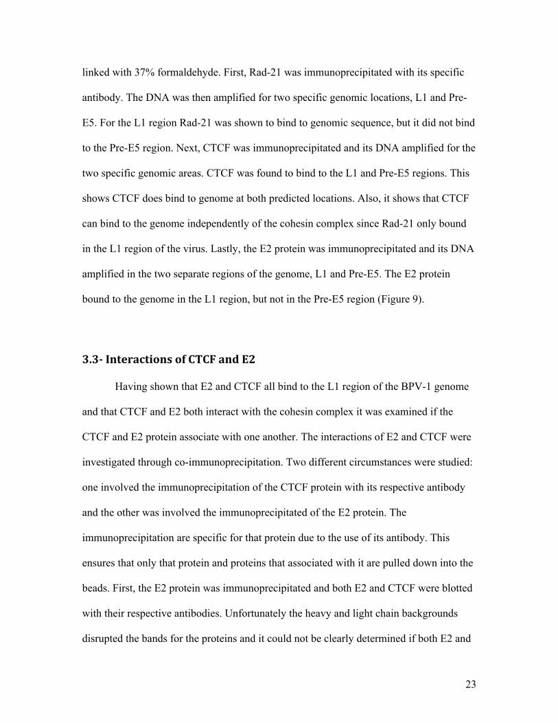
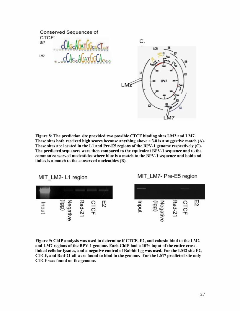
linked with 37% formaldehyde. First, Rad-21 was immunoprecipitated with its specific
antibody. The DNA was then amplified for two specific genomic locations, L1 and Pre-
E5. For the L1 region Rad-21 was shown to bind to genomic sequence, but it did not bind
to the Pre-E5 region. Next, CTCF was immunoprecipitated and its DNA amplified for the
two specific genomic areas. CTCF was found to bind to the L1 and Pre-E5 regions. This
shows CTCF does bind to genome at both predicted locations. Also, it shows that CTCF
can bind to the genome independently of the cohesin complex since Rad-21 only bound
in the L1 region of the virus. Lastly, the E2 protein was immunoprecipitated and its DNA
amplified in the two separate regions of the genome, L1 and Pre-E5. The E2 protein
bound to the genome in the L1 region, but not in the Pre-E5 region (Figure 9).
3.3InteractionsofCTCFandE2
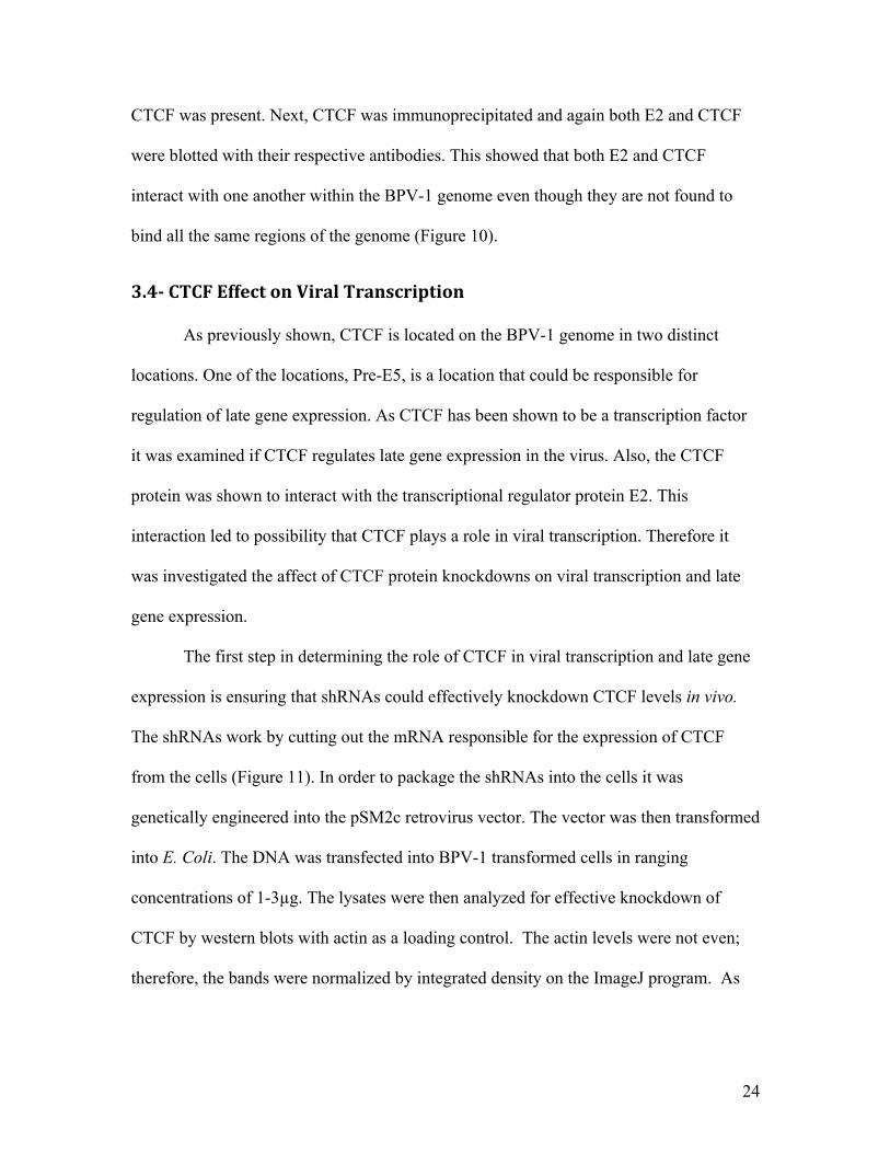
Having shown that E2 and CTCF all bind to the L1 region of the BPV-1 genome
and that CTCF and E2 both interact with the cohesin complex it was examined if the
CTCF and E2 protein associate with one another. The interactions of E2 and CTCF were
investigated through co-immunoprecipitation. Two different circumstances were studied:
one involved the immunoprecipitation of the CTCF protein with its respective antibody
and the other was involved the immunoprecipitated of the E2 protein. The
immunoprecipitation are specific for that protein due to the use of its antibody. This
ensures that only that protein and proteins that associated with it are pulled down into the
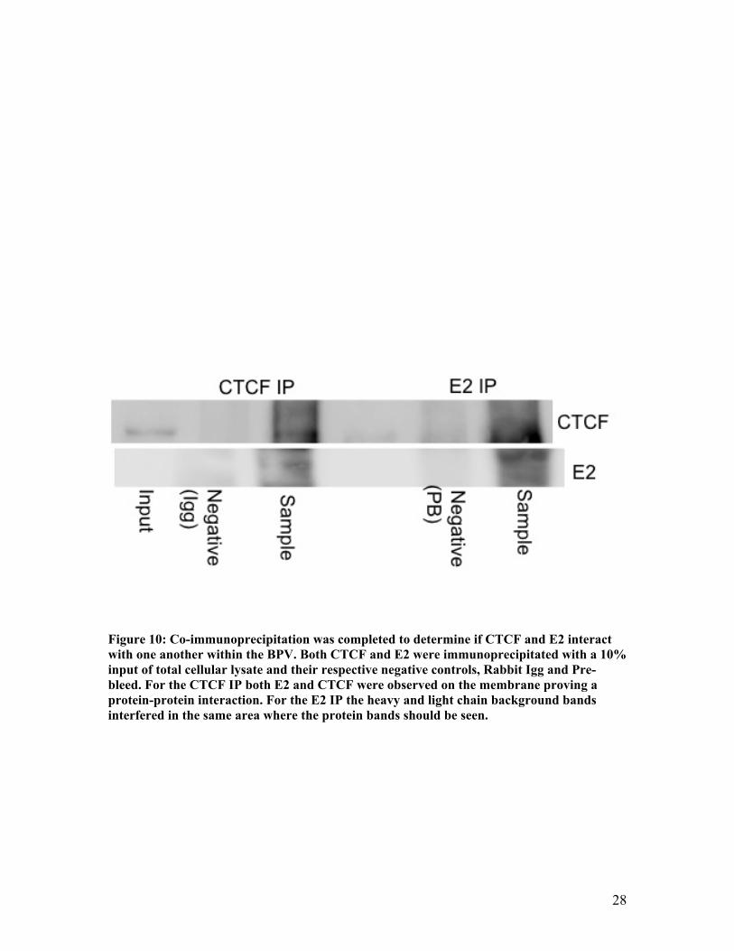
beads. First, the E2 protein was immunoprecipitated and both E2 and CTCF were blotted
with their respective antibodies. Unfortunately the heavy and light chain backgrounds
disrupted the bands for the proteins and it could not be clearly determined if both E2 and
24
CTCF was present. Next, CTCF was immunoprecipitated and again both E2 and CTCF
were blotted with their respective antibodies. This showed that both E2 and CTCF
interact with one another within the BPV-1 genome even though they are not found to
bind all the same regions of the genome (Figure 10).
3.4CTCFEffectonViralTranscription
As previously shown, CTCF is located on the BPV-1 genome in two distinct
locations. One of the locations, Pre-E5, is a location that could be responsible for
regulation of late gene expression. As CTCF has been shown to be a transcription factor
it was examined if CTCF regulates late gene expression in the virus. Also, the CTCF
protein was shown to interact with the transcriptional regulator protein E2. This
interaction led to possibility that CTCF plays a role in viral transcription. Therefore it
was investigated the affect of CTCF protein knockdowns on viral transcription and late
gene expression.
The first step in determining the role of CTCF in viral transcription and late gene
expression is ensuring that shRNAs could effectively knockdown CTCF levels in vivo.
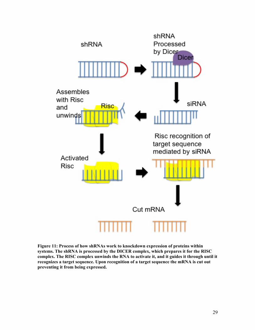
The shRNAs work by cutting out the mRNA responsible for the expression of CTCF
from the cells (Figure 11). In order to package the shRNAs into the cells it was
genetically engineered into the pSM2c retrovirus vector. The vector was then transformed
into E. Coli. The DNA was transfected into BPV-1 transformed cells in ranging
concentrations of 1-3µg. The lysates were then analyzed for effective knockdown of
CTCF by western blots with actin as a loading control. The actin levels were not even;
therefore, the bands were normalized by integrated density on the ImageJ program. As
25
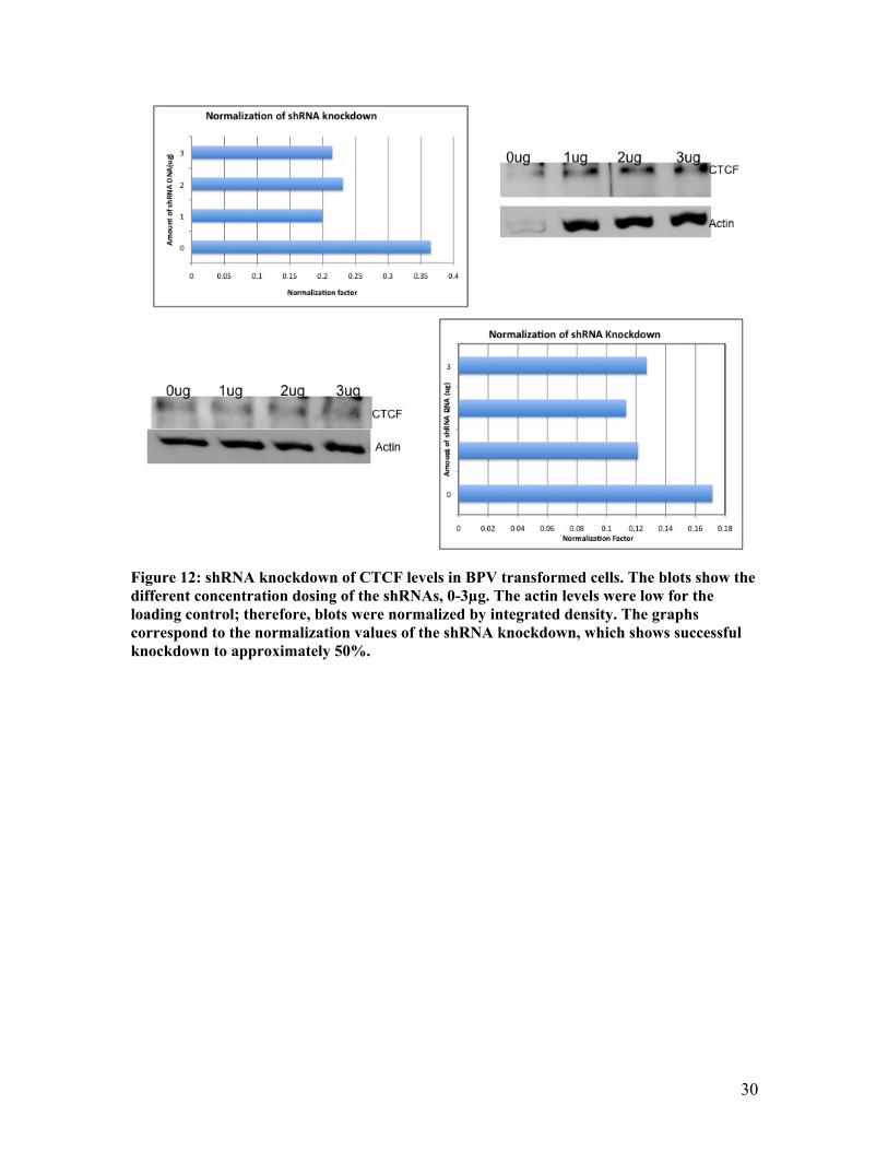
shown clearly in the bar graphs there was still a significant knockdown observed in the
cells ranging to near a 50% knockdown of CTCF in the cells (Figure 12).
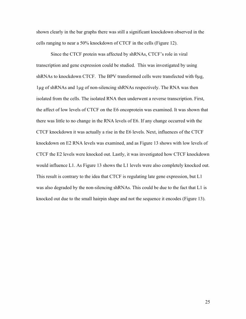
Since the CTCF protein was affected by shRNAs, CTCF’s role in viral
transcription and gene expression could be studied. This was investigated by using
shRNAs to knockdown CTCF. The BPV transformed cells were transfected with 0µg,
1µg of shRNAs and 1µg of non-silencing shRNAs respectively. The RNA was then
isolated from the cells. The isolated RNA then underwent a reverse transcription. First,
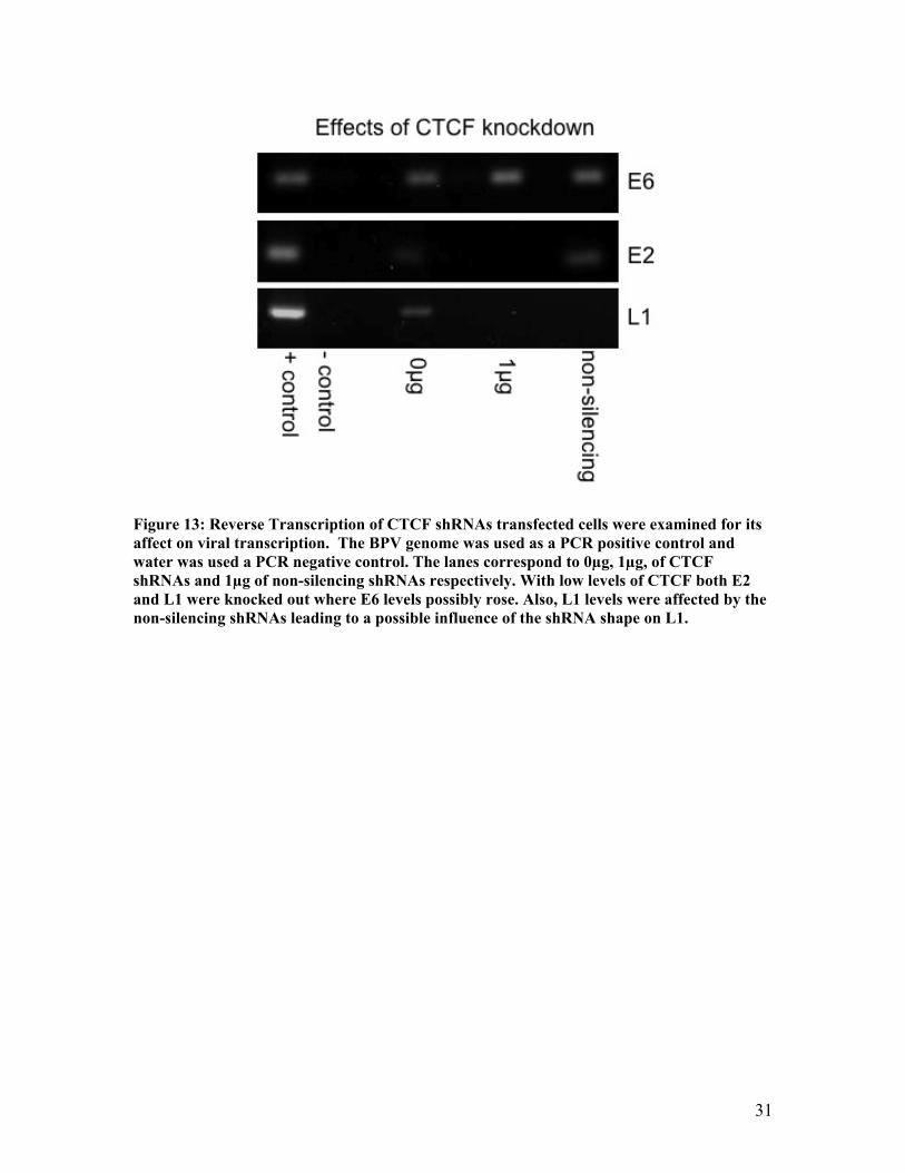
the affect of low levels of CTCF on the E6 oncoprotein was examined. It was shown that
there was little to no change in the RNA levels of E6. If any change occurred with the
CTCF knockdown it was actually a rise in the E6 levels. Next, influences of the CTCF
knockdown on E2 RNA levels was examined, and as Figure 13 shows with low levels of
CTCF the E2 levels were knocked out. Lastly, it was investigated how CTCF knockdown
would influence L1. As Figure 13 shows the L1 levels were also completely knocked out.
This result is contrary to the idea that CTCF is regulating late gene expression, but L1
was also degraded by the non-silencing shRNAs. This could be due to the fact that L1 is
knocked out due to the small hairpin shape and not the sequence it encodes (Figure 13).
26
MotifPWM InputSequence MotifSequence MotifStartLocation
MotifLength
Score
MIT_LM2 BPV‐1CompleteGenome
TTAACAGTGGGGGACAATA 6682 19 5.86352
MIT_LM7 BPV‐1CompleteGenome
GAACCAGGTGGTGGTGCAGT 3721 20 10.2766
LM7 Motif PMW GAA CCA GGT GGT GGT GCA GT BPV-1 seq. GAC AAG CAC AAA TAC TGA TC LM2 Motif PMW TTA ACA GTG GGG GAC AAT A BPV-1 seq. AAT GTA TAC CAT AGA CAT A
A.
B.
27
Figure 8: The prediction site provided two possible CTCF binding sites LM2 and LM7. These sites both received high scores because anything above a 3.0 is a suggestive match (A). These sites are located in the L1 and Pre-E5 regions of the BPV-1 genome respectively (C). The predicted sequences were then compared to the equivalent BPV-1 sequence and to the common conserved nucleotides where blue is a match to the BPV-1 sequence and bold and italics is a match to the conserved nucleotides (B).
Figure 9: ChIP analysis was used to determine if CTCF, E2, and cohesin bind to the LM2 and LM7 regions of the BPV-1 genome. Each ChIP had a 10% input of the entire cross-linked cellular lysates, and a negative control of Rabbit Igg was used. For the LM2 site E2, CTCF, and Rad-21 all were found to bind to the genome. For the LM7 predicted site only CTCF was found on the genome.
28
Figure 10: Co-immunoprecipitation was completed to determine if CTCF and E2 interact with one another within the BPV. Both CTCF and E2 were immunoprecipitated with a 10% input of total cellular lysate and their respective negative controls, Rabbit Igg and Pre-bleed. For the CTCF IP both E2 and CTCF were observed on the membrane proving a protein-protein interaction. For the E2 IP the heavy and light chain background bands interfered in the same area where the protein bands should be seen.
29
Figure 11: Process of how shRNAs work to knockdown expression of proteins within systems. The shRNA is processed by the DICER complex, which prepares it for the RISC complex. The RISC complex unwinds the RNA to activate it, and it guides it through until it recognizes a target sequence. Upon recognition of a target sequence the mRNA is cut out preventing it from being expressed.
30
Figure 12: shRNA knockdown of CTCF levels in BPV transformed cells. The blots show the different concentration dosing of the shRNAs, 0-3µg. The actin levels were low for the loading control; therefore, blots were normalized by integrated density. The graphs correspond to the normalization values of the shRNA knockdown, which shows successful knockdown to approximately 50%.
31
Figure 13: Reverse Transcription of CTCF shRNAs transfected cells were examined for its affect on viral transcription. The BPV genome was used as a PCR positive control and water was used a PCR negative control. The lanes correspond to 0µg, 1µg, of CTCF shRNAs and 1µg of non-silencing shRNAs respectively. With low levels of CTCF both E2 and L1 were knocked out where E6 levels possibly rose. Also, L1 levels were affected by the non-silencing shRNAs leading to a possible influence of the shRNA shape on L1.
32
4.0Discussion
CTCF has been shown in many different systems to play a role as a transcription
factor and/or a chromatin insulator. CTCF has been shown to associate with the cohesin
complex, and the cohesin complex has recently been revealed to interact with the E2 viral
protein of HPV. This study was the first to find that the CTCF protein associates with
HPV.
The first part of this study showed that CTCF has possible binding sites in the
BPV-1 genome in silico (Figure 8A-C). The predicted site provided guidance on where to
look for possible CTCF sites in vivo, but it doesn’t mean that those are the only possible
CTCF binding sites within the BPV-1 genome. It is possible that it binds to many other
locations within the genome or that within that one region it could bind numerous times.
Therefore, CTCF binding sites should be mapped throughout the genome this would
provide us with a better understanding of where CTCF binds and other possible
mechanisms for the protein.
The predicted sites in the L1 and Pre-E5 regions were then examined in vivo.
These sites were examined for the binding of the Rad-21 protein, subunit of cohesin, and
the E2 viral protein. It was shown that for L1 CTCF, E2, and Rad-21 all bind but for Pre-
E5 only CTCF bound (Figure 9). This is interesting because it provides the suggestion
that CTCF has two independent functions or mechanisms within the genome. The Rad-21
protein has been shown to be necessary for CTCF’s insulator function. The E2 protein
has been shown to associate with the cohesin complex throughout the cell cycle,
providing evidence E2 is essential for cohesin role in gene regulation. Also, E2 is
responsible for transcriptional repression of many viral proteins, which could aid in the
33
insulation process for CTCF. Therefore, it is has been presumed that E2 and cohesin are
important for the insulation function of CTCF. This provides significant evidence of
CTCF acting as an insulator in the L1 region and not in the Pre-E5 region. The role of
CTCF acting independently in the Pre-E5 region would need to be further examined due
to the fact CTCF is a versatile protein with a range of functions.
The CTCF protein was also found to associate with the viral protein E2 (Figure
10). This is interesting because as previously stated E2 did not bind to the Pre-E5 region
but CTCF did. Therefore this means that although the proteins do associate it is not
always the case. This provides further evidence that although the proteins do interact it
could be just for certain viral functions, such as the previously described insulation.
Also, due to CTCF known roles in transcription and E2 acting as the viral protein
responsible for transcription it is probable that CTCF plays a role in the HPV
transcription. This could mean that only when CTCF is affecting viral transcription, such
as chromatin insulation, do the CTCF and E2 protein interact with each other. The reason
this is plausible that the interactions could occur only at specific locations is due to the
fact the CTCF’s zinc fingers are do unique. The zinc fingers are able to allosterically
change for specific sequences. This could mean that when CTCF is bound to certain
genomic regions its binding region for E2 is experiencing a conformational changed and
the binding can’t occur, or it could work the other way so that when bound to E2 the zinc
fingers are in a conformation that the CTCF protein can only bind to distinct genomic
regions. Although, if E2 directly binds to CTCF or the association is by an indirect
mechanism stills needs to investigated.
34
With the ability to lower levels of CTCF through shRNA transfection the role of
CTCF in late gene expression was studied (Figure 12-13). With the knockdown of CTCF
levels, L1 levels were knocked out. This is contradictory to the idea of CTCF as an
insulator for late gene expression, which would have shown an increase in L1 with CTCF
knockdown. This is further contradicted by the presumption that for CTCF to be able to
function as an insulator both Rad-21 and E2 must be present, but in the Pre-E5 area that
precedes the late genes, only CTCF was bound to the genome. Another issue is that L1
was also affected by the non-silencing shRNAs. This could mean that the shape of the
shRNA degrades L1 independently of the sequence of the shRNA. Therefore, a further
study needs to pursue if the small hairpin shape of the shRNAs affect L1 levels
independently of their sequence.
CTCF ‘s role in viral transcription was also examined through shRNA
transfection. When CTCF levels were lowered E2 was knocked out where E6 levels rose.
This is because E2 controls the levels of E6, so with high levels of E2 the E6 protein is
regulated but with low levels of E2 the E6 is expressed in high levels. A possible reason
for this is CTCF is necessary for E2 transcription and possibly an enhancer for E2. The
reason CTCF can be an enhancer for E2 is because enhancers can act independently of
their location. Therefore, in the Pre-E5 region where only CTCF is bound it could be
acting with the E2 promoter for transcription. This it still a preliminary mechanism that
needs to be further studied for how CTCF and E2 associate during viral transcription.
A mechanism has been proposed for CTCF’s function in the L1 region, and it
entails CTCF insulating anti-sense transcription of the virus. This mechanism has
significant evidence starting with the binding of both E2 and Rad-21 in the L1 region,
35
which as previously discussed could be vital for CTCF chromatin insulation. Therefore it
is possible that when the CTCF levels are low the virus undergoes anti-sense
transcription, which leads to the pKR cellular death pathway. This could also explain the
knockout of L1 when there are low levels of CTCF because the anti-sense transcription
would lead to rapid degradation of L1. A cellular death assay should be completed to see
the affect of shRNA CTCF knockdown on the cells. This mechanism provides an
explanation for the role of CTCF in the L1 region taking into account all of the findings
of this study, but it still needs to be further examined.
36
References
1. Bartkuhn, M., Renkawitz, R. (2008). Long range chromatin interactions involved in gene regulation. Biochimica et Biophysica Acta, 1783, 2161-2166.
2. Gause, M., Schaaf, C., Dorsett, D. (2008). Cohesin and CTCF: Cooperating to control chromosome conformation?. Bioessays, 30 (8), 715-718.
3. Gondor, A., Ohlsson, R. (2008). Chromatin insulators and cohesins. EMBO reports , 9 (4), 327-329.
4. Hagstrom, K. A., Meyer, J. B. (2003). Condensin and Cohesin: More Than Chromosome Compactor and Glue. Nature Reviews , 4, 520-534.
5. Hanahan, D., Weinberg, R. (2000). Hallmarks of cancer. Cell, 100, 57-70. 6. Hebner, C., Laimins, L. (2006). Human papillomaviruses: Basic mechanisms of pathogenesis and
oncogenicity. Reviews in Medical Virology, 16, 83-97. 7. Kanodia, S., Fahey, L., Kast, M. (2007). Mechanisms used by human papillomaviruses to escape
the host immune response. Current Cancer Drug Targets, 7(1), 79-89. 8. Kim, T. H., Abdullaev, Z. K., Smith, A. D., Ching, K. A., Loukinov, D. I., Green, R. D., et al.
(2007). Analysis of the Vertebrate Insulator Protein CTCF-Binding Sites in the Human Genome. Cell , 128, 1231-1245.
9. Klenova, E., Nicolas, R., U, Sally., Carne, A. (1997). Molecular weight and abnormalities of the CTCF transcription factor: CTCF migrates aberrantly in SDS-PAGE and the size of the expressed protein is affected by the UTRs and the sequences within the coding region of the CTCF gene. Nucleic Acid Research, 25 (3), 466-473.
10. Knipe, D., Howley, P. (Eds.). (2001). Fields Virology (Fourth ed.). Philadelphia, PA: Lippincott Williams and Wilkins.
11. McBride, A., Romanczuk, I. (1991). The Papillomavirus E2 Regulatory Proteins. Journal of Biological Chemistry, 266(28), 18411- 18414.
12. Narisawa-Soto, M., Kiyono, T. (2007). Basic mechanisms of high-risk human papillomavirus-induced carcinogenesis: Roles of E6 and E7 proteins. Cancer Science, 98(10), 1505-1511.
13. Ohlsson, R., Renkawitz, R., Lobanenkov, V. (2001). CTCF is a uniquely varsatile transcription regulator linked to epigenetic and disease. Trends in Genetics , 17 (9), 520-527.
14. Parelho, V., Hadjur, S., Spivakov, M., Leleu, M., Sauer, S., Gregson, H. C., et al. (2008). Cohesin Functionally Associates with CTCF on Mammalian Chromosome Arms. Cell , 132, 422-433.
15. Parish, J., Melanson, S., Bean, A., Androphy, E., (Submitted for Publication). Association of cohesin with papillomavirus E2 protein and episomal viral genomes.
16. Psyrri, A., DiMaio, D. (2008). Human papillomavirus in cervical and head-and-neck cancer. Nature Clinical Practice: Oncology, 5(1), 24-31.
17. Schiffman, M., Castle, P., Jeronimo, J., Rodriguez, A., Wacholder, S. (2007). Human papillomavirus and cervical cancer. Lancet, 370, 890-907.
18. Steben, M., Duarte-Franco, E. (2007). Human papillomavirus infection: Epidemiology and pathophysiology. Gynecologic Oncology, 107, S2-S5.
19. Stedman, W., Kand, H., Lin, S., Kissi, J. L., Bartolomei, M. S., Lieberman, P. M. (2008). Cohesin localizes with CTCF at the KSHV latency control region and at cellular c-myc and H-19/Igf2 insulators. The EMBO Journal, 1-13.
20. Sun, Y., Lu, J., McCance, D. (1995). Mapping of HPV-11 Binding Site and Determination of Other Important cis Elements for Replication of the Origin. Virology, 216, 219-222.
21. Uhlmann, F. (2008). Cohesin Branches Out. Nature , 451, 777-778. 22. University of Tennessee Health Science Center (April 2009). “CTCFBSDB: a CTCF binding site
database for characterization of vertebrate genomic insulators.” Retrieved on 2009-04-09. 23. Wendt, K. S., Yoshida, K., Itoh, T., Bando, M., Koch, B., Schirghuber, E., et al. (2008). Cohesin
Mediates Transcriptional Insulation by CCCTC-binding factor. Nature , 451, 796-803. 24. Wilson, V., West, M. (2002). Papillomavirus E1 proteins: Form, function, and features. Virus
Genes, 24(3), 275-290. 25. World Health Organization (February 2006). "Fact sheet No. 297: Cancer". Retrieved on 2008-09-
12.)
37
26. Xie, Xiaohui., Mikkelsen, Tarjei., Gnirke, Andreas., Lindblad-Toh, Kerstin., et al. (2007). Systematic Discovery of regulatory motifs in conserved region of the human genome, including thousands of CTCF insulator sites. PNAS, 104 (17), 7145-7150.
27. Yang, L., Mohn, I., Fouts, E., Lim, D., Nohalie, M., & Botchan, M. (1993). The E1 protein of bovine papilloma virus 1 is an ATP-dependent DNA helicase. PNAS, 90, 5086-5090.