Embed Size (px)
Citation preview
Journal Pre-proof
Polymyositis and dermatomyositis – challenges in diagnosis and management
Shu-Han Yang, Christopher Chang, Zhe-Xiong Lian
PII: S2589-9090(19)30018-8
DOI: https://doi.org/10.1016/j.jtauto.2019.100018
Reference: JTAUTO 100018
To appear in: Journal of Translational Autoimmunity
Received Date: 4 October 2019
Accepted Date: 4 October 2019
Please cite this article as: S.-H. Yang, C. Chang, Z.-X. Lian, Polymyositis and dermatomyositis– challenges in diagnosis and management, Journal of Translational Autoimmunity, https://doi.org/10.1016/j.jtauto.2019.100018.
This is a PDF file of an article that has undergone enhancements after acceptance, such as the additionof a cover page and metadata, and formatting for readability, but it is not yet the definitive version ofrecord. This version will undergo additional copyediting, typesetting and review before it is publishedin its final form, but we are providing this version to give early visibility of the article. Please note that,during the production process, errors may be discovered which could affect the content, and all legaldisclaimers that apply to the journal pertain.
© 2019 Published by Elsevier B.V.

Polymyositis and dermatomyositis – 1
challenges in diagnosis and management 2
Shu-Han Yang1, Christopher Chang2,3 and Zhe-Xiong Lian1 * 3
4
1Chronic Disease Laboratory, Institutes for Life Sciences and School of Medicine, South China 5
University of Technology, Guangzhou 510006, China; 6
2Division of Rheumatology, Allergy and Clinical Immunology, University of California, Davis, 7
Davis, CA, USA 8
3Division of Pediatric Immunology and Allergy, Joe DiMaggio Children’s Hospital, Hollywood, 9
FL, USA 10
11
*Correspondence to: Zhe-Xiong Lian, M.D., Ph.D., Chronic Disease Laboratory, Institutes for 12
Life Sciences and School of Medicine, South China University of Technology, Guangzhou 510006, 13
China; Phone: +86-020-39380962; e-mail: [email protected] 14
15
1
Polymyositis and dermatomyositis – 1
challenges in diagnosis and management 2
Shu-Han Yang1, Christopher Chang2,3 and Zhe-Xiong Lian1 * 3 4
1Chronic Disease Laboratory, Institutes for Life Sciences and School of Medicine, South China 5
University of Technology, Guangzhou 510006, China; 6
2Division of Rheumatology, Allergy and Clinical Immunology, University of California, Davis, 7
Davis, CA, USA 8
3Division of Pediatric Immunology and Allergy, Joe DiMaggio Children’s Hospital, Hollywood, 9
FL, USA 10
11
*Correspondence to: Zhe-Xiong Lian, M.D., Ph.D., Chronic Disease Laboratory, Institutes for 12
Life Sciences and School of Medicine, South China University of Technology, Guangzhou 510006, 13
China; Phone: +86-020-39380962; e-mail: [email protected] 14
15
2
Abstract 16
Polymyositis (PM) and dermatomyositis (DM) are different disease subtypes of idiopathic 17
inflammatory myopathies (IIMs). The main clinical features of PM and DM include progressive 18
symmetric, predominantly proximal muscle weakness. Laboratory findings include elevated 19
creatine kinase (CK), autoantibodies in serum, and inflammatory infiltrates in muscle biopsy. 20
Dermatomyositis can also involve a characteristic skin rash. Both polymyositis and 21
dermatomyositis can present with extramuscular involvement. The causative factor is agnogenic 22
activation of immune system, leading to immunologic attacks on muscle fibers and endomysial 23
capillaries. The treatment of choice is immunosuppression. PM and DM can be distinguished from 24
other IIMs and myopathies by thorough history, physical examinations and laboratory evaluation 25
and adherence to specific and up-to-date diagnosis criteria and classification standards. Treatment 26
is based on correct diagnosis of these conditions. 27
28
3
Key words 29
idiopathic inflammatory myopathy; polymyositis; dermatomyositis; diagnosis criteria; treatment 30
31
Abbreviations 32
APC, antigen presenting cell; AZA, Azathioprine; CAM, cancer associated myositis; CK, creatine 33
kinase; DM, dermatomyositis; EMG, electromyography; HLA, human leukocyte antigen; IIM, 34
idiopathic inflammatory myopathies; ILD, interstitial lung disease; IV, intravenous; JDM, juvenile 35
dermatomyositis; MAA, myositis associated antibody; MAC, membrane attack complex; MHC, 36
major histocompatibility complex; MMF, mycophenolate mofetil; MRI, magnetic resonance 37
imaging; MSA, myositis specific antibody; MTX, methotrexate; MUAP, motor unit action 38
potential; NAM, necrotizing autoimmune myopathy; PM, polymyositis; sIBM, sporadic inclusion 39
body myositis; TNF, tumor necrosis factor; Treg, regulatory T cell; UVR, ultraviolet radiation. 40
41
4
1. Introduction 42
The idiopathic inflammatory myopathies (IIMs), also referred to generally as myositis, are 43
classified into polymyositis (PM), dermatomyositis (DM), sporadic inclusion body myositis 44
(sIBM) and necrotizing autoimmune myopathy (NAM). The classification is based on clinical and 45
histological features [1, 2]. The incidence of IIMs are fairly low as reported by different research 46
groups. Both children and adults may suffer from DM while PM mainly affect adults. 47
Manifestations of IIMs include chronic muscle weakness and fatigue, and skin exanthema in DM. 48
The pathophysiology involves agnogenic inflammation-mediated muscular and/or connective 49
tissue damage, along with other organ involvement including the heart, lung, joints and 50
gastrointestinal tract. IIMs are therefore characterized as a systemic autoimmune disorder. The 51
mainstay of therapy is immunosuppression with corticosteroids, steroid-sparing agents and other 52
immunosuppressive drugs [1, 3, 4]. History, physical examination, and multiple laboratory 53
examinations such as serological tests, neurological tests and most importantly, muscle biopsy, 54
help to discriminate between PM and DM, and also other autoimmune disorders or myopathies. 55
Improved knowledge of the pathogenesis will help to more accurately classify disease and 56
establish better diagnostic criteria, which will define treatment algorithms. Moreover, basic 57
researches and clinical trials are necessary to find and develop potential target and therapies in 58
order to improve the prognosis of patients [1-3, 5, 6]. 59
60
5
2. The etiology of Polymyositis and dermatomyositis 61
2.1 The epidemiology of PM and DM 62
PM is rare in childhood and presents mainly after the second decade of life. The most common 63
time of presentation is between 45-60 years of age. DM affects both children and adults with an 64
overall female/male ratio of about 2:1 [7]. In the last twenty years, epidemiological studies have 65
shown a higher incidence and prevalence than historically reported. This may be due to more 66
thorough diagnosis criteria and improved patient access and services. Data from recent 67
epidemiological studies are shown in Table 1. It should be noted that the reported prevalence and 68
incidence of PM and DM are quite variable depending on differences in study methodology, but 69
female gender and urban living appear to be consistent risk factors. 70
71
2.2 Hereditary susceptibility 72
It is well accepted that autoimmunity results from susceptible genes, environmental factors and a 73
dysregulated/dysfunctional immune system [8]. Evidence from case reports, family studies, 74
studies of animal models, candidate gene case-control studies, and whole genome investigations 75
supports a role for genetic factors in the etiology of autoimmune disease [9]. The earliest known 76
cases of familial IIM were reported in the 1950s [10, 11], and there is a scarcity of affected sibling 77
pairs and twins. To date, the major histocompatibility complex (MHC) has been shown to be the 78
strongest genetic association for the IIMs, and candidate gene studies have identified shared 79
genetic susceptibility with other autoimmune diseases [12-15]. In IIM, the strongest association is 80
6
with the 8.1 ancestral haplotype (8.1 AH, HLA-A1-B8-Cw7- DRB1*0301- 81
DQA1*0501-C4A*Q0), a large common haplotype in Caucasian populations in Northern and 82
Western Europe [16, 17]. This allele has also been found in other populations including 83
African-American, Japanese, and Spanish but not in Korean or Mexican-American [18-21]. 84
However, which gene or genes contribute to the pathogenesis is not clear. In addition, the 85
non-HLA genes UBE2L3, CD28, TRAF6, STAT4 [17], TNF-α [22], IKBL (NFKBIL1) [23, 24], 86
ACTN3 [25], BLK [26, 27], IRF5 [28] and PLCL1 [29] have also been reported as potential risk 87
factors for IIM. Moreover, differences in candidate genes among clinical subgroups have also 88
been identified. For example, PTPN22, IL18R1, RGS1, IFN-γ[30] and IFIH1 [31] have been 89
reported to be associated with PM, while GSDMB has been reported as a risk factor for DM [17]. 90
Studies have also been able to correlate HLA with serology, complications and responses to 91
therapy in IIMs. 92
93
2.3 Environmental factors 94
In recent years, evidence has shown that environmental factors play also play a role in the 95
development of autoimmunity. Environmental factors include infection, gut microbiota, drugs, 96
chemicals, pollutants and physical agents [32, 33]. Animal models of myositis have been 97
developed that are induced by viruses, drugs, or parasites, providing additional evidence for the 98
likely role of environmental agents in the pathogenesis of IIMs [34]. 99
100
An online survey of DM patients from the USA and Canada examined environmental factors in 101
7
patients with or without disease flares over a period of 6 months and found that sun exposure and 102
nonsteroidal anti-inflammatory drug (NSAIDs) were significant factors. In addition, urinary tract 103
infections, gastroenteritis, elevated blood pressure, use of anti-depressants, mood changes and 104
relocation were also risk factors for disease flares [35]. The association between ultraviolet 105
radiation (UVR) and DM has been reported by several groups, who have demonstrated that UVR 106
may modulate the clinical and immunologic expression of DM, including the levels of 107
autoantibodies [36-38]. Infection is thought to be an important contributor to immune system 108
activation, and it has been reported that there is a high frequency of opportunistic infections in 109
PM/DM, which may lead to an increase in mortality [39]. An association of viral infections and 110
IIM has also been reported. Coxsackie B virus is associated with increased muscle tropism and is 111
considered to be a potential trigger for PM/DM [40]. Human immunodeficiency virus (HIV) 112
infection has been reported to foster an environment favorable for the development of DM [41]. 113
Notably, PM and DM are associated with a high risk of malignancy [42] and it has been proposed 114
that hepatocellular carcinoma (HCC) and/or a chronic HBV infection may play a role in the 115
pathogenesis of DM through a paraneoplastic mechanism [43, 44]. Studies also suggest a possible 116
interaction between tobacco smoking and autoantibody phenotypes of PM/DM [45]. 117
118
3. The pathology of Polymyositis and Dermatomyositis 119
3.1 Animal models 120
Animal models are important tools for investigating the mechanisms of autoimmune diseases for a 121
number of reasons that include low numbers of patients, an inability to acquire patient samples, 122
8
ethical issues of doing particular types of studies on humans, variable phenotypes of the disease, 123
non-compliance with study protocols and cost. Compared to other well-researched autoimmune 124
disease such as rheumatoid arthritis and systemic lupus erythematosus, the development of animal 125
model research in PM/DM has been lagging. Dogs and mice are the only two nonhuman species 126
which have been reported to spontaneously develop myositis [46, 47]. SJL/J mice spontaneously 127
develop a chronic IIM resembling human myositis which presents as muscle inflammation, 128
centralized nuclei, and muscle fiber necrosis [48-50]. There is limited similarity to human 129
myositis. On the other hand, myositis may be induced in animals by injection with autologous or 130
heterologous muscle homogenates or C protein, purified muscle antigens, viruses, drugs, and 131
naked DNA constructs [34, 47]. There are several other animal models which reveal new insights 132
regarding the pathophysiology of IIM [47], but unfortunately, no single animal model fully 133
reproduces the clinical and pathologic features of human IIM. 134
135
3.2 Immunological mechanisms 136
The immunological signaling pathways and immunopathogenesis involved in PM and DM have 137
been extensively reviewed [3, 5, 51-53]. In PM, there is evidence of antigen-directed cytotoxic 138
CD8+ T cells surrounding and attacking MHC-I-antigen expressing muscle fibers [52, 54-57]. 139
Up-regulation of costimulatory molecules (BB1 and ICOSL) and their ligands (CD28, CTLA-4, 140
and ICOS), as well as ICAM-1 or LFA-1, stabilizes the synaptic interaction between CD8+ T cells 141
and MHC-I on muscle fibers, which means that these muscle fibers act as antigen-presenting cells 142
(APCs) [5, 58-60]. Upon activation, perforin granules are released by auto-aggressive CD8+ T 143
9
cells and mediate muscle-fiber necrosis [61]. In DM, the main target is the vascular endothelium. 144
Early activation of complement C3 by putative antibodies directed against endothelial cells leads 145
to the formation and deposition of C3b, C3bNEO, C4b fragments and C5b–9 membrane attack 146
complex (MAC) on the endothelial cells. These markers can be detected in the serum and muscle 147
of patients in the early phases of the disease [62, 63]. Sequentially, the complement deposits 148
induce swollen endothelial cells, vacuolization, capillary necrosis, perivascular inflammation, 149
ischemia, and destruction of muscle fibers, which results in the remaining capillaries developing 150
dilated lumens to compensate for the ischemia [64, 65]. At the same time, complement activation 151
leads to the release of proinflammatory cytokines and chemokines. [3]. As a result, both innate and 152
acquired immune cells are recruited to the perimysial and endomysial spaces through higher 153
expression of adhesion molecules on endothelial cells interacting with the integrins on immune 154
cells, leading to aggravated immune attack and antibody production [8]. 155
156
T cell infiltrates in the muscles of patients with PM/DM are dominated by CD28-null T Cells, 157
which are long-lived, proinflammatory and terminally differentiated T cells lacking CD28 [66]. 158
These T cells are linked to resistance against immunosuppression and poor clinical outcome [67]. 159
CD8+ T cells play a critical role in muscle fiber damage especially in PM. T-cell lines expanded 160
from muscle biopsy material of IIM patients consist predominantly of CD8+ T cells and are 161
cytotoxic to autologous myotubes [68]. Clonal expansion of peripheral blood CD8+ T cells with 162
activation of STAT and pZAP70 signaling [69] is more frequently seen in patients of PM than DM 163
[70, 71]. Analysis of T cell receptor (TCR) antigen-binding region sequences suggests that T-cell 164
expansion is driven by a common antigen, possibly an autoantigen [56, 72]. 165
10
166
In DM, complement activation induces capillary destruction and perivascular inflammation. This 167
process is mediated by CD4+ T cells [73]. STAT, forkhead box transcription factor (FoxP3), and 168
pZAP70 expression in peripheral CD4+ T cells is suppressed in active DM, but except for FoxP3, 169
are improved during periods of remission [56]. Immunohistochemical analysis reveals that FoxP3+ 170
regulatory T (Treg) cells predominately locate in perivascular and perimysial infiltrates of DM 171
muscle. In juvenile DM (JDM), Treg cells from peripheral blood show a lower expression of 172
CTLA-4 and are functionally compromised as well [74]. 173
174
B cells are detected in muscle biopsy specimens of IIM patients. B cells and plasma cells that 175
infiltrate into the perivasculature of DM patients are also found in all subtypes of IIMs [75, 76]. 176
Upregulated BAFF signaling and Toll like receptor (TLR) expression [77-82], decreased Breg 177
subset [83], and notably, multiple autoantibody production, demonstrate a highly activated 178
humoral immune state in DM and PM. Researchers analyzed the molecular characteristics of the 179
antigen (Ag) receptor on B cells from muscle and found that BCR affinity maturation and 180
oligoclonal expansion occurred, suggesting a B cell Ag-specific response in the muscle tissue of 181
patients with DM and PM [84, 85]. Accordingly, rituximab, a B cell-depleting agent, has been 182
proven to be helpful in some cases of DM and PM [86]. 183
184
The innate immune system also plays an important role in the pathogenesis of DM and PM. 185
Overexpression of interferon (IFN) -regulated proteins and cytokines have been found in the skin 186
and muscle of patients with DM and PM, suggesting an upregulated “type I IFN signature” 187
11
[87-90]. Moreover, plasmacytoid dendritic cells (pDC) are a possible source of upregulated IFN in 188
the muscle of DM patients [91]. Besides dendritic cells (DCs) [92], mast cells [93], neutrophils 189
[94] and macrophages [95-97] are also involved in the development of IIMs, mainly acting as 190
APCs and a source of pro-inflammatory cytokines to activate the T cell response and mediate 191
tissue inflammation [98]. 192
193
3.3 Changes in the target tissue 194
During active phases of PM/DM, muscle fibers and endomysial capillaries experience 195
pathological alteration. Firstly, MHC-I is ubiquitously upregulated in polymyositis, even on 196
muscle fibers that are remote from the site of inflammation [99], which is probably induced by 197
cytokines secreted by activated T cells [51, 55]. In addition, muscle fibers in PM also express 198
co-stimulatory factors including ICOSL, CD40L, CD80 and to form tight immune synapses with T 199
cells [100, 101]. Overexpression of myxovirus resistance A (MxA), a type I interferon–inducible 200
protein, is observed in a perifascicular distribution or sometimes diffusely in biopsied muscle 201
specimens of DM [91], and may help with differential diagnosis [102]. On the other hand, Fas 202
antigen on muscle fibers and FasL on autoinvasive CD8-positive T cells are identified, but the 203
Fas-FasL-dependent apoptotic process is not functionally normal [103, 104]. Expression of the 204
anti-apoptotic molecules BCL2, Fas associated death domain-like interleukin-1-convertingenzyme 205
inhibitory protein (FLICE), and human IAP-like protein (hILP) may confer resistance of muscle to 206
Fas mediated apoptosis [103, 105, 106]. 207
208
12
4. Clinical features 209
Both PM and DM present with a varying degree of muscle weakness, usually developing slowly 210
over weeks to months, but acutely in rare cases [107]. The weakness is relatively symmetric, 211
predominantly proximal and unassociated with sensory loss or ptosis with sparing of extraocular 212
muscles which are characteristics of myasthenia [3, 107]. In the late stages of PM and DM, distal 213
muscle weakness which affects fine motor movements can occur. In contrast, this feature is an 214
early and prominent finding in sIBM [108]. 215
216
In PM and DM, the neck extensor muscles may also be involved, causing difficulty in holding up 217
the head and rarely causing a dropped head syndrome (DHS) [109]. Primary weakness of the 218
diaphragm and accessory muscles, or pharyngeal muscles in advanced cases, may contribute to 219
respiratory insufficiency or dysphagia, nasal speech, hoarseness, nasal regurgitation, and 220
aspiration pneumonia [110, 111]. The tendon reflexes are usually preserved but may be absent in 221
severely weakened or atrophied muscles. Myalgia occurs in less than 30% patient with 222
polymyositis and dermatomyositis [112]. 223
224
There are many diseases whose symptoms resemble PM or DM, increasing the possibility of 225
misdiagnosis and introducing additional challenges in classification and management, especially 226
in PM. Patients with anti-synthetase autoantibodies may carry a diagnosis of DM or PM. 227
Presenting symptoms include myalgias, muscle weakness, and a combination of interstitial lung 228
disease (ILD), Raynaud phenomenon, seronegative arthritis of the distal joints, fever, mechanic’s 229
hands, and a skin rash that is different from the heliotrope erythema typically seen in DM 230
13
[113-115]. PM may be diagnosed erroneously in DM patients who present with isolated proximal 231
muscle weakness and develop the rash months later [116]. sIBM has been shown as the most 232
common disease misdiagnosed as PM, whereby sIBM is suspected retrospectively in patients who 233
do not respond to therapy for polymyositis. PM may also be diagnosed incorrectly in cases of 234
NAM, overlap syndrome associated with a connective tissue disease, muscular dystrophies, 235
myalgia syndromes, toxic and endocrine myopathies, and Kennedy’s disease (KD) [2, 107, 236
117-119]. In chronic DM, patients may suffer from fasciitis and thickening of the skin, which can 237
also occur in patients with eosinophilia-myalgia syndrome, eosinophilic fasciitis, or macrophagic 238
myofasciitis [120, 121]. 239
240
4.1 Polymyositis 241
PM is frequently misdiagnosed, as it lacks a unique clinical phenotype and remains a diagnosis of 242
exclusion [3, 122]. PM is best defined as a subacute proximal myopathy that evolves muscle 243
weakness over weeks to months, affects adults but rarely children, and excludes those who have a 244
rash, a family history of neuromuscular disease, exposure to myotoxic drugs (e.g., statins, 245
penicillamine, and zidovudine), involvement of facial and extraocular muscles, endocrinopathy, or 246
a clinical phenotype of sIBM [3, 5]. PM mimics many other myopathies and may also be 247
diagnosed incorrectly in cases of DM, sIBM, NAM, overlap syndrome associated with a 248
connective tissue disease, muscular dystrophies, myalgia syndromes, or toxic and endocrine 249
myopathies [118, 122-124]. 250
14
4.2 Dermatomyositis 251
DM is identified by a characteristic rash accompanying or preceding subacute muscle weakness 252
[3]. About 6% of patients have no or poorly recognized skin involvement. However, histologic 253
feature of the muscular biopsy sample may be helpful in diagnosing DM. In these cases, where 254
there is no skin involvement, the condition is termed dermatomyositis sine dermatitis [125]. Up to 255
20% patients with cutaneous features of DM and typical histopathologic features on muscle 256
biopsy but without clinical muscle weakness for more than 6 months are categorized as 257
amyopathic dermatomyositis (ADM) [126, 127]. The skin manifestations of DM include a 258
violaceous eruption (Gottron’s papules) on the knuckles, which is pathognomoic for DM; a 259
characteristic periorbital heliotrope (blue–purple) rash with edema; an erythematous rash on the 260
face, knees, elbows, malleoli, neck, anterior chest (in a V-sign), and back and shoulders (in a 261
shawl sign); and, which may evolve into a scaly discoloration. Dilated capillary loops at the base 262
of the fingernails, irregular and thickened cuticles, and cracked palmar fingertips (“mechanic’s 263
hands”) are characteristic of DM. These lesions are photosensitive and are commonly pruritic. 264
265
4.3 JDM 266
In children, DM is the most frequent inflammatory myopathy while PM is very rare [128]. DM in 267
children is referred to as Juvenile Dermatomyositis (JDM). 268
The average age of onset of JDM is approximately 7 years old and the female-to-male ratio is 269
about 2:1 [129-131]. The pathogenesis and major clinical and autoantibody phenotypes in children 270
are similar to those in adults. Skin rash, consisting of a heliotrope eyelid discoloration and 271
15
Gottron’s papules, and proximal muscle weakness are the most common manifestations of JDM, 272
while lesions include cutaneous calcinosis, which develops over pressure points, occurs more 273
commonly in JDM [132]. Other lesions that occur more commonly in JDM include subcutaneous 274
calcifications, sometimes extruding to the surface of the skin and causing ulcerations and 275
infections. Rare dermatologic findings include non-scarring alopecia, erythroderma, 276
vesiculobullous lesions, leukocytoclastic vasculitis, and livedo reticularis [3, 5, 127, 132]. 277
278
Other organ systems including the gastrointestinal tract, lungs, heart, articular, visual, and nervous 279
system can also be involved [131], although the prevalence of ILD and cancer is different between 280
JDM and adult DM. An autoantibody to a 155kDa protein with a second weaker 140kDa band, 281
called anti-p155/140, has been found in sera of 23-29% of JDM cases and is associated with a risk 282
of more severe skin involvement and generalized lipodystrophy [133-135]. The serological and 283
genetic differences between adult DM and JDM may provide insights into the pathogenic 284
mechanisms that underlie their clinical differences [136]. 285
286
4.4 Other extramuscular manifestations and involvements 287
IIM patients exhibit many other extramuscular manifestations. As a result, diagnosis and the 288
evaluation for other organ system involvement is imperative towards determining the optimal 289
treatment strategy for PM and DM. 290
291
4.4.1 Cardiac abnormalities. 292
In adult onset IIM, cardiovascular complications represent a major cause of clinical deterioration 293
16
and death [137]. Cardiac involvement may include inflammation of the myocardium, accelerated 294
coronary atherosclerosis, angina, dysrhythmias and more [138-140]. Heart abnormalities may 295
occur during any phase of PM/DM, even when PM/DM is in remission [141]. According to a 296
retrospective analysis of adults PM/DM patients in British Columbia, there is an increased risk of 297
myocardial infraction (MI) but not of stroke in patients with PM/DM compared with a control 298
cohort [142]. Electrocardiograms (ECG), echocardiogram (ECHO), cardiac magnetic resonance 299
(CMR) imaging and cardiac enzymes analysis may be helpful to diagnose subclinical heart 300
complications [143-145]. In addition, anti-mitochondrial antibodies (AMA) may be associated 301
with cardiovascular involvement in IIM [146]. The efficacy of glucocorticoids and 302
immunosuppressants in the treatment of cardiac complications is undetermined [147, 148]. 303
304
4.4.2 Pulmonary symptoms. 305
Interstitial lung disease (ILD) is a common extramuscular manifestation of myositis. PM/DM 306
patients with accompanying ILD have poorer prognosis than those without [149, 150] DM-ILD 307
usually demonstrates a more severe course with a poorer prognosis that is more resistant to 308
treatment than PM-ILD [151, 152]. The characteristics of PM/DM-ILD include a non-productive 309
inspiratory cough and dyspnea [153], lymphocyte or neutrophil alveolitis in bronchoalveolar 310
lavage (BAL), and interstitial pneumonia in lung biopsy samples [154]. High-resolution computed 311
tomography (HRCT) and pulmonary function tests (PFT) are important for diagnosis of lung 312
involvement in PM and DM [155, 156]. 313
314
Recently, two types of myositis-specific autoantibodies (MSAs), anti-aminoacyl transfer RNA 315
17
synthetases (ARS) and anti-CADM-140 (MDA-5/IFIH1) antibodies, have been shown to be 316
associated with ILD in myositis, suggesting separate clinical and serological phenotypes [157]. 317
For example, numerous reports have indicated a higher prevalence of rapidly progressive ILD 318
with ADM and anti-MDA-5 antibodies, with many patients being refractory to 319
immunosuppressive therapy [158-160]. In the case of anti-Jo-1 associated ILD, the presence of 320
high levels of anti-Ro52 antibodies predicts a more severe acute-onset disease and 321
non-responsiveness to immunosuppressive treatment [161, 162] . 322
323
4.4.3 Malignancy. 324
An association between PM/DM and malignant disease has been reported and confirmed. Studies 325
show that DM is strongly associated with ovarian, lung, pancreatic, stomach, colorectal cancers, 326
and non-Hodgkin lymphoma [163], while PM is associated with an increased risk of non-Hodgkin 327
lymphoma, lung and bladder cancers [164-168]. JDM has a 16-fold increased risk for 328
hematopoietic or lymphoid malignancy [169]. In addition, malignant disease is more common in 329
older patients (>50 years of age) and may occur before the onset of PM/DM, concurrently with 330
PM/DM, or after the onset of PM/DM. However, the risk of malignant disease is highest shortly 331
after myositis diagnosis in both DM and PM [164, 169, 170]. 332
333
Scientists have assessed the diagnostic values of serum tumor markers for the detection of solid 334
cancer in PM/DM patients and found that carcinoembryonic antigen CA125 and CA19-9 335
assessment may be useful markers [171]. On the other hand, there are several examples of 336
tumor-associated immune responses that target seemingly unrelated tissues in a predictable 337
18
fashion, which has been extensively described in the autoimmune paraneoplastic neurological 338
disorders (PNDs) [172]. Compared to IIM without cancer, cancer associated myositis (CAM) 339
shows different clinical and immunological features, as well as antigen expression [173, 174]. 340
Absent myositis-specific/associated autoantibodies and positive anti-155/140 antibody, anti-SAE1, 341
anti-TIF1-γ and anti-NXP2 antibodies are associated with a high risk of CAM [175-177]. 342
Moreover, the gene expression profile of IIM with malignancy is similar to that of DM rather than 343
PM, which suggests that humoral immunity plays a significant role in PM/DM [178]. 344
345
4.4.4 Other manifestations 346
Several studies show an increased prevalence of celiac disease in IIM [179, 180]. Renal 347
involvement develops in about one fifth of IIM patients [181-183]. PM and DM are frequently 348
associated with systemic sclerosis and mixed connective tissue disease in the context of an overlap 349
syndrome [184, 185]. It has also been reported that several cases of JDM have been complicated 350
by systemic capillary leak syndrome (SCLS), a rare, life-threatening disorder characterized by 351
severe hypotension, hypoalbuminaemia and hemoconcentration [186]. Lipodystrophy, 352
hypertriglyceridemia and insulin resistance have also been associated with JDM [187, 188]. 353
354
5. Prognosis 355
A United States study in 2012 of 160 PM/DM patients demonstrated a 10 year survival rate of 62% 356
[189]. Deaths are mainly caused by cardiac (22%) and pulmonary (22%) complications, infections 357
(15%), and cancer (11%) [189]. Prognostic factors including gender, age at time of diagnosis, 358
19
presence of Raynaud phenomenon, ILD, dysphagia, respiratory muscle involvement and cardiac 359
involvement at any time in the clinical course affect prognosis[137], while the prognostic role of 360
autoantibodies needs further long-term investigation. The long-term data for JDM are still scarce. 361
Compared to age-matched controls, adults who had JDM showed reduced quality of life and 362
reduced fitness measured by maximal oxygen uptake as a measure of muscle function [190, 191] 363
364
6. Diagnosis and classification 365
Bohan and Peter’s diagnostic criteria, proposed in 1975, have been widely accepted [1, 192]. 366
Patients complaining about muscle weakness, fatigue and myalgia, with or without skin rash 367
should be suspected as having PM or DM. Table 2 listed the Bohan and Peter’s diagnostic criteria 368
of PM/DM with the exclusion of family history of neuromuscular disorder, endocrine or 369
neurogenic diseases, myotoxic drug exposure, muscular dystrophies and metabolic myopathies, 370
sIBM, NAM or infection [1, 192, 193]. Because of studies with undersized patient cohort and 371
potentially erroneous disease classification, these criteria are not perfect, and often fail to rule out 372
IBM. 373
374
In 2003, Dalakas and Hohlfeld supplemented the existing criteria with pathologic features [3]. 375
First, they reiterated the crucial role of the muscle biopsy test, and proposed the following: 376
primary inflammation with the CD8/MHC-I complex and no vacuoles for definite PM, ubiquitous 377
MHC-I expression but no CD8+ cell infiltrates or vacuoles for probable PM, perifascicular, 378
perimysial or perivascular infiltrates, perifascicular atrophy and rash present for definite DM; no 379
20
rash present for probable DM. In addition, ADM is diagnosed when a rash is present but biopsy 380
findings are nonspecific or are diagnostic for DM, and no weakness is present. 381
382
In the MSG and ENMC international workshop in 2003, many neurologists, rheumatologists, and 383
statisticians worked together to propose classification criteria for IIMs [2], as shown in Table 3. 384
More importantly, this workshop provides information on how to apply these criteria to each 385
category of myositis, including definite and probable PM/DM, ADM, possible DM sine dermatitis, 386
non-specific myositis and NAM. At the same time, this workshop also pointed out the unmet 387
needs in treatment due to difficulties in the study design and the low incidence and prevalence of 388
patients. The workshop also promoted the development of valid, sensitive, and reliable outcome 389
measures for (randomized controlled trial) RCTs in myositis. 390
391
In recent decades, muscle immunopathology, myositis specific autoantibodies testing, and new 392
techniques of muscle imaging such as contrast-enhanced ultrasound or Magnetic Resonance 393
Imaging (MRI) have been used in the diagnosis of patients with IIM, contributing to improved 394
diagnostic capability [125]. 395
6.1 Clinical history and Physical examinations 396
Obtaining a clinical history is crucial and should include information on disease onset, pattern of 397
presentation, and possible inciting or environmental factors. Environmental factors including 398
recent infections, drugs (over-the-counter, prescription or recreational), work exposures, diet and 399
nutritional supplements. Family history can help determine the potential genetic contributors to 400
21
the myopathy. In addition, a good history should also elicit information regarding pattern of 401
weakness, including distal versus proximal, symmetric versus asymmetric, or bulbar involvement. 402
This portion of the history can be confirmed or supported by physical findings. Validated 403
patient/parent questionnaire of activities of daily living (Health Assessment Questionnaire, 404
HAQ/childhood Health Assessment Questionnaire CHAQ) and validated observational tools of 405
function, strength and endurance (Childhood Myositis Assessment Scale, CMAS) are 406
recommended by the International Myositis Assessment and Clinical Studies Group (IMACS) 407
[194, 195]. 408
6.2 Blood tests 409
6.2.1 Muscle enzymes 410
When the muscles are damaged, muscle enzyme elute from muscle fibers leading which can be 411
detectable in plasma or serum. Serum Creatine Kinase (CK) level is the most sensitive but not 412
specific indicator. The CK level does not normally correlate with the severity of the symptoms 413
among different patients, but can reflect changes in disease activity within an individual patient. 414
Elevated CK levels range from 5 to 50-fold above normal in PM. 70–80% of DM patients will 415
have up to 50-fold levels while the rest will have normal CK levels [196]. CK levels can be 416
extremely high and reach 100-fold of normal in NAM, whereas they are often normal or only 417
mildly elevated in sIBM patients [197]. Other identified elevated muscle enzymes include lactate 418
dehydrogenase (LDH), aspartate aminotransferase (AST) and alanine aminotransferase (ALT), 419
which are also markers of liver injury, and aldolase, which is a marker of muscle cell degeneration 420
or cell membrane damage. Other serum inflammatory biomarkers such as Erythrocyte 421
22
Sedimentation Rate (ESR) and C-reactive protein may also be increased during the active phase 422
[198]. Elevated Interleukin-1RA (IL-1RA) is considered to be a diagnostic clue in PM and DM, 423
and can be found in most patients even in the absence of CK elevation [199]. 424
425
6.2.2 Antibodies 426
Autoantibodies associated with IIM are subdivided into myositis specific autoantibodies (MSA) 427
and myositis associated autoantibodies (MAA), as shown in Table 4. MSAs are found in 428
approximately 50-60% of patients with IIM while MAAs are also found in other autoimmune 429
diseases [200]. As is the case in other autoimmune diseases, the pathogenic role of the antibodies 430
in PM and DM is unclear, although some appear to be specific for distinct clinical phenotypes and 431
HLA-DR genotypes. The MSAs might be divided into three broad groups (1) anti-tRNA 432
synthetases, (2) anti-signal recognition particle (anti-SRP), and (3) other antibodies against 433
cytoplasmic or nuclear components involved in the regulation of protein synthesis and 434
translocation, gene transcription, and viral recognition including anti-Mi-2 anti-PM-Scl and 435
anti-CADM-140 [201, 202]. 436
6.3 Muscle imaging 437
6.3.1 Magnetic Resonance Imaging. 438
MRI is a very useful imaging tool of choice for both assessment of disease activity and selection 439
of the biopsy site, providing a detailed anatomic view of the extent of muscle involvement [203]. 440
Muscle necrosis, degeneration, and inflammation can be detected by MRI and are characterized by 441
increased signal intensity on short tau inversion recovery (STIR) [204]. T1-weighted images are 442
23
useful for detecting muscle damage and for loss of volume and fatty replacement whereas 443
T2-weighted images are useful for distinguishing the fatty infiltration and edema seen in active 444
muscle inflammation. The latter correlates with disease activity [205-208]. It is recommended by 445
new diagnostic criteria that MRI can be used to evaluate JDM in order to avoid electromyography 446
(EMG) or muscle biopsy [209]. Although the edema in skeletal muscles on MRI is not specific for 447
myositis, it is more commonly seen in myositis in comparison to non-inflammatory myopathies. 448
449
6.3.2 Muscular Ultrasound 450
Ultrasound, specifically doppler sonography, contrast-enhanced ultrasound, and sonoelastography 451
may be also be used to differentiate between normal and pathologic muscle [210], but its 452
sensitivity and negative predictive value for diagnosis remain low compared to MRI. Acute 453
muscular inflammation is characterized by normal or increased size, low echogenicity, and 454
elevated perfusion of affected muscles, whereas in the chronic disease stage, muscle size and 455
perfusion are reduced and echogenicity is increased. Moreover, being widely available and cheap, 456
muscular ultrasound is a useful tool in the follow-up of muscle lesions especially when MRI is not 457
available, and it can reveal complications such as fibrosis, cystic hematomas, or myositis 458
ossificans [211]. 459
6.4 EMG 460
The characteristic EMG features of myositis patients are: (1) increased insertional and 461
spontaneous activity with fibrillation potentials, positive sharp waves, and occasionally 462
pseudomyotonic or complex repetitive discharges, (2) polyphasic motor unit action potentials 463
24
(MUAPs) of short duration and low amplitude, (3) early recruiting MUAPs [107]. Although it is 464
nonspecific, abnormalities may be observed in 70-90% of patients. The additional value of EMG 465
includes identifying the highest yield biopsy sites and assessing response to therapy. 466
6.5 Muscle biopsy 467
Although the pivotal role that muscle biopsy plays in the diagnosis of IIM is agreed upon by many 468
investigators, it was not until 1984 that histopathological features were clearly established by 469
Arahata and Engel [212]. Although very specific and important, muscle biopsy is not regarded as 470
obligatory for diagnosis when typical features such as skin changes or specific autoantibodies are 471
present and are consistent with clinical manifestations. Muscle histology allows distinguishing 4 472
main subtypes of IIM on the basis of distinct immunopathologic features: DM, PM, s-IBM, and 473
NAM. 474
475
In PM, perivascular inflammation is most typically concentrated in multiple foci within the 476
endomysium. CD8+ T cells invading healthy-appearing, nonnecrotic muscle fibers expressing 477
MHC class I antigen are typically involving the fascicles. In DM, histopathology typically shows 478
perivascular inflammation which is most prominently located in the interfascicular septae or the 479
periphery of the fascicles. The muscle fibers undergo necrosis and phagocytosis, leading to 480
hypoperfusion and perifascicular atrophy, which is characterized by layers of atrophic fibers at the 481
periphery of the fascicles with perivascular and interfascicular infiltrates. Capillary deposition of 482
the complement C5b-9 MAC with the presence of endothelial tubuloreticular inclusions and 483
microinfarcts can also be detected [64, 107, 122, 213, 214]. In contrast, vacuolated muscle fibers, 484
25
degeneration/regeneration areas, necrotized/phagocytized fibers, β-pleated-sheet amyloid 485
inclusions, and phosphorylated tau are typically found in biopsies of sIBM, while NAM is 486
characterized by abundant necrotic and regenerative fibers that contrast with modest inflammation 487
consisting of macrophage rather than T cells infiltration [6, 215]. 488
489
7. Treatment & Management 490
A clinical misdiagnosis attributed to cursory examinations or erroneous interpretation of the 491
biopsy usually leads to unnecessary, inappropriate or delayed therapies. For example, 492
dermatomyositis responds better to conventional treatment than polymyositis, and some cases of 493
“polymyositis unresponsive to therapies” should be suspected as being sIBM, NAM or other 494
myopathies [216]. Currently, the primary goal of therapy should be an objective increase in 495
strength and daily activities, as well as an improvement in systemic manifestations. Decreased 496
serum muscle enzymes may be observed after treatment in the absence of an improvement in 497
muscle strength, so called “chemical improvement”. Unfortunately, some clinicians may fall into 498
the habit of “chasing” or “treating” the CK concentration instead of the muscle weakness, ignoring 499
the fact that the treatment they are using may be clinically ineffective. The main concern about 500
drug therapy in IIMs is the lack of controlled trials and the absence of standardized outcome 501
measures to capture meaningful changes to identify correlations between disability and quality of 502
life [53, 217, 218]. When considering treatment options for patients with IIMs, great care should 503
be taken to ensure that optimal therapies are being used which can positively impact patient 504
quality of life. 505
26
506
The mainstay of therapy for DM and PM consists of immunosuppression, physical therapy, 507
monitoring for adverse events from medications, and prevention of complications [53]. The most 508
commonly used pharmacological therapies in clinical practice are listed in Table 5. 509
7.1 Immunosuppression medications 510
7.1.1 First-line therapy 511
Glucocorticoids remains the first-line therapy for IIMs with a standard oral prednisone dose of 1 512
mg per kilogram of body weight (high dose, up to 100 mg per day). It should be noted that this 513
choice of drug is based on experience and not on placebo controlled trials [53, 219]. A clinical trial 514
found no difference in efficiency between pulsed high-dose oral dexamethasone and daily 515
prednisolone as first line treatment of IIMs, although dexamethasone showed substantially fewer 516
side-effects [220]. In patients with rapidly worsening disease, intravenous methylprednisolone 517
administration is preferable at a dose of 1000 mg per day for 3 to 5 days before starting treatment 518
with oral glucocorticoids. Depending on efficacy and side effects, the daily dose is slowly reduced 519
or switched to an alternate-day program slowly over several weeks, until the lowest possible dose 520
that controls the disease is reached. 521
522
Some clinicians claim that other steroid sparing agents also should be used [53, 221]. Indeed, if 523
the patient is unresponsive to steroids, and objective signs of increased strength and ability to 524
perform activities in daily living in months are not observed, tapering should be accelerated so that 525
treatment with an alternate agent can be initiated. Importantly, patients with other co-morbidities 526
27
such as hypertension, diabetes, osteoporosis, and obesity, which will be exacerbated by 527
corticosteroid use, should be started on a second-line agent early and subsequently have their 528
prednisone tapered to a minimally effective dose. Adverse events need to be carefully monitored 529
while on chronic high dose corticosteroids. Treatment for bone and liver complications and gastric 530
mucosa protection should be considered to help minimize the adverse side effects of steroids. 531
532
7.1.2 Other immunosuppressive medications 533
Studies have demonstrated the efficacy of glucocorticoids in improving muscle strength and 534
achieving prolonged treatment-free remissions [222-224]. However, there is still a high percentage 535
of patients with IIM who fail to respond completely to glucocorticoids alone [225]. Intravenous 536
immune globulin therapy has been shown to be effective in severe and rapidly progressive or 537
refractory PM and DM in several clinical trials [219, 226, 227]. Azathioprine (AZA), a derivative 538
of 6-mercaptopurine, is administered orally at a dose of 2–3 mg/kg daily and is usually effective 539
after 4 to 8 months, according to a small number of case series or case studies [228-231]. 540
Methotrexate (MTX), an antagonist of folate metabolism, has been reported to be used in 541
steroid-refractory PM first in 1968 [232] and then in other studies [230, 233, 234]. MTX is often 542
given orally, starting at 3 doses of 2.5 mg every 12 h weekly for the first 3 weeks, with a gradual 543
increase by 2.5 mg per week up to a total of 15–20 mg weekly [235]. Mycophenolate mofetil 544
(MMF), a morpholinoethyl ester of mycophenolic acid that blocks de novo purine synthesis, is 545
administered orally at a dose of up to 3 g per day, but it can take up to 2–3 months to see the 546
benefits of treatment. MMF is well tolerated although it may be expensive [218]. Cyclosporine, 547
which affects T-cell-mediated immunity by inhibiting transcription of the IL2 gene, is given at 548
28
doses of 150 mg twice a day (not more than 5 mg/kg per day). Cyclosporine is useful in newly 549
diagnosed PM and DM although it has significant side effects [127]. Each of these 550
immunomodulating medicines exhibits efficacy at different stages of disease or in different 551
complications and are associated with significant side effects, including bone marrow suppression 552
and infections, so they need to be used under careful considering and monitoring. 553
7.2 New biological therapies 554
In refractory PM/DM, biological agents which have been approved for the treatment of other 555
immune diseases may be considered as experimental treatment options. 556
557
7.2.1 Rituximab 558
Rituximab, an anti-CD20 antibody, causes depletion of the circulating B cells for at least 6 months 559
[236]. It is reported that Rituximab administration can be effective for some patients with PM and 560
DM who are resistant to other therapies [237-241]. It has also been reported that rituximab may be 561
helpful in PM/DM-related ILD [242]. It follows that biologics directed against B cells should 562
theoretically be helpful in treating autoimmune diseases with MSAs, and indeed there is research 563
demonstrating that Rituximab is more effective in the presence of an anti-synthetase, anti-Mi-2, or 564
other autoantibody, with a shorter time to improvement, compared to the autoantibody negative 565
subset [243]. 566
567
7.2.2 Tumor necrosis factor inhibitors 568
TNF inhibitors (Infliximab, Adalimumab and Etanercept) have been approved to treat autoimmune 569
29
disorders including rheumatoid arthritis (RA), juvenile idiopathic arthritis and psoriatic arthritis. 570
Anecdotal reports suggest that TNF inhibitors can be helpful in the treatment of a subset of 571
patients with PM or DM [244, 245], although other reports show no such benefit and have even 572
been reported to induce flares. It should be noted that treatment of autoimmune diseases with TNF 573
inhibitors has been associated with the development of new autoimmune diseases, and there are 574
numerous case reports of IIMs induced by anti-TNF agents as well [246, 247]. In general, 575
anti-TNF therapy is not routinely used in myositis and further studies are needed to obtain more 576
data on efficacy and safety. 577
578
7.2.3 Newer agents 579
Other agents targeting cells or molecules that are involved in autoimmune diseases have been used 580
in the treatment of PM and DM, and are described predominantly as case reports only. These 581
regents and their mechanisms include Alemtuzumab, Efalizumab, tacrolimus and rapamycin, 582
which target T-cell intracellular signaling pathways. Alemtuzumab is a humanized anti-CD52 583
monoclonal antibody, which interferes with T-cell signaling and has shown to be effective in 584
improving muscle strength in a case of refractory PM [248] and has achieved comparable immune 585
ablation compared to a pre-hematopoietic stem cell transplantation (HSCT) conditioning regimen 586
in juvenile PM case [249]. Other strategies include targeting B-cell growth factors by inhibiting 587
B-cell activating factor (BAFF) and a related ligand, APRIL, targeting complement with 588
Eculizumab, a monoclonal antibody against C5, and targeting cellular adhesion and T-cell 589
migration with Natalizumab, a monoclonal antibody directed against the α4β1 integrin VLA4 [53, 590
127, 218, 250]. The involvement of activated complement, T cells, B cells, cytokines, adhesion 591
30
molecules, and transmigration molecules in the pathogenesis of PM and DM justifies the use of 592
several new biologic agents that target specific molecules, but there is still a need for further 593
clinical studies to evaluate efficacy and safety. 594
595
7.3 Nonpharmacological treatment 596
Nonpharmacological therapies must also be integrated into the care of patients with myositis. 597
Exercise and physical therapy are important components of treatment for patients with IIM. These 598
therapies are safe and may improve aerobic capacity and muscle strength [251-253]. In addition, 599
diet and lifestyle changes and modifications can have a positive effect. The role of dietary 600
supplementation hitherto in the treatment of IIMs is limited. However it has been shown in a 601
clinical trial that patients with DM or PM improved significantly with oral creatine supplements in 602
conjunction with exercise as compared with exercise alone, based on functional performance 603
times and better performance in laboratory examinations [254]. In addition, dietary modifications 604
such as a low-fat, low-carbohydrate, and low-salt diet need to be undertaken by patients receiving 605
corticosteroids to minimize effects of weight gain, hypertension, hyperglycemia and edema. 606
Calcium (1 g/d) and vitamin D (400–800 IU/d) supplementation to decrease the risk of osteopenia 607
is also recommended [53]. Thirdly, assistive devices, home modifications, precautions for 608
aspiration in patients with severe dysphagia and emotional support may be helpful for those 609
patients who suffer from more rapid disease progression and weakness [53]. 610
611
31
8. Conclusion 612
Unmet needs or challenges in PM and DM include better diagnostic algorithms and more effective 613
and safe treatment modalities. More specifically, how to improve diagnostic precision to avoid 614
misdiagnosis or delayed treatment due to the uncertainty of excluded diagnosis is critical. In 615
addition, a better understanding of the disease pathogenesis and the progression of disease may 616
help to guide future treatment and research strategies Great strides have been made in advancing 617
the diagnosis and treatment of other autoimmune diseases including rheumatoid arthritis, systemic 618
lupus erythematosus and Crohn’s disease, which regretfully have not yet been fully seen in 619
patients with IIMs. The incidence and prevalence of PM/DM is fairly low, and as a result, the 620
amount of basic and clinical research performed is much less than other autoimmune diseases, and 621
unified and authoritative diagnosis criteria and management outlines are not frequently updated. In 622
conclusion, a great deal of international cooperation still needs to be realized in order to improve 623
the lives of patients suffering from IIMs. 624
625
Contributions 626
Shu-Han Yang and Christopher Chang wrote the manuscript. Shu-Han Yang made the tables. 627
Christopher Chang and Zhe-Xiong Lian edited the manuscript. 628
629
Acknowledgements 630
This work is supported by the Program for Guangdong Introducing Innovative and Enterpreneurial 631
Teams (2017ZT07S054), the National Key R&D Program of China (2017YFA0205600), the 632
National Natural Science Foundation of China (81430034). 633

32
634
Conflict of interest 635
The authors have declared that no conflict of interest exists. 636
637

33
References 638
34
1. Bohan, A. and J.B. Peter, Polymyositis and dermatomyositis (first of two parts). N Engl J Med, 639
1975. 292(7): p. 344-7. 640
2. Hoogendijk, J.E., et al., 119th ENMC international workshop: trial design in adult idiopathic 641
inflammatory myopathies, with the exception of inclusion body myositis, 10-12 October 2003, 642
Naarden, The Netherlands. Neuromuscul Disord, 2004. 14(5): p. 337-45. 643
3. Dalakas, M.C. and R. Hohlfeld, Polymyositis and dermatomyositis. Lancet, 2003. 362(9388): 644
p. 971-82. 645
4. Castro, C. and M. Gourley, Diagnosis and treatment of inflammatory myopathy: issues and 646
management. Ther Adv Musculoskelet Dis, 2012. 4(2): p. 111-20. 647
5. Dalakas, M.C., Inflammatory muscle diseases. N Engl J Med, 2015. 372(18): p. 1734-47. 648
6. Dalakas, M.C., Review: An update on inflammatory and autoimmune myopathies. 649
Neuropathol Appl Neurobiol, 2011. 37(3): p. 226-42. 650
7. Jakubaszek, M., B. Kwiatkowska, and M. Maslinska, Polymyositis and dermatomyositis as a 651
risk of developing cancer. Reumatologia, 2015. 53(2): p. 101-5. 652
8. Ceribelli, A., et al., The Immune Response and the Pathogenesis of Idiopathic Inflammatory 653
Myositis: a Critical Review. Clin Rev Allergy Immunol, 2017. 52(1): p. 58-70. 654
9. Shamim, E.A. and F.W.J.C.R.R. Miller, Familial autoimmunity and the idiopathic 655
inflammatory myopathies. 2000. 2(3): p. 201-211. 656
10. Wedgwood, R.J., C.D. Cook, and J. Cohen, Dermatomyositis; report of 26 cases in children 657
with a discussion of endocrine therapy in 13. Pediatrics, 1953. 12(4): p. 447-466. 658
11. CHRISTIANSON, H.B., L.A. BRUNSTING, and H.O. PERRY, Dermatomyositis: Unusual 659
Features, Complications, and Treatment. JAMA Dermatology, 1956. 74(6): p. 581-589. 660
12. Rothwell, S., et al., Entering a new phase of immunogenetics in the idiopathic inflammatory 661
myopathies. Curr Opin Rheumatol, 2013. 25(6): p. 735-41. 662
13. Rothwell, S., J.A. Lamb, and H. Chinoy, New developments in genetics of myositis. Curr Opin 663
Rheumatol, 2016. 28(6): p. 651-6. 664
14. Chinoy, H., et al., Recent advances in the immunogenetics of idiopathic inflammatory 665
myopathy. Arthritis Res Ther, 2011. 13(3): p. 216. 666
15. Chinoy, H., et al., An update on the immunogenetics of idiopathic inflammatory myopathies: 667
major histocompatibility complex and beyond. Curr Opin Rheumatol, 2009. 21(6): p. 588-93. 668
16. Miller, F.W., et al., Genome-wide association study identifies HLA 8.1 ancestral haplotype 669
alleles as major genetic risk factors for myositis phenotypes. Genes Immun, 2015. 16(7): p. 670
470-80. 671
17. Rothwell, S., et al., Dense genotyping of immune-related loci in idiopathic inflammatory 672
myopathies confirms HLA alleles as the strongest genetic risk factor and suggests different 673
genetic background for major clinical subgroups. Ann Rheum Dis, 2016. 75(8): p. 1558-66. 674
18. O'Hanlon, T.P., et al., HLA polymorphisms in African Americans with idiopathic inflammatory 675
myopathy: allelic profiles distinguish patients with different clinical phenotypes and myositis 676
autoantibodies. Arthritis Rheum, 2006. 54(11): p. 3670-81. 677
19. Arnett, F.C., et al., Interrelationship of major histocompatibility complex class II alleles and 678
autoantibodies in four ethnic groups with various forms of myositis. Arthritis Rheum, 1996. 679
39(9): p. 1507-18. 680
20. O'Hanlon, T.P., et al., Immunogenetic Risk and Protective Factors for the Idiopathic 681
Inflammatory Myopathies: Distinct HLA-A, -B, -Cw, -DRB1 and -DQA1 Allelic Profiles and 682
35
Motifs Define Clinicopathologic Groups in Caucasians. 2005. 84(6): p. 338-349. 683
21. Rider, L.G., et al., Genetic risk and protective factors for idiopathic inflammatory myopathy in 684
Koreans and American whites: a tale of two loci. Arthritis Rheum, 1999. 42(6): p. 1285-90. 685
22. Werth, V.P., et al., Associations of Tumor Necrosis Factor α and HLA Polymorphisms with 686
Adult Dermatomyositis: Implications for a Unique Pathogenesi1. Journal of Investigative 687
Dermatology, 2002. 119(3): p. 617-620. 688
23. Chinoy, H., et al., Tumour necrosis factor-alpha single nucleotide polymorphisms are not 689
independent of HLA class I in UK Caucasians with adult onset idiopathic inflammatory 690
myopathies. Rheumatology (Oxford), 2007. 46(9): p. 1411-6. 691
24. Chinoy, H., et al., Genetic association study of NF-kappaB genes in UK Caucasian adult and 692
juvenile onset idiopathic inflammatory myopathy. Rheumatology (Oxford), 2012. 51(5): p. 693
794-9. 694
25. Sandoval-Garcia, F., et al., The ACTN3 R577X polymorphism is associated with inflammatory 695
myopathies in a Mexican population. Scand J Rheumatol, 2012. 41(5): p. 396-400. 696
26. Sugiura, T., et al., Association between a C8orf13-BLK polymorphism and 697
polymyositis/dermatomyositis in the Japanese population: an additive effect with STAT4 on 698
disease susceptibility. PLoS One, 2014. 9(3): p. e90019. 699
27. Sugiura, T., et al., Positive association between <em>STAT4</em> polymorphisms 700
and polymyositis/dermatomyositis in a Japanese population. Annals of the Rheumatic 701
Diseases, 2012. 71(10): p. 1646. 702
28. Chen, S., et al., Genetic association study of TNFAIP3, IFIH1, IRF5 polymorphisms with 703
polymyositis/dermatomyositis in Chinese Han population. PLoS One, 2014. 9(10): p. 704
e110044. 705
29. Wang, Q., et al., Positive association of genetic variations in the phospholipase C-like 1 gene 706
with dermatomyositis in Chinese Han. Immunol Res, 2016. 64(1): p. 204-12. 707
30. Chinoy, H., et al., Interferon-gamma and interleukin-4 gene polymorphisms in Caucasian 708
idiopathic inflammatory myopathy patients in UK. Annals of the Rheumatic Diseases, 2007. 709
66(7): p. 970. 710
31. Gono, T., et al., Interferon-induced helicase (IFIH1) polymorphism with systemic lupus 711
erythematosus and dermatomyositis/polymyositis. Mod Rheumatol, 2010. 20(5): p. 466-70. 712
32. Generali, E., et al., Lessons learned from twins in autoimmune and chronic inflammatory 713
diseases. J Autoimmun, 2017. 83: p. 51-61. 714
33. Rosser, E.C. and C. Mauri, A clinical update on the significance of the gut microbiota in 715
systemic autoimmunity. J Autoimmun, 2016. 74: p. 85-93. 716
34. Nagaraju, K. and P.H. Plotz, Animal models of myositis. Rheum Dis Clin North Am, 2002. 717
28(4): p. 917-33. 718
35. Mamyrova, G., et al., Environmental factors associated with disease flare in juvenile and 719
adult dermatomyositis. Rheumatology (Oxford), 2017. 56(8): p. 1342-1347. 720
36. Okada, S., et al., Global surface ultraviolet radiation intensity may modulate the clinical and 721
immunologic expression of autoimmune muscle disease. Arthritis Rheum, 2003. 48(8): p. 722
2285-93. 723
37. Love, L.A., et al., Ultraviolet radiation intensity predicts the relative distribution of 724
dermatomyositis and anti-Mi-2 autoantibodies in women. Arthritis Rheum, 2009. 60(8): p. 725
2499-504. 726
36
38. Shah, M., et al., Brief report: ultraviolet radiation exposure is associated with clinical and 727
autoantibody phenotypes in juvenile myositis. Arthritis Rheum, 2013. 65(7): p. 1934-41. 728
39. Marie, I., et al., Opportunistic infections in polymyositis and dermatomyositis. Arthritis 729
Rheum, 2005. 53(2): p. 155-65. 730
40. Bowles, N.E., et al., Dermatomyositis, polymyositis, and Coxsackie-B-virus infection. Lancet, 731
1987. 1(8540): p. 1004-7. 732
41. Carroll, M.B. and R. Holmes, Dermatomyositis and HIV infection: case report and review of 733
the literature. Rheumatol Int, 2011. 31(5): p. 673-9. 734
42. Fang, Y.F., et al., Malignancy in dermatomyositis and polymyositis: analysis of 192 patients. 735
Clin Rheumatol, 2016. 35(8): p. 1977-1984. 736
43. Yang, S.Y., et al., Dermatomyositis associated with hepatitis B virus-related hepatocellular 737
carcinoma. Korean J Intern Med, 2014. 29(2): p. 231-5. 738
44. Chou, J.W., et al., Dermatomyositis Induced by Hepatitis B Virus-related Hepatocellular 739
Carcinoma: A Case Report and Review of the Literature. Intern Med, 2017. 56(14): p. 740
1831-1837. 741
45. Schiffenbauer, A., et al., The effect of cigarette smoking on the clinical and serological 742
phenotypes of polymyositis and dermatomyositis. Semin Arthritis Rheum, 2018. 48(3): p. 743
504-512. 744
46. Evans, J.M., et al., Beyond the MHC: A canine model of dermatomyositis shows a complex 745
pattern of genetic risk involving novel loci. PLoS Genet, 2017. 13(2): p. e1006604. 746
47. Katsumata, Y. and D.P. Ascherman, Animal models in myositis. Curr Opin Rheumatol, 2008. 747
20(6): p. 681-5. 748
48. Rosenberg, N.L., Experimental models of inflammatory myopathies. Baillieres Clin Neurol, 749
1993. 2(3): p. 693-715. 750
49. Weller, A.H., et al., Spontaneous myopathy in the SJL/J mouse: pathology and strength loss. 751
Muscle Nerve, 1997. 20(1): p. 72-82. 752
50. Rosenberg, N.L., S.P. Ringel, and B.L. Kotzin, Experimental autoimmune myositis in SJL/J 753
mice. Clin Exp Immunol, 1987. 68(1): p. 117-29. 754
51. Dalakas, M.C., Mechanisms of disease: signaling pathways and immunobiology of 755
inflammatory myopathies. Nat Clin Pract Rheumatol, 2006. 2(4): p. 219-27. 756
52. Dalakas, M.C., Pathophysiology of inflammatory and autoimmune myopathies. Presse Med, 757
2011. 40(4 Pt 2): p. e237-47. 758
53. Dalakas, M.C., Immunotherapy of myositis: issues, concerns and future prospects. Nat Rev 759
Rheumatol, 2010. 6(3): p. 129-37. 760
54. Schmidt, J. and M.C. Dalakas, Pathomechanisms of inflammatory myopathies: recent 761
advances and implications for diagnosis and therapies. Expert Opin Med Diagn, 2010. 4(3): p. 762
241-50. 763
55. Wiendl, H., R. Hohlfeld, and B.C. Kieseier, Immunobiology of muscle: advances in 764
understanding an immunological microenvironment. Trends Immunol, 2005. 26(7): p. 373-80. 765
56. Bender, A., et al., T cell receptor repertoire in polymyositis: clonal expansion of 766
autoaggressive CD8+ T cells. J Exp Med, 1995. 181(5): p. 1863-8. 767
57. Hofbauer, M., et al., Clonal tracking of autoaggressive T cells in polymyositis by combining 768
laser microdissection, single-cell PCR, and CDR3-spectratype analysis. Proc Natl Acad Sci U 769
S A, 2003. 100(7): p. 4090-5. 770
37
58. Schmidt, J., et al., Upregulated inducible co-stimulator (ICOS) and ICOS-ligand in inclusion 771
body myositis muscle: significance for CD8+ T cell cytotoxicity. Brain, 2004. 127(Pt 5): p. 772
1182-90. 773
59. De Paepe, B., K.K. Creus, and J.L. De Bleecker, Role of cytokines and chemokines in 774
idiopathic inflammatory myopathies. Curr Opin Rheumatol, 2009. 21(6): p. 610-6. 775
60. Wiendl, H., et al., Muscle fibres and cultured muscle cells express the B7.1/2-related inducible 776
co-stimulatory molecule, ICOSL: implications for the pathogenesis of inflammatory 777
myopathies. Brain, 2003. 126(Pt 5): p. 1026-35. 778
61. Goebels, N., et al., Differential expression of perforin in muscle-infiltrating T cells in 779
polymyositis and dermatomyositis. J Clin Invest, 1996. 97(12): p. 2905-10. 780
62. Kissel, J.T., J.R. Mendell, and K.W. Rammohan, Microvascular deposition of complement 781
membrane attack complex in dermatomyositis. N Engl J Med, 1986. 314(6): p. 329-34. 782
63. Emslie-Smith, A.M. and A.G. Engel, Microvascular changes in early and advanced 783
dermatomyositis: a quantitative study. Ann Neurol, 1990. 27(4): p. 343-56. 784
64. Pestronk, A., Acquired immune and inflammatory myopathies: pathologic classification. Curr 785
Opin Rheumatol, 2011. 23(6): p. 595-604. 786
65. Lahoria, R., D. Selcen, and A.G. Engel, Microvascular alterations and the role of complement 787
in dermatomyositis. Brain, 2016. 139(Pt 7): p. 1891-903. 788
66. Fasth, A.E., et al., T cell infiltrates in the muscles of patients with dermatomyositis and 789
polymyositis are dominated by CD28null T cells. J Immunol, 2009. 183(7): p. 4792-9. 790
67. Pandya, J.M., et al., Effects of conventional immunosuppressive treatment on CD244+ 791
(CD28null) and FOXP3+ T cells in the inflamed muscle of patients with polymyositis and 792
dermatomyositis. Arthritis Res Ther, 2016. 18: p. 80. 793
68. Hohlfeld, R. and A.G. Engel, Coculture with autologous myotubes of cytotoxic T cells isolated 794
from muscle in inflammatory myopathies. Ann Neurol, 1991. 29(5): p. 498-507. 795
69. Shimojima, Y., et al., T-cell receptor-mediated characteristic signaling pathway of peripheral 796
blood T cells in dermatomyositis and polymyositis. Autoimmunity, 2017. 50(8): p. 481-490. 797
70. Nishio, J., et al., Clonal biases of peripheral CD8 T cell repertoire directly reflect local 798
inflammation in polymyositis. J Immunol, 2001. 167(7): p. 4051-8. 799
71. Benveniste, O., et al., Severe perturbations of the blood T cell repertoire in polymyositis, but 800
not dermatomyositis patients. J Immunol, 2001. 167(6): p. 3521-9. 801
72. O'Hanlon, T.P., et al., Predominant TCR-alpha beta variable and joining gene expression by 802
muscle-infiltrating lymphocytes in the idiopathic inflammatory myopathies. J Immunol, 1994. 803
152(5): p. 2569-76. 804
73. Waschbisch, A., et al., FOXP3+ T regulatory cells in idiopathic inflammatory myopathies. J 805
Neuroimmunol, 2010. 225(1-2): p. 137-42. 806
74. Vercoulen, Y., et al., Increased presence of FOXP3+ regulatory T cells in inflamed muscle of 807
patients with active juvenile dermatomyositis compared to peripheral blood. PLoS One, 2014. 808
9(8): p. e105353. 809
75. Greenberg, S.A., et al., Plasma cells in muscle in inclusion body myositis and polymyositis. 810
Neurology, 2005. 65(11): p. 1782-7. 811
76. Wang, D.X., et al., Clinical significance of peripheral blood lymphocyte subsets in patients 812
with polymyositis and dermatomyositis. Clin Rheumatol, 2012. 31(12): p. 1691-7. 813
77. Kryštůfková, O., et al., Increased serum levels of B cell activating factor (BAFF) in subsets of 814
38
patients with idiopathic inflammatory myopathies. Annals of the Rheumatic Diseases, 2009. 815
68(6): p. 836. 816
78. Baek, A., et al., The expression of BAFF in the muscles of patients with dermatomyositis. 817
Journal of Neuroimmunology, 2012. 249(1): p. 96-100. 818
79. Peng, Q.-L., et al., B-cell activating factor as a serological biomarker for polymyositis and 819
dermatomyositis. Biomarkers in Medicine, 2014. 8(3): p. 395-403. 820
80. Kryštůfková, O., et al., Expression of BAFF receptors in muscle tissue of myositis patients 821
with anti-Jo-1 or anti-Ro52/anti-Ro60 autoantibodies. Arthritis Research & Therapy, 2014. 822
16(5): p. 454. 823
81. Tournadre, A., V. Lenief, and P. Miossec, Expression of Toll-like receptor 3 and Toll-like 824
receptor 7 in muscle is characteristic of inflammatory myopathy and is differentially regulated 825
by Th1 and Th17 cytokines. Arthritis Rheum, 2010. 62(7): p. 2144-51. 826
82. Brunn, A., et al., Toll-Like Receptors Promote Inflammation in Idiopathic Inflammatory 827
Myopathies. Journal of Neuropathology & Experimental Neurology, 2012. 71(10): p. 855-867. 828
83. Kikuchi, Y., et al., Difference in B cell activation between dermatomyositis and polymyositis: 829
analysis of the expression of RP105 on peripheral blood B cells. Annals of the Rheumatic 830
Diseases, 2001. 60(12): p. 1137. 831
84. Bradshaw, E.M., et al., A local antigen-driven humoral response is present in the 832
inflammatory myopathies. J Immunol, 2007. 178(1): p. 547-56. 833
85. McIntyre, D., et al., The V(H) repertoire and clonal diversification of B cells in inflammatory 834
myopathies. Eur J Immunol, 2014. 44(2): p. 585-96. 835
86. Valiyil, R., et al., Rituximab therapy for myopathy associated with anti-signal recognition 836
particle antibodies: a case series. Arthritis Care Res (Hoboken), 2010. 62(9): p. 1328-34. 837
87. Hornung, T. and J. Wenzel, Innate immune-response mechanisms in dermatomyositis: an 838
update on pathogenesis, diagnosis and treatment. Drugs, 2014. 74(9): p. 981-98. 839
88. Wenzel, J., et al., Type I interferon-associated skin recruitment of CXCR3+ lymphocytes in 840
dermatomyositis. Clinical and Experimental Dermatology, 2006. 31(4): p. 576-582. 841
89. Wenzel, J., et al., Evidence for a role of type I interferons in the pathogenesis of 842
dermatomyositis. British Journal of Dermatology, 2005. 153(2): p. 462-463. 843
90. Walsh, R.J., et al., Type I interferon-inducible gene expression in blood is present and reflects 844
disease activity in dermatomyositis and polymyositis. Arthritis Rheum, 2007. 56(11): p. 845
3784-92. 846
91. Greenberg, S.A., et al., Interferon-α/β–mediated innate immune mechanisms in 847
dermatomyositis. Annals of Neurology, 2005. 57(5): p. 664-678. 848
92. Page, G., G. Chevrel, and P. Miossec, Anatomic localization of immature and mature dendritic 849
cell subsets in dermatomyositis and polymyositis: Interaction with chemokines and Th1 850
cytokine-producing cells. Arthritis Rheum, 2004. 50(1): p. 199-208. 851
93. Yokota, M., et al., Roles of mast cells in the pathogenesis of inflammatory myopathy. Arthritis 852
Res Ther, 2014. 16(2): p. R72. 853
94. Zhang, S., et al., Enhanced formation and impaired degradation of neutrophil extracellular 854
traps in dermatomyositis and polymyositis: a potential contributor to interstitial lung disease 855
complications. Clinical & Experimental Immunology, 2014. 177(1): p. 134-141. 856
95. Rostasy, K.M., et al., Monocyte/macrophage differentiation in dermatomyositis and 857
polymyositis. Muscle Nerve, 2004. 30(2): p. 225-30. 858
39
96. Shimizu, M., et al., Role of activated macrophage and inflammatory cytokines in the 859
development of calcinosis in juvenile dermatomyositis. Rheumatology (Oxford), 2014. 53(4): 860
p. 766-7. 861
97. Peng, Q.L., et al., Elevated Serum Levels of Soluble CD163 in Polymyositis and 862
Dermatomyositis: Associated with Macrophage Infiltration in Muscle Tissue. J Rheumatol, 863
2015. 42(6): p. 979-87. 864
98. Ascherman, D.P., et al., Critical requirement for professional APCs in eliciting T cell 865
responses to novel fragments of histidyl-tRNA synthetase (Jo-1) in Jo-1 antibody-positive 866
polymyositis. J Immunol, 2002. 169(12): p. 7127-34. 867
99. Karpati, G., Y. Pouliot, and S. Carpenter, Expression of immunoreactive major 868
histocompatibility complex products in human skeletal muscles. Ann Neurol, 1988. 23(1): p. 869
64-72. 870
100. Sugiura, T., et al., Increased CD40 Expression on Muscle Cells of Polymyositis and 871
Dermatomyositis: Role of CD40-CD40 Ligand Interaction in IL-6, IL-8, IL-15, and Monocyte 872
Chemoattractant Protein-1 Production. The Journal of Immunology, 2000. 164(12): p. 6593. 873
101. Xiaoyu, D., et al., Expression of B7-homolog 1 in Polymyositis. Ann Clin Lab Sci, 2011. 41(2): 874
p. 154-60. 875
102. Uruha, A., et al., Sarcoplasmic MxA expression: A valuable marker of dermatomyositis. 876
Neurology, 2017. 88(5): p. 493-500. 877
103. Behrens, L., et al., Cytotoxic mechanisms in inflammatory myopathies. Co-expression of Fas 878
and protective Bcl-2 in muscle fibres and inflammatory cells. Brain, 1997. 120 ( Pt 6): p. 879
929-38. 880
104. Schneider, C., et al., MHC class I-mediated cytotoxicity does not induce apoptosis in muscle 881
fibers nor in inflammatory T cells: studies in patients with polymyositis, dermatomyositis, and 882
inclusion body myositis. J Neuropathol Exp Neurol, 1996. 55(12): p. 1205-9. 883
105. Nagaraju, K., et al., The inhibition of apoptosis in myositis and in normal muscle cells. J 884
Immunol, 2000. 164(10): p. 5459-65. 885
106. Li, M. and M.C. Dalakas, Expression of human IAP-like protein in skeletal muscle: a possible 886
explanation for the rare incidence of muscle fiber apoptosis in T-cell mediated inflammatory 887
myopathies. J Neuroimmunol, 2000. 106(1-2): p. 1-5. 888
107. Dalakas, M.C., Polymyositis, dermatomyositis and inclusion-body myositis. N Engl J Med, 889
1991. 325(21): p. 1487-98. 890
108. Griggs, R.C., et al., Inclusion body myositis and myopathies. Annals of Neurology, 1995. 891
38(5): p. 705-713. 892
109. Finsterer, J., M. Frank, and E. Krexner, Steroid-responsive dropped-head-syndrome due to 893
polymyositis. Joint Bone Spine, 2010. 77(5): p. 485-6. 894
110. Ebert, E.C., Review article: the gastrointestinal complications of myositis. Aliment Pharmacol 895
Ther, 2010. 31(3): p. 359-65. 896
111. de Merieux, P., et al., Esophageal abnormalities and dysphagia in polymyositis and 897
dermatomyositis. Arthritis Rheum, 1983. 26(8): p. 961-8. 898
112. Greenberg, S.A., Inflammatory myopathies: evaluation and management. Semin Neurol, 2008. 899
28(2): p. 241-9. 900
113. Mozaffar, T. and A. Pestronk, Myopathy with anti-Jo-1 antibodies: pathology in perimysium 901
and neighbouring muscle fibres. J Neurol Neurosurg Psychiatry, 2000. 68(4): p. 472-8. 902

40
114. Mammen, A.L., Autoimmune myopathies: autoantibodies, phenotypes and pathogenesis. Nat 903
Rev Neurol, 2011. 7(6): p. 343-54. 904
115. Katzap, E., M.L. Barilla-LaBarca, and G. Marder, Antisynthetase syndrome. Curr Rheumatol 905
Rep, 2011. 13(3): p. 175-81. 906
116. Amato, A.A. and R.J. Barohn, Evaluation and treatment of inflammatory myopathies. J Neurol 907
Neurosurg Psychiatry, 2009. 80(10): p. 1060-8. 908
117. Chahin, N. and A.G. Engel, Correlation of muscle biopsy, clinical course, and outcome in PM 909
and sporadic IBM. Neurology, 2008. 70(6): p. 418-24. 910
118. Amato, A.A. and R.C. Griggs, Unicorns, dragons, polymyositis, and other mythological 911
beasts. Neurology, 2003. 61(3): p. 288-9. 912
119. Shi, W., et al., A case of Kennedy's disease misdiagnosed as polymyositis. Scand J Rheumatol, 913
2019. 48(2): p. 168-170. 914
120. Gherardi, R.K., et al., Macrophagic myofasciitis: an emerging entity. Groupe d'Etudes et 915
Recherche sur les Maladies Musculaires Acquises et Dysimmunitaires (GERMMAD) de 916
l'Association Francaise contre les Myopathies (AFM). Lancet, 1998. 352(9125): p. 347-52. 917
121. Hertzman, P.A., et al., Association of the eosinophilia-myalgia syndrome with the ingestion of 918
tryptophan. N Engl J Med, 1990. 322(13): p. 869-73. 919
122. van der Meulen, M.F., et al., Polymyositis: an overdiagnosed entity. Neurology, 2003. 61(3): p. 920
316-21. 921
123. Doyle, D.R., T.L. McCurley, and J.S. Sergent, Fatal polymyositis in D-penicillamine-treated 922
rheumatoid arthritis. Ann Intern Med, 1983. 98(3): p. 327-30. 923
124. Dalakas, M.C., et al., Mitochondrial myopathy caused by long-term zidovudine therapy. N 924
Engl J Med, 1990. 322(16): p. 1098-105. 925
125. Iaccarino, L., et al., The clinical features, diagnosis and classification of dermatomyositis. J 926
Autoimmun, 2014. 48-49: p. 122-7. 927
126. Bailey, E.E. and D.F. Fiorentino, Amyopathic dermatomyositis: definitions, diagnosis, and 928
management. Curr Rheumatol Rep, 2014. 16(12): p. 465. 929
127. Findlay, A.R., N.A. Goyal, and T. Mozaffar, An overview of polymyositis and dermatomyositis. 930
Muscle & Nerve, 2015. 51(5): p. 638-656. 931
128. Ramanan, A.V. and B.M. Feldman, Clinical features and outcomes of juvenile 932
dermatomyositis and other childhood onset myositis syndromes. Rheumatic Disease Clinics of 933
North America, 2002. 28(4): p. 833-857. 934
129. McCann, L.J., et al., The Juvenile Dermatomyositis National Registry and Repository (UK 935
and Ireland)--clinical characteristics of children recruited within the first 5 yr. Rheumatology 936
(Oxford), 2006. 45(10): p. 1255-60. 937
130. Compeyrot-Lacassagne, S. and B.M. Feldman, Inflammatory myopathies in children. Pediatr 938
Clin North Am, 2005. 52(2): p. 493-520, vi-vii. 939
131. Ramanan, A.V. and B.M. Feldman, Clinical features and outcomes of juvenile 940
dermatomyositis and other childhood onset myositis syndromes. Rheum Dis Clin North Am, 941
2002. 28(4): p. 833-57. 942
132. Khan, S. and L. Christopher-Stine, Polymyositis, dermatomyositis, and autoimmune 943
necrotizing myopathy: clinical features. Rheum Dis Clin North Am, 2011. 37(2): p. 143-58, v. 944
133. Targoff, I.N., et al., A novel autoantibody to a 155-kd protein is associated with 945
dermatomyositis. Arthritis Rheum, 2006. 54(11): p. 3682-9. 946
41
134. Bingham, A., et al., Predictors of acquired lipodystrophy in juvenile-onset dermatomyositis 947
and a gradient of severity. Medicine (Baltimore), 2008. 87(2): p. 70-86. 948
135. Gunawardena, H., et al., Clinical associations of autoantibodies to a p155/140 kDa doublet 949
protein in juvenile dermatomyositis. Rheumatology (Oxford), 2008. 47(3): p. 324-8. 950
136. Tansley, S.L., N.J. McHugh, and L.R. Wedderburn, Adult and juvenile dermatomyositis: are 951
the distinct clinical features explained by our current understanding of serological subgroups 952
and pathogenic mechanisms? Arthritis Res Ther, 2013. 15(2): p. 211. 953
137. Danko, K., et al., Long-term survival of patients with idiopathic inflammatory myopathies 954
according to clinical features: a longitudinal study of 162 cases. Medicine (Baltimore), 2004. 955
83(1): p. 35-42. 956
138. Schwartz, T., et al., Cardiac involvement in adult and juvenile idiopathic inflammatory 957
myopathies. RMD Open, 2016. 2(2): p. e000291. 958
139. Gupta, R., et al., Clinical cardiac involvement in idiopathic inflammatory myopathies: a 959
systematic review. Int J Cardiol, 2011. 148(3): p. 261-70. 960
140. Diederichsen, L.P., Cardiovascular involvement in myositis. Curr Opin Rheumatol, 2017. 961
29(6): p. 598-603. 962
141. Zhang, L., et al., Cardiac involvement in adult polymyositis or dermatomyositis: a systematic 963
review. Clin Cardiol, 2012. 35(11): p. 686-91. 964
142. Rai, S.K., et al., Risk of myocardial infarction and ischaemic stroke in adults with polymyositis 965
and dermatomyositis: a general population-based study. Rheumatology (Oxford), 2016. 55(3): 966
p. 461-9. 967
143. Rosenbohm, A., et al., Early diagnosis of cardiac involvement in idiopathic inflammatory 968
myopathy by cardiac magnetic resonance tomography. J Neurol, 2015. 262(4): p. 949-56. 969
144. Chen, F., Y. Peng, and M. Chen, Diagnostic Approach to Cardiac Involvement in Idiopathic 970
Inflammatory Myopathies. Int Heart J, 2018. 59(2): p. 256-262. 971
145. Hughes, M., et al., Cardiac troponin testing in idiopathic inflammatory myopathies and 972
systemic sclerosis-spectrum disorders: biomarkers to distinguish between primary cardiac 973
involvement and low-grade skeletal muscle disease activity. Ann Rheum Dis, 2015. 74(5): p. 974
795-8. 975
146. Albayda, J., et al., Inflammatory myopathy associated with anti-mitochondrial antibodies: A 976
distinct phenotype with cardiac involvement. Semin Arthritis Rheum, 2018. 47(4): p. 552-556. 977
147. Mavrogeni, S., et al., Myocarditis during acute inflammatory myopathies: evaluation using 978
clinical criteria and cardiac magnetic resonance imaging. Int J Cardiol, 2013. 164(1): p. e3-4. 979
148. Allanore, Y., et al., Effects of corticosteroids and immunosuppressors on idiopathic 980
inflammatory myopathy related myocarditis evaluated by magnetic resonance imaging. Ann 981
Rheum Dis, 2006. 65(2): p. 249-52. 982
149. Ye, S., et al., Adult clinically amyopathic dermatomyositis with rapid progressive interstitial 983
lung disease: a retrospective cohort study. Clin Rheumatol, 2007. 26(10): p. 1647-54. 984
150. Won Huh, J., et al., Two distinct clinical types of interstitial lung disease associated with 985
polymyositis-dermatomyositis. Respir Med, 2007. 101(8): p. 1761-9. 986
151. Hayashi, S., et al., High-resolution computed tomography characterization of interstitial lung 987
diseases in polymyositis/dermatomyositis. J Rheumatol, 2008. 35(2): p. 260-9. 988
152. Fujisawa, T., et al., Differences in clinical features and prognosis of interstitial lung diseases 989
between polymyositis and dermatomyositis. J Rheumatol, 2005. 32(1): p. 58-64. 990
42
153. Saketkoo, L.A., et al., Reconciling healthcare professional and patient perspectives in the 991
development of disease activity and response criteria in connective tissue disease-related 992
interstitial lung diseases. J Rheumatol, 2014. 41(4): p. 792-8. 993
154. Marie, I., et al., Interstitial lung disease in polymyositis and dermatomyositis. Arthritis Rheum, 994
2002. 47(6): p. 614-22. 995
155. Fujisawa, T., et al., Prognostic factors for myositis-associated interstitial lung disease. PLoS 996
One, 2014. 9(6): p. e98824. 997
156. Chen, I.J., et al., Interstitial lung disease in polymyositis and dermatomyositis. Clin 998
Rheumatol, 2009. 28(6): p. 639-46. 999
157. Mimori, T., R. Nakashima, and Y. Hosono, Interstitial lung disease in myositis: clinical 1000
subsets, biomarkers, and treatment. Curr Rheumatol Rep, 2012. 14(3): p. 264-74. 1001
158. Chua, F., et al., Idiopathic inflammatory myositis-associated interstitial lung disease: ethnicity 1002
differences and lung function trends in a British cohort. Rheumatology (Oxford), 2012. 51(10): 1003
p. 1870-6. 1004
159. Lee, C.S., et al., Idiopathic inflammatory myopathy with diffuse alveolar damage. Clin 1005
Rheumatol, 2002. 21(5): p. 391-6. 1006
160. Gono, T., et al., Brief report: Association of HLA-DRB1*0101/*0405 with susceptibility to 1007
anti-melanoma differentiation-associated gene 5 antibody-positive dermatomyositis in the 1008
Japanese population. Arthritis Rheum, 2012. 64(11): p. 3736-40. 1009
161. Bauhammer, J., et al., Rituximab in the Treatment of Jo1 Antibody–associated Antisynthetase 1010
Syndrome: Anti-Ro52 Positivity as a Marker for Severity and Treatment Response. The 1011
Journal of Rheumatology, 2016. 43(8): p. 1566. 1012
162. Betteridge, Z. and N. McHugh, Myositis-specific autoantibodies: an important tool to support 1013
diagnosis of myositis. Journal of Internal Medicine, 2016. 280(1): p. 8-23. 1014
163. Buchbinder, R., et al., Incidence of malignant disease in biopsy-proven inflammatory 1015
myopathy. A population-based cohort study. Ann Intern Med, 2001. 134(12): p. 1087-95. 1016
164. Hill, C.L., et al., Frequency of specific cancer types in dermatomyositis and polymyositis: a 1017
population-based study. Lancet, 2001. 357(9250): p. 96-100. 1018
165. Lee, S.W., et al., Malignancies in Korean patients with inflammatory myopathy. Yonsei Med J, 1019
2006. 47(4): p. 519-23. 1020
166. Fung, W.K., H.L. Chan, and W.M. Lam, Amyopathic dermatomyositis in Hong Kong -- 1021
association with nasopharyngeal carcinoma. Int J Dermatol, 1998. 37(9): p. 659-63. 1022
167. Chen, Y.J., C.Y. Wu, and J.L. Shen, Predicting factors of malignancy in dermatomyositis and 1023
polymyositis: a case-control study. Br J Dermatol, 2001. 144(4): p. 825-31. 1024
168. Marie, I., et al., Hematological malignancy associated with polymyositis and dermatomyositis. 1025
Autoimmun Rev, 2012. 11(9): p. 615-20. 1026
169. Huang, Y.L., et al., Malignancies associated with dermatomyositis and polymyositis in Taiwan: 1027
a nationwide population-based study. Br J Dermatol, 2009. 161(4): p. 854-60. 1028
170. Marie, I., et al., Influence of age on characteristics of polymyositis and dermatomyositis in 1029
adults. Medicine (Baltimore), 1999. 78(3): p. 139-47. 1030
171. Amoura, Z., et al., Tumor antigen markers for the detection of solid cancers in inflammatory 1031
myopathies. Cancer Epidemiol Biomarkers Prev, 2005. 14(5): p. 1279-82. 1032
172. Albert, M.L. and R.B. Darnell, Paraneoplastic neurological degenerations: keys to tumour 1033
immunity. Nat Rev Cancer, 2004. 4(1): p. 36-44. 1034
43
173. Ponyi, A., et al., Cancer-associated myositis: clinical features and prognostic signs. Ann N Y 1035
Acad Sci, 2005. 1051: p. 64-71. 1036
174. Levine, S.M., Cancer and myositis: new insights into an old association. Curr Opin 1037
Rheumatol, 2006. 18(6): p. 620-4. 1038
175. Hida, A., et al., Anti-TIF1-gamma antibody and cancer-associated myositis: A 1039
clinicohistopathologic study. Neurology, 2016. 87(3): p. 299-308. 1040
176. Yang, H., et al., Identification of multiple cancer-associated myositis-specific autoantibodies 1041
in idiopathic inflammatory myopathies: a large longitudinal cohort study. Arthritis Res Ther, 1042
2017. 19(1): p. 259. 1043
177. Chinoy, H., et al., The diagnostic utility of myositis autoantibody testing for predicting the risk 1044
of cancer-associated myositis. Ann Rheum Dis, 2007. 66(10): p. 1345-9. 1045
178. Noda, T., et al., Gene Expression Profile of Inflammatory Myopathy with Malignancy is 1046
Similar to that of Dermatomyositis rather than Polymyositis. Intern Med, 2016. 55(18): p. 1047
2571-80. 1048
179. Danielsson, O., et al., Increased prevalence of celiac disease in idiopathic inflammatory 1049
myopathies. Brain Behav, 2017. 7(10): p. e00803. 1050
180. Selva-O'Callaghan, A., et al., Celiac disease and antibodies associated with celiac disease in 1051
patients with inflammatory myopathy. Muscle Nerve, 2007. 35(1): p. 49-54. 1052
181. Yen, T.H., et al., Renal involvement in patients with polymyositis and dermatomyositis. 1053
International Journal of Clinical Practice, 2005. 59(2): p. 188-193. 1054
182. Couvrat-Desvergnes, G., et al., The spectrum of renal involvement in patients with 1055
inflammatory myopathies. Medicine (Baltimore), 2014. 93(1): p. 33-41. 1056
183. Cucchiari, D. and C. Angelini, Renal Involvement in Idiopathic Inflammatory Myopathies. 1057
Clin Rev Allergy Immunol, 2017. 52(1): p. 99-107. 1058
184. Clements, P.J., et al., Muscle disease in progressive systemic sclerosis: diagnostic and 1059
therapeutic considerations. Arthritis Rheum, 1978. 21(1): p. 62-71. 1060
185. Sharp, G.C., et al., Mixed connective tissue disease--an apparently distinct rheumatic disease 1061
syndrome associated with a specific antibody to an extractable nuclear antigen (ENA). Am J 1062
Med, 1972. 52(2): p. 148-59. 1063
186. Meneghel, A., et al., Life-threatening systemic capillary leak syndrome in juvenile 1064
dermatomyositis. Rheumatology (Oxford), 2017. 56(10): p. 1822-1823. 1065
187. Quecedo, E., et al., Partial lipodystrophy associated with juvenile dermatomyositis: report of 1066
two cases. Pediatr Dermatol, 1996. 13(6): p. 477-82. 1067
188. Huemer, C., et al., Lipodystrophy in patients with juvenile dermatomyositis--evaluation of 1068
clinical and metabolic abnormalities. J Rheumatol, 2001. 28(3): p. 610-5. 1069
189. Schiopu, E., et al., Predictors of survival in a cohort of patients with polymyositis and 1070
dermatomyositis: effect of corticosteroids, methotrexate and azathioprine. Arthritis Res Ther, 1071
2012. 14(1): p. R22. 1072
190. Tollisen, A., et al., Quality of life in adults with juvenile-onset dermatomyositis: a case-control 1073
study. Arthritis Care Res (Hoboken), 2012. 64(7): p. 1020-7. 1074
191. Mathiesen, P.R., et al., Aerobic fitness after JDM--a long-term follow-up study. Rheumatology 1075
(Oxford), 2013. 52(2): p. 287-95. 1076
192. Bohan, A. and J.B. Peter, Polymyositis and dermatomyositis (second of two parts). N Engl J 1077
Med, 1975. 292(8): p. 403-7. 1078
44
193. Milisenda, J.C., A. Selva-O'Callaghan, and J.M. Grau, The diagnosis and classification of 1079
polymyositis. J Autoimmun, 2014. 48-49: p. 118-21. 1080
194. Miller, F.W., et al., Proposed preliminary core set measures for disease outcome assessment in 1081
adult and juvenile idiopathic inflammatory myopathies. Rheumatology (Oxford), 2001. 40(11): 1082
p. 1262-73. 1083
195. Rider, L.G., Outcome assessment in the adult and juvenile idiopathic inflammatory 1084
myopathies. Rheum Dis Clin North Am, 2002. 28(4): p. 935-77. 1085
196. Bohan, A., et al., Computer-assisted analysis of 153 patients with polymyositis and 1086
dermatomyositis. Medicine (Baltimore), 1977. 56(4): p. 255-86. 1087
197. Malik, A., et al., Idiopathic Inflammatory Myopathies: Clinical Approach and Management. 1088
Front Neurol, 2016. 7: p. 64. 1089
198. Yuan, L., et al., Serum levels of soluble ST2 and interleukin-33 in patients with 1090
dermatomyositis and polymyositis. Clin Exp Rheumatol, 2013. 31(3): p. 428-32. 1091
199. Gabay, C., et al., Elevated serum levels of interleukin-1 receptor antagonist in 1092
polymyositis/dermatomyositis. A biologic marker of disease activity with a possible role in the 1093
lack of acute-phase protein response. Arthritis Rheum, 1994. 37(12): p. 1744-51. 1094
200. Ghirardello, A., et al., Clinical implications of autoantibody screening in patients with 1095
autoimmune myositis. Autoimmunity, 2006. 39(3): p. 217-21. 1096
201. Gazeley, D.J. and M.E. Cronin, Diagnosis and treatment of the idiopathic inflammatory 1097
myopathies. Ther Adv Musculoskelet Dis, 2011. 3(6): p. 315-24. 1098
202. Ghirardello, A., et al., Autoantibodies in Polymyositis and Dermatomyositis. Current 1099
Rheumatology Reports, 2013. 15(6): p. 335. 1100
203. Tomasova Studynkova, J., et al., The role of MRI in the assessment of polymyositis and 1101
dermatomyositis. Rheumatology (Oxford), 2007. 46(7): p. 1174-9. 1102
204. Adams, E.M., et al., The idiopathic inflammatory myopathies: spectrum of MR imaging 1103
findings. Radiographics, 1995. 15(3): p. 563-74. 1104
205. Schulze, M., et al., MRI findings in inflammatory muscle diseases and their noninflammatory 1105
mimics. AJR Am J Roentgenol, 2009. 192(6): p. 1708-16. 1106
206. Kimball, A.B., et al., Magnetic resonance imaging detection of occult skin and subcutaneous 1107
abnormalities in juvenile dermatomyositis. Implications for diagnosis and therapy. Arthritis 1108
Rheum, 2000. 43(8): p. 1866-73. 1109
207. Maillard, S.M., et al., Quantitative assessment of MRI T2 relaxation time of thigh muscles in 1110
juvenile dermatomyositis. Rheumatology, 2004. 43(5): p. 603-608. 1111
208. Del Grande, F., et al., Magnetic Resonance Imaging of Inflammatory Myopathies. 2011. 22(2): 1112
p. 39-43. 1113
209. Brown, V.E., et al., An international consensus survey of the diagnostic criteria for juvenile 1114
dermatomyositis (JDM). Rheumatology (Oxford), 2006. 45(8): p. 990-3. 1115
210. Adler, R.S. and G. Garofalo, Ultrasound in the evaluation of the inflammatory myopathies. 1116
Current Rheumatology Reports, 2009. 11(4): p. 302-308. 1117
211. Weber, M.-A., Ultrasound in the Inflammatory Myopathies. Annals of the New York Academy 1118
of Sciences, 2009. 1154(1): p. 159-170. 1119
212. Arahata, K. and A.G. Engel, Monoclonal antibody analysis of mononuclear cells in 1120
myopathies. I: Quantitation of subsets according to diagnosis and sites of accumulation and 1121
demonstration and counts of muscle fibers invaded by T cells. Ann Neurol, 1984. 16(2): p. 1122
45
193-208. 1123
213. Lazarou, I.N. and P.-A. Guerne, Classification, Diagnosis, and Management of Idiopathic 1124
Inflammatory Myopathies. The Journal of Rheumatology, 2013. 40(5): p. 550. 1125
214. Luo, Y.B. and F.L. Mastaglia, Dermatomyositis, polymyositis and immune-mediated 1126
necrotising myopathies. Biochim Biophys Acta, 2015. 1852(4): p. 622-32. 1127
215. Engel, W.K. and V. Askanas, Inclusion-body myositis: clinical, diagnostic, and pathologic 1128
aspects. Neurology, 2006. 66(2 Suppl 1): p. S20-9. 1129
216. Dalakas, M.C., Inflammatory muscle diseases: a critical review on pathogenesis and therapies. 1130
Curr Opin Pharmacol, 2010. 10(3): p. 346-52. 1131
217. Dalakas, M.C., Therapeutic advances and future prospects in immune-mediated inflammatory 1132
myopathies. Ther Adv Neurol Disord, 2008. 1(3): p. 157-66. 1133
218. Dalakas, M.C., Immunotherapy of inflammatory myopathies: practical approach and future 1134
prospects. Curr Treat Options Neurol, 2011. 13(3): p. 311-23. 1135
219. Mastaglia, F.L. and P.J. Zilko, Inflammatory myopathies: how to treat the difficult cases. J 1136
Clin Neurosci, 2003. 10(1): p. 99-101. 1137
220. van de Vlekkert, J., et al., Oral dexamethasone pulse therapy versus daily prednisolone in 1138
sub-acute onset myositis, a randomised clinical trial. Neuromuscul Disord, 2010. 20(6): p. 1139
382-9. 1140
221. Ernste, F.C. and A.M. Reed, Idiopathic inflammatory myopathies: current trends in 1141
pathogenesis, clinical features, and up-to-date treatment recommendations. Mayo Clin Proc, 1142
2013. 88(1): p. 83-105. 1143
222. Hoffman, G.S., et al., Presentation, treatment, and prognosis of idiopathic inflammatory 1144
muscle disease in a rural hospital. Am J Med, 1983. 75(3): p. 433-8. 1145
223. Joffe, M.M., et al., Drug therapy of the idiopathic inflammatory myopathies: predictors of 1146
response to prednisone, azathioprine, and methotrexate and a comparison of their efficacy. 1147
Am J Med, 1993. 94(4): p. 379-87. 1148
224. Drake, L.A., et al., Guidelines of care for dermatomyositis. American Academy of 1149
Dermatology. J Am Acad Dermatol, 1996. 34(5 Pt 1): p. 824-9. 1150
225. Troyanov, Y., et al., Novel classification of idiopathic inflammatory myopathies based on 1151
overlap syndrome features and autoantibodies: analysis of 100 French Canadian patients. 1152
Medicine (Baltimore), 2005. 84(4): p. 231-49. 1153
226. Danieli, M.G., et al., Subcutaneous immunoglobulin in polymyositis and dermatomyositis: a 1154
novel application. Autoimmun Rev, 2011. 10(3): p. 144-9. 1155
227. Dalakas, M.C., et al., A controlled trial of high-dose intravenous immune globulin infusions as 1156
treatment for dermatomyositis. N Engl J Med, 1993. 329(27): p. 1993-2000. 1157
228. Ramirez, G., et al., Adult-onset polymyositis-dermatomyositis: description of 25 patients with 1158
emphasis on treatment. Semin Arthritis Rheum, 1990. 20(2): p. 114-20. 1159
229. Hollingworth, P., et al., Intensive immunosuppression versus prednisolone in the treatment of 1160
connective tissue diseases. Ann Rheum Dis, 1982. 41(6): p. 557-62. 1161
230. Miller, J.A.L., Y. Walsh, and S. Saminaden, Randomised double blind controlled trial of 1162
methotrexate and steroids compared with azathioprine and steroids in the treatment of 1163
idiopathic inflammatory myopathy. Vol. 199. 2002. S53. 1164
231. Bunch, T.W., Prednisone and azathioprine for polymyositis: long-term followup. Arthritis 1165
Rheum, 1981. 24(1): p. 45-8. 1166
46
232. Malaviya, A.N., A. Many, and R.S. Schwartz, Treatment of dermatomyositis with methotrexate. 1167
Lancet, 1968. 2(7566): p. 485-8. 1168
233. Zieglschmid-Adams, M.E., et al., The value of methotrexate in dermatomyositis. J Am Acad 1169
Dermatol, 1998. 38(1): p. 130-2. 1170
234. Villalba, L., et al., Treatment of refractory myositis: a randomized crossover study of two new 1171
cytotoxic regimens. Arthritis Rheum, 1998. 41(3): p. 392-9. 1172
235. Dalakas, M.C., Inflammatory myopathies: management of steroid resistance. Curr Opin 1173
Neurol, 2011. 24(5): p. 457-62. 1174
236. Dalakas, M.C., B cells as therapeutic targets in autoimmune neurological disorders. Nat Clin 1175
Pract Neurol, 2008. 4(10): p. 557-67. 1176
237. Levine, T.D., Rituximab in the treatment of dermatomyositis: an open-label pilot study. 1177
Arthritis Rheum, 2005. 52(2): p. 601-7. 1178
238. Chung, L., M.C. Genovese, and D.F. Fiorentino, A pilot trial of rituximab in the treatment of 1179
patients with dermatomyositis. Arch Dermatol, 2007. 143(6): p. 763-7. 1180
239. Innami, K., et al., Successful treatment using rituximab in a patient with refractory 1181
polymyositis complicated by scleroderma renal crisis. BMJ Case Rep, 2017. 2017. 1182
240. Aggarwal, R., et al., Cutaneous improvement in refractory adult and juvenile dermatomyositis 1183
after treatment with rituximab. Rheumatology (Oxford), 2017. 56(2): p. 247-254. 1184
241. Nalotto, L., et al., Rituximab in refractory idiopathic inflammatory myopathies and 1185
antisynthetase syndrome: personal experience and review of the literature. Immunol Res, 1186
2013. 56(2-3): p. 362-70. 1187
242. Sem, M., et al., Rituximab treatment of the anti-synthetase syndrome: a retrospective case 1188
series. Rheumatology (Oxford), 2009. 48(8): p. 968-71. 1189
243. Aggarwal, R., et al., Predictors of clinical improvement in rituximab-treated refractory adult 1190
and juvenile dermatomyositis and adult polymyositis. Arthritis Rheumatol, 2014. 66(3): p. 1191
740-9. 1192
244. Hengstman, G.J., et al., Successful treatment of dermatomyositis and polymyositis with 1193
anti-tumor-necrosis-factor-alpha: preliminary observations. Eur Neurol, 2003. 50(1): p. 10-5. 1194
245. Schiffenbauer, A., et al., A randomized, double-blind, placebo-controlled trial of infliximab in 1195
refractory polymyositis and dermatomyositis. Semin Arthritis Rheum, 2018. 47(6): p. 1196
858-864. 1197
246. Dastmalchi, M., et al., A high incidence of disease flares in an open pilot study of infliximab in 1198
patients with refractory inflammatory myopathies. Ann Rheum Dis, 2008. 67(12): p. 1670-7. 1199
247. A randomized, pilot trial of etanercept in dermatomyositis. Ann Neurol, 2011. 70(3): p. 1200
427-36. 1201
248. Thompson, B., et al., Alemtuzumab (Campath-1H) for treatment of refractory polymyositis. J 1202
Rheumatol, 2008. 35(10): p. 2080-2. 1203
249. Reiff, A., et al., Anti-CD52 antibody-mediated immune ablation with autologous immune 1204
recovery for the treatment of refractory juvenile polymyositis. J Clin Immunol, 2011. 31(4): p. 1205
615-22. 1206
250. Marie, I., Therapy of polymyositis and dermatomyositis. Presse Med, 2011. 40(4 Pt 2): p. 1207
e257-70. 1208
251. Alexanderson, H., Exercise in inflammatory myopathies, including inclusion body myositis. 1209
Curr Rheumatol Rep, 2012. 14(3): p. 244-51. 1210
47
252. Wiesinger, G.F., et al., Improvement of physical fitness and muscle strength in 1211
polymyositis/dermatomyositis patients by a training programme. Br J Rheumatol, 1998. 37(2): 1212
p. 196-200. 1213
253. Johnson, L.G., et al., Improvement in aerobic capacity after an exercise program in sporadic 1214
inclusion body myositis. J Clin Neuromuscul Dis, 2009. 10(4): p. 178-84. 1215
254. Chung, Y.L., et al., Creatine supplements in patients with idiopathic inflammatory myopathies 1216
who are clinically weak after conventional pharmacologic treatment: Six-month, double-blind, 1217
randomized, placebo-controlled trial. Arthritis Rheum, 2007. 57(4): p. 694-702. 1218
255. Bendewald, M.J., et al., Incidence of dermatomyositis and clinically amyopathic 1219
dermatomyositis: a population-based study in Olmsted County, Minnesota. Arch Dermatol, 1220
2010. 146(1): p. 26-30. 1221
256. Rosa, J., et al., Incidence and Prevalence of Polymyositis and Dermatomyositis in a Health 1222
Management Organization in Buenos Aires. 2013. 19(6): p. 303-307. 1223
257. Vargas-Leguas, H., et al., [Polymyositis-dermatomyositis: incidence in Spain (1997-2004)]. 1224
Med Clin (Barc), 2007. 129(19): p. 721-4. 1225
258. Ohta, A., et al., Prevalence and incidence of polymyositis and dermatomyositis in Japan. Mod 1226
Rheumatol, 2014. 24(3): p. 477-80. 1227
259. Cho, S.-K., et al., Incidence and Prevalence of Idiopathic Inflammatory Myopathies in Korea: 1228
a Nationwide Population-based Study. J Korean Med Sci, 2019. 34(8). 1229
260. Bernatsky, S., et al., Estimating the prevalence of polymyositis and dermatomyositis from 1230
administrative data: age, sex and regional differences. Ann Rheum Dis, 2009. 68(7): p. 1231
1192-6. 1232
261. Smoyer-Tomic, K.E., A.A. Amato, and A.W. Fernandes, Incidence and prevalence of 1233
idiopathic inflammatory myopathies among commercially insured, Medicare supplemental 1234
insured, and Medicaid enrolled populations: an administrative claims analysis. BMC 1235
Musculoskelet Disord, 2012. 13: p. 103. 1236
262. Furst, D.E., et al., Epidemiology of adult idiopathic inflammatory myopathies in a U.S. 1237
managed care plan. Muscle Nerve, 2012. 45(5): p. 676-83. 1238
263. Kuo, C.F., et al., Incidence, cancer risk and mortality of dermatomyositis and polymyositis in 1239
Taiwan: a nationwide population study. Br J Dermatol, 2011. 165(6): p. 1273-9. 1240
264. Rose, M.R., 188th ENMC International Workshop: Inclusion Body Myositis, 2-4 December 1241
2011, Naarden, The Netherlands. Neuromuscul Disord, 2013. 23(12): p. 1044-55. 1242
265. Love, L.A., et al., Ultraviolet radiation intensity predicts the relative distribution of 1243
dermatomyositis and anti–Mi-2 autoantibodies in women. Arthritis & Rheumatism, 2009. 1244
60(8): p. 2499-2504. 1245
266. Hamaguchi, Y., et al., Clinical Correlations With Dermatomyositis-Specific Autoantibodies in 1246
Adult Japanese Patients With Dermatomyositis: A Multicenter Cross-sectional 1247
StudyDM-Specific Autoantibodies in Patients With DM. JAMA Dermatology, 2011. 147(4): p. 1248
391-398. 1249
267. Joffe, M.M., et al., Drug therapy of the idiopathic inflammatory myopathies: predictors of 1250
response to prednisone, azathioprine, and methotrexate and a comparison of their efficacy. 1251
The American Journal of Medicine, 1993. 94(4): p. 379-387. 1252
268. Hoshino, K., et al., Anti-MDA5 and anti-TIF1-γ antibodies have clinical significance for 1253
patients with dermatomyositis. Rheumatology, 2010. 49(9): p. 1726-1733. 1254

48
269. Kobayashi, I., et al., Anti-melanoma differentiation-associated gene 5 antibody is a diagnostic 1255
and predictive marker for interstitial lung diseases associated with juvenile dermatomyositis. J 1256
Pediatr, 2011. 158(4): p. 675-7. 1257
270. Ge, Y., et al., Clinical characteristics of anti-SAE antibodies in Chinese patients with 1258
dermatomyositis in comparison with different patient cohorts. Sci Rep, 2017. 7(1): p. 188. 1259
271. Chinoy, H., et al., The diagnostic utility of myositis autoantibody testing for predicting the risk 1260
of cancer-associated myositis. Annals of the Rheumatic Diseases, 2007. 66(10): p. 1345. 1261
272. Casciola-Rosen, L. and A.L. Mammen, Myositis autoantibodies. 2012. 24(6): p. 602-608. 1262
273. Gunawardena, H., et al., Autoantibodies to a 140-kd protein in juvenile dermatomyositis are 1263
associated with calcinosis. Arthritis & Rheumatism, 2009. 60(6): p. 1807-1814. 1264
274. Christopher-Stine, L., et al., A novel autoantibody recognizing 200-kd and 100-kd proteins is 1265
associated with an immune-mediated necrotizing myopathy. Arthritis Rheum, 2010. 62(9): p. 1266
2757-66. 1267
275. Tansley, S.L., et al., Anti-HMGCR Autoantibodies in Juvenile Idiopathic Inflammatory 1268
Myopathies Identify a Rare but Clinically Important Subset of Patients. J Rheumatol, 2017. 1269
44(4): p. 488-492. 1270
276. Kao, A.H., et al., Anti–signal recognition particle autoantibody in patients with and patients 1271
without idiopathic inflammatory myopathy. Arthritis & Rheumatism, 2004. 50(1): p. 209-215. 1272
277. Targoff, I.N., et al., Classification criteria for the idiopathic inflammatory myopathies. 1997. 1273
9(6): p. 527-535. 1274
278. Lega, J.C., et al., The clinical phenotype associated with myositis-specific and associated 1275
autoantibodies: a meta-analysis revisiting the so-called antisynthetase syndrome. Autoimmun 1276
Rev, 2014. 13(9): p. 883-91. 1277
279. Coppo, P., et al., Inflammatory myositis associated with anti-U1-small nuclear 1278
ribonucleoprotein antibodies: a subset of myositis associated with a favourable outcome. 1279
Rheumatology (Oxford), 2002. 41(9): p. 1040-6. 1280
280. Plestilova, L., et al., THU0253 ANTI-PM-SCL autoantibodies in polymyositis and 1281
dermatomyositis. Annals of the Rheumatic Diseases, 2013. 71(Suppl 3): p. 240. 1282
281. Marie, I., et al., Long-term outcome of patients with polymyositis/ dermatomyositis and 1283
anti-PM-Scl antibody. Br J Dermatol, 2010. 162(2): p. 337-44. 1284
282. Rigolet, A., et al., Inflammatory myopathies with anti-Ku antibodies: a prognosis dependent 1285
on associated lung disease. Medicine (Baltimore), 2012. 91(2): p. 95-102. 1286
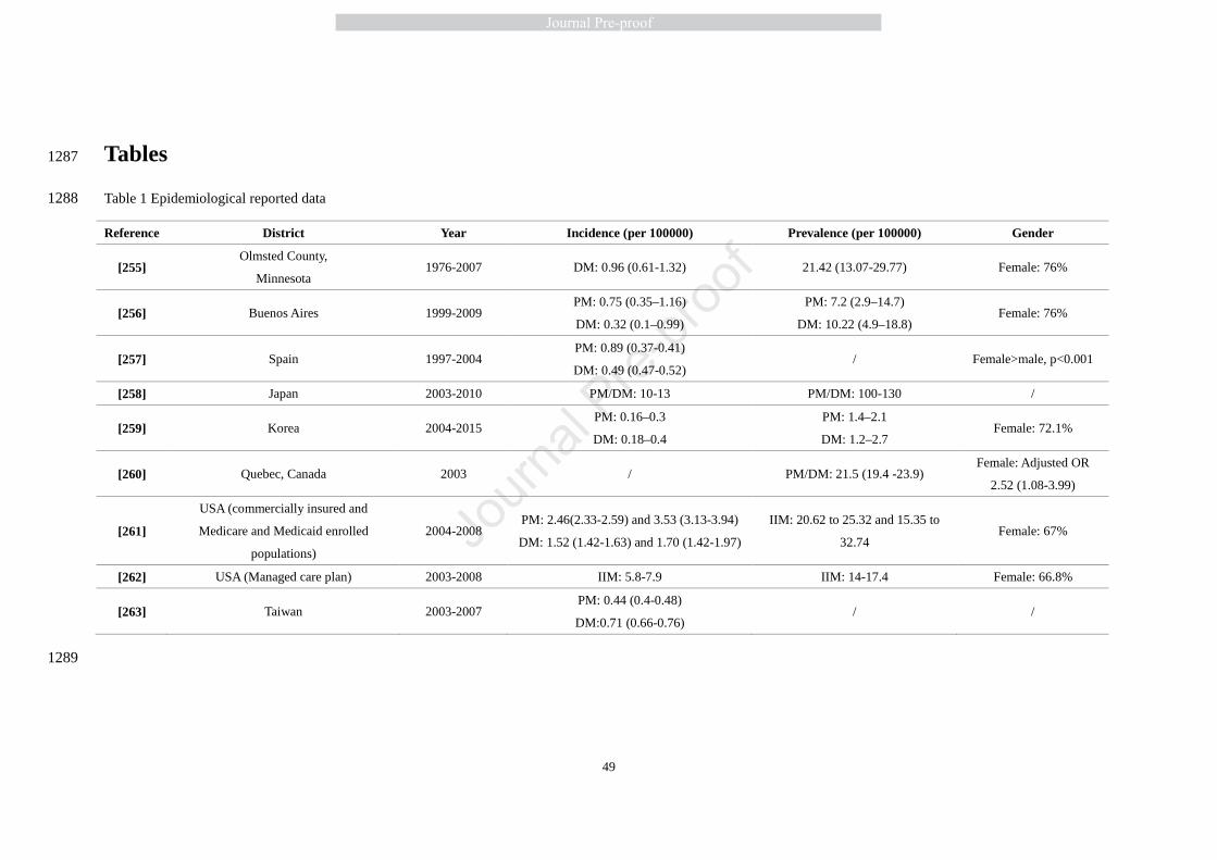
49
Tables 1287
Table 1 Epidemiological reported data 1288
Reference District Year Incidence (per 100000) Prevalence (per 100000) Gender
[255] Olmsted County,
Minnesota 1976-2007 DM: 0.96 (0.61-1.32) 21.42 (13.07-29.77) Female: 76%
[256] Buenos Aires 1999-2009 PM: 0.75 (0.35–1.16)
DM: 0.32 (0.1–0.99)
PM: 7.2 (2.9–14.7)
DM: 10.22 (4.9–18.8) Female: 76%
[257] Spain 1997-2004 PM: 0.89 (0.37-0.41)
DM: 0.49 (0.47-0.52) / Female>male, p<0.001
[258] Japan 2003-2010 PM/DM: 10-13 PM/DM: 100-130 /
[259] Korea 2004-2015 PM: 0.16–0.3
DM: 0.18–0.4
PM: 1.4–2.1
DM: 1.2–2.7 Female: 72.1%
[260] Quebec, Canada 2003 / PM/DM: 21.5 (19.4 -23.9) Female: Adjusted OR
2.52 (1.08-3.99)
[261]
USA (commercially insured and
Medicare and Medicaid enrolled
populations)
2004-2008 PM: 2.46(2.33-2.59) and 3.53 (3.13-3.94)
DM: 1.52 (1.42-1.63) and 1.70 (1.42-1.97)
IIM: 20.62 to 25.32 and 15.35 to
32.74 Female: 67%
[262] USA (Managed care plan) 2003-2008 IIM: 5.8-7.9 IIM: 14-17.4 Female: 66.8%
[263] Taiwan 2003-2007 PM: 0.44 (0.4-0.48)
DM:0.71 (0.66-0.76) / /
1289
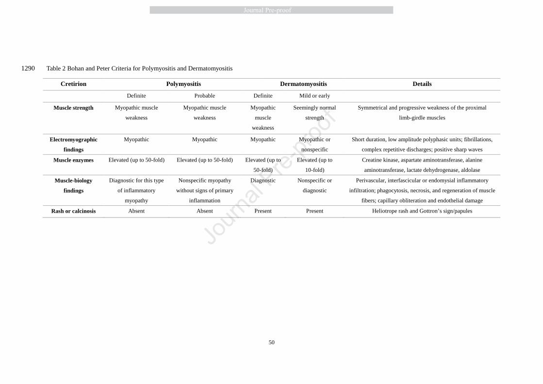
50
Table 2 Bohan and Peter Criteria for Polymyositis and Dermatomyositis 1290
Cretirion Polymyositis Dermatomyositis Details
Definite Probable Definite Mild or early
Muscle strength Myopathic muscle
weakness
Myopathic muscle
weakness
Myopathic
muscle
weakness
Seemingly normal
strength
Symmetrical and progressive weakness of the proximal
limb-girdle muscles
Electromyographic
findings
Myopathic Myopathic Myopathic Myopathic or
nonspecific
Short duration, low amplitude polyphasic units; fibrillations,
complex repetitive discharges; positive sharp waves
Muscle enzymes Elevated (up to 50-fold) Elevated (up to 50-fold) Elevated (up to
50-fold)
Elevated (up to
10-fold)
Creatine kinase, aspartate aminotransferase, alanine
aminotransferase, lactate dehydrogenase, aldolase
Muscle-biology
findings
Diagnostic for this type
of inflammatory
myopathy
Nonspecific myopathy
without signs of primary
inflammation
Diagnostic Nonspecific or
diagnostic
Perivascular, interfascicular or endomysial inflammatory
infiltration; phagocytosis, necrosis, and regeneration of muscle
fibers; capillary obliteration and endothelial damage
Rash or calcinosis Absent Absent Present Present Heliotrope rash and Gottron’s sign/papules
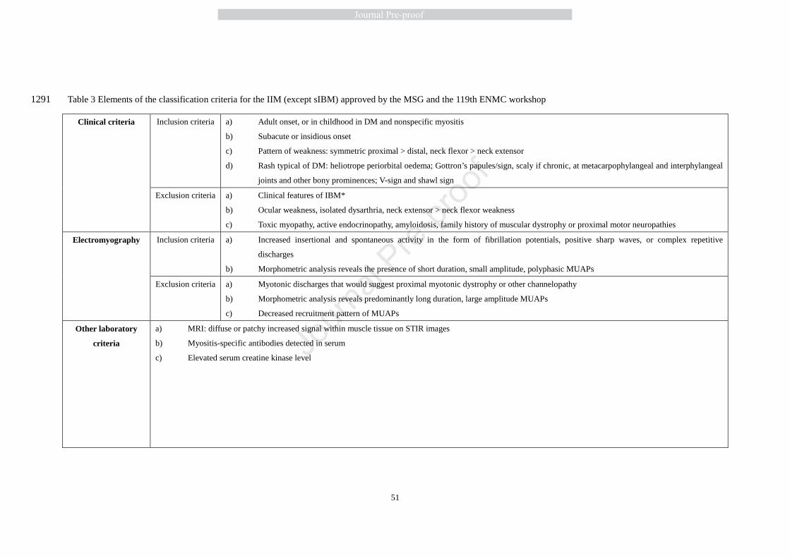
51
Table 3 Elements of the classification criteria for the IIM (except sIBM) approved by the MSG and the 119th ENMC workshop 1291
Clinical criteria Inclusion criteria a) Adult onset, or in childhood in DM and nonspecific myositis
b) Subacute or insidious onset
c) Pattern of weakness: symmetric proximal > distal, neck flexor > neck extensor
d) Rash typical of DM: heliotrope periorbital oedema; Gottron’s papules/sign, scaly if chronic, at metacarpophylangeal and interphylangeal
joints and other bony prominences; V-sign and shawl sign
Exclusion criteria a) Clinical features of IBM*
b) Ocular weakness, isolated dysarthria, neck extensor > neck flexor weakness
c) Toxic myopathy, active endocrinopathy, amyloidosis, family history of muscular dystrophy or proximal motor neuropathies
Electromyography Inclusion criteria a) Increased insertional and spontaneous activity in the form of fibrillation potentials, positive sharp waves, or complex repetitive
discharges
b) Morphometric analysis reveals the presence of short duration, small amplitude, polyphasic MUAPs
Exclusion criteria a) Myotonic discharges that would suggest proximal myotonic dystrophy or other channelopathy
b) Morphometric analysis reveals predominantly long duration, large amplitude MUAPs
c) Decreased recruitment pattern of MUAPs
Other laboratory
criteria
a) MRI: diffuse or patchy increased signal within muscle tissue on STIR images
b) Myositis-specific antibodies detected in serum
c) Elevated serum creatine kinase level
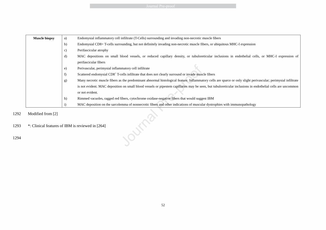
52
Muscle biopsy a) Endomysial inflammatory cell infiltrate (T-Cells) surrounding and invading non-necrotic muscle fibers
b) Endomysial CD8+ T-cells surrounding, but not definitely invading non-necrotic muscle fibers, or ubiquitous MHC-I expression
c) Perifascicular atrophy
d) MAC depositions on small blood vessels, or reduced capillary density, or tubuloreticular inclusions in endothelial cells, or MHC-I expression of
perifascicular fibers
e) Perivascular, perimysial inflammatory cell infiltrate
f) Scattered endomysial CD8+ T-cells infiltrate that does not clearly surround or invade muscle fibers
g) Many necrotic muscle fibers as the predominant abnormal histological feature. Inflammatory cells are sparce or only slight perivascular; perimysial infiltrate
is not evident. MAC deposition on small blood vessels or pipestem capillaries may be seen, but tubuloreticular inclusions in endothelial cells are uncommon
or not evident.
h) Rimmed vacuoles, ragged red fibers, cytochrome oxidase-negative fibers that would suggest IBM
i) MAC deposition on the sarcolemma of nonnecrotic fibers and other indications of muscular dystrophies with immunopathology
Modified from [2] 1292
*: Clinical features of IBM is reviewed in [264] 1293
1294
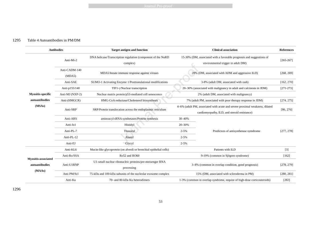
53
Table 4 Autoantibodies in PM/DM 1295
Antibodies Target antigen and function Clinical association References
Myositis-specific
autoantibodies
(MSAs)
Anti-Mi-2 DNA helicase/Transcription regulation (component of the NuRD
complex)
15-30% (DM, associated with a favorable prognosis and suggestions of
environmental trigger in adult DM) [265-267]
Anti-CADM-140
(MDA5) MDA5/Innate immune response against viruses 20% (DM, associated with ADM and aggressive ILD) [268, 269]
Anti-SAE SUMO-1 Activating Enzyme 1/Posttranslational modifications 3-8% (adult DM, associated with rash) [162, 270]
Anti-p155/140 TIF1-γ/Nuclear transcription 20–30% (associated with malignancy in adult and calcinosis in JDM) [271-273]
Anti-MJ (NXP-2) Nuclear matrix protein/p53-mediated cell senescence 2% (adult DM, associated with malignancy)
Anti-(HMGCR) HMG-CoA reductase/Cholesterol biosynthesis 7% (adult PM, associated with poor therapy response in JDM) [274, 275]
Anti-SRP SRP/Protein translocation across the endoplasmic reticulum 4–6% (adult PM, associated with acute and severe proximal weakness, dilated
cardiomyopathy, ILD, and steroid resistance) [86, 276]
Anti-ARS aminoacyl-tRNA synthetases/Protein synthesis 30–40%
Predictors of antisynthetase symdrome [277, 278]
Anti-Jo1 Histidyl 20–30%
Anti-PL-7 Threonyl 2-5%
Anti-PL-12 Alanyl 2-5%
Anti-EJ Glycyl 2-5%
Myositis-associated
autoantibodies
(MAAs)
Anti-KL6 Mucin-like glycoprotein (on alveoli or bronchial epithelial cells) Patients with ILD [3]
Anti-Ro/SSA Ro52 and RO60 9-19% (common in Sjögren syndrome) [162]
Anti-U1RNP U1-small nuclear ribonucleic proteins/pre-messenger RNA
processing 3–8% (common in overlap condition, good prognosis) [278, 279]
Anti PM/Scl 75-kDa and 100-kDa subunits of the nucleolar exosome complex 15% (DM, associated with scleroderma in PM) [280, 281]
Anti-Ku 70- and 80-kDa Ku heterodimers 1-3% (common in overlap syndrome, require of high-dose corticosteroids) [282]
1296
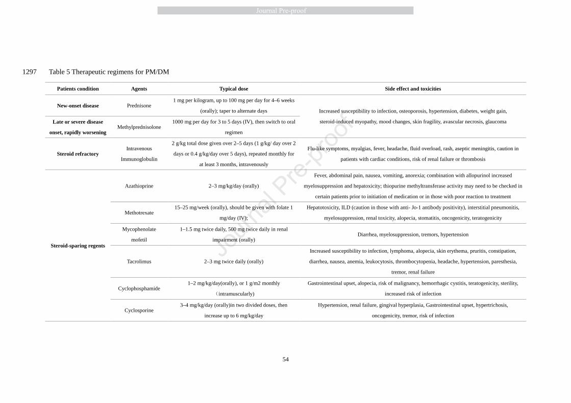
54
Table 5 Therapeutic regimens for PM/DM 1297
Patients condition Agents Typical dose Side effect and toxicities
New-onset disease Prednisone 1 mg per kilogram, up to 100 mg per day for 4–6 weeks
(orally); taper to alternate days Increased susceptibility to infection, osteoporosis, hypertension, diabetes, weight gain,
steroid-induced myopathy, mood changes, skin fragility, avascular necrosis, glaucoma Late or severe disease
onset, rapidly worsening Methylprednisolone
1000 mg per day for 3 to 5 days (IV), then switch to oral
regimen
Steroid refractory Intravenous
Immunoglobulin
2 g/kg total dose given over 2–5 days (1 g/kg/ day over 2
days or 0.4 g/kg/day over 5 days), repeated monthly for
at least 3 months, intravenously
Flu-like symptoms, myalgias, fever, headache, fluid overload, rash, aseptic meningitis, caution in
patients with cardiac conditions, risk of renal failure or thrombosis
Steroid-sparing regents
Azathioprine 2–3 mg/kg/day (orally)
Fever, abdominal pain, nausea, vomiting, anorexia; combination with allopurinol increased
myelosuppression and hepatoxicity; thiopurine methyltransferase activity may need to be checked in
certain patients prior to initiation of medication or in those with poor reaction to treatment
Methotrexate 15–25 mg/week (orally), should be given with folate 1
mg/day (IV);
Hepatotoxicity, ILD (caution in those with anti- Jo-1 antibody positivity), interstitial pneumonitis,
myelosuppression, renal toxicity, alopecia, stomatitis, oncogenicity, teratogenicity
Mycophenolate
mofetil
1–1.5 mg twice daily, 500 mg twice daily in renal
impairment (orally) Diarrhea, myelosuppression, tremors, hypertension
Tacrolimus 2–3 mg twice daily (orally)
Increased susceptibility to infection, lymphoma, alopecia, skin erythema, pruritis, constipation,
diarrhea, nausea, anemia, leukocytosis, thrombocytopenia, headache, hypertension, paresthesia,
tremor, renal failure
Cyclophosphamide 1–2 mg/kg/day(orally), or 1 g/m2 monthly
(intramuscularly)
Gastrointestinal upset, alopecia, risk of malignancy, hemorrhagic cystitis, teratogenicity, sterility,
increased risk of infection
Cyclosporine 3–4 mg/kg/day (orally)in two divided doses, then
increase up to 6 mg/kg/day
Hypertension, renal failure, gingival hyperplasia, Gastrointestinal upset, hypertrichosis,
oncogenicity, tremor, risk of infection
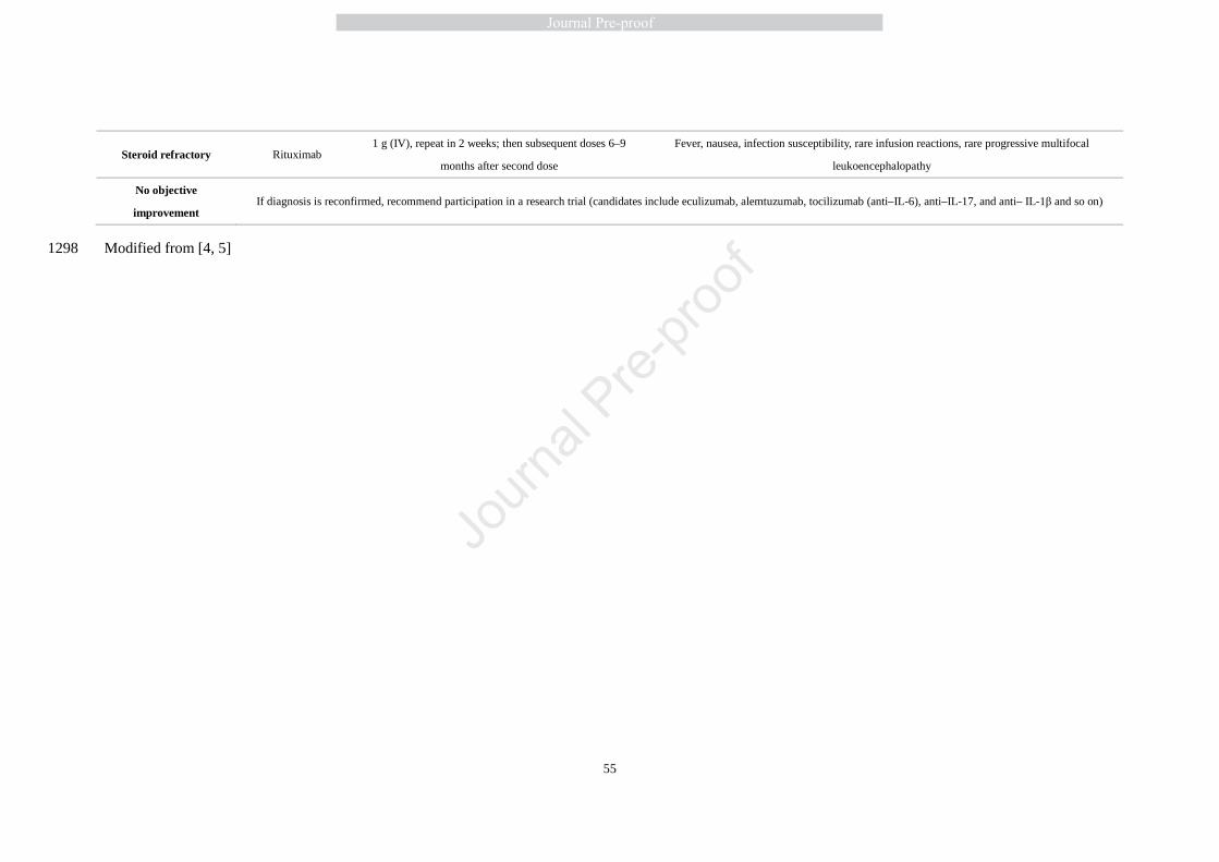
55
Steroid refractory Rituximab 1 g (IV), repeat in 2 weeks; then subsequent doses 6–9
months after second dose
Fever, nausea, infection susceptibility, rare infusion reactions, rare progressive multifocal
leukoencephalopathy
No objective
improvement If diagnosis is reconfirmed, recommend participation in a research trial (candidates include eculizumab, alemtuzumab, tocilizumab (anti–IL-6), anti–IL-17, and anti– IL-1β and so on)
Modified from [4, 5] 1298
High light
� Challenges of diagnosis and management are probably the biggest problem in clinical
research and practice of Polymyositis (PM) and dermatomyositis (DM).
� Diagnostic criteria have been updated and novel therapies have been developed in PM/DM.
� To improve diagnostic precision and to better understand the pathogenesis and the
progression of PM/DM may help to guide future treatment and research strategies.
� International cooperation is needed in order to improve the lives of patients suffering from
PM/DM.